ashok seth - summitmd.comsummitmd.com/pdf/pdf/1-5.ashokseth.pdf · ashok seth chairman & chief...
TRANSCRIPT

Ashok SethChairman & Chief Cardiologist
Chairman Cardiology Council, Fortis Group of Hospitals
Fortis Escorts Heart InstituteNew Delhi
How To Innovate in CV Technology – TCT AP 2011, Seoul

Disclosures Honorary Advisor to Merill LifeSciences

Innovation
Unbridled enthusiasm
Harsh realities
Ultimate applicability
Time
BioMime/Mitsu entry point
Efficacy
ST
Thoughtful adaptations
Safety

Stent Architecture
Drug Delivery methods Drug
Inflammatory reaction
Release kinetics
Incomplete healing
• Stent architecture, Polymer and Drug are the most polarized facets in DES construction
DES: A mechanical & pharmacological Approach to CAD. Paul Dobesh et al Pharmacotherapy, 2004
Penrose’s impossible triangle

Drivers of DES Safety Acute drivers
Vessel injury Complete apposition Biodegradable polymers or no polymers! Premature cessation of antiplatelet Rx
Long term drivers Re‐endothelialisation Functional endothelium No vessel wall inflammation

Thin Struts and Restenosis• Thin Struts allow for-
– Low blood flow perturbance
– Easy struts nesting to the vessel wall
– Added flexibility and conformability
• Improved clinical outcome*
• Improved, faster endothelialization **
• * Kastrati A, Schömig A, Dirschinger J, et al. Strut Thickness Effect on Restenosis Outcome (ISAR STEREO Trial). Circulation 2001; 103:2816-2821
• ** Simon C, Palmaz JC, Sprague EA. Influence of topography on endothelialization of stents: clues for new designes. J Lon Term Eff Med Implants. 2000;10:143-151
NO Stent: Laminar Flow
Stent Thickness S2>S1: Turbulent Flow
Stent Thickness S1:

Thin is In!P=0.003
ISAR‐STEREO Trial. Circulation 2001;103:2816‐2821
P=0.002
ISAR‐STEREO 2 Trial. JACC 2003;41:1283‐8
P=0.03
P<0.001

An “ultra‐thin” stent.
Hybrid stent design = optimal geometry.
New sirolimus analogue with preferred dosing and drug release kinetics.
Polymer ‐ free drug delivery.
Lowest possible vessel wall injury (↓inflammation)
Superior deliverability and conformability (“glove‐like” fit and apposition)
Improved healing = less stent thrombosis
Lowest possible restenosis
Early discontinuation of DAPT
Mitsu – Technology ApproachFeatures Benefits

Strut thicknessStrut thickness
Coating thicknessCoating thickness
PolymerPolymer
DrugDrug
3rd Gen3rd Gen
BioMime
65 µm65 µm
2 µm2 µm
PLLA + PLGAPLLA + PLGA
Sirolimus1.25 µg/mm2Sirolimus
1.25 µg/mm2
Cypher
140 µm140 µm
12.6 µm12.6 µm
PEVA‐PBMAPEVA‐PBMA
Sirolimus1.4 µg/mm2
Sirolimus1.4 µg/mm2
1st Gen
Taxus
132 µm132 µm
16 µm16 µm
SIBBSSIBBS
Paclitaxel1.0 µg/mm2
Paclitaxel1.0 µg/mm2
1st Gen
Endeavor
91 µm91 µm
5.3 µm5.3 µm
PCPC
Zotarolimus10.0 µg/mmZotarolimus10.0 µg/mm
2nd Gen
Xience V
81 µm81 µm
7.6 µm7.6 µm
FluoroFluoro
Everolimus1.0 µg/mm2
Everolimus1.0 µg/mm2
2nd Gen 4th Gen4th Gen
Mitsu
40 µm40 µm
< 2 µm< 2 µm
NoneNone
Merilimus0.45 µg/mm2Merilimus
0.45 µg/mm2
3.0 mm diameter stents, 500X magnification

Clo
sed
cel
ls
The “hybrid” design coupled with strut width variability eliminates the need for high strut thicknesses as required in
earlier stent technologies
Closed Cells 140µm
Open Cells 132µm
Open Cells 132µm
Open Cells 90µm
Open Cells 81µm
Hybrid Cells 65µm
Hybrid Cells 40µm
Bx Velocity
Express Liberte Driver Multi‐Link
BioMime Mitsu
Ope
n ce
lls
2000 2011

BioMime Stent Architecture Cobalt chromium (L605) platform with 65µm strut thickness.
Hybrid cell design comprising of an intelligent mix of open and close cells resulting in excellent radial strength with a high flexibility.
Unique strut width variability that ensure a <3% recoil and 0.29% foreshortening
Special electro‐polishing technique eliminates surface nickel oxides
Hybrid designOpen cell in mid ‐ segmentClosed cell at edges
SEM image of crimped BioMime SES at 50x

Mitsu – Cell Architecture
Open cells at the middleClose cells at the edges
40µm4040µµmm
Strut thickness
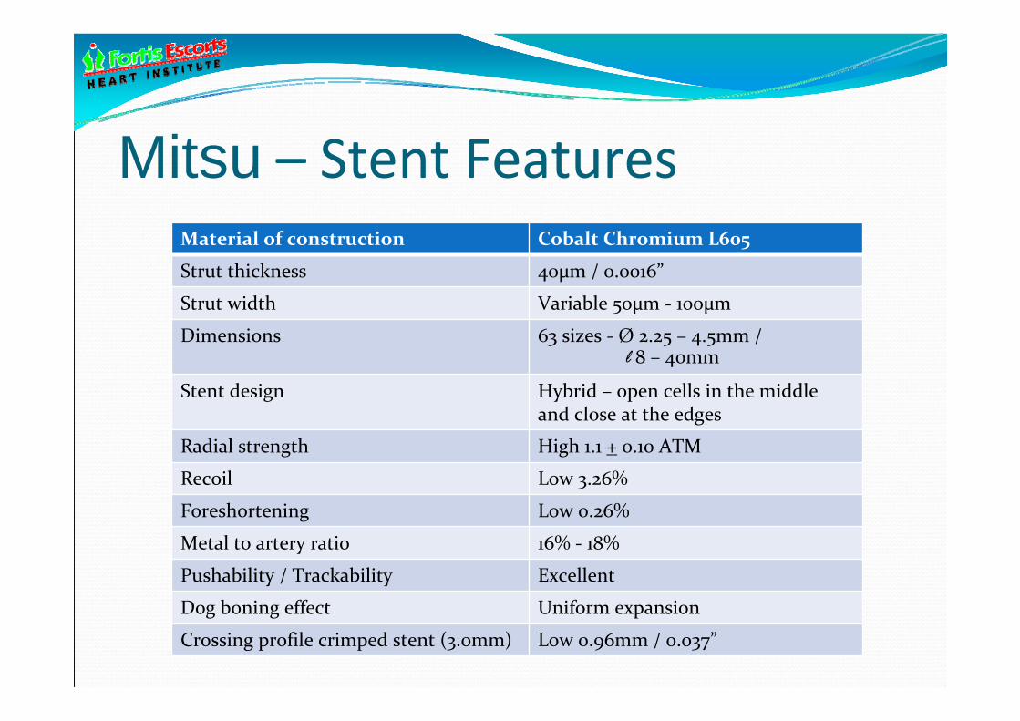
Mitsu – Stent FeaturesMaterial of construction Cobalt Chromium L605
Strut thickness 40µm / 0.0016”
Strut width Variable 50µm ‐ 100µm
Dimensions 63 sizes ‐ Ø 2.25 – 4.5mm / l 8 – 40mm
Stent design Hybrid – open cells in the middle and close at the edges
Radial strength High 1.1 + 0.10 ATM
Recoil Low 3.26%
Foreshortening Low 0.26%
Metal to artery ratio 16% ‐ 18%
Pushability / Trackability Excellent
Dog boning effect Uniform expansion
Crossing profile crimped stent (3.0mm) Low 0.96mm / 0.037”

Low injury design Conventional edge‐flaring stent designs allow the stent to dog‐bone during deployment.
This dog‐boning coupled with balloon overhang may cause edge injury.
BioMime has struts with design variability which results in morphology mediated expansionTM, having a propensity to minimize stent edge injury.
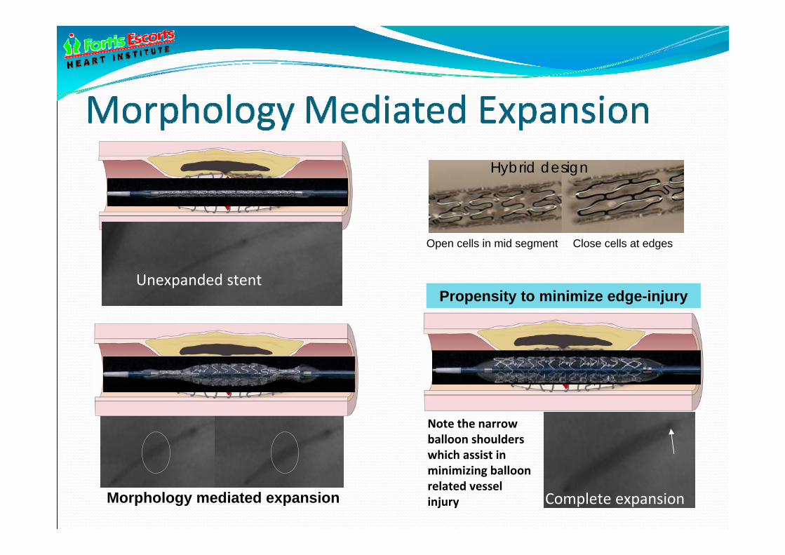
Propensity to minimize edge-injury
Open cells in mid segment Close cells at edges
Unexpanded stent
Hybrid design
Morphology mediated expansion Complete expansion
Note the narrow balloon shoulders which assist in minimizing balloon related vessel injury

Excellent Side Branch Access
The expanded BIOMIME 3.0 x 16 mm stent after side branch expansion
The area of the largest circle circumscribable in the cell of the stent expanded to the nominal diameter:Tc = 0.71 mm2
Expanded cell perimeter that ensures side branch access: KSBA = 11.29 mm
Expanded cell area that ensures side branch access: TSBA = 8.00 mm2Data on file with Meril Life Sciences.

Following Crimping Following Expansion
Following Crimping Following Expansion

Equivalent radio-opacity due to stent design
Driver Vision Mitsu
90µm 81µm 40µm

fkBP-12 receptor site adaptation by Merilimus
molecule
• Merilimus is a Meril Life Sciences invention.
• Heterogeneous 5 member ring on the parent limus molecule.
• Better toxicological profile than Sirolimus and a wider therapeutic window (more lipophilic).
• Low drug dosing of 0.4µg/mm2
possible to get optimal anti-proliferative effect.

Unique Formulation - Solid lipid nano-spheres (SLN)consisting of Merilimus + Lipid (<500 nm)
SEM Image of Stent struts coated with
nano-formulation
SLN rapidly leave stent and enter vessel wall with prolonged tissue residence time

Nanotech for Drug Delivery
Control and maintain in‐tissue drug release Improved PK stability Enhanced cellular uptake Feasibility of carrying both lipophilic and hydrophilic drugs
Lipids are biodegradable (excellent biocompatibility) Potential for lowering doses Potential to target different vascular layers by using differential NP size
Potential Benefits of Solid-Lipid Nanoparticles

Avg. Particle Size & Size Distribution
Size distribution of Drug-Lipid Nanoparticles
between 50nm and 500nm(80% ~127nm)
Average Particle Size (Z-Average) : 165.4 nm
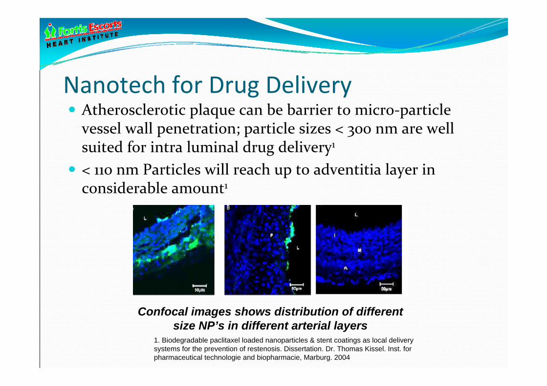
Nanotech for Drug Delivery Atherosclerotic plaque can be barrier to micro‐particle vessel wall penetration; particle sizes < 300 nm are well suited for intra luminal drug delivery1
< 110 nm Particles will reach up to adventitia layer in considerable amount1
Confocal images shows distribution of different size NP’s in different arterial layers
1. Biodegradable paclitaxel loaded nanoparticles & stent coatings as local delivery systems for the prevention of restenosis. Dissertation. Dr. Thomas Kissel. Inst. for pharmaceutical technologie and biopharmacie, Marburg. 2004

Zeta potential is the charge that develops at the interface between a solid Nanoparticles surface and aqueous medium. This potential, which is measured in MilliVolts, these are the dissociation of ionogenic groups in the particle surface and the differential adsorption of solution ions into the surface region.
The Zeta potential value (-53.9 mV) indicates Good Stability of Drug-lipid nano
particles in suspension.

Mitsu – The formulation• Nanotechnology based coating.• Designed for coating uniformity and lower drug dosage.• Controlled and reproducible drug release kinetics.• Rapid release of drug into tissue with “in‐tissue” drug depot or reservoir effect.
• Renders the stent “coating‐free” within a short time Rapid tissue absorption (SLN has high tissue diffusion co‐efficient) Formulation ensures drug availability for 1 month

Optical Microscopic Images (Coating)

Stent System
• 40 µm thin stent
• Cobalt Chromium L605
• Hybrid stent design
• Variable strut width
• Tapered balloon shoulders
Drug
• Merilimus – new limus
analogue
• Cytostatic, anti-inflammatory
and lipophilic
• Wide therapeutic window
Drug Delivery
• Novel SLN polymer-free
formulation
• Low drug for same effect
• Fast release kinetics
• Completely biocompatible
and anti-inflammatory
A unique differentiated new DES!