asian cellular therapy organization program & abstracts
TRANSCRIPT

Asian Cellular Therapy Organization
The 5th meeting of
http://acto2014.info
Osaka, 2014
Program & Abstracts
Date
Nov.10th(Mon)~12th(Wed),2014
VenueHyatt Regency Osaka, Japan
Regency Ballroom( 3F)1-13-11 Nanko-Kita, Suminoe-Ku, Osaka City, Osaka, JapanTEL: +81-6-6614-7821
PresidentTeruo Okano, Ph.D.
ProfessorInstitute of Advanced Bio-medical Engineering and Science
Tokyo Women’ s Medical University
Asian Cellular Therapy Organization

Title of Meeting
Date
Venue
President
ACTO Chair
Official Language
Website
Executive Secretary
Executive Secretariat
Inquiry
Committe Names………………………………………Welcome Remarks……………………………………Osaka MAP………………………………………………
Floor Guide ………………………………………………
Guideline for Participants …………………………
Detail of Program ● Monday,November 10th ………………………… ● Tuesday ,November 11th………………………… ● Wednesday,November 12th………………………
Abstract……………………………………………………
Poster Information……………………………………
Sponsors …………………………………………………
The 5th meeting of Asian Cellular Therapy Organization
November 10th(Mon)to 12th(Wed),2014
Hyatt Regency Osaka, JapanRegency Ballroom, 3F, 1-13-11 Nanko-Kita, Suminoe-Ku, Osaka City, Osaka, JapanTEL: +81-6-6614-7821http://osaka.regency.hyatt.jp/en/hotel/home.html
Teruo Okano, Ph.D.Professor, Institute of Advanced Bio-medical Engineering and Science, Tokyo Women’s Medical University
Akihiro Shimosaka, Ph.D.Director, Research & Development Division, Research Foundation for Community Medicine
English
http://acto2014.info
Yuji Heike, M.D., Ph.D.Unit Leader, Department of Cancer Immunotherapy, Exploratory Oncology Research & Clinical Trial Center, National Cancer Center
Nikkei Business Publications, Inc.
Secretariant OfficeSecretariat of ACTO 2014
c/o PCO Works, Inc.2F., Kanda Urban Bldg., 2-4-2 Kanda-Tsukasamachi, Chiyoda-ku, Tokyo 101-0048 JAPANTel: +81-3-6869-0347 / Fax: +81-3291-3635 / E-mail: [email protected]
Meeting Information
Contents
Asian Cellular Therapy Organization
Program & Abstracts
The 5th meeting of
4
5
6
7
8
1012
14
16
47
58

4 5
Committee Names
President
Vice Presidents
Secretary General
Committee Members
Industry Memebrs
Akihiro Shimosaka (Japan)
Yoichi Takaue (Japan)
Mickey Koh (Singapore)
Xiao-Jun Huang (China)
Khattry Navin (India)
Saengsuree Jootar (Thailand)
Yao-Chang Chen (Taiwan)
Hee Young Shin (Korea)
Chi Dung Phu (Vietnam)
Abdalla Awidi Abbadi (Jordan)
Bin Koming Ya’Akop (Malaysia)
Mohiuddin Ahmed Khan (Bangladesh)
Abbas Ghaderi (Iran)
Ferry Sandra (Indonesia)
Yuji Heike (Japan)
Kai-Yan Liu (China)
Jun Ren (China)
Shuichi Taniguchi (Japan)
Keiya Ozawa (Japan)
Takayuki Asahara (Japan)
Shinji Miyake (Japan)
Kiyoshi Okada (Japan)
T. J Hwang (Korea)
Hee-Je Kim (Korea)
Jay Lee (Korea)
Kam Man Hui (Singapore)
Kellathur N. Srinivasan (Singapore)
Artit Ungkanont (Thailand)
Suradej Hongeng (Thailand)
Xue-Tau Cao (China)
Hu Chen (China)
Iisrasena Nipan (Thailand)
Udomsak Bunworasate (Thailand)
Dinesh Pendharkar (India)
Kazuto Takesako (Japan)
Ryuji Maekawa (Japan)
Takahito Nakamura (Japan)
Sei-ichi Yusa (Japan)
Jaeseung Lim (Korea)
Min Liang (China)
Shing-Mou Lee (Taiwan)
Welcome Remarks
Asian Cellular Therapy Organization (ACTO) will hold its annual meeting. It is our great pleasure to welcome you to Osaka for the 5th Annual Meeting. This year’s event will be held in Japan for the first time after three years since our 2nd Meeting in Miyazaki. The participants of ACTO Annual Meeting has grown every year, and our gatherings are now welcoming people from the US and EU, in addition to over 10 Asian countries. The ACTO Annual Meeting serves as a great platform bringing together cell-based treatment technology researchers,
regulatory authorities and related businesses for sharing overall study outcomes and acquired knowledge on using cells and cultured tissues for treating patients as well as exchanging ideas and promoting friendships among researchers. Over the recent years, cell-based medical technologies are advancing and evolving at remarkable pace in such fields as cell pharmaceuticals, gene therapies, immune cell therapies and regenerative medicine using cells and cultured tissues, in addition to hematopoietic stem cell transplantation. Regarding cells used for medical purposes, ES cell and iPS cell technologies are edging closer to practical use, as well as therapies based on blood stem cells and somatic stem cells, which are gathering high expectations for further development in the future. This year’s ACTO Meeting will invite Dr. Masayo Takahashi from RIKEN as a plenary session speaker. Dr. Takahashi is a pioneer in developing iPS cell-based therapies for retinal degenerative diseases, and she is also scheduled to conduct the world’s first-ever iPS cell-based cell transplant surgery. Other lecturers include some researchers from Center for iPS Cell Research and Application (CiRA) of Kyoto University. We also invite notable academics from other Asian countries in the field of regenerative medicine using somatic stem cells, which is leading the way far ahead for practical use. Coincidentally on the occasion of the 5th ACTO Annual Meeting in this November, two new laws concerning cell and tissue regenerative therapies will be enacted in Japan. With these new law enforcements, Japan is expected to be the world’s fastest country in issuing approvals for cell and tissue regenerative therapies. It will be a significant experience that such country hosts our annual event bringing together regulatory authorities from other countries to discuss the future of regulations on cellular therapy technologies. Osaka, a host city to this year’s meeting, is a home to Osaka University which is one of the core bases for Japan’s regenerative medicine. The city is also located close to CiRA in Kyoto and Riken in Kobe. So to speak, this Annual Meeting is held in the active base of Japan’s cellular and regenerative therapy studies. We also hope all the participants to enjoy the attractiveness of Japan while they stay in this beautiful country. Osaka has unique food culture as seen in Okonomiyaki (a pizza-like savory pancake) and Udon (thick noodles), while also providing a showcase of popular entertainment such as traditional Bunraku puppet theater and Manzai stand-up comedy. You can also visit the Universal Studios Japan within a 10-minute drive from the Meeting venue, to enjoy a new attraction “The Wizarding World of Harry Potter.” Moreover, November is the fall foliage season in Kyoto, the neighboring city to Osaka, so it is also a good idea to take a day trip for admiring the beautiful scenery to your heart’s content. We hope you to enjoy a glimpse of Japan before or after the ACTO Meeting, and are sincerely looking forward to welcoming all of you in this November.
Since 2010, ACTO started as ISCT Asian Region and changed name in 2011 as Asian Cellular Therapy Organization. ACTO established as an independent society from ISCT but close collaboration with ISCT. ISCT and ACTO have been collaborating in the past ACTO meetings. ISCT and ACTO agreed to continue collaboration at the 20th ISCT meeting in Paris, April 2014. ACTO will maintain unique activity as mediator among three key players, academy, industry and regulatory agent, in the field of cellular therapy. ACTO organize meetings in 2010 and 2011 in Miyazaki, Japan, 2012 in Chen Mai in Thailand
and 2013 in Shinchu in Taiwan. ACTO organizing committee is consisted of representatives from member country and making decisions for ACTO activity. This year the 5th ACTO meeting will be held in Osaka on November 10-12 organized by Prof. Teruo Okano, Tokyo Women’s medical College in Tokyo. This year new Japanese regulations for cellular therapy will become fully effective which will accelerate the research and development of cellular therapy in Japan. We will organize special symposium on regulations attached to Annual meeting on November 9 to introduce Japanese new regulation as well as other Asian countries regulation and EU regulation. Also application of iPS technology will be discussed as well as tissue engineering, immuno cellular therapy and advances in manufacturing technology. Following ACTO meeting will be held in Korea, 2015 and China in 2016. Please join and learn and enjoy annual meeting in Osaka.
Teruo Okano, Ph. D.ProfessorInstitute of Advanced Bio-medical Engineering and ScienceTokyo Women’s Medical University
Akihiro Shimosaka, Ph. D. / ChairpersonDirectorResearch & Development DivisionResearch Foundation for Community Medicine

6 7
Route map (Osaka and Osaka City)
Men Women
Elevators
Cloak room Cloak room
Escalators
Prefunction
Stage
MeetingArea
PartyArea
Exhibition Hall(Sponsors, Poster)
Level 3(BANQUEST TOWER)
Men Women
Elevators
Prefunction Prefunction
To INTEX OSAKA To WTC
To Car Park
MeetingRoom
ManagingO�ce
Level 2(BANQUEST TOWER)
Floor Guide

8 9
Guideline for Participans
On-site RegistrationOn-site registration will be conducted as follows:Registration counter: front of Regency Ball roomDate & Time: All time available
On-site Registration FeeMember: 30,000 JPY Non-member:35,000JPYStudent: 5,000 JPY (all day), 3,000JPY(one day)All payment must be made in Japanese yen by cash or credit card.
Name cardName card will be distributed at registration desk.Name card should be worn at all time, if not admit all venue.
Prohibited matterRecording, photography is prohibitedDuring the lecture, if that act to interfere with the lecture has been discovered, you might get sent off.
Exhibition time & Poster Viewing11/10(Mon) 10:30-20:00 11/11(Tue) 10:00-21:00 11/12(Wed) 9:30-13:30
Meeting RoomSAKI(Level2 BanquetTower)
Luncheon SeminarManaging office will distribute the tickets in the Registration on the morning of 11/10,11/11. If there is no ticket, you will not be able to receive the LunchBox.
Welcome PartyCharge:5000JPY Please pay at front of party room.
Coffee ServingManaging office will prepare the coffee service at exhibition area (Room C)Serving time: 11/10 10:30-11:00 16:45-17:15 11/11 15:15-15:45
Managing OfficeMAI (Level2 BanquetTower)
Program at a Glance
8:00
9:00
10:00
11:00
12:00
13:00
14:00
15:00
16:00
17:00
18:00
19:00
20:00
21:00
November. 10th November. 11th November. 12th
Opening Ceremony
Closing Remark
Opening Session 1
Plenary Session 1ISCT-ACTO Joint Session
Plenary Session 2
Regenerative Medicine
Regulatory Session
Best abstract Presentation
Welcome Party
Somatic Cell BasedTissue Engineering 1
Plenary Sesion 3iPS cell for organ regenetation
Support for Cellular therapyby Japanese government
Panel Discussionon Regulatory Topics
Somatic Cell BasedTissue Engineering 2
Luncheon Seminar
CD 19 CAR
Manufacturing
iPS Cell BasedTissue Engineering 1
Industrialization of TissueEngineering
8:30-9:008:00-10:00
10:15-11:00
11:00-12:30
10:30-11:45
18:15-18:45
19:00-20:30
8:15-8:30
8:00-8:15
12:45-13:00
9:00-10:30
8:00-8:45
8:45-10:15
11:45-12:45
11:00-12:30
12:45-13:30
13:45-15:15
15:45-18:15
Sponsored by Lonza
Sponsored by Medinet
Sponsored by Lonza
13:45-16:45
17:15-19:45
Break
Break
Break
Break
Lunch
Break

10 11
Detail of Program
November 10 (Mon.)
Opening Ceremony
Speaker : Teruo Okano (Tokyo Women's Medical University)
8:00-8:15
Opening Session
OS-1Chair : Teruo Okano (Tokyo Women's Medical University) Speaker : Tsutomu Tomioka (The House of Representatives)
8:15–8:30
Luncheon Seminar Sponsored by Lonza
LS-1 Speaker : Thomas Fellner (Lonza)
12:45–13:30
Plenary Session 1
PLS-1Chair : Akihiro Shimosaka (ACTO) Speaker : Teruo Okano (Tokyo Women's Medical University)
8:30–9:00
Somatic Cell Based Tissue Engineering 1
GS-1Chair : Yoichi Takaue (St Luke's International Hospital) Saengsuree Jootar (Mahidol University)
Speaker : Shigeyuki Wakitani (Hiroshima University) Kohji Nishida (Osaka University) Yoshiki Sawa (Osaka University)
9:00–10:30
9:00–10:30
9:30–10:00
10:00–10:30
Somatic Cell Based Tissue Engineering 2
GS-2Chair : Hong-Nerng Ho (National Taiwan University) Yao-Chang Chen (National Taiwan University)
Speaker : Keun-Hong Park (Cha University) Tatsuya Shimizu (Tokyo Women's Medical University) Masato Satou (Tokai University)
11:00–12:30
11:00–11:30
11:30–12:00
12:00–12:30
iPS Cell Based Tissue Engineering
Industrialization of Tissue Engineering
GS-3
GS-4
Chair : Mickey Koh (St. Georges Hospital) Hiromitsu Nakauchi (University of Tokyo)
Chair : Koichi Nakayama (Saga University) Suradej Hongeng (Mahidol University)
Speaker : Jun Takahashi (Kyoto University)
Speaker : Koichi Nakayama (Saga University)
Kouji Etou (Kyoto University)
So Ra Park (Inha University)
Jun Yamashita (Kyoto University)
Kenichirou Hata (Japan Tissue Engineering)
HongKui Deng (Peking University)
Setsuko Hashimoto (CellSeed)
Suradej Hongeng (Mahidol University)
Masanori Murayama (Regience)
Noriyuki Tsumaki (Kyoto University)
13:45–16:45
17:15–19:45
13:45–14:15
13:45–14:15
14:15–14:45
14:15–14:45
14:45–15:15
14:45–15:15
15:15–15:45
15:15–15:45
15:45–16:15
15:45–16:15
16:15–16:45

12 13
November 11 ( Tue.)
ISCT-ACTO Joint Session
Regenerative Medicine
Plenary Session 2
SS-1
GS-5
GS-6
PLS-2
Chair : Kazuhiro Kakimi (University of Tokyo) Kurt Gunter (ISCT)
Chair : Yoichi Takaue (St Luke's International Hospital) Masayo Takahashi (RIKEN)
Chair : Keiya Ozawa (University of Tokyo) Bruce Levine (University of Pennsylvania)
Chair : Hee Young Shin (Seoul National University)Speaker : Masayo Takahashi (RIKEN)
Speaker : Kurt Gunter (ISCT)
Speaker : Hiromi Kojima (Jikei University School of Medicine)
Speaker : Bruce Levine (University of Pennsylvania)
Duanqin Pei (Chinese Academy of Sciences)
Bonghee Lee (Gachon University)
Marco Davila (Venderbilt University)
Kazuhiro Kakimi (University of Tokyo)
Kyung-Ha Ryu(Ewha Womans University College of Medicine)
Keiya Ozawa (University of Tokyo)
Jun Ren (Capital Medical University)
8:00–10:00
11:00–12:30
13:45–15:15
10:15–11:00
8:00–8:30
11:00–11:30
13:45–14:15
8:30–9:00
11:30–12:00
14:15–14:45
9:00–9:30
12:00–12:30
14:45–15:15
9:30–10:00Best abstract Presentation
Welcome Party
GS-7
SS-2
Chair : David Smith (Lonza) Kunihiko Suzuki (Medinet)
Chair : Akihiro Shimosaka (ACTO) Kai Yan Liu (Peking University)
Speaker : Hidetoshi Shibuya (Shibuya Kogyo)
Speaker : To be determined
Masahiro Kino-oka (Osaka University)
To be determined
David Smith (Lonza)
To be determined
Ying-Ku Lu (EMO Biomedicine) DirkBalshüsemann (Miltenyi Biotec)
15:45-18:15
18:15–18:45
19:00–20:30
15:45–16:15
18:15–18:25
16:15–16:45
18:25–18:35
16:45–17:15
18:35–18:45
17:15–17:45
17:45–18:15
Manufacturing Sponsored by Lonza
CD 19 CAR Sponsored by Medinet
14
November 12 (Wed.)
Plenary Session 3 iPS Cell for Organ Regeneration
Panel Discussion on Regulatory Topics
Support for Cellular Therapy by Japanese Government
Regulatory Session
PLS-3
PLS-3
GS-8
GS-9
Chair : Yoichi Takaue (St Luke's International Hospital)Speaker : Hiromitsu Nakauchi (University of Tokyo)
Chair : Akihiro Shimosaka (ACTO) Kiyoshi Okada (PMDA)
Speaker : Akihiro Shimosaka (ACTO)
Chair : Akihiro Shimosaka (ACTO) Toshio Miyata (Health and Global Policy Institute)
Chair : Ryousuke Maruyama (PMDA) Kellathur Srinivasan (Health Science Authority Singapore)
Speaker : Yoshihide Esaki (MITI)
Speaker : Ryousuke Maruyama (PMDA)
Yutaka Hishiyama (Japanese Cabinet Secretariat)
Kellathur Srinivasan (HSA/Singapore)
Toshio Miyata (Health and Global Policy Institute)
Won Shin (Korean FDA) Ying-Hsien Fu. (Taiwan FDA) Maria Christina Gali (EMA)
8:00–8:45
11:45–12:45
8:45–10:15
10:30–11:45
8:45–9:15
10:30–10:45
9:15–9:45
10:45–11:00
9:45–10:15
11:00–11:15
11:15–11:30
11:30–11:45
Closing Remark19:00–20:30
Asian Cellular Therapy Organization
Abstracts
16 17
10th Nov. 8:15-8:30 Opening Session
10th Nov. 8:15-8:30
Development of laws for promotion of regenerativemedicine in Japan
Tsutomu Tomioka
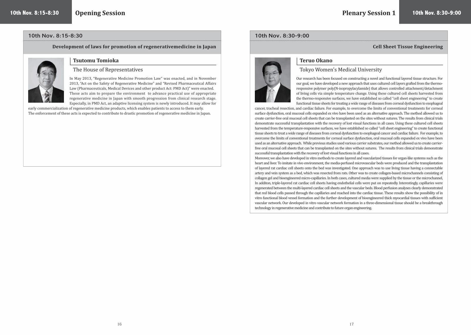
The House of RepresentativesIn May 2013, “Regenerative Medicine Promotion Law” was enacted, and in November 2013, “Act on the Safety of Regenerative Medicine” and “Revised Pharmaceutical Affairs Law (Pharmaceuticals, Medical Devices and other product Act: PMD Act)” were enacted. These acts aim to prepare the environment to advance practical use of appropriate regenerative medicine in Japan with smooth progression from clinical research stage. Especially, in PMD Act, an adaptive licensing system is newly introduced. It may allow for
early commercialization of regenerative medicine products, which enables patients to access to them early.The enforcement of these acts is expected to contribute to drastic promotion of regenerative medicine in Japan.
10th Nov. 8:30-9:00
10th Nov. 8:30-9:00
Cell Sheet Tissue Engineering
Teruo Okano
Tokyo Women's Medical UniversityOur research has been focused on constructing a novel and functional layered tissue structure. For our goal, we have developed a new approach that uses cultured cell layers grafted from the thermo-responsive polymer poly(N-isopropylacylamide) that allows controlled attachment/detachment of living cells via simple temperature change. Using these cultured cell sheets harvested from the thermo-responsive surfaces, we have established so called “cell sheet engineering” to create functional tissue sheets for treating a wide range of diseases from corneal dysfunction to esophageal
cancer, tracheal resection, and cardiac failure. For example, to overcome the limits of conventional treatments for corneal surface dysfunction, oral mucosal cells expanded ex vivo have been used as an alternative approach. The method allowed us to create carrier-free oral mucosal cell sheets that can be transplanted on the sites without sutures. The results from clinical trials demonstrate successful transplantation with the recovery of lost visual functions in all cases. Using these cultured cell sheets harvested from the temperature-responsive surfaces, we have established so called “cell sheet engineering” to create functional tissue sheets to treat a wide range of diseases from corneal dysfunction to esophageal cancer and cardiac failure. For example, to overcome the limits of conventional treatments for corneal surface dysfunction, oral mucosal cells expanded ex vivo have been used as an alternative approach. While previous studies used various carrier substrates, our method allowed us to create carrier-free oral mucosal cell sheets that can be transplanted on the sites without sutures. The results from clinical trials demonstrate successful transplantation with the recovery of lost visual functions in all cases. Moreover, we also have developed in vitro methods to create layered and vascularized tissues for organ-like systems such as the heart and liver. To imitate in vivo environment, the media-perfused microvascular beds were produced and the transplantation of layered rat cardiac cell sheets onto the bed was investigated. One approach was to use living tissue having a connectable artery and vein system as a bed, which was resected from rats. Other was to create collagen-based microchannels consisting of collagen gel and bioengineered micro-capillaries. In both cases, cultured media were supplied by the tissue or the microchannel, In additon, triple-layered rat cardiac cell sheets having endothelial cells were put on repeatedly. Interestingly, capillaries were regenerated between the multi-layered cardiac cell sheets and the vascular beds. Blood perfusion analyses clearly demonstrated that red blood cells passed through the capillaries and reached into the cardiac tissue. These results show the possibility of in vitro functional blood vessel formation and the further development of bioengineered thick myocardial tissues with sufficient vascular network. Our developed in vitro vascular network formation in a three-dimensional tissue should be a breakthrough technology in regenerative medicine and contribute to future organ engineering.
Plenary Session 1

18 19
10th Nov. 9:00-10:30 Somatic Cell Based Tissue Engineering 1
10th Nov. 9:00-9:30
Cartilage repair with autologous culture-expanded bone marrow mesenchymal stem cell Transplantation –over 10-year follow-up–
Shigeyuki Wakitani
Hiroshima UniversityWe focused on autologous culture-expanded bone marrow mesenchymal stem cells (BMSC), which can proliferate without losing their capacity for differentiation. Firstly, we transplanted BMSC into the defective articular cartilage of rabbit and succeeded in regenerating osteochondral tissue. We then applied this transplantation in human. Our previous reports showed that treatment with BMSC relieves the clinical symptoms of chondral defects in the knee and elbow joint. We investigated the efficacy of BMSC for osteoarthritic knee treated
with high tibial osteotomy, by comparing 12 BMSC-transplanted patients with 12 cell-free patients. At the 16-month follow-up, although the difference in clinical improvement between both groups was not significant, the arthroscopic and histological grading score was better in the cell-transplanted group. At the over 10-year follow- up, Hospital for Special Surgery knee scores improved to 76 and 73 in the BMSC-transplanted and cell-free groups, respectively, which were better than preoperative scores. Although we have never observed calcification above the tidemark in rabbit model and human histologically, the repair cartilage was not completely hyaline cartilage. Additionally, in all patients, and in the clinical study, we have never observed hypertrophy of repaired tissue, thereby guaranteeing the clinical safety of this therapy.
10th Nov. 9:30-10:00
Development of cell sheet-based therapy for corneal diseases-from tissue stem cell to iPS cell
Kohji Nishida
Osaka UniversityCorneal epithelial stem cells are known to be localized to the basal layer of the limbal epithelium. This corneal stem cell concept has been first reported in 1980s, based on the findings that label-retaining cells are located in the limbal basal epithelium. Since then, several investigators reported the specific characteristics for corneal epithelial stem/progenitor cells, including high colony-forming potential, p63 positive and so on. We have recently demonstrated that corneal epithelial stem/progenitor cells can be enriched
in integrin α6bri/CD71dim fraction by FACS.Complete loss of corneal epithelial stem cells because of severe trauma eye disease leads to corneal vascularization and opacification with severe visual loss. For corneal reconstruction in patients with such limbal stem cell deficiencies, we previously developed a unique method using tissue-engineered epithelial cell sheets comprising only the patient’s autologous oral mucosal epithelium. We are currently studying the potential of pluripotent stem cells for the treatment of corneal diseases. In this presentation, I will talk about the recent progress of stem cell therapy for corneal diseases.
10th Nov. 10:00-10:30
Autologous stem cell-sheet transplantation therapy for treating cardiomyopathy
Yoshiki Sawa
Osaka University We have developed the cell-sheet method in which scaffold-free cell-sheets are attached on the epicardial surface to maximize the paracrine effects. Based upon the “proof-of-concept” studies, Phase I Clinical Trial was launched to test the hypothesis that autologous skeletal muscle-derived cell-sheets transplantation may be feasible, safe and effective for treating severe congestive heart failure. All patients insisted marked symptomatic improvement post-treatment evaluated
by SAS (3.75±8.5 vs 5±1.4) with much decrease of Pulmonary artery pressure and Pulmonary vein resistance. In addition, 6-minute walk test showed a significant improvement (418.3±187.0 vs 522.8±129.1m). Multi-slice CT scanning revealed that in ICM patients whose LVESVI was between 100 and 130 showed LV reverse remodeling 6 months after sheet implantation compared with pre-value (EF; 27.9±1.6 vs 31.8±8.2%, ESVI; 118.5±12.1 vs 109.5±6.5ml/m2) and End systolic share stress (ESS) was decreased in the cell sheet received patients. In this Phase I study, cell-sheet transplantation as a sole therapy was feasible and safe for treating cardiomyopathy. Promising results in functional recovery warrant further clinical follow-up and accumulation of the cases to prove the therapeutic efficacy of this treatment for severe congestive heart failure.

20 21
10th Nov. 11:00-12:30 Somatic Cell Based Tissue Engineering 2
10th Nov. 11:00-11:30
Transfection of specific genes into normal human-derived dermal fibroblast cells for direct conversion
Keun-Hong Park
CHA UniversityWounded tissues and cells may be treated with growth factors and specific genes for the purpose of tissue repair and regeneration. To deliver specific genes into tissues and cells, this study presents the use of fabricated poly (DL-lactic-co-glycolic acid) (PLGA) nanoparticles (NPs) complexed with the cationic polymer poly (ethleneimine) (PEI). Through complexation with PEI, several types of genes (SOX9, Cbfa1, and C/EBP-α) were complexed with PLGA NPs, which enhanced gene uptake into normal human-derived
dermal fibroblast cells (NFDHCs)in vitro andin vivo. Several cell types (293T, HeLa, and fibroblast cells) were transfected with fluorescence-tagged PEI/SOX9, PEI/Cbfa1, and PEI/ C/EBP-α gene-complexed PLGA NPs. The gene and protein expression levels in the cells were evaluated by RT-PCR, real-time quantitative PCR, Western blotting, and confocal laser microscopy. Fibroblast cells encapsulated in fibrin gels were transfected with the gene-complexed NPs plus specific growth factors (TGF-b 3, BMP-2, or IGF/bFGF), which induced chondrogensis, osteogenesis, or adipogenesis both in vitro and after transplantation into nude mouse.
10th Nov. 11:30-12:00
Cell Sheet-Based 3D Tissue Fabrication
Tatsuya Shimizu
Tokyo Women's Medical UniversityWe have developed “cell sheet-based tissue engineering”. A cell sheet is harvested from a temperature-responsive culture dish only by lowering temperature. Various 3D tissues have been successfully fabricated by stacking cell sheets. For scaling-up, functional blood vessels within cell sheet constructs is inevitable. To enhance vascular formation, endothelial cells were co-cultured with cardiac cell sheets. These endothelial cells formed network structure in vitro and the network changed into perfusable tubular
structure in vivo. However, primary ischemia still limited the final tissue thickness. Therefore, we repeatedly transplanted 3-layer cell sheets in subcutaneous tissue with some interval for enough vascularization within the first grafts. Ten-time operation realized 1 mm-thick beating cardiac tissue in vivo. As a next challenge, triple-layer co-cultured cardiac cell sheets were stacked on in vitro vascular bed, which was ex vivo tissue with a connectable artery and vein or collagen gel including micro channels. The whole constructs were perfused with culture media by bioreactor systems. Perfusable blood vessels were formed between cell sheets and vascular beds and multi-step procedure has also realized functional vascularized thick cardiac tissues in vitro. Cell sheet-based tissue engineering have enourmous potential for functional 3D tissue fabrication and should contribute to future advanced regenerative therapy.
10th Nov. 12:00-12:30
Clinical Application of Chondrocyte Sheet and Future Perspectives
Masato Satou
Tokai UniversityRecent advances in tissue engineering have prompted research on techniques to repair articular cartilage damage using a variety of cell transplantation methods. We have investigated the repair and regeneration of cartilage damage using layered chondrocyte sheets prepared on a temperature-responsive culture dish. We confirmed the safety and efficacy of these chondrocyte sheets and then submitted a report to the Ministry of Health, Labour and Welfare in Japan. The Ministry gave us approval to perform a
clinical study of joint repair using these cell sheets. We have implanted these cell sheets to treat patients at Tokai University Hospital. 11 patients enrolled in this study, and we performed implantation of autologous cell sheets into 8 of these 11 patients. After 1 year, we conducted a second follow-up examination and evaluated the properties of the newly regenerated cartilage using a photoacoustic method to measure viscoelasticity and a biopsy to assess histology. Everything has worked out so far. But some problems still exist in using autografts. We are currently developing a strategy that involves using allografts instead of autografts to meet clinical needs. I will also introduce our strategy using allogeneic cell sheets.

22 23
10th Nov. 12:45-13:30 10th Nov. 13:45-16:45Lunceon Seminar Sponsored by Lonza iPS Cell Based Tissue Engineering
10th Nov. 12:45-13:30
Building Bridges from Research to Therapy: A Roadmap for the Successful Generation of Clinical-Grade iPSCs
10th Nov. 14:15-14:45
Transfusion products by iPS cell technology
Koji Etou
Kyoto UniversityThere is a risk of oncogenesis by iPS cells (iPSCs) or iPSC-derived regenerative products post transplantation. Platelets or erythrocytes (red blood cells) are anucleate cells, which can be γ-irradiated prior to their use. This procedure seems to be advantageous in view of killing or stopping growth of contaminated cells with nucleus including undifferentiated iPSCs. Japanese Ministry of Health and Labor reported that 20% of required blood
products for transfusion therapy in 2027 would be deficient by decreasing younger donors. We therefore propose novel system to constantly provide platelets or erythrocytes using iPSC technology. For instance, to supply huge amount of platelets over 1011 levels (i.e., for platelets) with a given quality, self-renewing megakaryocytes, platelet precursor, could be a master cell / working cell stock as a freezing vial while platelets must be maintained at room temperature associated with short shelf-life. However, there are still some issues to be resolved towards clinical application: liquid culture with large scale, cell separation, cell condensation, and medium for storage of platelets. Accordingly, the collaborative development with industries is truly required for commercialization.
Thomas Fellner
LonzaIn 2007, Dr. Shinya Yamanaka became the first to successfully convert adult human cells to induced pluripotent stem cells (iPSCs). These cells have similar characteristics to embryonic stem cells (ESCs) including the potential to become any cell type in the body. Therefore, it is thought that human iPSCs (hiPSCs) can be utilized as the starting material for the manufacture of cell therapies to treat a multitude of diseases. While human ESCs are limited to allogeneic therapies, hiPSCs can be used for the development
of both allogeneic and autologous therapies; the latter having the advantage of using a patient’s own cells for the generation of hiPSCs. However, before iPSC-based therapies can become technically and economically viable, several hurdles need to be overcome. From a clinical manufacturing perspective, the challenges are quite different depending on the type of therapy (allogeneic vs. autologous), phase of development (clinical phase I, phase II, phase III and commercial phase), and the clinical indication. Our presentation will expand on these challenges and address the critical steps necessary to enable the use of hiPSC-derived cells in a clinical setting. Additionally, we will provide a status update on the clinical manufacturing of iPSCs we are working on as part of a contract Lonza has in place with the National Institutes of Health (USA).
10th Nov. 13:45-14:15
Challenges towards stem cell therapy for Parkinson’s disease
Jun Takahashi
Kyoto UniversityHuman embryonic stem cells (ESCs) and induced pluripotent stem cells (iPSCs) can provide a promising source of midbrain dopaminergic (DA) neurons for cell replacement therapy for Parkinson’s disease (PD). To evaluate safety and efficacy of the human ESC-derived DA neurons, we induced neural progenitor cells from human ESCs by a modified SDIA (stromal cell-derived inducing activity) method. When the cells were transplanted into the bilateral striatum of monkey models of PD, they did not form tumors and
survived as DA neurons as long as 12 months proved by immunofluorescence and PET studies. In addition, the monkeys showed behavioral improvement after 3 months post-transplantation. These results support the idea that human ESCs/iPSCs can be used as a source for cell replacement therapy of PD. However, ESC/iPSC-derived donor cells may inevitably contain tumorigenic or inappropriate cells. Therefore, as a next step, we have developed a method for 1) scalable DA neuron induction on human laminin fragment and 2) sorting DA progenitor cells using a floor plate marker. The sorting of DA progenitor cells is favorable in terms of both safety and efficacy of the transplantation, and we are preparing for the clinical application of human iPSCs to treat PD.

24 25
10th Nov. 15:15-15:45 10th Nov. 16:15-16:45
Reprogramming cell fate and function Use of cell reprogramming technologies in cartilage regeneration
Hongkui Deng
Peking UniversityNoriyuki Tsumaki
Kyoto UniversityPluripotent stem cells can be induced from somatic cells, providing an unlimited cell resource, with potential for studying disease and use in regenerative medicine. We demonstrated that pluripotent stem cells can be generated from mouse somatic cells using a combination of seven small-molecule compounds (1). The chemically induced pluripotent stem cells resemble embryonic stem cells in terms of their gene expression profiles, epigenetic status, and potential for differentiation and germline transmission.
This chemical reprogramming strategy has potential use in generating functional desirable cell types for clinical applications. On the other hand, obtaining fully functional cell types is a major challenge for drug discovery and regenerative medicine (2-4). Currently, a fundamental solution to his key problem is still lacking. Recently, we demonstrated that functional human induced hepatocytes (hiHeps) can be generated from fibroblasts by overexpressing the hepatic fate conversion factors along with the maturation factors (5). hiHeps express a spectrum of phase I and II drug-metabolizing enzymes and phase III drug transporters. Importantly, the metabolic activities are comparable between hiHeps and freshly isolated primary human hepatocytes. Transplanted hiHeps repopulate up to 30% of the livers of Tet-uPA/Rag2(-/-)/γc(-/-) mice. Our data demonstrate that human hepatocytes with drug metabolic function can be generated by lineage reprogramming.
Articular cartilage covers the ends of bones and provides shock absorption and lubrication.The repair of cartilage injury with healthy hyaline cartilage continues to be a challenging clinical problem. There is a significant need to develop sources of chondrocytes that can be used for cell transplantation in regenerative medicine. We have been trying to generate chondrocytic cells using cell reprogramming technologies. One approach is to convert somatic cells, such as dermal fibroblasts or blood cells, into
induced pluripotent stem cells (iPSCs), followed by inducing their differentiation into chondrocytes. To establish the differentiation method that yield hyaline cartilage, rather than fibrocartilage, we generated a human iPSC lines that expresses EGFP when the cells have differentiated into chondrocytes, and have been using them to determine the optimal conditions for the chondrogenic differentiation of human iPSCs. Another approach is direct cell type conversion. We found that the transduction of dermal fibroblasts with two reprogramming factors (c-Myc and Klf4) and one chondrogenic factor (Sox9) results in the direct induction of chondrogenic cells without the need for them to go through the iPS cell state. This approach may also provide a candidate cell source for chondrocytes that can be used for cartilage regeneration.
10th Nov. 14:45-15:15 10th Nov. 15:45-16:15
Next Generation Strategies for Cardiac Regeneration with Pluripotent Stem Cells, Small Molecules, and Tissue Engineering
GENETIC CORRECTION OF ABERRANT SPLICING OF β-THALASSEMIC ERYTHROID CELLSWITH IVS-2 654/βE DERIVED FROM PATIENT-SPECIFIC IPS EXPRESSING ANTISENSE U7 SNRNA
Jun Yamashita
Kyoto UniversitySuradej Hongeng
Mahidol UniversityWe have been investigating cardiovascular cell differentiation and regeneration with the use of pluripotent stem cells. Previously, we established a systematic cardiovascular cell differentiation system using Flk1+cells as common progenitors.We screened small molecules for cardiomyocyte differentiation with the system and found a potent compound working at nanomolar level. Currently, we are examining the regenerative potential of the compound with systemic administration to rat myocardial infarction
model. We are also examining cell therapy strategies combining the cell sheets technology (collaboration with Tokyo Women’s Medical University) and stem cell biology. Transplantation of cardiac tissue sheet re-assembled with defined cardiovascular populations to rat myocardial infarction model significantly improved systolic function through paracrine effects but not with direct cellular contribution. To increase cellular engraftment after transplantation, we recently developed a novel tissue engineering method to generate large viable cardiac cell mass (more than 1mm thickness) by stacking up the cell sheets (up to 15 sheets). Moreover, we succeeded in identifying a novel human cardiomyocyte-committed progenitor population during human iPS cell differentiation that showed highly selective differentiation to cardiomyocytes. We are, thus, trying to explore novel cardiac regenerative strategies through integrative combinations of stem cell and chemical biology together with tissue engineering.
Induced pluripotent stem (iPS) cells are promising tools in medical research exclusively with human disease modeling, drug screening, gene and cell replacement therapy. One such genetic disorder is β thalassemia, one of the most common genetic diseases among the people living in Southeast Asia. Here, we report successful generation of iPS cells derived from a β-thalassemic patient carrying IVS-2 654/βE mutations fully characterized by pluripotent marker expressions and β teratoma formation. The C →T substitution
at position IVS-2 nt 654 causes to alternative aberrant spliced β-globin mRNA, consequently leading to the reduction in the production of β-globin chain. In this present study, we successfully created the iPS cells to express antisense U7 snRNA targeting the aberrant splice site of the thalassemic pre-mRNA. Subsequently, the established thalassemic iPS cells were found to differentiate to erythroid cells and stably express the antisense snRNA. To demonstrate the restoration of correct splicing pattern, the β-globin gene expression and hemoglobin synthesis will be further analyzed. Once proven, various mutations with abnormal splicing β-globin pre-transcripts could be corrected in thalassemic iPS cells and differentiated to hematopoietic stem cells (HSCs) before transplantation to the patients using this gene therapy approach.

26 27
10th Nov. 17:45-18:15 10th Nov. 18:45-19:15
Stem Cell & Regenerative Medicine in Korea: Current Status & Future Perspectives
Business Development in Regenerative Medicine Using Cell Sheet Engineering
So Ra Park
Inha University
Setsuko Hashimoto
CellSeed Combined with the small success of commercializing the world first stem cell therapeutics, the huge potential of stem cell is strong enough to inspire confidence in mind of the national science and technology board. In spite of our small success in commercialization, like the developed world there are many difficulties in industrializing stem cell therapy and regenerative medicine in Korea. Not only to accelerate the translation of scientific discovery into medical solution but
also promote the clinical adoption and make these expensive advanced therapeutics accessible to the public the Global Stem Cell & Regenerative medicine Acceleration Center has been trying to devise and implement strategic initiatives. In this respect, today’s talk deals with our agenda on which GSRAC is focusing since having commissioned by the Korean ministry of health & welfare in order to work as strategic think tank at a national level.
CellSeed Inc. is strived in research and development of various regenerative medicine products that utilize cell sheet engineering to treat patients with diseases that cannot be treated with the conventional therapies. CellSeed offers an innovative and versatile technology in regenerative medicine. The technology was develop by Prof. Teruo Okano of Tokyo Women’s Medical University. Cells grown to confluency on the temperature-responsive cell cultureware can be detached from the surface of the cultureware just by
lowering temperature as a single sheet without enzymatic digestion. The sheets with intact cells can be used for various therapeutic applications in regenerative medicine. CellSeed’s business is divided in two parts; “regenerative medicine business” where various types of cell sheet tissues are developed for therapy, and “regenerative medicine support business” where temperature-responsive cell cultureware are manufactured and marketed globally. In collaboration with academia, CellSeed has been developing several therapeutic applications which includes but not limited to regeneration of corneal epithelial tissue and esophageal epithelium tissue using cells obtained from the oral mucosa and regeneration of cartilage tissue. With the implementation of a new regulatory system for regenerative medicine products in Japan in November, 2014, CellSeed believes a rapid growth in regenerative medicine and related industry.
10th Nov. 17:15-19:45 Industrialization of Tissue Engineering
10th Nov. 17:15-17:45 10th Nov. 18:15-18:45
A New Approach for 3D Tissue & Organ Fabrication Inspired From Orthopedic Surgery
Koichi Nakayama
Saga University
Kenichirou Hata
Japan Tissue EngineeringFabrication of transplantable 3D tissue or organ in vitro is one of the major goals in regenerative medicine. Several scaffold-free systems have been developed to avoid potential side effects caused by scaffold mainly used to build three-dimensional tissue construct. They seemed to be still unable to produce fine structures without contamination from exogenous biochemical materials. Inspired from bone fracture treatments in orthopedic surgery, we established a simple
method to fabricate 3D scaffold-free cell construct. This method use spheroids and temporal fixator which enable placement of various types of three-dimensional cells into desired xyz positions without need of hydrogels or biochemical reactive materials. We also developed a robotic system for scaffold-free cell construction. The prototype can handle two different type of cells and able to fabricate 10mm3 scaffold free cell construct. Due to its simplicity and scalability, this unique system is considered easy to clinically introduce. Near future, with combination of the robotic technology and the bio technology, we may be able to build living organs for autologous transplantation. And this multi-cell construct may be useful research tools for drug development.
J-TEC is a bio-venture founded in 1999 to develop a tissue regenerative medical treatment based on the science of tissue engineering. We had been focusing on the commercialization of autologous cultured epidermis and autologous cultured cartilage since we had launched the business. We obtained marketing approval for autologous cultured epidermis as the first tissue-engineered medicinal product in Japan. This product is named JACE and has been listed as an item covered by the National Health
Insurance since January 2009. Our autologous cultured cartilage product, JACC, also obtained marketing approval in July 2012, and has been listed the insurance system since April 2013. We have applied more than 450 cases of JACE and have accumulated various experiences on the supply of tissue-engineered medicinal products utilizing patients’ own cells. After we obtained manufacture and sales approval, we needed to take different actions from ones in the supply of drugs or medical devices to promote the business. In my presentation, based on our own experiences, I will explain about the recent situation of our products JACE and JACC. And I would like to bring the issues around the industrialization of regenerative medicine in Japan up.
Challenge for innovation on regenerative medicine using autologous cells in Japan.-From our experience as supplier of cultured epidermis and cartilag

28 29
11th Nov. 8:30-9:00
Principles and Practices for Somatic Cell Reprogramming
Duanqin Pei
Chinese Academy of SciencesThe path from somatic cells to pluripotent ones remains poorly understood. We have shown that Vitamin C (Vc), known for its anti-scurvy activity and required for human health, promotes the generation of mouse and human iPSCs by the Yamanaka factors (1). Vc promotes reprogramming in part by suppressing the ROS generated during reprogramming and protecting the cells from senescence in culture (1). However, the main function of Vc in reprogramming is to promote cellular demethylations at both
H3K36 and H3K9 through histone demethylases Jhdm1a/1b (Kdm2a/2b) and Kdm3/4 (2,3). Dramatically, Jhdm1b appears to be able to replace three Yamanaka factors except Oct4 to mediate robust reprogramming, suggesting that H3k36 might be a major barrier for reprogramming (2). Recently, we and others have began to investigate the role of Vc in stimulating DNA demethylases Tet1/2/3 in the context of cell fate switching. We will discuss the role of Vc in regulating the activities of Tet enzymes during reprogramming.
11th Nov. 8:00-10:00ISCT-ACTO Joint Session
10th Nov. 19:15-19:45 11th Nov. 8:00-8:30
Towards a leading company in the regenerative medicineCellular therapy advances in post-transplant immune
reconstitution and lymphoma treatment
Masanori Murayama
RegienceKurt Gunter
ISCTOur mission is to establish leading company in this regenerative medicine field which has a number of robust pipelines. To begin with, we acquired "Corneal,cultured autologous mucosal oral sheet transplant" projects from Foundation for Biomedical Research and Innovation in Kobe. We are developing other products such as 1. Liver Cirrhosis remodeling projects by using hepatoblast-like cells differentiated from iPS Cell. 2. Liver Cirrhosis remodeling projects by using liver stellate cell. 3. anti- AGA projects
by producing new Hair follicle using cultured cells. We are establishing CPC in Kobe for our clinical trials of our products. We also made many contracts with so many Universities for research projects.
Based on advances in immunological science and manufacturing technology, immune cell therapy is rapidly becoming a reality. The immunology of T cells is well understood, and these cells can be expanded and differentially directed in vitro and in large scale, GMP manufacturing suites. Unlike standard drugs and biologics, following in vivo administration, T cells can expand, as well as persist long term. Moreover, T cells can be used to both facilitate immune reconstitution in patients with impaired immunity,
and can also be engineered genetically or activated to attack tumor targets. In this session, I will review selected current examples of advances in this field, and present recent in vitro and clinical data from our laboratories. I will consider technical and clinical challenges the field must address and engage in some speculation and prediction regarding the future of the field.

30 31
11th Nov. 9:30-10:00
Jun Ren
Capital Medical UniversityThe oncolytic vaccine composed of recombinant hGM-CSF herpes simplex virus were undertaken intratumor injection to deploy the phase I clinical trial in Beijing Shijitan Hospital, Capital Medical University Cancer Center. The biological antitumor activities of injected (OrienX010) could express GM-CSF to promote antitumor immune response. We have conducted the dose escalation of 6 doses among 4 groups in the study from 106 pfu single dosing to 4×10⁸ pfu multiple dosing. Among 18 evaluable patients, 1 had PR,
10 had SD, 7 had PD. The adverse events included transient fever, fatigue, pain of local injection sites. Most of them were grade 1-2. There were no grade 3-4 adverse reactions. MTD and DLT were not observed. This phase I trial provided the promising data to pursue the phase II study.
Phase I study of HSV-GM-CSF vaccine (OrienX010) in the treatment of advanced solid tumors
11th Nov. 10:15-11:00Plenary Session 2
11th Nov. 10:15-11:00
Application of iPS cells to retinal disease
Masayo Takahashi
RIKENWe have started clinical research using iPS cells for Age-related macular degeneration (AMD) and we are now preparing the first patinet’s iPSC-derived retinal pigment epithelial (hiPSC-RPE) cell sheets optimized to meet clinical requirements including quality, quantity, consistency, and safety. They have the necessary quality, such as expression of typical RPE markers, tight junction formation, polarized secretion of growth factors and phagocytotic ability. Furthermore, autologous primate iPSC-RPE cell
sheets showed no immune rejection or tumor formation after transplantation into the subretinal space of the cynomologus monkeys, while allogeneic transplantation showed immune rejection. In the clinical research, patients with active wet type AMD after existing treatment such as anti-VEGF drug injection into the eye will be enrolled. The primary endpoint is safety of the treatment. We will follow the patients for more than three years. The efficacy, the secondary endpoint, will be examined one year after the surgery. Thus, iPS cell-derived retinal cell transplantation is promising. However, the effect of the treatments will be limited for the first decade. We should know precisely about the possibility and the limitation of the therapy.
11th Nov. 9:00-9:30
CTLs regulate tumor growth via cytostatic effects rather than cytotoxicity; a few T cells can affect growth of many times more tumor cells.
Kazuhiro Kakimi
The University of Tokyo
To investigate the mechanisms responsible for effective immunotherapy, we utilized a murine model with B16 melanoma cells expressing fluorescent ubiquitination-based cell cycle indicator (B16-fucci) and pmel-1-TCR transgenic T cells. Tumors grew progressively in untreated B16-bearing mice, but this was prevented after the animals had received CTLs by adoptive transfer. CTLs were detected in the tumor as early as day 1, peaked on day 3 to 5 after CTL transfer. Diffuse infiltration of CTLs into the tumor was associated with large
numbers of tumor cells arrested at G1 of the cell cycle, in addition to the presence of spotty apoptotic or necrotic areas. The CTLs in vivo or IFN-γ treatment in vitro could induce G1 cell cycle arrest. This effect failed to occur if IFN-γ was blocked with an antibody in vivo or if a B16-fucci tumor with a defective IFN-γ receptor was used. The G1 arrest was associated with down-regulation of Skp2 and accumulation of its target p27 cyclin-dependent kinase inhibitor. These results can help explain how a few T cells can affect growth of many times more tumor cells, and also how T cells can suppress growth of even bystander tumor cells that may not express the target antigen.
32 33
11th Nov. 11:30-12:00
Bonghee Lee
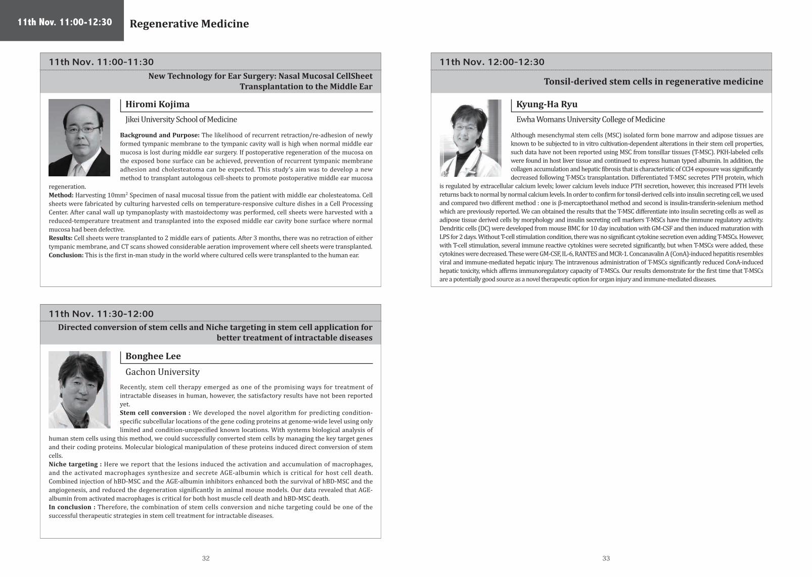
Gachon UniversityRecently, stem cell therapy emerged as one of the promising ways for treatment of intractable diseases in human, however, the satisfactory results have not been reported yet. Stem cell conversion : We developed the novel algorithm for predicting condition-specific subcellular locations of the gene coding proteins at genome-wide level using only limited and condition-unspecified known locations. With systems biological analysis of
human stem cells using this method, we could successfully converted stem cells by managing the key target genes and their coding proteins. Molecular biological manipulation of these proteins induced direct conversion of stem cells.Niche targeting : Here we report that the lesions induced the activation and accumulation of macrophages, and the activated macrophages synthesize and secrete AGE-albumin which is critical for host cell death. Combined injection of hBD-MSC and the AGE-albumin inhibitors enhanced both the survival of hBD-MSC and the angiogenesis, and reduced the degeneration significantly in animal mouse models. Our data revealed that AGE-albumin from activated macrophages is critical for both host muscle cell death and hBD-MSC death. In conclusion : Therefore, the combination of stem cells conversion and niche targeting could be one of the successful therapeutic strategies in stem cell treatment for intractable diseases.
Directed conversion of stem cells and Niche targeting in stem cell application for better treatment of intractable diseases
11th Nov. 11:00-12:30 Regenerative Medicine
11th Nov. 12:00-12:30
Tonsil-derived stem cells in regenerative medicine
Kyung-Ha Ryu
Ewha Womans University College of Medicine
Although mesenchymal stem cells (MSC) isolated form bone marrow and adipose tissues are known to be subjected to in vitro cultivation-dependent alterations in their stem cell properties, such data have not been reported using MSC from tonsillar tissues (T-MSC). PKH-labeled cells were found in host liver tissue and continued to express human typed albumin. In addition, the collagen accumulation and hepatic fibrosis that is characteristic of CCl4 exposure was significantly decreased following T-MSCs transplantation. Differentiated T-MSC secretes PTH protein, which
is regulated by extracellular calcium levels; lower calcium levels induce PTH secretion, however, this increased PTH levels returns back to normal by normal calcium levels. In order to confirm for tonsil-derived cells into insulin secreting cell, we used and compared two different method : one is β-mercaptoethanol method and second is insulin-transferin-selenium method which are previously reported. We can obtained the results that the T-MSC differentiate into insulin secreting cells as well as adipose tissue derived cells by morphology and insulin secreting cell markers T-MSCs have the immune regulatory activity. Dendritic cells (DC) were developed from mouse BMC for 10 day incubation with GM-CSF and then induced maturation with LPS for 2 days. Without T-cell stimulation condition, there was no significant cytokine secretion even adding T-MSCs. However, with T-cell stimulation, several immune reactive cytokines were secreted significantly, but when T-MSCs were added, these cytokines were decreased. These were GM-CSF, IL-6, RANTES and MCR-1. Concanavalin A (ConA)-induced hepatitis resembles viral and immune-mediated hepatic injury. The intravenous administration of T-MSCs significantly reduced ConA-induced hepatic toxicity, which affirms immunoregulatory capacity of T-MSCs. Our results demonstrate for the first time that T-MSCs are a potentially good source as a novel therapeutic option for organ injury and immune-mediated diseases.
11th Nov. 11:00-11:30
New Technology for Ear Surgery: Nasal Mucosal CellSheet Transplantation to the Middle Ear
Hiromi Kojima
Jikei University School of Medicine
Background and Purpose: The likelihood of recurrent retraction/re-adhesion of newly formed tympanic membrane to the tympanic cavity wall is high when normal middle ear mucosa is lost during middle ear surgery. If postoperative regeneration of the mucosa on the exposed bone surface can be achieved, prevention of recurrent tympanic membrane adhesion and cholesteatoma can be expected. This study’s aim was to develop a new method to transplant autologous cell-sheets to promote postoperative middle ear mucosa
regeneration. Method: Harvesting 10mm2 Specimen of nasal mucosal tissue from the patient with middle ear cholesteatoma. Cell sheets were fabricated by culturing harvested cells on temperature-responsive culture dishes in a Cell Processing Center. After canal wall up tympanoplasty with mastoidectomy was performed, cell sheets were harvested with a reduced-temperature treatment and transplanted into the exposed middle ear cavity bone surface where normal mucosa had been defective. Results: Cell sheets were transplanted to 2 middle ears of patients. After 3 months, there was no retraction of either tympanic membrane, and CT scans showed considerable aeration improvement where cell sheets were transplanted. Conclusion: This is the first in-man study in the world where cultured cells were transplanted to the human ear.

34 35
11th Nov. 14:15-14:45
Marco Davila
Venderbilt UniversityWe report on 16 patients with relapsed or refractory B cell acute lymphoblastic leukemia (B-ALL) that we treated with autologous T cells expressing the 19-28z chimeric antigen receptor (CAR) specific to the CD19 antigen. The overall complete response rate is 88%, which allowed us to transition most of these patients to a standard-of-care allogeneic hematopoietic stem cell transplant (allo-SCT). Through systematic analysis of clinical data and serum cytokine levels over the first 21 days after T cell infusion, we have
defined diagnostic criteria for a severe cytokine release syndrome (sCRS), with the goal of better identifying the subset of patients who will likely require therapeutic intervention with corticosteroids or interleukin-6 receptor blockade to curb the sCRS.Additionally, we found that serum C-reactive protein, a readily available laboratory study, can serve as a reliable indicator for the severity of the CRS.
CD19-targeted CAR T cells are effective at mediating remissions in adultswith chemotherapy-refractory B-ALL
11th Nov. 13:45-15:15 CD 19 CAR Sponsored by Medinet
11th Nov. 13:45-14:15 11th Nov. 14:45-15:15
CHIMERIC ANTIGEN RECEPTOR (CAR) MODIFIED T CELLS TARGETED AGAINST RELAPSED/REFRACTORY LEUKEMIAS INDUCE SUSTAINED REMISSIONS
CD19-targeted CAR (chimeric antigen receptor)-expressing T-cell gene therapy for B-cell lymphoma
Bruce Levine
University of Pennsylvania
Keiya Ozawa
The University of Tokyo
While adoptive immunotherapy with T cells has been under investigation for decades, several technologic advances and new strategies have resulted in potent clinical effects. T lymphocytes can be endowed with novel functions ex vivo through gene transfer. By inserting chimeric antigen receptors to redirect T cells to tumor, significant and durable clinical responses in leukemia have been observed in patients who are relapsed or refractory to all other available treatment including stem cell transplantation. A massive in
vivo expansion of chimeric antigen receptor T cells directed against CD19 (CTL019) has been observed, along with homing to disease sites, and long-term functional persistence. In relapsed/refractory Acute Lymphocytic Leukemia, we have observed a 90% Complete Response rate. This technology recently received Breakthrough Designation from the US Food and Drug Administration. Redirection of T cell immunity through this platform technology is now under investigation in other refractory cancers, including solid tumors.
Engineered T-cell therapy with CD19-targeted chimeric antigen receptors (CARs) is promising for treatment of relapsed/refractory B-cell non-Hodgkin lymphoma. Tumor targeting of CAR-expressing T-cells is likely to contribute therapeutic potency. We examined the relationship between the ability of CD19-targeted CAR (CD19-CAR)-transduced T-cells to accumulate at CD19+ tumor lesions, and their ability to provide antitumor effects in xenograft mouse models. Normal human peripheral blood lymphocytes were transduced
with retroviral vectors that encode CD19-CAR and then selectively propagated on the NIH3T3 fibroblasts expressing human CD19. Expanded CD19-CAR T-cells lysed both Raji and Daudi CD19+ human B-cell lymphoma cell lines in a 51Cr release assay. These cells efficiently accumulated at Raji tumor lesions where they suppressed tumor progression and prolonged survival in tumor-bearing immunodeficient mice compared to control cohorts. These results show that the ability of CD19-CAR T-cells to accumulate at tumor lesions is pivotal for their anti-tumor effects in our xenograft models, and therefore may enhance the efficacy of adoptive T-cell gene therapy for relapsed/refractory B-cell lymphoma. Recently, our clinical protocol for CD19-CAR-T gene therapy of malignant lymphoma has been approved by MHLW (Ministry of Health, Labour and Welfare) of Japan.

36 37
11th Nov. 15:45-16:15 11th Nov. 16:45-17:15
11th Nov. 16:15-16:45 11th Nov. 17:15-17:45
Hidetoshi Shibuya
Shibuya KogyoDavid Smith
Lonza
Masahiro Kino-oka
Osaka UniversityYing-Ku Lu
EMO Biomedicine
Since early 1990’s, SHIBUYA developed Isolator technology for the pharmaceutical industry, andsuccessfully installed and validated first system in Japan. The system enables operator access from normal room air conditioned environment into the aseptic space through the half-suit and gloves in order to aseptically assemble Kit-product at that time. We have also delivered Isolator for the operation of ES Cell to Osaka medical center in 2004 and the other several regenerative medicine and cellular therapy purposes until
today. “Bio 3D printer” is one of our automation system to heap up the cell aggregations to 3D structure such as knee cartridge and blood vessel, and “CellPRO” will be the key to next step that is our automatic cell culture system by using aseptic robot.Some of cellular therapies are positively in the clinical study or the commercialization stage nowadays. We hope that our system will support aseptic operation, preparation of GMP documents, systemization of SOP and production management, and lead to safer and more secured study and manufacturing.
Allogeneic cell therapies often require large cell doses for each patient, and when treating diseases with large patient populations, the total number of cells to be manufactured per year at commercial scale can reach into the trillions. It has been challenging to establish a large-scale production platform for adherent cells such as MSCs. However, good progress has been made in developing a bioreactor-based expansion process using microcarrier beads. Process modeling was used for a cost analysis of a bioreactor-based
process compared to planar cultures currently in use. Bioreactor technology, paired with appropriate downstream processing, enabled manufacturing at a scale to reach the trillions of cells needed, at a lower cost than planar cultures. Moreover, costs continue to decrease with increases in cell yield expected as bioreactor processes are further developed and refined.
The serial processes for cell processing affect the quality of the cells, and it can be said that “the process is the product. Thus, the automation is expected not only to maintain an aseptic environment but also to lead to stable processing in cell processing facility (CPF), and cell processing isolators can enable cell processing to be conducted in a closed aseptic environment, which may reduce the equipment and maintenance/operation costs while providing a reliable aseptic environment which reduces product losses and helps
ensure patient safety. In addition, for autologous cell processing, the operators in CPFs are expected to cultivate the cells collected from respective patients parallel (parallel production for multi-patients). Therefore, cell processing isolators (close-chamber system) have the advantage in providing a more reliable aseptic environment with the prevention of cross-contamination. In the present study, a novel isolator system based on a flexible Modular Platform (fMP) is proposed to realize that the individual modules can connect and disconnect flexibly with keeping the aseptic environment in each module, applying the preparation of cell products.
EMO Biomedicine Corporation has long focused on cell-based bioassays and provided testing services to become expert in this field. Recently, we advanced to cell-product manufacturing. The experiences that we gained and the principles that we followed in the testing field can be effectively applied to cell-product manufacturing. The quality of cell products cannot be guaranteed according to the results of quality control (QC) tests; rigorous manufacturing processes must be implemented to ensure quality. Based
on this theory, each step of the manufacturing process can affect the product and therefore should be controlled for quality assurance. The basic tool typically used for quality assurance is the plan–do–check–act (PDCA) cycle. The PDCA cycle can be applied to numerous steps of the manufacturing process to help ensure that the quality of products satisfies the requirements. The examples that we will present to explain the application of the PDCA cycle include_Management of facilities and equipment to provide appropriate environments in separate areas and necessary functions for various manufacturing processes. _Management of personnel to meet the special requirements of sterile-product manufacturing; this type of manufacturing depends highly on the skill, training, and attitudes of personnel. _Development of the QC tests to provide a reliable safeguard and prevent the release of nonconforming products.
Development of Automated Clinical Grade Cell Culture System Bioreactors in Cell Therapy: Manufacturing Scale and Cost Drivers
Cell production system based on flexible modular platform The Quality Assurance of Quality Management System for Cell-BasedProducts -from Testing Lab to Manufacturing Facility
11th Nov. 15:45-18:15 Manufacturing Sponsored by Lonza

38 39
11th Nov. 17:45-18:15
Dirk Balshüsemann
Miltenyi BiotecCellular therapies are being investigated for an increasing range of applications raising the demand for cost effectiveness, automation and standardization of the complex manufacturing processes in cell therapy. An integrated cell-processing device was developed for fully automated GMP-compliant density gradient centrifugation and magnetic cell separations, as well as cell culture, and final formulation. Single-use disposable tubing sets have been designed to ensure a sterile fluid path without the
need for manual sample transfer steps. An integral component of these sets is a new type of centrifugation chamber which also works as a cell culture container. The range of current applications comprises separation of stem cells from bone marrow for tissue regeneration, isolation of multi-virus-specific T cells for adoptive T cell therapy, monocyte isolation and subsequent generation of dendritic cells, active and passive T cell depletion in allogeneic stem cell transplantation, and various options for general processing and fractionation of cellular products. Further applications are in development such as complete workflows for generation and expansion of genetically transduced cells, e.g., CAR T cells.
"Fully automated and standardized cell processing for personalized cell therapy"
Best Abstract Presentation
Best Abstract will be announced on 9th Nov.

40 41
12th Nov. 8:00-8:45 12th Nov. 8:45-9:15
12th Nov.9:15-9:45
Hiromitsu Nakauchi
The University of TokyoYoshihide Esaki
MITI
Yutaka Hishiyama
Japanese Cabinet Secretariat
Recent development of induced pluripotent stem cell (iPSC) technology has enabled generation of PSCs from individual patients, opening up the way to regenerative medicine using the patient’s own PSC-derived cells. However, current stem cell therapy mainly targets diseases that can be treated by cell transplantation. Faced with absolute deficiency of donor organs to treat patients with organ failure, regenerative medicine has as one of its ultimate goals to generate organs using the patient’s own PSCs and
to transplant those organs into the patient. We recently demonstrated in mouse the generation of functionally normal rat pancreas by injecting rat PSCs into Pdx1-/- (pancreatogenesis-disabled) mouse embryos, providing proof of principle for organogenesis from xenogenic PSCs in an embryo unable to form a specific organ. To apply this principle to generate human organs, we need to use larger animals such as pigs. We first generated pig fetuses genetically lacking pancreata. Embryos prepared by somatic cell nuclear transfer using cells from apancreatic pig fetus were then complemented with blastomeres from wild type embryos expressing huKO fluorescent protein to produce chimeric pigs. These chimeric pigs had pancreata and survived to adulthood. The pancreata formed in chimerißc pigs were about the size of human pancreas, functioned normally, and were composed of huKO-positive donor-derived cells. Demonstration of generation of a functional organ from PSCs in pigs is a very important step toward generation of human organs from individual patients’ own PSCs in large animals.
There are high expectations for regenerative medicine in many countries, both as a way to solve various kinds of medical problems and provide better solutions that will help address the budget deficit of national health insurance program by curing chronic diseases. It is crucial for Japan that is world's fastest aging society to improve regenerative medicine. Although there are a plenty of top-level basic life science researches in Japan, practical applications of regenerative medicine are far behind the
United States and Europe.Based on such situation Japan decided to change the legal system to make it more conducive to regenerative medicine. A new legal framework for regenerative medicine will come into force by the end of November. This new framework will make Japan a very attractive market for both R&D and the practical application of regenerative medicine. Japanese government expects that the life science companies will provide the highest quality product at low cost in safety exploiting their sophisticated high technology. In this lecture, I would like to introduce the new legal framework for regenerative medicine including its back ground. In addition, I will explain the measures and policies to cultivate the related industries.
The Government of Japan is strongly promoting to reform the system of medical research and development policy. “Act on Promotion of Healthcare Industries and Advancement of Healthcare Technologies and “Act on the Independent Administrative Agency of Japan Agency for Medical Research and Development” were approved by the Diet in May 2014 after intensive discussions and deliberations.
Researchers in academia are likely to contribute to the development of new drugs with cutting edge technologies. In the field of stem research a lot of scientists have come up with excellent results such as iPS cells. Medical Doctors in the field of regenerative medicine have addressed cell therapy to treat difficult deseases..
In the new system we strengthen to bridge between bed and bench. Though basic researches in Japan have produced a lot of quality papers, the results often have not been utilized in clinical researches nor industries due to lack of management.
The Japan Agency for Medical Research and Development (AMED), which will be newly established and start its business on April 1, 2015, is expected to play roles as a funding agency and a bridge between academia and industry.
“From Cells to Organs” – Generation of Functional Organs from Pluripotent Stem Cells Establishing A Legal Framework Conducive To Practical Use Of Regenerative Medicine
The New Initiative for Medical Research and Development in Japan
12th Nov. 8:00-8:45 12th Nov. 8:45-10:15Plenary Session 3iPS Cell for Organ Regeneration
Support for Cellular Therapyby Japanese Government

42 43
12th Nov. 9:45-10:15
Toshio Miyata
Health and Global Policy InstituteWith a specialized approval system for regenerative medicine the effectiveness of treatments can be anticipated, and if proven safe they will go on to have a global impact. Currently, Dr. Masayo Takahashi of the Riken Kobe Institute conducted a first-in-human trial in patients with age-related macular degeneration, a serious condition of the retina in 2014. As global competition in regenerative medicine intensifies there is clearly a need for stronger public-private frameworks to support projects like this. With Japan's new
structure for regenerative medicine, it is necessary for relevant ministries to create a framework for more unified support and to merge their related budgets, make an effort to hire the best personnel, and create exit strategies for projects while managing their progress.
Next steps the government needs to take
12th Nov. 10:15-10:30
Kellathur Srinivasan
HSA/SingaporeSrinivasan Kellathur received his Bachelors and Masters in Biochemistry from India; and a PhD from the National University of Singapore. He was a Postdoctoral Fellow at the Department of Anatomy, National University of Singapore and then a Postdoctoral Fellow and Research Associate at the Department of Pharmacology and Molecular Sciences, Johns Hopkins University School of Medicine and Division of Biomedical Sciences, Johns Hopkins in Singapore. He joined Health Sciences Authority Singapore in 2007
as a Senior Regulatory Specialist, Advanced Therapy Products Unit, Health Products Regulation Group, Health Sciences Authority and is currently the Head of the unit. As Head, he has been involved in the development of policies, guidelines, processes and framework for regulation of human cell- and tissue-based therapeutic products in Singapore. He is also member of the Executive Committee, Asian Cellular Therapy Organization, Member of Ministry of Health’s Advisory Committee on Biobanking and Member of the Advisory Board, APEC Harmonization Centre for Cellular Therapies.
12th Nov. 10:30-11:45Regulatory Session
12th Nov. 10:30-10:45
Ryousuke Maruyama
PMDA

44 45
12th Nov. 11:00-11:15 12th Nov. 11:30-11:45
12th Nov. 11:15-11:30
Won Shin
Korean FDAMaria Chrstina Gali
EMA
Ying-Hsien Fu
Taiwan FDA
Dr. Galli holds a University degree in Biological Sciences and a PhD in Molecular Medicine; currently she is in-staff senior researcher, Cell Biology and Neurosciences Department, Istituto Superiore di Sanità, Roma, Italy. Dr. Galli was active for more than 25 yrs as basic researcher in experimental oncology, cellular biology, molecular immunology, with 60 publications on international journals. Dr. Galli has been active for the past 20 yrs in the field of translational medicine as quality assessor for gene therapy and biotechnology
medicines in national as well as European procedures; she has also been active for the past 18 yrs as GMP and GLP inspector. Dr. Galli was member of CAT/EMA in 2009-2011 and chair of CAT/EMA Gene Therapy Working Party in 2009-2012, in which she participated since its first meeting; she also participated in the EMA inspectors/experts group on revision of GMP annex 2 guideline. Dr. Galli is currently co-chair of the ATMP platform in the European infrastructure for translational medicine EATRIS-ERIC. Dr. Galli’s main expertise is in regulatory sciences for translational medicine (as shown by 20 yrs experience and latest publications) supported by scientific education and research experience in experimental oncology, cellular biology, molecular immunology (as shown by most of
Ying-Hsien received her master in Medical Technology from National Taiwan University. In 2006, she joined Bureau of Food and Drug Analysis (former TFDA). She was the reviewer and analyst in Section of Blood Products and In Vitro Diagnostics, Division of Drug Biology. She has extensive working experience in review of BLA/PMA submissions. Also, she was an active member of GTP /Human Organ Bank inspection cadre, participating in advanced therapy drug product inspections.
In 2010, Ying-Hsien joined Division of Drug and New Biotechnology Products (former Division of Medicinal Products). Her primary responsibility now is to promote the biotech industry development in Taiwan and to enact legislation governing biotech products (“The Promotion Plan for the Biotech Industry”). Also, she formulates the regulations and compliance policy relating to new drugs and biologics.
Ministry of Health and Welfare Food and Drug AdministrationDivision of Medicinal Products

4746
Cardiosphere-derived cells (CDCs) isolated from postnatal heart have been shown to be a convenient and an efficient source for therapeutic treatment of myocardial infarction. However, poor retention of CDCs in the infarcted heart often causes insufficient therapeutic outcomes. In this study, we combined cell-sheet technology with CDCs to prolong the survival of grafted cells in the infarcted heart to obtain better cardiac regeneration after implantation. A mouse model of a chronically infarcted heart was established by ligation of left anterior descending artery (LAD) at 1 month before sheet implantation. Mouse CDC monolayer sheet was delivered onto infarcted area, and the therapeutic effects on the left ventricular (LV) function and morphology were estimated at 4 weeks following sheet implantation. On the basis of macroscopic analysis, CDC monolayer sheets remained intact on infarcted heart for at least 4 weeks after implantation. The LV ejection fraction (LVEF) and the LV fractioning shortening (LVFS) were significantly improved in the sheet-implanted hearts as well as an increase of anterior wall thickness of left ventricle. Moreover, we also investigated whether hypoxic pretreatment increases therapeutic efficacy of CDC sheet on old myocardial infarction. Mouse CDC monolayer sheet was pre-incubated in 2% oxygen for 24 hours and then delivered into infarcted heart. Hypoxic pretreatment significantly increased secretion of vascular endothelial growth factor (VEGF) from CDC sheet through the PI3K-Akt signaling pathway, resulting in higher LVEF and LVFS in the infarcted heart following implantation than the group without hypoxic pretreatment. These results indicate that hypoxic preconditioning is potential “booster shot” for CDC sheet and the combination of cell sheet and hypoxic preconditioning may become novel therapeutic strategy for myocardial infarction.
Cell culture processing is one of the most important steps to expand human induced pluripotent stem cells (hiPSCs). For industrialization, it is essential to develop low cost cell manufacturing processes under high safety and stability. 2-dimentional (2D) cell culture of hiPS with planar culture dish, disposable culture bag etc., or several types of 3-dimentional (3D) culture with suspension culture vessel are known as a scalable cultivation. In the present study, to estimate culture efficiency or contents of the costs, 2D cultivation in the planar culture dish with manual operation, the culture bag with automated passaging system, and 3D aggregate suspension culture were performed. After seeding, cell density of hiPS in the vessel at t = 0 h, Xi, and t = 24 h, X24, were measured with a hematocytometer after enzymatic treatment and trypan blue exclusion. Cell-attachment ratio in 2D culture or cell viability to form aggregate in 3D, α (-), was estimated as the ratio of X24 to Xi. At t = 48 h, the live cell density, X48, was counted to calculate apparent specific growth rate, μApp (h-1). The planar culture, bag culture and aggregate suspension culture were α = 78, 78 and 56, μ = 0.033 h-1, 0.029 h-1 and 0.019 h-1 respectively. The culture costs in each method were estimated from α, μApp, labor costs, costs for experimental reagents and disposable vessels. Cost for suspension culture was significantly reduced due to less of extracellular matrix which is indispensable in the 2D culture. The suspension culture system and the bag culture system could reduce labor cost, suggesting that these bioreactors are promising tools for iPSC expansion.
Wounded tissues and cells may be treated with growth factors and specific genes for the purpose of tissue repair and regeneration. To deliver specific genes into tissues and cells, this study presents the use of fabricated poly (DL-lactic-co-glycolic acid) (PLGA) nanoparticles (NPs) complexed with the cationic polymer poly (ethleneimine) (PEI). Through complexation with PEI, several types of genes (SOX9, Cbfa1, and C/EBP-α) were complexed with PLGA NPs, which enhanced gene uptake into normal human-derived dermal fibroblast cells (NFDHCs)in vitro andin vivo. Several cell types (293T, HeLa, and fibroblast cells) were transfected with fluorescence-tagged PEI/SOX9, PEI/Cbfa1, and PEI/ C/EBP-α gene-complexed PLGA NPs. The gene and protein expression levels in the cells were evaluated by RT-PCR, real-time quantitative PCR, Western blotting, and confocal laser microscopy. Fibroblast cells encapsulated in fibrin gels were transfected with the gene-complexed NPs plus specific growth factors (TGF-b 3, BMP-2, or IGF/bFGF), which induced chondrogensis, osteogenesis, or adipogenesis both in vitro and after transplantation into nude mouse.
Cha Award for Best Abstract Selection Committee
Chair Kyung-Ha Ryu,Korea Udomsak Bunworasate,Thailand Abbas Ghaderi, Iran Mickey Koh,Singapore Kaiyan Liu, China Yuji Heike, Japan
12th Nov. 11:45-12:45
Tohru Hosoyama / Yamaguchi University Graduate School of Medicine
hiPSC culturing cost reduction effect by the cell culture apparatus introduction
Transfection of specific genes into normal human-derived dermal fibroblast cells for direct conversion
Hypoxic preconditioning enhances therapeutic efficacy of cardiosphere-derived cell sheet on chronically infarcted heart
12th Nov. 11:45-12:45
No.1
No.2
No. 3
Panel Discussion on TopicsPoster Information
Hideki Kitajima / Graduate school of Engineering, Osaka University.
Keun-Hong Park / CHA University

4948
Purpose: To investigate a possible therapeutic mechanism for cell therapy in the field of neurological disorders using G-CSF-mobilized peripheral blood mononuclear cells (mPBMCs), we compared the expression of inflammatory cytokines and neurotrophic factors in PBMCs and mPBMCs and cord blood (CB). Methods: We evaluated the intracellular expression of neurotrophic factors and inflammatory cytokines in PBMCs and mPBMCs from 14 CP children and 14 healthy adult volunteer donors as well as CB mononuclear cells donated from healthy newborns. Both PBMC collected prior to G-CSF administration and apheresed mPBMC were cryopreserved. We performed flow cytometric analysis with intracellular staining for various cytokines after thawing PBMCs and mPBMCs as well as CBs. Results: No significant differences in expression of neurotrophic factors were found between PBMC and mPBMC. However, in cells from CP children, the expression of IL-6 was significantly increased in mPBMC as compared to PBMC, and IL-3 was significantly decreased in mPBMC as compared to PBMC. In healthy adults, the expression of both IL-1β and IL-6 were significantly increased in mPBMC as compared to PBMC. The expression of BDNF in mPBMC from CP children was significantly higher than in CB or mPBMC from healthy adults. The expression of G-CSF in mPBMC from CP children was comparable to that in CB but significantly higher than in mPBMC from healthy adults. Lower expression of IL-1β, IL-3, and IL-6 and higher expression of IL-8 and IL-9 was observed from CB and mPBMCs from CP children rather than in healthy adults. Lower expression of IL-1β, and higher expression of IL-8 was observed from mPBMCs from CP children rather than in healthy adults.Conclusion: The altered expression of neurotrophic factors and anti-inflammatory cytokines in mPBMC in CP children and CB from healthy children could provide a new potential source for cellular therapy for individuals with neurologic diseases.
Mesenchymal stem cell (MSC) therapies hold great potential to treat a wide range of diseases and tissue defects. MSCs are site regulated and secrete trophic factors at various concentrations in response to local surrounding microenvironmental cues, which is critical for tissue regeneration. A systematic study of regenerative potential of trophic factors released from different MSCs, however, has not been performed. We have previously demonstrated significantly larger volume of regenerated pulp tissue by transplantation of dental pulp MSCs, CD31- side population (SP) cells compared to transplantation of bone marrow and adipose MSCs from the same individual in ectopic tooth transplantation models for pulp regeneration. Thus, in the present study, pulp regenerative potential of conditioned medium (CM) of these three populations from the same individual was also compared in an ectopic tooth transplantation model. Transplantation of pulp CM yielded higher volume of pulp regeneration, higher BrdU-positive migrated cells from surrounding tissues and less Caspase 3-positive cells in the regenerated pulp compared to bone marrow and adipose CM. Significantly higher activities of pulp CM on migration, anti-apoptosis and angiogenesis were also demonstrated in C2C12 myoblasts. Up-regulated genes in pulp CD31- SP cells compared with bone marrow and adipose CD31- SP cells, CXCL14, MCP1 and IL6, were identified as candidate trophic factors by microarray analysis and real-time RT-PCR. The stimulatory effects on migration and angiogenesis of CXCL14 and MCP1 and anti-apoptosis of IL6 were demonstrated in vitro. The BrdU-positive migrated cells expressed CXCR4 and CCR2, cognate receptors of CXCL14 and MCP1, respectively, and most of IL6-positive cells were Caspase 3-negative in the regenerated tissue. These results demonstrate that higher regenerative potential of pulp SP cells may be due to trophic effects on migration, angiogenesis and anti-apoptosis by potent trophic factors including CXCL14, MCP1 and IL6.
Regeneration of maxillofacial bone defects such as maxillary alveolar clefts and bone resorption resulting from marginal periodontitis pose a significant clinical challenge. We focused on the new cell transplantation technique in which bone marrow-derived mesenchymal stem cells were cultured as cell sheets with osteogenic potential (designated osteogenic matrix cell sheets; OMCSs). This study was performed to evaluate the usefulness of OMCSs in maxillofacial regeneration, using the mandibular nonunion model in rats.We obtained bone marrow cells by flushing out the femoral shafts of Fischer 344 rats with medium. The released cells were collected in the culture flasks containing regular medium supplemented with 15% fetal bovine serum and cultured. After reaching confluence, the primary cultured cells were released from the culture substratum using trypsin–EDTA. To prepare OMCSs, released cells were subcultured with osteoinductive medium until confluence and lifted as a cell sheet using a scraper. Different from human mandible, rat mandible is formed by a paired bone and the central portion consists of fibrous tissue. Therefore it is interpreted as a nonunion bone defect. In the experimental group (five rats), the defects were filled with OMCSs. In the control group (five rats), the mandibles were sham-operated. Eight weeks after implantation, micro computed tomography and histological analyses were taken. Both analyses demonstrated that bone union of the mandible occurred in all rats of experimental group, but in none of those of control group.These results suggest that OMCSs may regenerate the maxillofacial bone. The characteristics of maxillofacial bone defects are small and low load-bearing, but are quite complicated, especially compared with bone defects in the orthopedic surgery field. OMCSs are characterized by a good operability and shaping property, thus they may be an optimal application for maxillofacial regeneration.
Young-Ho Lee / Hanyang University
The tissue specificity of trophic factors from mesenchymal stem cells (MSCs) responsible for distinct regenerative potential
0steogenic potential of the osteogenic matrix cell sheets in maxillofacial regeneration
Intracellular expression of neurotrophic factors in mononuclear cells of cord blood and G-CSF-mobilized peripheral blood as a potential source for cellular therapyNo.4
No.5
No.6
No.7
Yuki Hayashi / Aichi-Gakuin University
Yoshihiro Ueyama / Nara Medical University
Treatment of severe burns remains a challenge where there are insufficient donor sites. Use of cultured epithelial autografts (CEAs) is one technology developed to address this problem. Currently, CEA culture involves the expansion of skin keratinocytes (SKs) on lethally-irradiated murine feeder cells in the presence of bovine serum - for optimal growth and stem cell maintenance. Although no major adverse events were reported in clinical practice for 30 years, this technology is restricted to critically-ill patients due to presence of animal products. Here, we evaluated the performance of SKs cultured on a chemically-defined, serum-free (SF) and feeder-free culture system developed in-house (SGH-NBF system) compared to the conventional culture system (Green’s system). The newly-developed SF medium (NB medium) was initially screened on several protein coatings known to support SK growth. These proteins include collagen I, collagen IV, gelatin, fibronectin and laminin. Fibronectin was found to be most conducive for SK growth and this combination was used to compare with Green’s system. SKs (n=5) cultured in these 2 systems were compared in terms of growth, morphology, colony forming efficiency (CFE), stem cell phenotype, genetic stability and ability to form a fully stratified epidermis on organotypic cultures. SKs in both systems were observed to form colonies within 5-7 days and reach confluency within 9-10 days. SKs in the 2 groups exhibited comparable average CFEs [14% ± 0.4% (Green’s) versus 18% ± 1.1% (SGH-NBF)] with no observable differences in the expressions of epidermal stem cell and basal cell markers. The karyotype of SKs at each passage (up to passage 12) cultured in both systems was observed to be normal. Finally, SKs in both systems were able to form a fully stratified epidermal layer on a dermal equivalent. The data augur well for a xeno-free system to culture good quality CEA in future therapeutic application.
Alvin Chuak / Singapore General Hospital
Evaluation of a xeno-free and chemically defined culture system to expand skin keratinocytes for potential therapeutic use
Tissue engineering techniques, including cytokines and bio-scaffolds, were used to regenerate simple tissues such as skin and bone, but complicated organs such as kidney or liver are not achieved yet. For tissue regeneration, spatial and temporal expression of cytokines and their roles are determined recent years, however, appropriate extracellular matrix (ECM) is still under discussion. Changes of original ECM structures and shifts of collagen types during kidney development was reported, indicated that renal generation in vitro may be regulated by plural types and structures of collagen. Therefore, in vivo ECM expression may be very useful information for tissue generation in vitro. Here we suggest that type V collagen (Col V) fibril, a minor component in ECM, may be used as a new niche for kidney regeneration. Col V was reported to play roles in cell migration and detachment, which provide micro-environment for cell detachment and migration during tissue development and wound healing. In this study, we clarified the spatial and temporal expressions of Col V fibril structures during kidney development in vivo, and cultured mouse embryonic kidneys with Col V fibrils in vitro. In vivo results shows that higher Col V fibrils, some co-exist with Col I, are observed in immature/developing renal tissues including uretic buds, metanephric mesenchymes, new-forming renal vessels, and glomeruli, and decreases in mature areas. In vitro results indicated that, compared to control samples, Col V fibrils provide better micro-environment for cell mobility and outgrowth extension during the earlier stage of kidney culture. In later stage of culture, better nephron formation and extended ureteric bud elongation were observed. These results indicated that Col V fibrils may provide cell mobility, regulate Col I fibrils formation, and support tubulogenesis, which may be used as a pioneer bio-scaffold for kidney organogenesis in vitro.
Bisphosphonate-related osteonecrosis of the jaw (BRONJ) is a severe complication in patients who receive bisphosphonates. Recent studies suggested that intravenous infusion of multipotent mesenchymal stromal cells (MSC) was effective for bone exposure in BRONJ-like disease models. However, it is suggested that the stability of injected MSC at the diseased area is relatively low, resulting in pulmonary embolism or even death in some cases. To overcome the problem, our laboratory developed “Cell Sheet Engineering” using temperature-responsive culture dishes in which intact cells and extracellular proteins can be harvested as a sheet by simple temperature reduction. In this study, we investigated the effect of MSC sheet transplantation on a BRONJ-like disease model in rats. Zoledronate and dexamethasone were administered to SD rats and maxillary first molars were extracted to induce BRONJ-like disease. MSC were isolated from bone marrow of EGFP SD rats. MSC at passage 3 were seeded on temperature-responsive culture dishes (UpCell®) and cultured for 7 days to produce cell sheets. After surgical debridement, MSC sheets were transplanted to the lesion of bone exposure (transplant group) or sutured only (control group). Two weeks after the transplantation, the transplant group showed wound healing in most cases (87.5%; 7/8). In contrast, the control group showed the presence of bone exposures without soft tissue (80%; 12/15). Micro CT analysis showed bone regeneration in the extraction socket of the transplant group. TRAP staining showed that the number of osteoclasts was significantly increased in the transplant group (25.67±3.38) compared the control group (7.00±5.13)(p<0.05). Immunohistochemical analysis showed that EGFP- positive cells were observed in the transplanted sub-mucosa 2 weeks after the transplant. The number of blood vessels in the transplant group was significantly higher than the natural mucosal healing (p<0.05). MSC sheet transplantation can be an alternative approach to treat BRONJ.
Backgrounds: Transplantation of autologous somatic tissue-derived cells into the heart has been shown to yield functional recovery of the failing heart via “paracrine effects” that enhance the native regenerative process. We have introduced skeletal myoblast cell (SMBc) sheet methods for treating severe heart failure, in which scaffold-free cell-sheets are attached on the epicardial surface to maximize the paracrine effects. In this study, we investigated the safety and efficacy of SMBc sheet implantation.Methods: This study enrolled fifteen patients (mean age, 48±14 years, 8 DCM and 7 ICM patients) with severe heart failure despite the maximum medical and/or interventional therapies. Scaffold-free cell-sheets containing SMBc (average: 3.5±1.9×10⁸ cells) was transplanted over the LV free wall via the left thoracotomy.Results: All patients were discharged without procedure-related mortalities or major complications. Their NYHA classification and 6-minute walk test were significantly improved 6 months after treatment (NYHA class: 3.2±0.7 vs. 2.3±1.1 p<0.01, 6MW: 476±148 vs. 547±102m p<0.05). LV reverse remodeling was achieved in 8 of 15 patients, which was more frequent in ICM etiology (37.5% vs. 71.4% p=0.19). Improvement of systolic function was observed in 5 of 15 patients (EF:34.4±7.0 vs. 40.8±8.3% p<0.05), which was correlated with decreasing of regional end-systolic wall stress. The freedom from cardiac mortality was 90.0% at 3 years and the freedom from recurrence of heart failure was 78.8% at 3 years. The incidence of heart failure was significantly decreased after SMBc sheet implantation (1.11 vs. 0.24 event/year p<0.05).Conclusions: SMBc sheet implantation was safe and effective for improvement of symptom, recurrence of heart failure and reverse remodeling. It has a potential to improve both systolic and diastolic function in selected patients. Promising results in functional recovery warrant further clinical follow-up and accumulation of the cases to prove the therapeutic efficacy of this treatment for severe heart failure.
Han-Hsiu Hsu / University of Tsukuba
Allogeneic multipotent mesenchymal stromal cell (MSC) sheet transplantation for bisphosphonate-related osteonecrosis of the jaw in rats
Safety and Efficacy of Cardiac Regenerative Therapy with Skeletal Myoblast Cell Sheet Implantation for Severe Heart Failure
Type V Collagen Fibrils Applied as Bio-scaffold for Renal Tissue RegenerationNo.8
No.9
No.10
No.11
Nobuyuki Kaibuchi / Tokyo Women's Medical University School of Medicine
Keitaro Domae / Osaka University, Graduate school of Medfical
[Objectives] Safe and Stable cryopreservation is very important in clinical application of human iPS cells. DMSO is a useful cryoprotective agent, but it has cytotoxicity and influence on cell differentiation those are not ignorable. We reported that we developed anti-freeze polyamino acid based cryopreservation agents (CPLL-CPA) of slow-freezing hiPS cells, which was xeno-free and contained 6% DMSO (ISSCR, Boston, 2013). In this study, we used the same component CPLL-CPA at that time, but reduced DMSO to 5%, and tested pluripotency of cryopreserved hiPS cells.[Methods] CPLL-CPA contained carboxylated-poly-L-lysine, recombinant human serum albumin, 5%DMSO and basal medium. Cryopreserved hiPS cells with CPLL-CPA were thawed and cultured about 10 passages, and observed their morphology, differentiated colonies, and tested alkaline phosphatase (AP) staining, FACS analysis and RT-PCR profiles for their pluripotency. We further added hydroxyethyl starch (HES) or conditioned medium for raising cryopreservation efficacy. [Result] The iPS cells cryopreserved with newly developed cryopreservation liquid showed the same clear cytomorphology as the iPS cells which used 10% of DMSO as a control. And their AP staining and FACS stained against Tra-1-60 were also showed their pluripotency, but we couldn’t find remarkable agents with additional cryopreservation effect, we should search for other reagents which has high effecacy. [Conclusion] Slow freezing is essential for iPS-cell culture without feeder cells, and for clinical application and cell therapy, we developed 5%DMSO-contained CPLL-CPA, and it was a useful tool for slow freezing. We tried to reduce DMSO concentration, but hiPS cells were very weak at cryopreservation step without DMSO. We need further survey in order to overcome this problem.
Akemi Ota / Kyoto University
Improvement of anti-freeze polyamino acid based cryopreservation agents for slow-freezing hiPS-cells
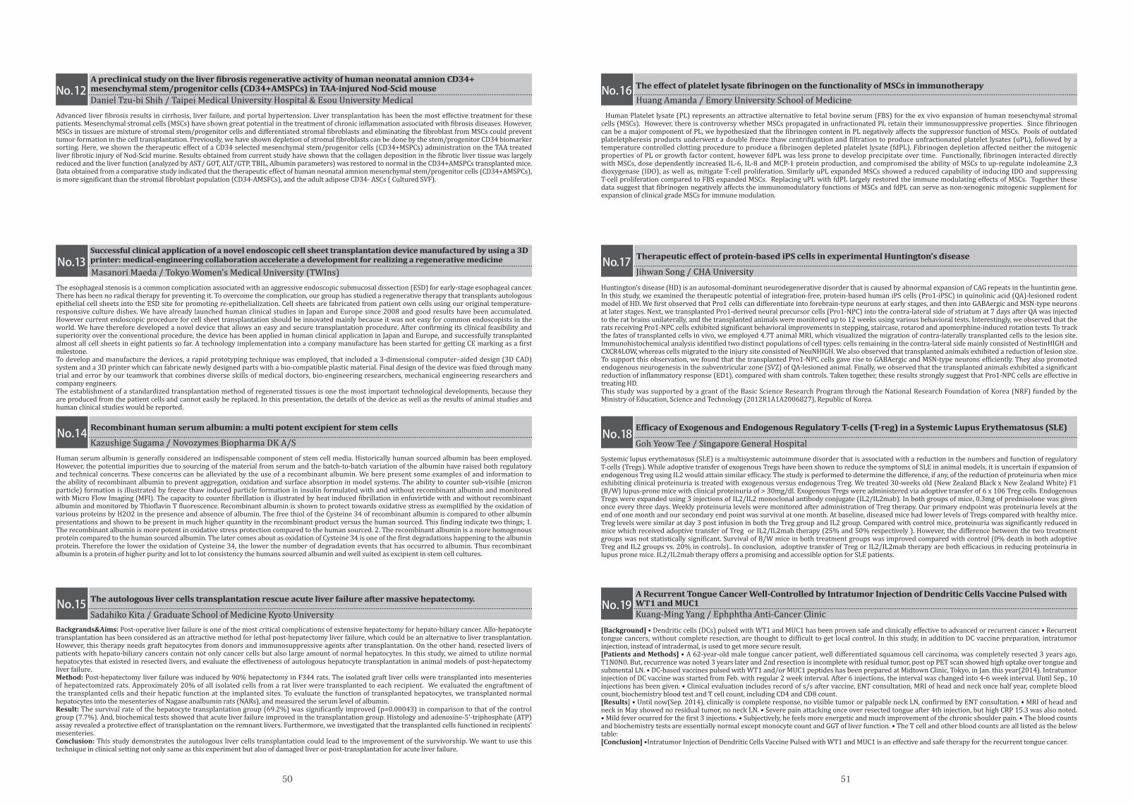
5150
Human serum albumin is generally considered an indispensable component of stem cell media. Historically human sourced albumin has been employed. However, the potential impurities due to sourcing of the material from serum and the batch-to-batch variation of the albumin have raised both regulatory and technical concerns. These concerns can be alleviated by the use of a recombinant albumin. We here present some examples of and information to the ability of recombinant albumin to prevent aggregation, oxidation and surface absorption in model systems. The ability to counter sub-visible (micron particle) formation is illustrated by freeze thaw induced particle formation in insulin formulated with and without recombinant albumin and monitored with Micro Flow Imaging (MFI). The capacity to counter fibrillation is illustrated by heat induced fibrillation in enfuvirtide with and without recombinant albumin and monitored by Thioflavin T fluorescence. Recombinant albumin is shown to protect towards oxidative stress as exemplified by the oxidation of various proteins by H2O2 in the presence and absence of albumin. The free thiol of the Cysteine 34 of recombinant albumin is compared to other albumin presentations and shown to be present in much higher quantity in the recombinant product versus the human sourced. This finding indicate two things; 1. The recombinant albumin is more potent in oxidative stress protection compared to the human sourced. 2. The recombinant albumin is a more homogenous protein compared to the human sourced albumin. The later comes about as oxidation of Cysteine 34 is one of the first degradations happening to the albumin protein. Therefore the lower the oxidation of Cysteine 34, the lower the number of degradation events that has occurred to albumin. Thus recombinant albumin is a protein of higher purity and lot to lot consistency the humans sourced albumin and well suited as excipient in stem cell cultures.
Backgrands&Aims: Post-operative liver failure is one of the most critical complications of extensive hepatectomy for hepato-biliary cancer. Allo-hepatocyte transplantation has been considered as an attractive method for lethal post-hepatectomy liver failure, which could be an alternative to liver transplantation. However, this therapy needs graft hepatocytes from donors and immunosuppressive agents after transplantation. On the other hand, resected livers of patients with hepato-biliary cancers contain not only cancer cells but also large amount of normal hepatocytes. In this study, we aimed to utilize normal hepatocytes that existed in resected livers, and evaluate the effectiveness of autologous hepatocyte transplantation in animal models of post-hepatectomy liver failure. Method: Post-hepatectomy liver failure was induced by 90% hepatectomy in F344 rats. The isolated graft liver cells were transplanted into mesenteries of hepatectomized rats. Approximately 20% of all isolated cells from a rat liver were transplanted to each recipient. We evaluated the engraftment of the transplanted cells and their hepatic function at the implanted sites. To evaluate the function of transplanted hepatocytes, we transplanted normal hepatocytes into the mesenteries of Nagase analbumin rats (NARs), and measured the serum level of albumin. Result: The survival rate of the hepatocyte transplantation group (69.2%) was significantly improved (p=0.00043) in comparison to that of the control group (7.7%). And, biochemical tests showed that acute liver failure improved in the transplantation group. Histology and adenosine-5'-triphosphate (ATP) assay revealed a protective effect of transplantation on the remnant livers. Furthermore, we investigated that the transplanted cells functioned in recipients’ mesenteries. Conclusion: This study demonstrates the autologous liver cells transplantation could lead to the improvement of the survivorship. We want to use this technique in clinical setting not only same as this experiment but also of damaged liver or post-transplantation for acute liver failure.
Recombinant human serum albumin: a multi potent excipient for stem cells
The autologous liver cells transplantation rescue acute liver failure after massive hepatectomy.
No.14
No.15
Kazushige Sugama / Novozymes Biopharma DK A/S
Sadahiko Kita / Graduate School of Medicine Kyoto University
Human Platelet lysate (PL) represents an attractive alternative to fetal bovine serum (FBS) for the ex vivo expansion of human mesenchymal stromal cells (MSCs). However, there is controversy whether MSCs propagated in unfractionated PL retain their immunosuppressive properties. Since fibrinogen can be a major component of PL, we hypothesized that the fibrinogen content in PL negatively affects the suppressor function of MSCs. Pools of outdated plateletpheresis products underwent a double freeze thaw centrifugation and filtration to produce unfractionated platelet lysates (uPL), followed by a temperature controlled clotting procedure to produce a fibrinogen depleted platelet lysate (fdPL). Fibrinogen depletion affected neither the mitogenic properties of PL or growth factor content, however fdPL was less prone to develop precipitate over time. Functionally, fibrinogen interacted directly with MSCs, dose dependently increased IL-6, IL-8 and MCP-1 protein production, and compromised the ability of MSCs to up-regulate indoleamine 2,3 dioxygenase (IDO), as well as, mitigate T-cell proliferation. Similarly uPL expanded MSCs showed a reduced capability of inducing IDO and suppressing T-cell proliferation compared to FBS expanded MSCs. Replacing uPL with fdPL largely restored the immune modulating effects of MSCs. Together these data suggest that fibrinogen negatively affects the immunomodulatory functions of MSCs and fdPL can serve as non-xenogenic mitogenic supplement for expansion of clinical grade MSCs for immune modulation.
Advanced liver fibrosis results in cirrhosis, liver failure, and portal hypertension. Liver transplantation has been the most effective treatment for these patients. Mesenchymal stromal cells (MSCs) have shown great potential in the treatment of chronic inflammation associated with fibrosis diseases. However, MSCs in tissues are mixture of stromal stem/progenitor cells and differentiated stromal fibroblasts and eliminating the fibroblast from MSCs could prevent tumor formation in the cell transplantation. Previously, we have shown depletion of stromal fibroblasts can be done by the stem/progenitor CD34 biomarker sorting. Here, we shown the therapeutic effect of a CD34 selected mesenchymal stem/progenitor cells (CD34+MSPCs) administration on the TAA treated liver fibrotic injury of Nod-Scid murine. Results obtained from current study have shown that the collagen deposition in the fibrotic liver tissue was largely reduced and the liver function (analyzed by AST/ GOT, ALT/GTP, TBIL, Albumin parameters) was restored to normal in the CD34+AMSPCs transplanted mice. Data obtained from a comparative study indicated that the therapeutic effect of human neonatal amnion mesenchymal stem/progenitor cells (CD34+AMSPCs), is more significant than the stromal fibroblast population (CD34-AMSFCs), and the adult adipose CD34- ASCs ( Cultured SVF).
Systemic lupus erythematosus (SLE) is a multisystemic autoimmune disorder that is associated with a reduction in the numbers and function of regulatory T-cells (Tregs). While adoptive transfer of exogenous Tregs have been shown to reduce the symptoms of SLE in animal models, it is uncertain if expansion of endogenous Treg using IL2 would attain similar efficacy. The study is performed to determine the difference, if any, of the reduction of proteinuria when mice exhibiting clinical proteinuria is treated with exogenous versus endogenous Treg. We treated 30-weeks old (New Zealand Black x New Zealand White) F1 (B/W) lupus-prone mice with clinical proteinuria of > 30mg/dl. Exogenous Tregs were administered via adoptive transfer of 6 x 10⁶ Treg cells. Endogenous Tregs were expanded using 3 injections of IL2/IL2 monoclonal antibody conjugate (IL2/IL2mab). In both groups of mice, 0.3mg of prednisolone was given once every three days. Weekly proteinuria levels were monitored after administration of Treg therapy. Our primary endpoint was proteinuria levels at the end of one month and our secondary end point was survival at one month. At baseline, diseased mice had lower levels of Tregs compared with healthy mice. Treg levels were similar at day 3 post infusion in both the Treg group and IL2 group. Compared with control mice, proteinuria was significantly reduced in mice which received adoptive transfer of Treg or IL2/IL2mab therapy (25% and 50% respectively ). However, the difference between the two treatment groups was not statistically significant. Survival of B/W mice in both treatment groups was improved compared with control (0% death in both adoptive Treg and IL2 groups vs. 20% in controls).. In conclusion, adoptive transfer of Treg or IL2/IL2mab therapy are both efficacious in reducing proteinuria in lupus prone mice. IL2/IL2mab therapy offers a promising and accessible option for SLE patients.
[Background] • Dendritic cells (DCs) pulsed with WT1 and MUC1 has been proven safe and clinically effective to advanced or recurrent cancer. • Recurrent tongue cancers, without complete resection, are thought to difficult to get local control. In this study, in addition to DC vaccine preparation, intratumor injection, instead of intradermal, is used to get more secure result.[Patients and Methods] • A 62-year-old male tongue cancer patient, well differentiated squamous cell carcinoma, was completely resected 3 years ago, T1N0N0. But, recurrence was noted 3 years later and 2nd resection is incomplete with residual tumor, post op PET scan showed high uptake over tongue and submental LN. • DC-based vaccines pulsed with WT1 and/or MUC1 peptides has been prepared at Midtown Clinic, Tokyo, in Jan. this year(2014). Intratumor injection of DC vaccine was started from Feb. with regular 2 week interval. After 6 injections, the interval was changed into 4-6 week interval. Until Sep., 10 injections has been given. • Clinical evaluation includes record of s/s after vaccine, ENT consultation, MRI of head and neck once half year, complete blood count, biochemistry blood test and T cell count, including CD4 and CD8 count.[Results] • Until now(Sep. 2014), clinically is complete response, no visible tumor or palpable neck LN, confirmed by ENT consultation. • MRI of head and neck in May showed no residual tumor, no neck LN. • Severe pain attacking once over resected tongue after 4th injection, but high CRP 15.3 was also noted. • Mild fever ocurred for the first 3 injections. • Subjectively, he feels more energetic and much improvement of the chronic shoulder pain. • The blood counts and biochemistry tests are essentially normal except monocyte count and GGT of liver function. • The T cell and other blood counts are all listed as the below table:[Conclusion] •Intratumor Injection of Dendritic Cells Vaccine Pulsed with WT1 and MUC1 is an effective and safe therapy for the recurrent tongue cancer.
Huang Amanda / Emory University School of MedicineDaniel Tzu-bi Shih / Taipei Medical University Hospital & Esou University Medical
Efficacy of Exogenous and Endogenous Regulatory T-cells (T-reg) in a Systemic Lupus Erythematosus (SLE)
A Recurrent Tongue Cancer Well-Controlled by Intratumor Injection of Dendritic Cells Vaccine Pulsed with WT1 and MUC1
The effect of platelet lysate fibrinogen on the functionality of MSCs in immunotherapyA preclinical study on the liver fibrosis regenerative activity of human neonatal amnion CD34+ mesenchymal stem/progenitor cells (CD34+AMSPCs) in TAA-injured Nod-Scid mouse No.16No.12
No.17No.13
No.18
No.19
Goh Yeow Tee / Singapore General Hospital
Kuang-Ming Yang / Ephphtha Anti-Cancer Clinic
Huntington’s disease (HD) is an autosomal-dominant neurodegenerative disorder that is caused by abnormal expansion of CAG repeats in the huntintin gene. In this study, we examined the therapeutic potential of integration-free, protein-based human iPS cells (Pro1-iPSC) in quinolinic acid (QA)-lesioned rodent model of HD. We first observed that Pro1 cells can differentiate into forebrain-type neurons at early stages, and then into GABAergic and MSN-type neurons at later stages. Next, we transplanted Pro1-derived neural precursor cells (Pro1-NPC) into the contra-lateral side of striatum at 7 days after QA was injected to the rat brains unilaterally, and the transplanted animals were monitored up to 12 weeks using various behavioral tests. Interestingly, we observed that the rats receiving Pro1-NPC cells exhibited significant behavioral improvements in stepping, staircase, rotarod and apomorphine-induced rotation tests. To track the fates of transplanted cells in vivo, we employed 4.7T animal MRI, which visualized the migration of contra-laterally transplanted cells to the lesion site. Immunohistochemical analysis identified two distinct populations of cell types: cells remaining in the contra-lateral side mainly consisted of NestinHIGH and CXCR4LOW, whereas cells migrated to the injury site consisted of NeuNHIGH. We also observed that transplanted animals exhibited a reduction of lesion size. To support this observation, we found that the transplanted Pro1-NPC cells gave rise to GABAergic and MSN-type neurons efficiently. They also promoted endogenous neurogenesis in the subventricular zone (SVZ) of QA-lesioned animal. Finally, we observed that the transplanted animals exhibited a significant reduction of inflammatory response (ED1), compared with sham controls. Taken together, these results strongly suggest that Pro1-NPC cells are effective in treating HD. This study was supported by a grant of the Basic Science Research Program through the National Research Foundation of Korea (NRF) funded by the Ministry of Education, Science and Technology (2012R1A1A2006827), Republic of Korea.
The esophageal stenosis is a common complication associated with an aggressive endoscopic submucosal dissection (ESD) for early-stage esophageal cancer. There has been no radical therapy for preventing it. To overcome the complication, our group has studied a regenerative therapy that transplants autologous epithelial cell sheets into the ESD site for promoting re-epithelialization. Cell sheets are fabricated from patient own cells using our original temperature-responsive culture dishes. We have already launched human clinical studies in Japan and Europe since 2008 and good results have been accumulated. However current endoscopic procedure for cell sheet transplantation should be innovated mainly because it was not easy for common endoscopists in the world. We have therefore developed a novel device that allows an easy and secure transplantation procedure. After confirming its clinical feasibility and superiority over the conventional procedure, the device has been applied in human clinical application in Japan and Europe, and successfully transplanted almost all cell sheets in eight patients so far. A technology implementation into a company manufacture has been started for getting CE marking as a first milestone.To develop and manufacture the devices, a rapid prototyping technique was employed, that included a 3-dimensional computer–aided design (3D CAD) system and a 3D printer which can fabricate newly designed parts with a bio-compatible plastic material. Final design of the device was fixed through many trial and error by our teamwork that combines diverse skills of medical doctors, bio-engineering researchers, mechanical engineering researchers and company engineers.The establishment of a standardized transplantation method of regenerated tissues is one the most important technological developments, because they are produced from the patient cells and cannot easily be replaced. In this presentation, the details of the device as well as the results of animal studies and human clinical studies would be reported.
Jihwan Song / CHA UniversityMasanori Maeda / Tokyo Women's Medical University (TWIns)
Therapeutic effect of protein-based iPS cells in experimental Huntington’s diseaseSuccessful clinical application of a novel endoscopic cell sheet transplantation device manufactured by using a 3D printer: medical-engineering collaboration accelerate a development for realizing a regenerative medicine
5352
NK cell-based adoptive immunotherapy is a promising approach for the treatment of cancer, and is expected to apply for the combination therapies with therapeutic antibodies, such as Trastuzumab and Cetuximab. However, there are few efficient methods to provide a large amount of functional NK cells and evaluate their in vivo ADCC activities. We developed a large-scale NK-cell expansion method using a combination of RetroNectin® induced T-cells (RN-T cells), OK-432 and IL-2(RN-T cells are defined as T cells expanded by the co-stimulation of anti-CD3mAb and RetroNectin®). In this method, we investigated the NK-cell expansion from 31 of cancer patients, using PBMCs and autologous plasma from 20-40mL of blood as start materials. As results, 688±76-fold expansion was achieved in this system and the proportion of CD3-CD56+ NK cells in the expanded cells was 84.7±3.6%. To confirm the significance of RN-T cells as stimulator, we compared the effects on NK-cell proliferation between RN-T cells and aCD3-T cells which expanded by anti-CD3 mAb only. As a result, the stimulation by RN-T cells led to preferable NK-cell proliferation rather than the one by aCD3-T cells. In addition, almost all expanded cells highly expressed functional markers such as NKG2D and CD16 implicated in cytotoxicity and ADCC activity. Subsequently, we assessed in vivo ADCC activity on HER2-positive gastric cancer tumor cells, NCI-N87 bearing hIL-2 Tg NOG mice (supplied by CIEA) model. Combination treatment with the expanded NK cells plus Trastuzumab almost completely suppressed the tumor growth, while modest activities were observed in the case of each treatment alone. The infused NK cells were maintained in peripheral blood during an observation period of about 3 months and also infiltrated into the tumor tissues. Thus, this expansion method is an innovative tool for cancer immunotherapy and evaluation of in vivo ADCC activity of therapeutic antibodies.
Background: There are limited choices for the treatment among advanced cancer patients who failed and/or untolerated to conventional anti-cancer treatments. Therefore this study aimed to deploy the benefits and clinical efficacy of continuous dendritic cells-cytokine induced killer cells infusions. Methods: A total of 381 infusions (from 67 advanced cases recruited) were included in this study. All patients were undertaken peripheral blood mononuclear cells apheresis for the following cellular therapy and dendritic cells-cytokine induced killer cells (DC-CIKs) were expanded in vitro. Peripheral blood T lymphocyte subsets were quantified through flow cytometry to reflect the cellular immunity status. Clinical efficacy was evaluated by RECIST criteria and ECOG statuses were recorded. Logistic regression model was used to estimate the association between cellular infusions and clinical benefits.Results: Average 5.7±2.94×10⁹ of induced cells were infused each time and patients were exposed to 6 infusions. Of 67 cases 2 patients (3.0%) reached partial response, 26 patients (38.8%) remained stable, 39 cases (58.2%) progressed, and the disease control rate was 41.8%. Cell immunity was improved that CD8+CD28+T lymphocytes were increased by 74% and CD8+CD28-T lymphocytes were inclined by 16% (p<0.05). Continuous infusions of DC-CIKs were associated with improvement of both ECOG scores and CD8+CD28-T lymphocytes (p<0.05). Median 6 infusions were capable of reducing risk of progression by 70% (95% CI 0.10-0.91).Conclusions: To the advanced cancer patients, continuous DC-CIKs infusions were capable of recovering the cellular immunity, ECOG score and improving clinical efficacy.
Dendritic cells (DCs) are well known as the most potent professional antigen presenting cells (APCs). Nonetheless, the use of these cells in immunotherapy has been limited due to the time consuming and laborious steps required to generate DCs from monocytes in vitro. Therefore, alternative APCs has drawn much attention. In this study, the efficacy of B cells as APCs in induction of cytotoxic T lymphocytes (CTLs) against Epstein-Barr virus (EBV)-specific antigens was evaluated. B cells were isolated by depletion of peripheral blood mononuclear cell (PBMCs) from healthy individuals with MACS system, loaded with α-galactosylceramide (α-GalCer) for inducing B cell activation, and nucleofected with EBV-antigen coding plasmid DNA, pCK-E1dGLMP2-IRES-BZLF1. Ag-nucleofected B cells were cocultured with T cells for 14 days in vitro. The cells were harvested and subsequently immunoassayed. Proliferation of cells was expanded 1.93 folds in EBV-CTLs induced by B cells. Immunogenicity of CTLs were identified by IFN-γ ELISPOT (Enzyme-linked immunospot) assay and cytotoxicity assay. The CTLs induced by α-GalCer-loaded B cells induced powerful cytotoxicity against EBV antigen in vitro. The EBV-CTLs by α-GalCer-loaded B cells recognized EBV antigen LMP2 (median 121 SFC/10^5) and BZLF1 (median 112 SFC/10^5). The EBV-CTLs by α-GalCer-loaded B cells have killing activity against EBV antigen LMP2 (100%, at E:T ratio 10:1) and BZLF1 (100%, at E:T ratio 10:1). These observations suggest that α-GalCer-loaded B cells could be used in general as APCs. Using the B cells as APCs have several benefits such as cost-effectiveness, less time-consuming, and less laborious compared to when DCs are used. Furthermore, nucleofection technique might be useful in delivering antigen-coding DNA, not only for virus antigens but also for tumor antigens, directly into the nucleus. Our results demonstrate that α-GalCer-loaded B cells could be potent APCs in generating EBV-specific CTLs for cellular vaccines and adoptive immunotherapy.
We have previously reported the therapeutic potential of rat fetal membrane(FM)-derived mesenchymal stem cells (MSCs) using various rat models including hindlimb ischemia, autoimmune myocarditis, glomerulonephritis, renal ischemia-reperfusion injury, and myocardial infarction. In this study, 1) we isolated and characterized MSCs from human amnion and chorion; 2) we examined their differences in the expression profile of growth factors and cytokines; and 3) we investigated the therapeutic potential and difference of these MSCs using murine hindlimb ischemia and acute graft-versus-host disease (GVHD) models. Isolated MSCs from both amnion and chorion layers of FM showed similar morphological appearance, multipotency, and cell-surface antigen expression. Conditioned media obtained from amnion- and chorion-derived MSCs inhibited cell death caused by serum starvation or hypoxia in endothelial cells and cardiomyocytes. Amnion and chorion MSCs secreted significant amounts of angiogenic factors including HGF, IGF-1, VEGF, and bFGF, although differences in the cellular expression profile of these soluble factors were observed. Transplantation of human amnion or chorion MSCs significantly increased blood flow and capillary density in a murine hindlimb ischemia model. In addition, compared to human chorion MSCs, human amnion MSCs markedly reduced T-lymphocyte proliferation with the enhanced secretion of PGE2, and improved the pathological situation of a mouse model of GVHD disease. Our results highlight that human amnionand chorion-derived MSCs, which showed differences in their soluble factor secretion and angiogenic/immuno-suppressive function, could be ideal cell sources for regenerative medicine.
Mitsuko Ideno / Takara Bio INC. Xinna Zhou / Capital Medical University(CMU)
Hyoung Jin Kang / Seoul National University College of Medicine Kenichi Yamahara / National Cerebral and Cardiovascular Center
A robust and selective expansion method for functional NK cells and its application for in vivo ADCC evaluation system
Continuous DC-CIK infusions sufficiently restore CD8+ cellular immunity and improve ECOG status in advanced cancers patients failed and untolerated to conventional anti-cancer treatments
Generation of EBV-specific cytotoxic T lymphocytes with activated B cellsAngiogenic, cytoprotective, and immunosuppressive properties of human amnion- and chorion-derived mesenchymal stem cells
No.20 No.24
No.22 No.26
No.21 No.25
No.23 No.27
The inhibitory activities of the Cordyceps pruinosa butanol fraction (Cp-BF) were investigated by determining inflammatory responses of lipopolysaccharide (LPS)-treated RAW264.7 macrophage cells and by evaluating HCl/ethanol (EtOH)-triggered gastric ulcers in mice. The molecular mechanisms of the inhibitory effects of Cp-BF were investigated by identifying target enzymes using biochemical and molecular biological approaches. Cp-BF strongly inhibited the production of NO and TNF-α, release of reactive oxygen species (ROS), phagocytic uptake of FITC-dextran, and mRNA expression levels of interleukin (IL)-6, inducible NO synthase (iNOS), and tumour necrosis factor-alpha (TNF)-α in activated RAW264.7 cells. Cp-BF also strongly downregulated the NF-κB pathway by suppressing IKKβ according to luciferase reporter assays and immunoblot analysis. Furthermore, Cp-BF blocked both increased levels of NF-κB-mediated luciferase activities and phosphorylation of p65/p50 observed by IKKβ overexpression. Finally, orally administered Cp-BF was found to attenuate gastric ulcer and block the phosphorylation of IκBα induced by HCl/EtOH. Therefore, these results suggest that the anti-inflammatory activity of Cp-BF may be mediated by suppression of IKKα and its downstream NF-κB activation. Since our group has established the mass cultivation conditions by developing culture conditions for Cordyceps pruinosa, the information presented in this study may be useful for developing new anti-inflammatory agents.
Although the introduction of high-dose therapy with hematopoietic stem cell transplantation and the development of novel molecular targeting agents have resulted in a marked improvement in overall survival, multiple myeloma (MM) still remains incurable. Alternative approaches are clearly needed to prolong both disease-free and overall survival of patients with MM. Cellular immunotherapy with dendritic cells (DCs) is emerging as a useful immunotherapeutic tool to treat MM. Ex vivo-generated DCs can be loaded with myeloma-associated antigens as vaccines for patients with MM. The use of immature DCs or mature DCs, method used to induce DC maturation, types of tumor antigens, techniques used to load tumor antigens onto DCs, routes of administration, and dosing schedules are being investigated. Our group tried to develop the potent DCs having a capacity of high IL-12p70 production and high migratory character for inducing effective tumor-specific type I immune responses. In this presentation, I will show how the efficacy of DC therapy in MM can be improved. In addition, I will briefly present to our ongoing phase I/IIa clinical trial using DCs in patients with relapsed or refractory MM.
ntroduction: The purpose of this study were to determine antitumor efficacy of γδ+ T cells in children with hematologic malignancy after αβ+ T-cell-depleted haploidentical hematopoietic stem cell transplantation (haplo-HSCT).
Methods: Ten children (6 males, 4 females, median age 12.3 years) with hematologic malignancy received haplo-HSCT after ex vivo depletion of αβ+ T cells between May 2012 and December 2013 at AMCCH. Of 10 patients, 2 had ALL (1 in CR2, 1 in CR3), 5 had AML [2 CR1, 2 CR2, 1 non-remission (NR)], 2 had JMML (1 CR1, 1 NR), and 2 had NHL (1 CR2 and 1 CR3). The median number of CD34+, αβ+, γδ+ and CD3+ cells infused was 10.6 x 10⁶/Kg, 6.3 x 10⁵/Kg, 2.1 x 10⁷/Kg and 22.9 x 10⁶/Kg, respectively.
Results: All 10 patients engrafted and 1 patient experienced grade IV acute GVHD without treatment-related mortality. At a median follow-up period of 387 days (range, 208 - 766), 5 patients (2 ALL, 2 AML, 1 NHL) relapsed at the median 131 days (range, 42-234) after haplo-HSCT. The median dose of infused γδ+ T cell for 5 patients without relapse and 5 with relapse were 2.2 x 10⁷/Kg and 1.8 x 10⁷/Kg. The medianproportion of γδ+ T cells after HSCT was higher in non-relapsed patients than in relapsed patients (69.4% vs 29.3% at 1 month, 44.2% vs 6.2% at 2 months and 36.8% vs 9.1% at 3 months post-transplant). At a median follow-up of 12 months probability of 1 year relapse-free survival was 48.0%, and probability of 1 year overall survival was 78.8%.
Conclusion: These findings suggest a correlation between an increase in the proportion of γδ+ T cells and improved relapse-free survival after αβ+ T-cell-depleted haplo-HSCT. However, further studies including prospective large numbered trial are needed to confirm our results.
Adoptive transfer of T cells expressing a chimeric antigen receptor (CAR) is a promising cell-based anticancer therapy. Although clinical studies of this approach show therapeutic efficacy, additional genetic modification is necessary to enhance the efficacy and safety of CAR-T cells. For example, producing an antitumor cytokine from CAR-T cells can enhance their tumor-killing activity, but there are concerns that constitutive expression of anticancer molecules will cause systemic side effects. Therefore, it is important that exogenous gene expression is confined to the tumor locality. Such an approach may lead to therapeutic strategies that are safe and effective. In this study, we aimed to develop a switch promoter driven by activation signals from a CAR. We prepared a switch cassette that was arranged in the order of two modified SV40 early polyA sequences as a background reduction signal, four NFAT-responsive elements, a minimal interleukin-2 (IL-2) promoter, ZsGreen1, and a BGH polyA sequence. Transgene expression in peripheral blood mononuclear cells (PBMCs) transduced with the CD19-targeted CAR and switch cassette (PBMCs/CAR/iReporter) was only induced strongly by co-culture with CD19-positive target cells. Furthermore, after antigen stimulation, PBMCs/CAR/iReporter produced approximately the same amounts of IL-2 and interferon-γ (IFN-γ) as PBMCs expressing the CAR only. The cells also showed redirected cytolysis toward CD19-positive, but not CD19-negative, tumor cells. These results indicated that the switch promoter was selectively driven by activation signals from the CAR. Furthermore, transduction with the switch cassette did notaffect the original effector activity including IL-2 and IFN-γ production and antitumor activity of CAR-redirected cytotoxic T lymphocytes. In summary, we developed a retroviral vector that incorporates a CAR-derived, activation signal-dependent promoter to drive exogenous gene expression. This switch cassette permits visualization and quantification of the activation status in CAR-expressing PBMCs.
Sungjae Yang / Sungkyunkwan University Je-Jung Lee / Chonnam National University Hwasun Hospital
Kyung-Nam Koh / University of Ulsan, College of Medicine Ryosuke Uchibori / Jichi Medical University
Anti-Inflammatory Activities of a Butanol Fraction of Cordyceps pruinosa Fruit Bodies Cellular immunotherapy using dendritic cells against multiple myeloma: from bench to clinic
Clinical Implication of γδ+ T Cell Recovery after αβ+ T-Cell-Depleted HaploidenticalHematopoietic Cell Transplantation in Children with Hematologic Malignancy
Analysis of function and response specificity of a switch promoter driven by activation signals from a CD19-targeted chimeric antigen receptor.
5554
A stable cell aseptic processing method that lead to both high-quality and low-cost will be a significant technological breakthrough. Isolators, as an alternative for biological safety cabinets, are notable technology and provide high-quality manufacturing environment. However, achievement of stable quality and cost-saving using the isolator for the current manual processing methods is difficult. We have tried two approaches to construct of cell manufacturing and processing system for industrialization of regenerative medicine. One is the mechanization/automation for manual operations, and another is developing a novel method for process assembly, called a "flexible modular platform" (fMP). We had already designed and fabricated a first prototype of an automated production system, called a "Tissue Factory" (T-Factory). A T-Factory adopted a modular architecture by fMP. A key technique of fMP is the aseptic connection interfaces between modules which can be decontaminated so that they can be joined in a single process. We have tried a full-scale manufacturing of multi-layered myoblast sheets from a skeletal muscle tissue of a swine, consisting of automated processes of cell isolation, cell expansion, and cell sheet forming/stacking. In this presentation, we would report details of the trial production processes. This research is funded by grants from Funding Program for World-Leading Innovative R&D on Science and Technology (FIRST), and New Energy and Industrial Technology Development Organization (NEDO), grant number P14006.
Flavonoids are the plant pigments that have been revealed to have various physiological effects including anti-cancer, anti-bacterial and anti-inflammatory activities. Fisetin, a polyphenolic flavonoid, which is widely found in many fruits and vegetables such as strawberry, mango and onion. Several groups reported anti-inflammatory activity of fisetin, but it is not well studied with regard to how fisetin regulates the inflammatory signaling pathway. To evaluate immunomodulatory properties of fisetin, we checked the production of nitric oxide (NO), one of the pro-inflammatory mediators. This compound significantly inhibited NO production in RAW264.7 cells without affecting their cell viability. In addition, mRNA expression levels of pro-inflammatory genes, such as inducible nitric oxide synthase(iNOS), cyclooxygenase-2 (COX-2), and pro-inflammatory cytokines including tumor necrosis factor α(TNF-α). Results showed that of iNOS, COX-2 and TNF-α were also decreased. Additionally, the translocation of transcription factor p65, a subunit of NF-κB, into the nucleus was remarkably inhibited. Futhermore, the phosphorylation levels of inflammatory signaling proteins (IκBα and Src), upstream regulatory molecules of p65, was decreased under fisetin-treated condition. Therefore, these results suggest that fisetin can be considered as a bioactive immunomodulatory compound with anti-inflammatory property. Further studies will include detail mechanistic understanding by using reporter gene luciferase assay and immunoprecipitation as well as several in vivo experiments including peritonitis and hepatitis to judge its total anti-inflammatory efficacy
Luteolin is flavonoids which are polyphenolic compounds found in numerous plants. It exhibits anti-inflammatory, anti-tumor, antioxidant properties and reduces cardiovascular disease risks. However, the molecular mechanism of action against inflammation in RAW 264.7 cells is only partially explored. The effect of the Luteolin on the production of inflammatory mediators [nitric oxide (NO) and prostaglandin E2 (PGE2)] and the molecular mechanism of Luteolin-mediated inhibition, including target enzymes, were studied with RAW264.7. Luteolin clearly inhibited the production of NO and PGE2 in lipopolysaccharide (LPS)-activated RAW264.7 cells and in a dose-dependent manner. According to RT-PCR and immunoblotting analyses and a kinase assay with mRNA, whole cell extract, and nucleus lysates from RAW264.7 cells, it was revealed that Luteolin was capable of suppressing the activation of both nuclear factor (NF)-kB by directly targeting Syk/Src. Luteolincould have anti-inflammatory properties by suppressing Syk/Src/NF-kB pathways and will be further developed as a herbal remedy for preventive and/or curative purposes in various inflammatory diseases.
Manabu Mizutani / Graduate School of Engineering, Osaka University Junho Kim / Sungkyunkwan University
Jeong Deok / Sungkyunkwan University
Trial productions of multi-layered myoblast sheets containing a primary and an expansion culture by an automation system for future clinical practice in regenerative medicine Anti-inflammatory effects of fisetin by inhibition of Src kinase activity
Luteolin modulates inflammatory response on LPS-mediated macrophage
No.28 No.32
No.30
No.29 No.33
No.31
While human pluripotent stem cells are attractive sources for regenerative medicine, a major concern remains regarding their tumorigenic potential. Safety assessment of human pluripotent stem cell-based products in terms of tumorigenicity is thus critical. Through comprehensive glycome analysis using high-density lectin microarray, we have identified a pluripotent stem cell-specific lectin probe rBC2LCN recognizing hyperglycosylated podocalyxin as a cell surface ligand. We found that hyperglycosylated podocalyxin is secreted from human pluripotent stem cells into cell culture supernatants. We established a sandwich assay system, named the GlycoStem test, targeting the soluble hyperglycosylated podocalyxin using rBC2LCN. The GlycoStem test is sufficiently sensitive and quantitative to detect residual human pluripotent stem cells using cell culture supernatants. This work provides a proof of concept for the noninvasive and quantitative detection of tumorigenic human pluripotent stem cells using cell culture supernatants. The developed method should increase the safety of human pluripotent stem cell-based cell therapies.Ref: Tateno et al. Scientific Reports 2014
Mitochondrial DNA (mtDNA) is an extranuclear and multicopy genome that is essential for mitochondrial energy production. Mutations in mtDNA cause a wide spectrum of human diseases. The A3243G mutation is the most common mtDNA mutation and causes 80% of all cases of MELAS (mitochondrial myopathy, encephalopathy, lactic acidosis, and stroke-like episodes), which is progressive and fatal. However, there is no fundamental cure for MELAS so far.In patients with MELAS, wild-type (normal) and mutated mtDNA coexist in the same cells. The energy generation is impaired under certain threshold levels of wild-type mtDNA. Therefore, the selective increase of wild-type mtDNA levels above the threshold can be a definitive therapeutic strategy for the patients.The A3243G mutation is located in mitochondrial transcription termination sequence called a “tridecamer sequence” within mTERF (mitochondrial transcription termination factor) binding site. In theory, the inhibition with the interaction between mTERF and wild-type sequence was thought to promote wild-type mtDNA replication by unlocking replication arrest adjacent to mTERF binding region.In the present study, a molecular structure of pyrrole-imidazole polyamide, an artificial DNA-binding compound, was designed to target the tridecamer sequence of wild-type mtDNA and synthesized (ML1 polyamide). The data showed that ML1 polyamide permeated cell membrane and mitochondrial lipid bilayers in no need of special drug delivery systems and inhibited the mTERF binding to the mtDNA sequence in living cells.In subsequent experiments using human cybrid cells carrying the A3243G mutation (MELAS model cells), the treatment of the MELAS cybrid cells with ML1 polyamide over two weeks successfully demonstrated a significant increase of wild-type mtDNA levels without cell toxicity.We believe that the strategy of wild-type mtDNA replication activation over mutant mtDNA leads to a promising therapeutics and ML1 polyamide can be a realistic curative drug for patients with the MELAS A3243G gene mutation.
4-isopropyl-2,6-bis(1-phenylethyl)aniline 1 (KTH13-AD1) is a metabolite of Cordyceps bassiana that has been traditionallyused to treat various inflammatory disease. Even though it has the magnificent pharmaceutical potential, there was not much understanding on its anti-inflammatory actions. Therefore, this study was aimed to determine the anti-inflammatory effects of KTH13-AD1. We found that KTH13-AD1 suppressed nitric oxide (NO) and reactive oxygen species (ROS) production in lipopolysaccharide (LPS)- or sodium nitroprusside (SNP)-treated macrophages (RAW264.7 cells). Similarly, mRNA expression of inducible NO synthase (iNOS) and tumor necrosis factor-α (TNF-α) analyzed by RT-PCR and real-time PCR was also reduced by this compound. Interestingly, KTH13-AD1 also strongly diminished the levels of NF-κB-mediated luciferase activities and nuclear translocated NF-κB family proteins. In agreement with these, KTH13-AD1 suppressed the upstream signaling pathway for NF-κB activation including IκBα, IKKα/β, AKT, p85/PI3K and Src in time- and dose-dependent manners. Therefore, these results strongly suggest that KTH13-AD1 has a strong anti-inflammatory activity via suppression of the NF-κB signaling pathway.
Hiroaki Tateno / National Institute of Advanced Industrial Science and Technology (AIST) Takamitsu Yano / Ibaraki Prefectural University of Health Sciences
Yong Kim / SungKyunKwan University
Development of a noninvasive and quantitative detection system of tumorigenic human pluripotent stem cells using cell culture supernatants
Synthetic pyrrole-imidazole polyamide targeting mitochondrial transcription termination sequence demonstrated effectiveness of increasing levels of normal mitochondrial DNA by a selective its replication activation over mutated DNA in mitochondrial disease MELAS cybrid cells
4-isopropyl-2,6-bis(1-phenylethyl)aniline 1 Originated from Cordyceps bassiana in MacrophagesHas Anti-inflammatory Activities

56 57

58 59
Sponsors
Accelrys K.K.COREFRONT CorporationDAIICHI SANGYO Co., LTD.DS Pharma Biomedical Co., Ltd.EMO Biomedicine Corp.FUKOKU CO.,LTD.JX Nippon Oil & Energy CorporationKohjin Bio Co., Ltd.MEDICAL & BIOLOGICAL LABORATORIES CO., LTD.NIKON ENGINEERING CO., LTD.
Alfresa Pharma CorporationAsahi Kasei Pharma Corp.ASKA Pharmaceutical. Co., Ltd.Astellas Pharma Inc.AstraZeneca K.K.Bayer Yakuhin, Ltd.Bristol-Myers SquibbChugai Pharmaceutical Co., Ltd.DAIICHI SANKYO COMPANY, LIMITEDEIKEN CHEMICAL CO., LTD.,Eisai Co., Ltd.Eli Lilly Japan K.K. ELMED EISAI Co., Ltd. Fuso Pharmaceutical Industries,Ltd.GlaxoSmithKline K.K.Japan Tobacco Inc.KAKEN PHARMACEUTICAL Co., Ltd.KISSEI PHARMACEUTICAL CO., LTD.Kowa Company, Ltd.Kracie Pharma, Ltd.KYORIN Pharmaceutical Co., Ltd.Kyowa Hakko Kirin Co., Ltd.Maruho Co.,Ltd.Maruishi Pharmaceutical Co.LtdMeiji Seika Pharma Co., LtdMinophagen Pharmaceutical Co., Ltd.Mitsubishi Tanabe Pharma CorporationMOCHIDA PHARMACEUTICAL CO., LTD.MSD K.K.NIHON PHARMACEUTICAL CO., LTD.Nippon Boehringer Ingelheim Co ., Ltd.Nippon Chemiphar Co., Ltd.Nippon Kayaku Co., Ltd.
NIPPON SHINYAKU CO.,LTD.Nippon Zoki Pharmaceutical Co., Ltd.NIPRO PHARMA CORPORATIONNovartis Pharma K.K.ONO PHARMACEUTICAL CO., LTD.OSAKA GOVERNMENT TOURISM BUREAUOtsuka Pharmaceutical Co., Ltd.Otsuka Pharmaceutical Factory, Inc.Pfizer Japan Inc.ROHTO Pharmaceutical Co., Ltd.Sanofi K.K. Santen Pharmaceutical Co.,Ltd.SANWA KAGAKU KENKYUSHO CO., LTD.SATO PHARMACEUTICAL CO.,LTD. Sawai Pharmaceutical Co.,Ltd.SHIONOGI & CO., LTD.SSP Co., Ltd.Sumitomo Dainippon Pharma Co., Ltd. TAIHO PHARMACEUTICAL CO., LTD.Taisho Pharmaceutical Co., Ltd.Takara Bio Inc.Takeda Pharmaceutical Company Limited. TEIJIN PHARMA LIMITED. Terumo CorporationTOA EIYO LTD.TORII PHARMACEUTICAL CO., LTD.TOWA PHARMACEUTICAL CO., LTD.TOYAMA CHEMICAL CO., LTD.Tsumura & Co.Wakamoto Pharmaceutical Co., Ltd.YAKULT HONSHA CO., LTD.Zeria Pharmaceutical Co., Ltd.
FUJIFILM Corporation MINERVA TECH K.K. (In alphabetical order)
Sponsorship
Exhibition
Donation
Advertisement
Nippi incorporatedSCREEN HOLDINGS CO.,LTD.SHIBUYA KOGYO CO., LTD.SHIN NIPPON BIOMEDICAL LABORATORIES,LTD.Sony Corporation STREX, Inc. SUMITOMO BAKELITE CO., LTD.WAKENBTECH CO.,LTD.
