assessment of contemporary root canal … · (endodontics). chapel hill ... chapter 1 review of...
TRANSCRIPT

ASSESSMENT OF CONTEMPORARY ROOT CANAL FILE SYSTEMS: SIZES, SHAPES
AND THE EFFECTIVENESS OF 31-GAUGE IRRIGATION NEEDLE IN INTRACANAL
BACTERIAL REDUCTION
Melita Islambasic
A thesis submitted to the faculty at the University of North Carolina at Chapel Hill in partial
fulfillment of the requirements for the degree of Master of Science in the School of Dentistry
(Endodontics).
Chapel Hill
2015
Approved by:
Peter Z. Tawil
Roland Arnold
Mary Pettiette

ii
©2015
Melita Islambasic
ALL RIGHTS RESERVED

iii
ABSTRACT
Melita Islambasic: Assessment of Contemporary Root Canal File Systems: Sizes, Shapes and the
Effectiveness of 31-Gauge Irrigation Needle In Intracanal Bacterial Reduction
(Under the direction of Peter Z. Tawil)
In Part I, the effects of irrigant delivered via a 31-gauge irrigation needle to E. faecalis
infected teeth instrumented to different apical sizes with six different nickel titanium (NiTi)
rotary instruments were examined in an in-vitro model using 165 single canaled extracted teeth.
In Part II, the accuracy and deformation of NiTi rotary files before and after use were examined
via SEM imaging and ImageJ software. Statistical analysis was done using r-ANOVA and
Tukey’s test for Part I, and proportional odds model and Mantel-Haenszel row mean score for
Part II. Smaller preparation sizes showed higher likelihood of having a larger bacterial count
compared to larger preparation sizes. Most files’ nominal diameter and taper did not conform to
their advertised size showing a lack of standardization between manufacturers. Pilot tip lengths
vary greatly between different rotary files.

iv
ACKNOWLEDGEMENTS
I would like to acknowledge my awesome mentors and committee members, Dr. Peter Z.
Tawil, Dr. Roland Arnold and Dr. Mary Pettiette and thank them for their guidance and support
throughout my research. I would also like to thank Dr. Ceib Phillip and Mr. Adane Wogu for
their assistance with statistical analysis, Mr. Eric Simmons for his assistance in the lab and Mr.
Wallace Ambrose for his assistance with the SEM imaging.

v
TABLE OF CONTENTS
LIST OF TABLES ........................................................................................................................ vii
LIST OF FIGURES ..................................................................................................................... viii
LIST OF ABBREVIATIONS ........................................................................................................ ix
CHAPTER 1: REVIEW OF LITERATURE ................................................................................. 1
INTRODUCTION ....................................................................................................................... 1
REFERENCES ............................................................................................................................ 7
CHAPTER 2 MANUSCRIPT #1: ASSESSMENT OF CONTEMPORARY ROOT CANAL
FILE SYSTEMS: SIZES, SHAPES AND THE EFFECTIVENESS OF 31-GAUGE
IRRIGATION NEEDLE IN INTRACANAL BACTERIAL REDUCTION .............................. 11
INTRODUCTION ..................................................................................................................... 11
EXPERIMENTAL METHOD .................................................................................................. 13
Extracted teeth preparation .................................................................................................... 13
Bacterial suspension and root infection ................................................................................. 14
Pilot study .............................................................................................................................. 14
Experimental method ............................................................................................................. 16
Statistical analysis.................................................................................................................. 17
RESULTS.................................................................................................................................. 17
DISCUSSION ........................................................................................................................... 19
REFERENCES .......................................................................................................................... 22
CHAPTER 3 MANUSCRIPT #2: NOMINAL SIZE AND TAPER ANALYSIS OF NOVEL
METALLURGY NiTi FILES ....................................................................................................... 25

vi
INTRODUCTION .................................................................................................................... 25
MATERIALS AND METHODS .............................................................................................. 26
RESULTS.................................................................................................................................. 29
DISCUSSION ........................................................................................................................... 33
REFERENCES .......................................................................................................................... 37

vii
LIST OF TABLES
Table
1.1. Brands and sizes of NiTi rotary files used ............................................................ 16
1.2. Pairwise comparison of odds ratios. A 20.04, B 20.07,
C 25.06, D 25.08, E 30.06, F 35.06 ...................................................................... 18
2.1. Diameter values (before and after) compared to the advertised
nominal size. ......................................................................................................... 29
2.2. Percentage of files within the ANSI/ADA and NRG (new
recommended guideline) diameter tolerance limit. Numbers in
parenthesis show percentage of files below and above the tolerance
limit, respectively. ................................................................................................. 30
2.3. Taper values (before and after) compared to the advertised nominal
taper sizes. ............................................................................................................. 31
2.4. Percentage of files within the ANSI/ADA and NRG taper tolerance
limit. Numbers in parenthesis show percentage of files below and
above the tolerance limit, respectively. ................................................................. 32
2.5. True pilot tip (TPT) lengths (before and after) ..................................................... 33

viii
LIST OF FIGURES
Figure
1.1. Microtube storage box with prepared teeth. ......................................................... 14
1.2. Percentage of bacterial infection between days 3 and 14 for
experimental groups and their respective controls.
(1) A 20.04, (2) E 30.06, (3) F 35.06. ................................................................... 19
2.1. SEM image with measurement schematics. D0 – distance from
cutting edge across the file. True D0 – location of the advertised
file size. D2 – diameter 2mm from D0. Pilot tip – distance from
the tip of the file to D0. True pilot tip – distance from the tip
of the file to True D0. ........................................................................................... 27

ix
LIST OF ABBREVIATIONS
ANSI/ADA American National Standards Institute/American Dental Association
EDTA Ethylene Diamine Tetraacetic Acid
ISO International Standards Organization
NaOCl Sodium Hypochlorite
NiTi Nickel-Titanium
NRG New Recommended Guideline
TPT True Pilot Tip
TSA Tryptic Soy Agar
TSB Tryptic Soy Broth

1
CHAPTER 1 REVIEW OF LITERATURE
INTRODUCTION
The etiology of pulpal and periapical disease is the bacteria and their by-products (1, 2).
The goal of endodontic therapy is to eliminate bacteria, pulpal tissue and debris from the root
canal system using mechanical instrumentation and chemical disinfection while maintaining
aseptic techniques (3). This is followed by a root canal filling that should seal the root canal
system thus preventing future influx of bacteria from the coronal and/or apical aspect, as well as
to entomb any residual microorganisms that might remain (4). A number of protocols have been
suggested to accomplish this task: some support larger apical size (5), while others prefer greater
continuous taper (6).
The debate over what is the appropriate apical preparation size and taper has been going
on for the past couple of decades. Even though numerous publications are available on the
subject of root canal preparation, there is still no conclusive scientific evidence as to what is the
best instrumentation and technique protocol (7). Partially, this could be attributed to the fact that
mechanical cleaning and shaping of the root canal is only 65% effective in touching all of the
canal walls (8), thus leaving a lot of debris and untouched surfaces which may harbor the
pathogens we are trying to eliminate. It is known that the irrigant plays a major role in
disinfecting the root canal space (9), thus one of the purposes of the mechanical preparation is to
allow the irrigant to reach the most apical aspect of the root canal system. Multiple articles have
been published suggesting a necessary size of the root canal enlargement in order to disinfect it.
Card et al.(5) showed that by enlarging the mandibular cuspids and bicuspids to a size 80 (by

2
using LightSpeed rotary), negative cultures were obtained from the roots prior to obturation
when using a 28-gauge needle. Peters et al. (10) found that it may not be necessary to obtain a
negative culture prior to obturation in order to have a favorable endodontic outcome during a
five year follow up study. Multiple articles have reported on numerous pathogens responsible
for the development of apical periodontitis (11, 12). In our quest to eliminate the pathogens in
the apical deltas and ramifications, increasing the canal preparation size allows the irrigant to
reach the most apical aspects of the root, but it also weakens the root and makes it more
susceptible to dentinal defects that can lead to cracks, fractures and unfavorable long term
prognosis (13-15).
Khademi et al. (16) showed that when using a 27-gauge irrigation needle, the minimal
apical preparation size of 30.06 was required for the irrigant to reach the apical third of the root.
It is important to be able to effectively clean, shape and disinfect the apical third of the root due
to the numerous deltas and ramifications that are commonly encountered in this region of the
root canal systems, especially of premolars and molars (17). A number of papers have been
published evaluating the irrigant penetration in the root utilizing 23, 27, 28 and 30-gauge needles
(5, 9, 16, 18-20). When talking about different gauge sizes, it is important to keep in mind that a
lower number gauge corresponds to a broader needle diameter, and a higher number gauge
needle corresponds to a narrower or smaller needle diameter (for example, 23-gauge needle
(lower number gauge) has a diameter of 0.637mm, and a 31-gauge needle (higher number gauge)
has a diameter of 0.2605mm). A publication that governs tubing dimensions is ISO 9626 (21).
Albrecht et al. (20) reported that when evaluating the distance between the 28-gauge
irrigation needle (designated metric size 0.36mm, with the outside diameter range of 0.349-
0.370mm) and the apices of anterior and premolar teeth that have been instrumented to a size 40
3
with a .04 taper, the irrigation needle was approximately 2.25mm short of the working length
when inserted in the canal without binding. The suggested explanations were that this may be
due to the parallelism of the canal walls in a .04 taper preparation, as well as to a slight but
sufficient amount of canal curvature which might have impeded the full needle penetration. It
has been shown that delivering the irrigant with a higher number gauge needle which has a
narrow diameter is more desirable (18, 22, 23) because it allows for more apical placement of the
irrigation needle. It has also been shown that the reduction of the microbes is directly correlated
to the total volume of the irrigant used (24). However, we need to be aware that with the higher
number gauge needle, it will take longer to extrude the same amount of volume when compared
to the lower number gauge needle (23). This is due to the flow resistance in needles with a
higher number gauge.
With the intracanal instrument size of 30, if there are no obstacles, a 31-gauge needle
should be able to reach within a millimeter from the apex in a canal that has been prepared to a
minimal size of 25.04 or 25.06. Many publications evaluated and compared several different
rotary instruments and irrigation protocol effectiveness in bacterial reduction (5, 16, 24-27).
With our present study, we wanted to add to this body of knowledge by evaluating the amount of
bacterial reduction using smaller apical sizes of rotary preparation in conjunction with higher
numbered (31-gauge, 0.2605mm) irrigation needle with dual side port vents.
Recent improvements and innovations in the manufacturing process of the nickel
titanium (NiTi) rotary instrumentation have been able to provide greater flexibility of the files,
thus increasing their torsional strength and ability to negotiate a challenging root canal system
with lesser chance of separation (28). If we add to this a narrow diameter of a 31-gauge
irrigation needle, we may postulate that it may be possible to place the irrigation needle deeper

4
into the root while maintaining conservative root canal preparation. Historically, the advocates
of the “standardized technique” or apical stop preparation (29) suggested that the canal should be
prepared to at least three sizes larger than the first file that binds at the working length. Some of
the downsides of this technique include inaccurate determination of working length if the canal is
not enlarged coronally prior to making working length determination (31), unnecessary
enlargement of small canals, apical perforation and transportation. On the other hand, the
advocates of the tapered apical control zone technique (32) want to maintain the anatomy and the
position of the apical foramen by leaving the apical preparation size as small as possible while
creating a more tapered canal preparation (30). This creates a “linear resistance form” (6) which
minimizes the potential of gutta percha extrusion, as well as canal transportation and perforation.
Regardless of which instrumentation technique is used in the endodontic procedure, what
may be more important is the knowledge and awareness of the file sizes that are being introduced
into the root canal system. Thanks to the efforts of Green (33), Ingle (29, 34) and others who in
the mid-1950’s proposed standardization of endodontic files, we have the privilege of combining
different rotary systems while achieving and/or maintaining a desired size. Manufacturers of
endodontic files rely on the specifications set by the International Standards Organization (ISO
3630-1) (35) and American National Standards Institute/American Dental Association
(ANSI/ADA Spec 101) (36) to ensure their files specifications fall within the allowable tolerance
for their advertised sizes. However, even with the guidelines in place, reports of files not fitting
the requirements are available. Evaluation of stainless steel H- and K-files, as well as rotary
NiTi files by Zinelis et al. (37) showed that none of the files tested had the advertised nominal
diameter. Lask et al. (38) evaluated diameter and taper of four different brands of size 30.04
NiTi rotary files, and found that files tended to be larger than the nominal size, but concluded

5
that the differences were minute and probably not clinically relevant. Kim et al. (39) evaluated
dimensional standard of several NiTi rotary files, and found that the diameter values were mostly
not in compliance with the advertised value.
With newer more flexible rotary file systems becoming available, we need to be
cognizant of the amount of deformity that occurs with each file and its effect on the final size of
root canal preparation we are trying to achieve. Nickel-titanium files have a unique property of
shape memory (the ability of a deformed file to bounce back into the original shape) and
superelasticity (the ability to return to original shape upon unloading before deformation occurs)
(40). One of the characteristics of the newer files is their ability to undergo “pseudoelastic
recoverable deformation” (41), which allows them to unwind when placed under torsional stress
thus increasing their fatigue resistance. The manufacturers’ state that the unwinding feature is a
safety mechanism which increases the resistance of a file to separation. To our knowledge, there
have been no reports on the amount of unwinding that occurs, as well as on the resulting size of
the file when it unwinds. The unwinding feature implies that the file size changes as the file is
stretched. When correlating this feature to a clinical situation, it would appear that the shape and
size of the canal preparation may not be the same as the original size of the file used, due to its
deformity during use. It is also important to consider how this change affects the depth of
irrigation needle penetration, and thus the efficacy of the irrigant that is being delivered. In our
present study, we evaluated the accuracy and the amount of file deformity that occurred during
instrumentation. Nominal diameter, taper and pilot tip lengths were recorded for each file
before and after use, and analyzed for statistical relevance.
The null hypothesis was that there will be no difference in using smaller vs. larger size
rotary instruments in conjunction with a 31-gauge irrigation needle in achieving bacterial

6
reduction, and that file sizes will not change before and after use. The alternative hypothesis was
that there will be a difference in bacterial reduction between smaller and larger rotary
instruments, and that the file deformation will occur.

7
REFERENCES
1. Kakehashi S, Stanley HR, Fitzgerald RJ. The Effects of Surgical Exposures of Dental
Pulps in Germ-Free and Conventional Laboratory Rats. Oral surgery, oral medicine, and oral
pathology 1965;20:340-349.
2. Moller AJ, Fabricius L, Dahlen G, Ohman AE, Heyden G. Influence on periapical tissues
of indigenous oral bacteria and necrotic pulp tissue in monkeys. Scandinavian journal of dental
research 1981;89(6):475-484.
3. Peters OA, Peters CI. Cleaning and shaping of the root canal system. In: Cohen S,
Pathways of the Pulp. 10th edition, St. Louis, MO: Mosby Company, 2011: 283-348.
4. Sundqvist G, Figdor D. Endodontic treatment of apical periodontitis. In: Orstavik D, Pitt
Ford TR, eds. Essential Endodontology: Prevention and Treatment of Apical Periodontitis.
London, UK. Blackwell Science Ltd. 1998:242-269.
5. Card SJ, Sigurdsson A, Orstavik D, Trope M. The effectiveness of increased apical
enlargement in reducing intracanal bacteria. Journal of endodontics 2002;28(11):779-783.
6. Buchanan LS. Tapered shaping objectives can make your life easier! Dentistry Today
2010;29(1):112, 114-119.
7. Hulsmann M, Peters OA, Dummer PMH. Mechanical Preparation of Root Canals:
Shaping Goals, Techniques and Means. Endodontic Topics 2005(10):30-76.
8. Peters OA, Schonenberger K, Laib A. Effects of four Ni-Ti preparation techniques on
root canal geometry assessed by micro computed tomography. International endodontic journal
2001;34(3):221-230.
9. Siqueira JF, Jr., Rocas IN, Favieri A, Lima KC. Chemomechanical reduction of the
bacterial population in the root canal after instrumentation and irrigation with 1%, 2.5%, and
5.25% sodium hypochlorite. Journal of endodontics 2000;26(6):331-334.
10. Peters LB, Wesselink PR. Periapical healing of endodontically treated teeth in one and
two visits obturated in the presence or absence of detectable microorganisms. International
endodontic journal 2002;35(8):660-667.
11. Fujii R, Saito Y, Tokura Y, Nakagawa KI, Okuda K, Ishihara K. Characterization of
bacterial flora in persistent apical periodontitis lesions. Oral microbiology and immunology
2009;24(6):502-505.
12. Nair PN. Pathogenesis of apical periodontitis and the causes of endodontic failures.
Critical reviews in oral biology and medicine : an official publication of the American
Association of Oral Biologists 2004;15(6):348-381.

8
13. Shemesh H, Bier CA, Wu MK, Tanomaru-Filho M, Wesselink PR. The effects of canal
preparation and filling on the incidence of dentinal defects. International endodontic journal
2009;42(3):208-213.
14. Liu R, Kaiwar A, Shemesh H, Wesselink PR, Hou B, Wu MK. Incidence of apical root
cracks and apical dentinal detachments after canal preparation with hand and rotary files at
different instrumentation lengths. Journal of endodontics 2013;39(1):129-132.
15. Tawil PZ, Saraiya VM, Galicia JC, Duggan DJ. Periapical microsurgery: the effect of
root dentinal defects on short- and long-term outcome. Journal of endodontics 2015;41(1):22-27.
16. Khademi A, Yazdizadeh M, Feizianfard M. Determination of the minimum
instrumentation size for penetration of irrigants to the apical third of root canal systems. Journal
of endodontics 2006;32(5):417-420.
17. De Deus QD. Frequency, location, and direction of the lateral, secondary, and accessory
canals. Journal of endodontics 1975;1(11):361-366.
18. Guerreiro-Tanomaru JM, Loiola LE, Morgental RD, Leonardo Rde T, Tanomaru-Filho
M. Efficacy of four irrigation needles in cleaning the apical third of root canals. Brazilian dental
journal 2013;24(1):21-24.
19. Hockett JL, Dommisch JK, Johnson JD, Cohenca N. Antimicrobial efficacy of two
irrigation techniques in tapered and nontapered canal preparations: an in vitro study. Journal of
endodontics 2008;34(11):1374-1377.
20. Albrecht LJ, Baumgartner JC, Marshall JG. Evaluation of apical debris removal using
various sizes and tapers of ProFile GT files. Journal of endodontics 2004;30(6):425-428.
21. International Standards Organization. Stainless steel needle tubing for the manufacture of
medical devices. ISO 9626:1991/Amd.1:2001(E)
22. Abou-Rass M, Piccinino MV. The effectiveness of four clinical irrigation methods on the
removal of root canal debris. Oral surgery, oral medicine, and oral pathology 1982;54(3):323-
328.
23. Chow TW. Mechanical effectiveness of root canal irrigation. Journal of endodontics
1983;9(11):475-479.
24. Sedgley CM, Nagel AC, Hall D, Applegate B. Influence of irrigant needle depth in
removing bioluminescent bacteria inoculated into instrumented root canals using real-time
imaging in vitro. International endodontic journal 2005;38(2):97-104.
25. Baratto-Filho F, Leonardi DP, Zielak JC, Vanni JR, Sayao-Maia SM, Sousa-Neto MD.
Influence of ProTaper finishing files and sodium hypochlorite on cleaning and shaping of

9
mandibuldar central incisors--a histological analysis. Journal of applied oral science : revista
FOB 2009;17(3):229-233.
26. Coldero LG, McHugh S, MacKenzie D, Saunders WP. Reduction in intracanal bacteria
during root canal preparation with and without apical enlargement. International endodontic
journal 2002;35(5):437-446.
27. Falk KW, Sedgley CM. The influence of preparation size on the mechanical efficacy of
root canal irrigation in vitro. Journal of endodontics 2005;31(10):742-745.
28. Shen Y, Qian W, Abtin H, Gao Y, Haapasalo M. Effect of environment on fatigue failure
of controlled memory wire nickel-titanium rotary instruments. Journal of endodontics
2012;38(3):376-380.
29. Ingle JI. A standardized endodontic technique utilizing newly designed instruments and
filling materials. Oral surgery, oral medicine, and oral pathology 1961;14:83-91.
30. Schilder H. Cleaning and shaping the root canal. Dental clinics of North America
1974;18(2):269-296.
31. Weine FS, Kelly RF, Lio PJ. The effect of preparation procedures on original canal shape
and on apical foramen shape. Journal of endodontics 1975;1(8):255-262.
32. Serota KS, Nahmias Y, Barnett F, Brock M, Senia ES. Predictable endodontic success.
The apical control zone. Dentistry today 2003;22(5):90-97.
33. Green EN. Microscopic investigation of root canal file and reamer widths. Oral surgery,
oral medicine, and oral pathology 1957;10(5):532-540.
34. Ingle JI. The need for endodontic instrument standardization. Oral surgery, oral medicine,
and oral pathology 1955;8(11):1211-1213.
35. International Standards Organization. Dentistry - Root-canal instruments. Part 1: General
Requirements and Test Methods. ISO 3630-1:2008(E) 2008.
36. American Dental Association Council on Scientific Affairs. ANSI/ADA Specification
No. 101. Root Canal Instruments: General Requirements.. 2001. Reaffirmed Oct 2010.
37. Zinelis S, Magnissalis EA, Margelos J, Lambrianidis T. Clinical relevance of
standardization of endodontic files dimensions according to the ISO 3630-1 specification.
Journal of endodontics 2002;28(5):367-370.
38. Lask JT, Walker MP, Kulild JC, Cunningham KP, Shull PA. Variability of the diameter
and taper of size #30, 0.04 nickel-titanium rotary files. Journal of endodontics 2006;32(12):1171-
1173.
10
39. Kim KW, Cho KM, Park SH, Choi KY, Karabucak B, Kim JW. A comparison of
dimensional standard of several nickel-titanium rotary files. Restorative dentistry & endodontics
2014;39(1):7-11.
40. Thompson SA. An overview of nickel-titanium alloys used in dentistry. International
endodontic journal 2000;33(4):297-310.
41. Peters OA, Gluskin AK, Weiss RA, Han JT. An in vitro assessment of the physical
properties of novel Hyflex nickel-titanium rotary instruments. International endodontic journal
2012;45(11):1027-1034.
11
CHAPTER 2 MANUSCRIPT #1
ASSESSMENT OF CONTEMPORARY ROOT CANAL FILE SYSTEMS: SIZES,
SHAPES AND THE EFFECTIVENESS OF 31-GAUGE IRRIGATION NEEDLE IN
INTRACANAL BACTERIAL REDUCTION
INTRODUCTION
The etiology of pulpal and periapical disease is the bacteria and their by-products (1, 2).
The goal of endodontic therapy is to eliminate bacteria, pulpal tissue and debris from the root
canal system using mechanical instrumentation and chemical disinfection (3). This is followed
by a root canal filling which should seal the root canal system thus preventing future influx of
bacteria from the coronal and/or apical aspect, as well as to entomb any residual micro-
organisms that might remain (4).
Mechanical cleaning and shaping of the root canal touches about 65% of the canal walls
(5), leaving debris and untouched surfaces which may harbor pathogens we are trying to
eliminate. Instrumentation without the use of an irrigant leaves approximately 70% more debris
in the canal space when compared to the canals instrumented with irrigation (6). Irrigant plays a
major role in disinfecting the root canal space (7) and one of the purposes of the mechanical
preparation is to allow the irrigant to reach the most apical aspect of the root canal system.
Multiple articles have been published suggesting a necessary size of the root canal enlargement
in order to disinfect it. Card et al. (8) showed that enlarging the mandibular cuspids and
bicuspids to a size 80 (by using LightSpeed rotary) provided negative cultures prior to obturation
when using a 28-gauge needle. Peters et al. (9) found that it may not be necessary to obtain a
negative culture prior to obturation in order to have a favorable endodontic outcome

12
during a five year follow up study. Multiple articles have reported on numerous pathogens
responsible for the development of apical periodontitis (10, 11). In our quest to eliminate or at
least reduce the pathogens in the apical deltas and ramifications, increasing the canal preparation
size allows the irrigant to reach the most apical aspects of the root, but it also weakens the root
and makes it more susceptible to dentinal defects that can lead to cracks and fractures (12-14).
Khademi et al. (15) showed that when using a 27-gauge irrigation needle, the minimal
apical preparation size of 30.06 was required in order for the irrigant to reach the apical third of
the root and remove debris and smear layer. It is important to be able to effectively clean, shape
and disinfect the apical third of the root due to the numerous deltas and ramifications that are
commonly encountered in this region of the root canal systems, especially of premolars and
molars (16). A number of papers have been published evaluating the irrigant penetration in the
root utilizing 23, 27, 28 and 30-gauge needles (7, 8, 15, 17-19).
Delivering the irrigant with a higher number gauge needle which has a narrow diameter is
more desirable (17, 20, 21) because it allows more apical placement of the irrigation needle. It
has also been shown that the reduction of the microbes is directly correlated to the total volume
of the irrigant used (22). Studies have been done evaluating and comparing several different
rotary instruments and irrigation protocol effectiveness in bacterial reduction (8, 15, 22-25).
With our present study, we wanted to add to this body of knowledge using a newer and smaller
31-gauge needle.
The purpose of this in-vitro study was to evaluate the amount of intracanal bacterial
reduction after instrumentation with different sizes of NiTi rotary files and irrigation with a 31-
gauge needle.
13
EXPERIMENTAL METHOD
Extracted teeth preparation
A total of 165 extracted single canaled teeth were used. Teeth were stored in 0.5%
NaOCl. Inclusion criteria: single canaled teeth with narrow and restricted canal space
(maxillary and mandibular anteriors and premolars) where a size #15 K-file met resistance
during negotiation; minimal curvature (<15°); acceptable restoration. Exclusion criteria:
multiple canals, fracture lines, curvature beyond 15 degrees. Teeth were radiographed
buccolingually and mesiodistally to evaluate compliance with inclusion/exclusion criteria. The
length of the teeth was standardized to 20mm via enameloplasty with a high speed diamond bur
(SS White, Lakewood, NJ). Teeth were accessed using a carbide FG 2R bur (SS White,
Lakewood, NJ), negotiated with 6-8-10 C-files (Dentsply Tulsa, Tulsa, OK) and instrumented
0.5mm beyond the apical foramen with a size #15 K-file (Dentsply Tulsa, Tulsa, OK) under a
dental operating microscope (Global Surgical Co, St. Louis, MO). Tap water irrigation with
NaviTip 31-gauge double sideport side-vented irrigation needle (Ultradent Products Inc., South
Jordan, UT) was used during the canal negotiation. After preparation, teeth were placed in 0.5%
NaOCl for 24 hrs, rinsed with sterile ionized distilled water (5 volume changes, 2 min each) and
air dried. Entire root surface and the apical foramina were sealed with nail varnish to prevent
bacterial leakage. Teeth were placed vertically in a hinged lid microtube storage box (Argos
Tech, Elgin, IL) with Miltex replacement sponge (Integra Miltex, York, PA) on the bottom (roots
were not allowed to touch the sponge) and sterilized overnight by ethylene oxide gas.

14
Figure 1.1. Microtube storage box with prepared teeth.
Bacterial suspension and root infection
Bacterial suspension was prepared by placing a pure isolated 24-hour clinical colonies of
E. faecalis grown on trypticase soy agar (TSA) 5% sheep blood plates (Spectrum Scientific,
Philadelphia, PA) for 24 hours into a vial containing 15ml of tryptic soy broth (TSB). After 8
hours, one drop of the suspension was transferred to a new vial with fresh TSB in order to avoid
saturation. This process was repeated twice every 12 hours to allow for bacterial acclimation.
Bacterial suspension was adjusted to match the turbidity of 1.5 x 10⁸ colony-forming units
(CFUs)/mL (equivalent to 0.5 McFarland standard) and diluted to 106 CFU. 1ml of 10
6
suspension was used for the inoculation of each experimental and positive control tooth using
sterile 10-ml syringe (BD Co, Franklin Lakes, NJ) with a 31-gauge needle. The inoculum was
pumped in the root canal space with a sterile #15 K-file. Excess inoculum was wiped off the
tooth with an alcohol saturated gauze. The box sponges on the bottom of the boxes were
saturated with 50ml of sterile ionized water for humidity and minimization of root and bacteria
dehydration. Boxes were placed in the anaerobic chamber (80%N2, 10% H2, 10% CO2) at 37°C
for 36 hrs, when the infection challenge was repeated with an additional 1ml of inoculum, and
the boxes were placed back in the anaerobic chamber for another 36 hrs.
Pilot study
Pilot study was conducted to insure the consistency of bacterial infection. The smallest
file size (20.04) was tested with positive and negative controls (5 teeth in each group, experiment

15
repeated twice; total n=30). Negative control groups remained negative throughout the
experiment, and were omitted from future inclusions. Sampling occurred before instrumentation,
immediately after instrumentation, 3, 7, 10 and 14 days after instrumentation. Before
instrumentation, results showed 80% success rate of establishing infection with single infection
challenge, thus in future experiments, the infection challenge was repeated twice. There was no
bacterial recovery immediately after instrumentation in any group. As the sampling days
progressed, so did the number of bacteria in each consecutive sampling. Thus, sampling before
and immediately after instrumentation were omitted from future experiments.
Inhibitory effects of the irrigants (NaOCl, EDTA, sodium thiosulfate, TSB and phosphate
buffered saline solution) on E. faecalis were evaluated using agar diffusion test on Mueller-
Hinton agar plate (Spectrum Scientific, Philadelphia, PA). It showed that NaOCl had the largest
diameter of zone of inhibition of bacterial growth, followed by EDTA which showed half the
diameter of NaOCl. The rest of the irrigants had no effect on the bacterial growth.
In deciding which bacteria to use, we compared stock culture of E. faecalis (American
Type Culture Collection CC18) to a clinical isolate (total n=30). CC18 was easily eradicated
from the teeth compared to the clinical isolate. Therefore, clinical isolate was used in future
experiments.
One round of the experiment (total n=30) experienced a humidity issue in the anaerobic
chamber, causing dehydration and inhibition of bacterial growth. Therefore, the results of that
round were excluded from the final results, and the humidity was adjusted for future
experiments.

16
Experimental method
Sterilized boxes were randomly assigned to experimental or control groups. Brands and
sizes used are listed in Table 1.1.
Group Brand Manufacturer Size
Experimental
A
TF Adaptive
SybronEndo, Orange, CA
20.04
B ProTaper Gold
Dentsply Tulsa, Tulsa, OK 20.07
C TF Adaptive
SybronEndo, Orange, CA 25.06
D ProTaper Gold
Dentsply Tulsa, Tulsa, OK 25.08
E Vortex
Dentsply Tulsa, Tulsa, OK 30.06
F
Positive control
Typhoon
TF Adaptive
Clinician’s Choice, New Milford, CT
SybronEndo, Orange, CA
35.06
20.04
A-PC
E-PC
Vortex
Dentsply Tulsa, Tulsa, OK
30.06
F-PC
Typhoon
Clinician’s Choice, New Milford, CT
35.06
Table 1.1. Brands and sizes of NiTi rotary files used
The experiment was repeated three times (total n=75). Instrumentation protocol followed
manufacturer’s guidelines for each file system used. The irrigation protocol was as follows:
Experimental Groups: 3ml 6%NaOCl (during instrumentation) + 1.5ml 17% EDTA + 3ml 6%
NaOCl + 2ml 5% sodium thiosulfate solution + 1ml of 0.9% phosphate buffered saline solution
Positive Control Groups: 7.5ml phosphate buffered saline solution +2ml 5% sodium thiosulfate
solution + 1ml of 0.9% phosphate buffered saline solution.

17
Sample collection occurred 3, 7, 10 and 14 days after instrumentation. Three paper
points (Maillefer, Patterson Dental, St. Paul, MN) size “fine” were used per root. Paper points
were transferred to vials containing liquid TSB with microbeads, vortexed for 30 seconds, and
immediately plated on non-selective media agar plates (TSA 5% SB, Spectrum Scientific,
Philadelphia, PA) using model D spiral plater. Plates were incubated aerobically for 24hrs at
37°C. Growth on the plates was counted using ProtoCOL automated colony counter and plate
reader.
Teeth were replenished with 1ml of TSB every 36-48 hours (either immediately after
sample collection, or in between sampling days) in order to allow E. faecalis that might have
been shocked during the irrigation protocol to recover.
Statistical analysis
Outcome variable (bacterial count) was classified into three ordinal categories [<2.77 (no
detectable growth), 2.78-4.99 (transitional growth), >5.0 (established infection)] and the bacterial
count was evaluated twice (day 3 and 14). To evaluate for differences between six experimental
groups over time, data was analyzed using proportional odds model for repeated measures with
independent working correlation. In order to see if there was a difference between the
experimental group and its respective control group, the change in bacterial count score from day
3 to day 14 was compared using the Mantel-Haenszel row mean score statistic with modified
ridit scores. The significance level was set at 5% (p < 0.05).
RESULTS
The pattern of change over time did not differ between the six experimental groups (p-
value=0.0870). There was a significant overall group difference, averaged over time (i.e., at
least two of the groups differed from each other significantly) (p-value= 0.0133) and there was a
18
significant overall difference between the two time points (averaged over the six groups) (p-
value <0.0001) with respect to the likelihood of higher bacteria count.
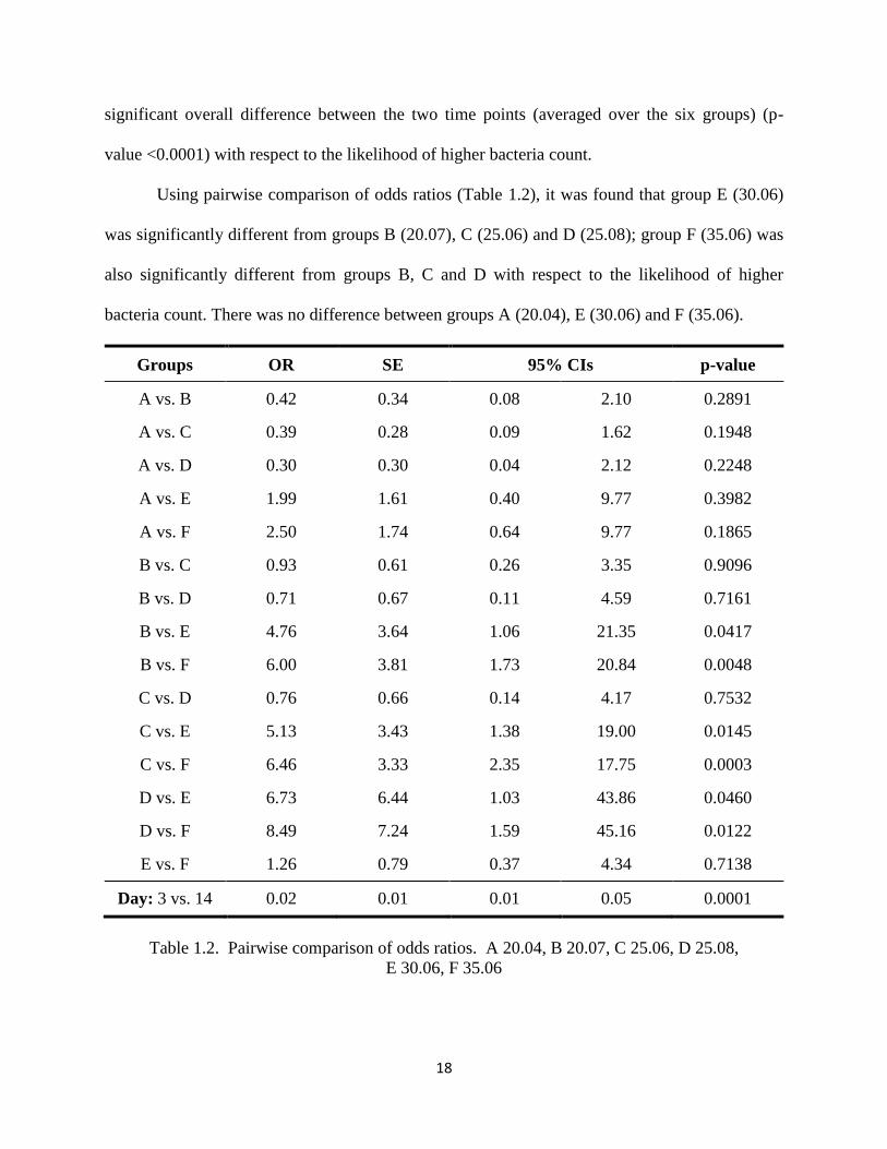
Using pairwise comparison of odds ratios (Table 1.2), it was found that group E (30.06)
was significantly different from groups B (20.07), C (25.06) and D (25.08); group F (35.06) was
also significantly different from groups B, C and D with respect to the likelihood of higher
bacteria count. There was no difference between groups A (20.04), E (30.06) and F (35.06).
Groups OR SE 95% CIs p-value
A vs. B 0.42 0.34 0.08 2.10 0.2891
A vs. C 0.39 0.28 0.09 1.62 0.1948
A vs. D 0.30 0.30 0.04 2.12 0.2248
A vs. E 1.99 1.61 0.40 9.77 0.3982
A vs. F 2.50 1.74 0.64 9.77 0.1865
B vs. C 0.93 0.61 0.26 3.35 0.9096
B vs. D 0.71 0.67 0.11 4.59 0.7161
B vs. E 4.76 3.64 1.06 21.35 0.0417
B vs. F 6.00 3.81 1.73 20.84 0.0048
C vs. D 0.76 0.66 0.14 4.17 0.7532
C vs. E 5.13 3.43 1.38 19.00 0.0145
C vs. F 6.46 3.33 2.35 17.75 0.0003
D vs. E 6.73 6.44 1.03 43.86 0.0460
D vs. F 8.49 7.24 1.59 45.16 0.0122
E vs. F 1.26 0.79 0.37 4.34 0.7138
Day: 3 vs. 14 0.02 0.01 0.01 0.05 0.0001
Table 1.2. Pairwise comparison of odds ratios. A 20.04, B 20.07, C 25.06, D 25.08,
E 30.06, F 35.06

19
In order to see whether there was a difference between an experimental group and its
respective control group, the change in bacterial count score from day 3 to day 14 was compared.
The experimental group differed significantly from the control group for all 3 comparisons
(Figure 1.2).
(1) (2) (3)
Figure 1.2. Percentage of bacterial infection between days 3 and 14 for experimental groups and
their respective controls. (1) A 20.04, (2) E 30.06, (3) F 35.06.
DISCUSSION
In this study, we evaluated the differences in bacterial reduction between different sizes
of rotary preparations while using a 31-gauge irrigation needle. A number of studies have been
done evaluating the debris removal and bacterial reduction and elimination using irrigation
needles ranging in size from 22- to 30-gauge (7, 8, 17, 26-28). They all concluded that the
higher number gauge narrower diameter needle is better because it allows more apical placement
within the root, thus delivering the irrigant to the most apical portion. This was why we decided
to test the 31-gauge needle. According to the ISO 9626 tubing specification (29), size 31-gauge
needle has a designated metric size of 0.25, with the outside diameter range of 0.254-0.267mm,
and a minimal inside diameter of 0.114mm. Therefore, in a canal that has been prepared to an
apical size of 30, a 31-gauge needle should be able to reach all the way to the apex. In testing of
this hypothesis, we wanted to see if it would be possible to instrument the canals to a smaller size
than is currently recommended (15) and achieve a desired goal.
20
Enterococcus faecalis is a Gram+ aerotolerant bacteria commonly found in the
gastrointestinal tract. It is also frequently encountered in secondary endodontic infections (30-
32). E. faecalis is known for its resilient nature and capability of surviving under extreme
conditions and low nutritional availability. It is an exceptionally challenging bacteria to
eradicate, which is why it was chosen for this experiment. Besides being in a planktonic state, E.
faecalis has the ability to form a biofilm within the root (33), and it has been shown that
microorganisms in the biofilm have different phenotypes then their planktonic counterparts (34).
Dunavant et al. (35) showed when E. faecalis was put under stress, it induced a more resilient
biofilm. Stress has also been shown to induce increased E. faecalis penetration into the dentinal
tubules (33). All of these findings contribute to the difficulties in eradication attempts of E.
faecalis.
The results of our study showed that smaller canal preparations harbored more bacteria
over the tested time period compared to the larger sizes, however, the pattern of change over
time was the same for all the groups. This is because E. faecalis grows exponentially, so as long
as there is at least one colony of E. faecalis left after instrumentation and irrigation, if the
conditions are favorable, it will rebound.
Groups 20.04, 20.07, 25.06 and 25.08 showed no difference between each other when
evaluating for likelihood of higher vs. lower bacterial count controlling for days (day 3 and day
14). The same was found between groups 30.06 and 35.06. There was a statistically significant
difference between these two groups and all the smaller sizes groups except for 20.04 group.
This could be explained by the fact that with the size 20.04, the 31-gauge needle reached within
three millimeter from the apex. Therefore, the TSB replenishment may not have been adequate,
and it took longer for the bacteria to rebound after the initial shock of instrumentation and

21
irrigation. Therefore, our bacterial recovery during sampling may not have been as successful.
For the purpose of comparing different preparation sizes and the amount of bacterial reduction,
we decided to consider the 20.04 group an outlier.
Controlling for group, the likelihood of higher bacteria count (vs. lower bacteria) at day 3
was 0.02 times less likely than that at day 14 (95% CI: 0.01–0.05), which was statistically
significant.
When comparing the experimental groups to their matching controls, the results showed
significant difference between the two. Positive control groups showed positive bacterial growth
early on, but the experimental groups caught up over time. This was another proof that the
bacteria are not eliminated during instrumentation and irrigation regardless of the type of irrigant
used, and if the conditions for the bacterial growth are favorable, they will regain their ground
and proliferate.
In conclusion, within the limitations of this study, our results support those by Khademi
et al. (15) in stating that 30.06 was the minimal size of root canal preparation needed to notice a
statistically significant difference in bacterial reduction when compared to smaller sizes during
the testing period. The use of a 31-gauge irrigation needle did not show an increased effect on
bacterial elimination in smaller preparation sizes. Since E. faecalis cannot be eradicated from
the root canal system, future research could evaluate the same sizes and 31-gauge needle without
the bacterial infection (similar to Khademi’s 2006 study) (15), and evaluate the amount of debris
and smear layer removal in the apical portion of the root under the SEM.

22
REFERENCES
1. Kakehashi S, Stanley HR, Fitzgerald RJ. The Effects of Surgical Exposures of Dental
Pulps in Germ-Free and Conventional Laboratory Rats. Oral surgery, oral medicine, and oral
pathology 1965;20:340-349.
2. Moller AJ, Fabricius L, Dahlen G, Ohman AE, Heyden G. Influence on periapical tissues
of indigenous oral bacteria and necrotic pulp tissue in monkeys. Scandinavian journal of dental
research 1981;89(6):475-484.
3. Peters OA, Peters CI. Cleaning and shaping of the root canal system. In: Cohen S,
Pathways of the Pulp. 10th edition, St. Louis, MO: Mosby Company, 2011: 283-348.
4. Sundqvist G, Figdor D. Endodontic treatment of apical periodontitis. In: Orstavik D, Pitt
Ford TR, eds. Essential Endodontology: Prevention and Treatment of Apical Periodontitis.
London, UK. Blackwell Science Ltd. 1998:242-269.
5. Peters OA, Schonenberger K, Laib A. Effects of four Ni-Ti preparation techniques on
root canal geometry assessed by micro computed tomography. International endodontic journal
2001;34(3):221-230.
6. Baker NA, Eleazer PD, Averbach RE, Seltzer S. Scanning electron microscopic study of
the efficacy of various irrigating solutions. Journal of endodontics 1975;1(4):127-135.
7. Siqueira JF, Jr., Rocas IN, Favieri A, Lima KC. Chemomechanical reduction of the
bacterial population in the root canal after instrumentation and irrigation with 1%, 2.5%, and
5.25% sodium hypochlorite. Journal of endodontics 2000;26(6):331-334.
8. Card SJ, Sigurdsson A, Orstavik D, Trope M. The effectiveness of increased apical
enlargement in reducing intracanal bacteria. Journal of endodontics 2002;28(11):779-783.
9. Peters LB, Wesselink PR. Periapical healing of endodontically treated teeth in one and
two visits obturated in the presence or absence of detectable microorganisms. International
endodontic journal 2002;35(8):660-667.
10. Fujii R, Saito Y, Tokura Y, Nakagawa KI, Okuda K, Ishihara K. Characterization of
bacterial flora in persistent apical periodontitis lesions. Oral microbiology and immunology
2009;24(6):502-505.
11. Nair PN. Pathogenesis of apical periodontitis and the causes of endodontic failures.
Critical reviews in oral biology and medicine : an official publication of the American
Association of Oral Biologists 2004;15(6):348-381.
12. Shemesh H, Bier CA, Wu MK, Tanomaru-Filho M, Wesselink PR. The effects of canal
preparation and filling on the incidence of dentinal defects. International endodontic journal
2009;42(3):208-213.

23
13. Liu R, Kaiwar A, Shemesh H, Wesselink PR, Hou B, Wu MK. Incidence of apical root
cracks and apical dentinal detachments after canal preparation with hand and rotary files at
different instrumentation lengths. Journal of endodontics 2013;39(1):129-132.
14. Tawil PZ, Saraiya VM, Galicia JC, Duggan DJ. Periapical microsurgery: the effect of
root dentinal defects on short- and long-term outcome. Journal of endodontics 2015;41(1):22-27.
15. Khademi A, Yazdizadeh M, Feizianfard M. Determination of the minimum
instrumentation size for penetration of irrigants to the apical third of root canal systems. Journal
of endodontics 2006;32(5):417-420.
16. De Deus QD. Frequency, location, and direction of the lateral, secondary, and accessory
canals. Journal of endodontics 1975;1(11):361-366.
17. Guerreiro-Tanomaru JM, Loiola LE, Morgental RD, Leonardo Rde T, Tanomaru-Filho
M. Efficacy of four irrigation needles in cleaning the apical third of root canals. Brazilian dental
journal 2013;24(1):21-24.
18. Hockett JL, Dommisch JK, Johnson JD, Cohenca N. Antimicrobial efficacy of two
irrigation techniques in tapered and nontapered canal preparations: an in vitro study. Journal of
endodontics 2008;34(11):1374-1377.
19. Albrecht LJ, Baumgartner JC, Marshall JG. Evaluation of apical debris removal using
various sizes and tapers of ProFile GT files. Journal of endodontics 2004;30(6):425-428.
20. Abou-Rass M, Piccinino MV. The effectiveness of four clinical irrigation methods on the
removal of root canal debris. Oral surgery, oral medicine, and oral pathology 1982;54(3):323-
328.
21. Chow TW. Mechanical effectiveness of root canal irrigation. Journal of endodontics
1983;9(11):475-479.
22. Sedgley CM, Nagel AC, Hall D, Applegate B. Influence of irrigant needle depth in
removing bioluminescent bacteria inoculated into instrumented root canals using real-time
imaging in vitro. International endodontic journal 2005;38(2):97-104.
23. Baratto-Filho F, Leonardi DP, Zielak JC, Vanni JR, Sayao-Maia SM, Sousa-Neto MD.
Influence of ProTaper finishing files and sodium hypochlorite on cleaning and shaping of
mandibuldar central incisors--a histological analysis. Journal of applied oral science : revista
FOB 2009;17(3):229-233.
24. Coldero LG, McHugh S, MacKenzie D, Saunders WP. Reduction in intracanal bacteria
during root canal preparation with and without apical enlargement. International endodontic
journal 2002;35(5):437-446.

24
25. Falk KW, Sedgley CM. The influence of preparation size on the mechanical efficacy of
root canal irrigation in vitro. Journal of endodontics 2005;31(10):742-745.
26. Siqueira JF, Jr., Rocas IN, Santos SR, Lima KC, Magalhaes FA, de Uzeda M. Efficacy of
instrumentation techniques and irrigation regimens in reducing the bacterial population within
root canals. Journal of endodontics 2002;28(3):181-184.
27. Cohenca N, Paranjpe A, Heilborn C, Johnson JD. Antimicrobial efficacy of two irrigation
techniques in tapered and non-tapered canal preparations. A randomized controlled clinical trial.
Quintessence international 2013;44(3):217-228.
28. Huang TY, Gulabivala K, Ng YL. A bio-molecular film ex-vivo model to evaluate the
influence of canal dimensions and irrigation variables on the efficacy of irrigation. International
endodontic journal 2008;41(1):60-71.
29. International Standards Organization. Stainless steel needle tubing for the manufacture of
medical devices. ISO 9626:1991/Amd.1:2001(E)
30. Molander A, Reit C, Dahlen G, Kvist T. Microbiological status of root-filled teeth with
apical periodontitis. International endodontic journal 1998;31(1):1-7.
31. Peciuliene V, Maneliene R, Balcikonyte E, Drukteinis S, Rutkunas V. Microorganisms in
root canal infections: a review. Stomatologija / issued by public institution "Odontologijos
studija" ... [et al.] 2008;10(1):4-9.
32. Rocas IN, Siqueira JF, Jr., Santos KR. Association of Enterococcus faecalis with
different forms of periradicular diseases. Journal of endodontics 2004;30(5):315-320.
33. George S, Kishen A, Song KP. The role of environmental changes on monospecies
biofilm formation on root canal wall by Enterococcus faecalis. Journal of endodontics
2005;31(12):867-872.
34. Li YH, Chen YY, Burne RA. Regulation of urease gene expression by Streptococcus
salivarius growing in biofilms. Environmental microbiology 2000;2(2):169-177.
35. Dunavant TR, Regan JD, Glickman GN, Solomon ES, Honeyman AL. Comparative
evaluation of endodontic irrigants against Enterococcus faecalis biofilms. Journal of endodontics
2006;32(6):527-531.

25
CHAPTER 3 MANUSCRIPT #2
NOMINAL SIZE AND TAPER ANALYSIS OF NOVEL METALLURGY NiTi FILES
INTRODUCTION
Manufacturers of endodontic files rely on the specifications set by the International
Standards Organization (ISO 3630-1) (1) and American National Standards Institute/American
Dental Association (ANSI/ADA Spec 101) (2) to ensure their files specifications fall within the
allowable tolerance for their advertised sizes. These guidelines exist thanks to the pioneering
efforts of Green (3), Ingle (4, 5), and others who called for standardization of stainless steel 02
taper endodontic instruments and obturation materials in the mid-1950’s. Current guidelines for
nickel-titanium (NiTi) rotary files of any size set the diameter tolerance to be within 50% of the
difference between the next smaller and/or larger instrument size (2), while the allowable taper
tolerance is set to be within 0.05 of the advertised taper size (2). In other words, for the
diameter, if the file’s size increases by 0.05mm, then the allowable tolerance would be
+0.025mm from the advertised size. For the taper, the generous allowance of +0.05 means that
if the file is advertised as having a 06 taper, the actual file taper can be anywhere from 01 taper
to 11 taper, and still satisfy the ANSI/ADA guideline.
In 2002, Zinelis et al. (6) reported on the clinical relevance of standardization rules of
ISO 3630-1 specification, and concluded that due to the large amount of allowable tolerance,
even though the tested files were not the size they were advertised to be, they still fell within the
acceptable tolerance range. A number of studies looked at the continuous taper NiTi rotary files
diameters and tapers (6-11), but to our knowledge, there has not been a publication comparing

26
dimensional values of conventional NiTi rotary files with the newer heat-treated NiTi files.
When a file is heat-treated, the flexibility of the file is increased, which may lead to a greater
affinity for distortion during use (12). If this happens, then the prepared canal size may not be
the size of the file that was used, and it may pose a disinfection challenge and unnecessary
frustration to the practitioner when choosing a proper size gutta percha for obturation (13).
The ISO and ANSI/ADA publications did not set a guideline for the file’s pilot tip length;
they left it up to manufacturer’s discretion. Pilot tip (Figure 2.1) is the area of the file from the
very tip of the file to the first cutting edge. Its purpose is to enlarge the canal and to help guide
the file through it (14). The distance from the tip of the file to where the advertised size of the
file is actually located, is what we termed a true pilot tip length. To our knowledge, there have
been no reports on evaluation of the true pilot tip length, and we hope to bridge that gap in
knowledge.
The purpose of this study was to evaluate the nominal tip diameter, taper and true pilot
tip length of NiTi rotary files before and after use, and to evaluate manufacturer’s compliance
with the ANSI/ADA guidelines as well as our stricter, new recommended guideline (NRG).
MATERIALS AND METHODS
Five different brands of files were used; three in size 25.08: ProTaper Universal
(Dentsply, Tulsa Dental, Tulsa, OK), ProTaper Gold (Dentsply, Tulsa Dental, Tulsa, OK), and
Channels Progressive Taper (Insight Endo for Henry Schein, Melville, NY), and two in size
35.06: Vortex Blue (Dentsply, Tulsa Dental, Tulsa, OK) and Typhoon Infinite Flex NiTi files
(Clinician’s Choice, Dental Products Inc, New Milford, CT). Ten files were used from each
brand (total n=50). Prior to measurements, files were steam sterilized, conditioned for at least
ten hours at 20+5° C, and imaged under SEM microscope (FEI Quanta 200F Environmental

27
Scanning Electron Microscope (ESEM), FEI Inc. Hillsboro, OR) at 50x magnification and a
resolution of 0.1 micron.
All images were evaluated and measured by two independent and equilibrated examiners
using ImageJ software (National Institute of Health, Bethesda, MD). The agreement was set at
0.001mm.
Nominal diameter (D0) was measured from the cutting tip of the file (Figure 2.1). If the
obtained value did not correspond to the advertised size of the file, a measurement was made
down the length of the file until the measurement of the advertised size was reached. This was
termed “True D0”. This value was used for true pilot tip length determination (from the tip of
the file to the “True D0”).
Figure 2.1. SEM image with measurement schematics. D0 – distance from cutting edge across
the file. True D0 – location of the advertised file size. D2 – diameter 2mm from D0. Pilot tip
– distance from the tip of the file to D0. True pilot tip – distance from the tip of the file to True
D0.

28
Taper of the file was measured using the following formula set by ANSI/ADA Spec 101:
Taper =Distance of diameters
Distance between diameters
The guideline states that the diameters included in the above equation are D0 and D16 (or
alternatively D3 and D16). We made a protocol adjustment here due to the variable taper of the
ProTaper and Channels files beyond the initial 4mm of the file length. Therefore, the diameters
included in our analysis were D0 and D2. Differences between the measured taper and the
advertised taper were determined.
Pilot tip length was measured from the tip of the file to D0. True pilot tip length was
measured from the tip of the file to where the advertised size of the file was recorded (True D0).
After initial measurements, instrumentation was performed on plastic endodontic training
blocks (Brasseler, Savannah, GA) according to each file’s manufacturer’s recommendation.
After instrumentation, files were conditioned and imaged under the SEM again. The same
measurements were repeated as in the initial set up.
Data analysis was conducted using repeated measures analysis of variance (r-ANOVA)
and Tukey’s test to evaluate the differences between the diameter, taper and true pilot tip length
before and after use and compared to their advertised sizes. The P-value was set at <0.05. An
observation was made of the percentages of files that satisfied the ANSI/ADA tolerance level
(+0.025mm for diameter, and +0.05 for the taper), and our new recommended guideline (NRG)
(+0.0125mm for the diameter, and +0.005 for the taper).

29
RESULTS
The mean diameter values are recorded in Table 2.1. None of the files had the advertised
nominal diameter size. The mean measure of D0 in each group was significantly different from
the advertised size; the mean D0 measure was less than the advertised size for all brands except
for the Channels files. There was no significant mean difference between the before and after
measures of D0 in each group except for the ProTaper Universal group.
Table 2.1. Diameter values (before and after) compared to the advertised nominal size.
30
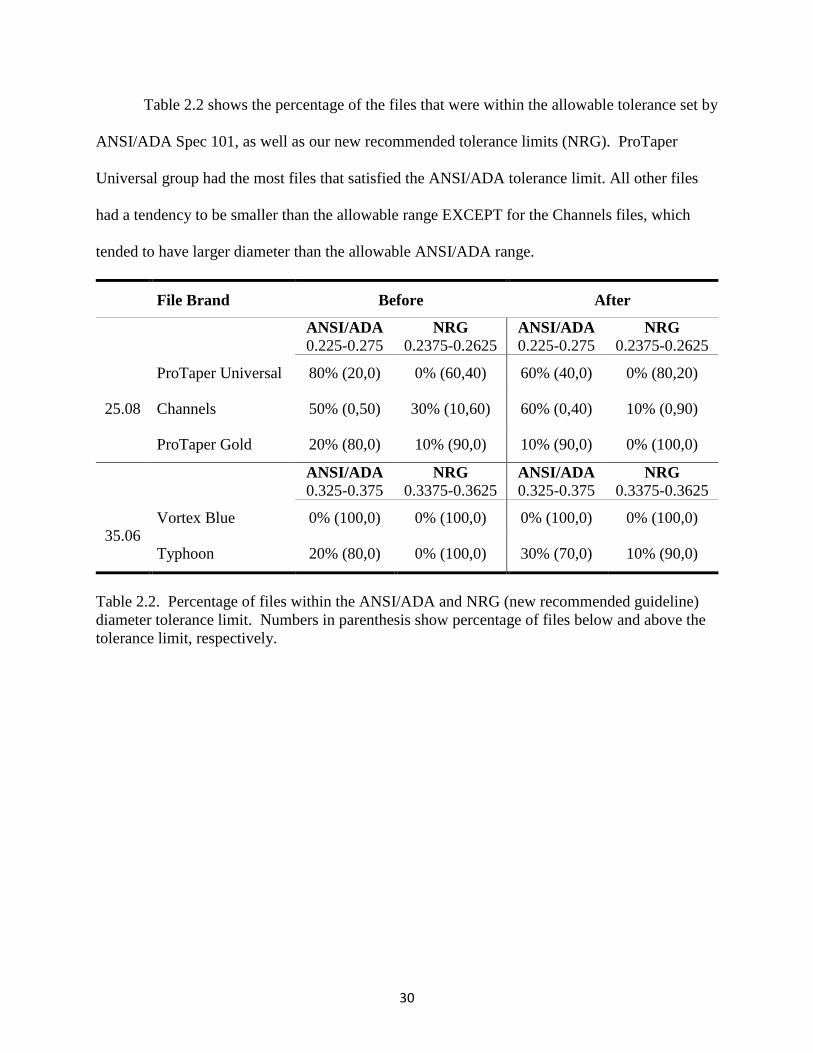
Table 2.2 shows the percentage of the files that were within the allowable tolerance set by
ANSI/ADA Spec 101, as well as our new recommended tolerance limits (NRG). ProTaper
Universal group had the most files that satisfied the ANSI/ADA tolerance limit. All other files
had a tendency to be smaller than the allowable range EXCEPT for the Channels files, which
tended to have larger diameter than the allowable ANSI/ADA range.
File Brand Before After
ANSI/ADA
0.225-0.275 NRG
0.2375-0.2625 ANSI/ADA
0.225-0.275 NRG
0.2375-0.2625
25.08
ProTaper Universal 80% (20,0) 0% (60,40) 60% (40,0) 0% (80,20)
Channels 50% (0,50) 30% (10,60) 60% (0,40) 10% (0,90)
ProTaper Gold 20% (80,0) 10% (90,0) 10% (90,0) 0% (100,0)
ANSI/ADA
0.325-0.375 NRG
0.3375-0.3625 ANSI/ADA
0.325-0.375 NRG
0.3375-0.3625
35.06 Vortex Blue 0% (100,0) 0% (100,0) 0% (100,0) 0% (100,0)
Typhoon 20% (80,0) 0% (100,0) 30% (70,0) 10% (90,0)
Table 2.2. Percentage of files within the ANSI/ADA and NRG (new recommended guideline)
diameter tolerance limit. Numbers in parenthesis show percentage of files below and above the
tolerance limit, respectively.

31
Taper values indicated that the mean measure of taper in each group was significantly
different from the advertised taper both before and after use except for the ProTaper Gold group.
In the ProTaper Universal, Channels and the Typhoon groups, the mean taper measure (before
and after) were less than the advertised size; whereas in the ProTaper Gold and the Vortex Blue
groups, the mean taper measure (before and after) was greater than the advertised size. These
findings are summarized in Table 2.3. There was no statistically significant mean change (before
vs. after) in any group.
Table 2.3. Taper values (before and after) compared to the advertised nominal taper sizes.

32
Table 2.4 shows the percentages of files within each tolerance level (ISO and NRG). All
the files in all groups were within the allowable ISO taper tolerance. 40% of ProTaper Universal
files, 20% of ProTaper Gold files, 20% of Vortex Blue files and 30% of Typhoon files were the
exact taper as advertised. None of the Channels files had the advertised nominal taper size.
When compared to the NRG range, ProTaper Universal, ProTaper Gold, Vortex Blue and
Typhoon files were within the allowable range 30% of the time. 100% of Channels files were
smaller than NRG range. Findings are summarized in the table below (Table 2.4).
File Brand Before After
ANSI/ADA
0.03-0.13 NRG
0.075-0.085 ANSI/ADA
0.03-0.13 NRG
0.075-0.085
25.08
ProTaper Universal 100% (0,0) 30% (70,0) 100% (0,0) 20% (80,0)
Channels 100% (0,0) 0% (100,0) 100% (0,0) 0% (100,0)
ProTaper Gold 100% (0,0) 30% (10,60) 100% (0,0) 40% (10,50)
ANSI/ADA
0.01-0.11 NRG
0.055-0.065 ANSI/ADA
0.01-0.11 NRG
0.055-0.065
35.06 Vortex Blue 100% (0,0) 30% (0,70) 100% (0,0) 10% (0,90)
Typhoon 100% (0,0) 30% (70,0) 100% (0,0) 40% (60,0)
Table 2.4. Percentage of files within the ANSI/ADA and NRG taper tolerance limit. Numbers
in parenthesis show percentage of files below and above the tolerance limit, respectively.
33
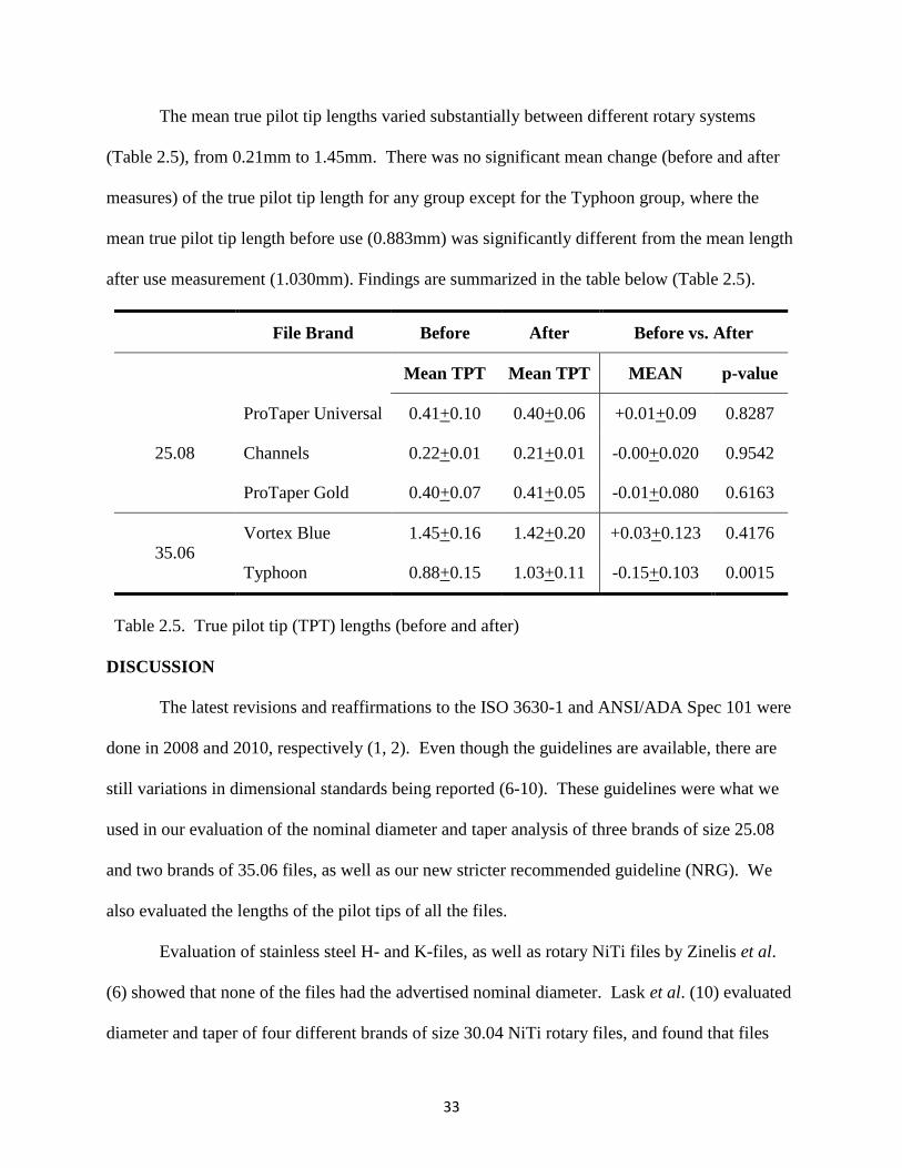
The mean true pilot tip lengths varied substantially between different rotary systems
(Table 2.5), from 0.21mm to 1.45mm. There was no significant mean change (before and after
measures) of the true pilot tip length for any group except for the Typhoon group, where the
mean true pilot tip length before use (0.883mm) was significantly different from the mean length
after use measurement (1.030mm). Findings are summarized in the table below (Table 2.5).
File Brand Before After Before vs. After
Mean TPT Mean TPT MEAN p-value
25.08
ProTaper Universal 0.41+0.10 0.40+0.06 +0.01+0.09 0.8287
Channels 0.22+0.01 0.21+0.01 -0.00+0.020 0.9542
ProTaper Gold 0.40+0.07 0.41+0.05 -0.01+0.080 0.6163
35.06 Vortex Blue 1.45+0.16 1.42+0.20 +0.03+0.123 0.4176
Typhoon 0.88+0.15 1.03+0.11 -0.15+0.103 0.0015
Table 2.5. True pilot tip (TPT) lengths (before and after)
DISCUSSION
The latest revisions and reaffirmations to the ISO 3630-1 and ANSI/ADA Spec 101 were
done in 2008 and 2010, respectively (1, 2). Even though the guidelines are available, there are
still variations in dimensional standards being reported (6-10). These guidelines were what we
used in our evaluation of the nominal diameter and taper analysis of three brands of size 25.08
and two brands of 35.06 files, as well as our new stricter recommended guideline (NRG). We
also evaluated the lengths of the pilot tips of all the files.
Evaluation of stainless steel H- and K-files, as well as rotary NiTi files by Zinelis et al.
(6) showed that none of the files had the advertised nominal diameter. Lask et al. (10) evaluated
diameter and taper of four different brands of size 30.04 NiTi rotary files, and found that files

34
tended to be larger than their nominal sizes, but concluded that the differences were minute and
probably not clinically relevant. Kim et al. (9) evaluated dimensional standard of several NiTi
rotary files, and found that the diameter values were mostly not in compliance with the
advertised values. In our current investigation, none of the files tested had the advertised
nominal tip diameter (p<0.05). For the 25.08 size, the range was from 21mm to 28mm.
ProTaper Universal and ProTaper Gold files tended to be smaller than the nominal size, and the
Channels files tended to be larger than the advertised nominal size. A statistically significant
difference was noted in the ProTaper Universal group between before and after measurement,
however, with the mean diameter difference of 0.006mm and a standard deviation of +0.007mm,
this deformation would probably not be clinically relevant.
For the 35.06 size, both groups displayed smaller nominal diameter sizes than advertised,
ranging from 24mm for the Vortex Blue group, to 31mm for the Typhoon group.
The percent of files that were within the ANSI/ADA tolerance limit was 80% for the
ProTaper Universal, 50% for the Channels, 20% for both the ProTaper Gold and the Typhoon
groups, and none for the Vortex Blue group. There was a tendency for the files to be on the
smaller size for all of the groups except for the Channels group, where a consistently larger than
the highest ANSI/ADA limit was observed. The same trend was noted within our new tolerance
limit as well, with the smaller percentage of files satisfying the tolerance range. It is interesting
to observe that the austenite phase files (ProTaper Universal and Channels) had more files that
were within the ANSI/ADA tolerance limit compared to the newer metallurgy heat-treated
(ProTaper Gold and Vortex Blue) and martensite phase files (Typhoon). It is also of interest that
even though files may be advertised as the exact replicas of one another (ProTaper Universal and
Channels), practitioner should be aware of a potential size difference, especially if using a hybrid
35
technique with rotary instrumentation. The mean nominal size of ProTaper Universal was
23mm, and for the Channels was 28mm. Zinelis et al. (6) pointed out the issue of file size
overlap, where the next bigger size may be the same as the last smaller size. Practitioner should
be aware of this possibility in order to minimize potential iatrogenic mishap during endodontic
treatment. It should also be pointed out that if the file is not the size that it is advertised to be,
the shaping of the canal space may not be adequate and it may prevent full irrigant delivery to
the root canal system.
With the objective of endodontic therapy being thorough cleaning and shaping of the root
canal system followed with an apical seal, it is important to have the obturation material of the
same size as the prepared canal space, i.e. as the last instrument used in the canal preparation.
Cunningham et al. (13) found significant variability between different brands of gutta percha size
30.04 in both diameter and taper, and none of the brands tested had the nominal advertised size.
Practitioner should be aware that not only files may be of different sizes than advertised, but the
matching gutta percha may not be a match at all. Therefore, a stricter adherence by the
manufacturers to the set guidelines should be recommended.
Taper evaluation indicated that only ProTaper Gold had the mean taper that was not
statistically different from the advertised taper. However, all the files satisfied the ANSI/ADA
tolerance range. This is in agreement with previous reports (7, 8). In the 25.08 group, mean
taper for the ProTaper Universal was 07 and for the Channels was 06. For the 35.06 group, the
mean taper for the Vortex Blue was 07 and for the Typhoon group was 05.
When evaluating compliance with our new recommended guidelines (NRG), 30%
agreement was noted for all the files except for the Channels group. When not in compliance,
ProTaper Universal, Channels and Typhoon files were generally smaller than the set tolerance

36
range, and ProTaper Gold and Vortex Blue files were generally larger. Again a pattern was
observed, where the austenite phase files acted in the similar fashion (ProTaper Universal and
Channels), as did the heat-treated files (ProTaper Gold and Vortex Blue). Further investigation
might be warranted. It should also be pointed out that the current ANSI/ADA Spec 101
guidelines for taper tolerance of +0.05 are rather generous, and a stricter updated guideline
should be recommended.
Evaluation of the true pilot tip length pointed to the complete lack of standardization
between different files and manufacturers. The range in true pilot tip length was truly
extraordinary, from 0.21mm for the Channels group to 1.45mm for the Vortex Blue group.
ProTaper Universal and ProTaper Gold displayed almost identical true pilot tip lengths of
0.41mm and 0.40mm, respectively. Typhoon files were the only ones that displayed a significant
difference in the before vs. after measurement (0.883mm to 1.030mm). This is in agreement
with Peters’ (12) report of martensite file deformation under stress. Such large variations in true
pilot tip lengths might be of clinical importance and warrants further research.
In summary, the present study showed that the nominal diameter sizes of the files tested
and most of their taper values are not in agreement with their advertised sizes. Clinical relevance
might be argued, but stricter expectations from the manufacturers should be recommended in
order to minimize potential for sizing overlap, practitioner frustration and iatrogenic
complications. We would also like to propose that a guideline should be set to standardize the
length of the pilot tip. Future studies should look at other sizes and brands of files and evaluate
for differences between different metallurgies.

37
REFERENCES
1. International Standards Organization. Dentistry - Root-canal instruments. Part 1: General
Requirements and Test Methods. ISO 3630-1:2008(E). 2008.
2. American Dental Association Council on Scientific Affairs. ANSI/ADA Specification
No. 101. Root Canal Instruments: General Requirements. 2001. Reaffirmed Oct 2010.
3. Green EN. Microscopic investigation of root canal file and reamer widths. Oral surgery,
oral medicine, and oral pathology. 1957;10(5):532-40.
4. Ingle JI. The need for endodontic instrument standardization. Oral surgery, oral medicine,
and oral pathology. 1955;8(11):1211-3.
5. Ingle JI. A standardized endodontic technique utilizing newly designed instruments and
filling materials. Oral surgery, oral medicine, and oral pathology. 1961;14:83-91.
6. Zinelis S, Magnissalis EA, Margelos J, Lambrianidis T. Clinical relevance of
standardization of endodontic files dimensions according to the ISO 3630-1 specification.
Journal of endodontics. 2002;28(5):367-70.
7. Gergi R, Abou Rjeily J, Osta N, Sader J, Naaman A. Taper preparation variability
compared to current taper standards using computed tomography. International journal of
dentistry. 2012;2012:265695.
8. Hatch GW, Roberts S, Joyce AP, Runner R, McPherson JC, 3rd. Comparative study of
the variability of 0.06 tapered rotary endodontic files to current taper standards. Journal of
endodontics. 2008;34(4):463-5.
9. Kim KW, Cho KM, Park SH, Choi KY, Karabucak B, Kim JW. A comparison of
dimensional standard of several nickel-titanium rotary files. Restorative dentistry & endodontics.
2014;39(1):7-11.
10. Lask JT, Walker MP, Kulild JC, Cunningham KP, Shull PA. Variability of the diameter
and taper of size #30, 0.04 nickel-titanium rotary files. Journal of endodontics.
2006;32(12):1171-3.
11. Stenman E, Spangberg LS. Root canal instruments are poorly standardized. Journal of
endodontics. 1993;19(7):327-34.
12. Peters OA, Gluskin AK, Weiss RA, Han JT. An in vitro assessment of the physical
properties of novel Hyflex nickel-titanium rotary instruments. International endodontic journal.
2012;45(11):1027-34.

38
13. Cunningham KP, Walker MP, Kulild JC, Lask JT. Variability of the diameter and taper
of size #30, 0.04 gutta-percha cones. Journal of endodontics. 2006;32(11):1081-4. Epub
2006/10/24.
14. McSpadden JT. Mastering Endodontic Instrumentation (1st edition). Chattanooga, TN:
Cloudland Institute. 2007.