endodontic prognosisdownload.e-bookshelf.de/download/0007/8249/96/l-g...endodontically treated teeth...
TRANSCRIPT

Nadia ChugalLouis M. Lin Editors
123
Clinical Guide for Optimal Treatment Outcome
Endodontic Prognosis

Endodontic Prognosis

Nadia Chugal • Louis M. LinEditors
Endodontic Prognosis
Clinical Guide for Optimal Treatment Outcome

EditorsNadia ChugalSection of EndodonticsUCLA School of DentistryLos AngelesCaliforniaUSA
Louis M. LinDept of EndodonticsNYU College of DentistryNew York New YorkUSA
ISBN 978-3-319-42410-1 ISBN 978-3-319-42412-5 (eBook)DOI 10.1007/978-3-319-42412-5
Library of Congress Control Number: 2016960548
© Springer International Publishing Switzerland 2017This work is subject to copyright. All rights are reserved by the Publisher, whether the whole or part of the material is concerned, specifically the rights of translation, reprinting, reuse of illustrations, recita-tion, broadcasting, reproduction on microfilms or in any other physical way, and transmission or infor-mation storage and retrieval, electronic adaptation, computer software, or by similar or dissimilar methodology now known or hereafter developed.The use of general descriptive names, registered names, trademarks, service marks, etc. in this publica-tion does not imply, even in the absence of a specific statement, that such names are exempt from the relevant protective laws and regulations and therefore free for general use.The publisher, the authors and the editors are safe to assume that the advice and information in this book are believed to be true and accurate at the date of publication. Neither the publisher nor the authors or the editors give a warranty, express or implied, with respect to the material contained herein or for any errors or omissions that may have been made.
Printed on acid-free paper
This Springer imprint is published by Springer NatureThe registered company is Springer International Publishing AG SwitzerlandThe registered company address is Gewerbestrasse 11, 6330 Cham, Switzerland

To my family, friends, and mentors for their love, support, and encouragement.
Nadia Chugal

vii
Foreword
It is indeed a privilege to write this foreword for an Endodontics book with progno-sis as its main emphasis. While all practitioners aspire to achieve the highest levels of success of treatment, the definition of this success and the factors that affect it receive very little attention among clinicians. Complicating this matter is that, with the exception of resolution of pain or purulent drainage, true and complete end-odontic success is not demonstrable clinically until a long period has passed after treatment, typically measured in years. The difficulty in establishing an effective follow-up program for all patients, especially that they are typically asymptomatic, has led many practitioners to rely only on surrogate measures of success like the quality of the obturation and the resolutions of symptoms. While there are some population-based data in the literature to support reliance on these parameters, they clearly provide an incomplete assessment of prognosis.
As one reflects on this book’s working definition of endodontics, as the preven-tion and/or elimination of apical periodontitis, it is reasonable to reconsider whether this is still consistent with recent information as noted in the relevant chapters. For example, the word “prevention” is used in a discipline in which home care is not thought to affect the outcome of treatment. The intent likely arose from the need to diagnose irreversible pathosis more vigilantly, in order to perform the endodontic treatment at this stage, and avoid pathogenesis of apical periodontitis. However, recent advances in vital pulp therapy leads one to question whether the priority is still to remove the vital inflamed pulp at all costs to assure the goal of preventing apical periodontitis. The growing interest in pulp and dentin regeneration, the advent of more biocompatible reparative materials, and the presence of good out-come studies on vital pulp therapy make one reflect more on this classic definition of endodontic therapy.
This book also provides an excellent discussion in several chapters of the radio-graphic detection of emergent and residual disease, as it has evolved in the last 60 years or so. Today, tools like CBCT allow us to visualize this disease earlier in the diagnostic process, and for a longer period after treatment. Therefore, there is more of an overlap in the pulpitis/apical periodontitis spectrum of diagnosis, and perhaps a longer period when teeth with apical radiolucencies may be considered healing. There are even questions as to whether teeth with long-standing small
viii
radiolucencies, and no other abnormalities, should be retreated or subjected to root end surgery. There is more realization that complete bone regeneration may not be achievable in many of these asymptomatic cases, the way it is not achievable in cases with marginal periodontitis.
Postoperative factors that affect the prognosis are also of particular interest. The profession has in the last decade transitioned from relying on bench-top laboratory studies to clinical outcome studies in making many clinical decisions that are related to coronal leakage. The question remains as to who controls the prognosis to a larger extent: is it the practitioner that did the endodontic therapy or the one who restored the tooth?
Finally, this book eloquently addresses the emerging concept of personalized endodontics, in which the prognosis may be affected by a combination of the unique and complex microbiota that causes the disease, together with the systemic health of the patient, as well as genetic and epigenetic variability among patients. This area promises to provide us in future more detailed predictors for outcomes, which can help the provider with treatment planning and help the patient with decision making.
Ashraf F. Fouad, DDS, MS
Foreword

ix
Preface
This book distinguishes itself from endodontic textbooks because it is the first text-book completely focused on the prognosis of endodontic treatments. Our goal for this book was to make recent results at the forefront of endodontics accessible for clinical practice.
The book is intended to serve as a clinical guide to help practitioners in their clinical decision-making process and ultimately improve endodontic treatment outcomes.
The goal of endodontic treatment is to prevent and/or eliminate apical periodon-titis, a disease entity occurring as a result of microbiologic challenge to the pulp and periradicular tissues. Like many other human diseases, endodontic treatment out-comes are profoundly affected by a multitude of prognostic factors. These determi-nants of treatment success or failure can exert their effect preoperatively, intraoperatively, and postoperatively. Therefore, it is important for the clinician to be familiar with the favorable predictors of outcome as well as prognostic risk fac-tors. This knowledge is essential to effectively circumvent and manage risks in order to achieve the desired treatment result.
We first outlined the theme of every chapter that we considered important for the book. We then invited experts in their respective areas to write on the specific topics. These topics include both basic and clinical sciences and cover several key aspects of endodontic prognosis. The multidisciplinary authorship by highly respected cli-nicians and scientists reflects the multifactorial nature of endodontic outcome.
Outcome assessment of endodontic therapy has evolved from Strindberg’s strin-gent criteria that emphasized the absence of clinical symptoms/signs and restoration of normal structure of the periapical tissues to newer patient-centered criteria focus-ing on the absence of clinical symptoms/signs and survivability and functionality of endodontically treated teeth even with the presence of small and stable periapical lesions. However, as pulpal and periapical pathosis is a disease, a tooth with a per-sistent inflammatory periapical lesion after treatment, regardless of its size, should be considered as unsuccessful elimination of the disease. Therefore, complete elim-ination of the disease still remains the ultimate goal of root canal treatment.
We hope that the readers will enjoy this book and benefit from it, as much as we have enjoyed spending our time and energy working on it.
Nadia ChugalLouis M. Lin

xi
Acknowledgments
We thank all contributors for their time, support, and contributions to this book. Without their generosity and dedication, this book would not have been published. We wish to express our deep appreciation for the many cases, radiographic images, photographs, and histologic slides provided by our colleagues. We also thank the editors from Springer for believing in us and for their support, understanding, and patience.
Nadia ChugalLouis M. Lin
xiii
Contents
1 Introduction: Endodontic Prognosis and Outcome . . . . . . . . . . . . . . . . . 1Nadia Chugal, Louis M. Lin, and Bill Kahler
2 Microbiology and Immunology of Endodontic Infections . . . . . . . . . . 13Luis E. Chávez de Paz and Gunnar Dahlén
3 Diagnosis of Pulpal and Periradicular Disease. . . . . . . . . . . . . . . . . . . . 29Katsushi Okazaki, Matthew Malek, Nadia Chugal, and Louis M. Lin
4 Endodontic Treatment of Mature Teeth . . . . . . . . . . . . . . . . . . . . . . . . . 43Louis M. Lin, Simona Loghin, and Domenico Ricucci
5 Regenerative Approaches in Endodontic Therapies of Immature Teeth . . . . . . . . . . . . . . . . . . . . . . . . . . . . . . . . . . . . . . . . . . . 65Mo K. Kang and George Bogen
6 Endodontic Pharmacotherapeutics . . . . . . . . . . . . . . . . . . . . . . . . . . . . . 87Helaine De Brito-Gariepy, Thereza Cristina Botelho- Dantas, and Jennifer Lynn Gibbs
7 Anatomy, Access, and Length Determination . . . . . . . . . . . . . . . . . . . 115Frederick Barnett
8 Instrumentation and Disinfection of Root Canals . . . . . . . . . . . . . . . . 131Frederic Barnett and José F. Siqueira Jr.
9 Obturation of Root Canals . . . . . . . . . . . . . . . . . . . . . . . . . . . . . . . . . . . 141Dag Ørstavik
10 Restoration of Endodontically Treated Teeth . . . . . . . . . . . . . . . . . . . . 161Nadim Z. Baba, Shane N. White, and George Bogen
11 Local, Systemic, and Genetic Considerations of Endodontic Treatment Prognosis . . . . . . . . . . . . . . . . . . . . . . . . . . . . . . . . . . . . . . . . 193Matthew Malek and Louis M. Lin

xiv
12 Criteria for Outcome Assessment of Nonsurgical Endodontic Treatment . . . . . . . . . . . . . . . . . . . . . . . . . . . . . . . . . . . . . . 211Nadia Chugal, Sanjay M. Mallya, and Bill Kahler
13 From Tooth Retention Through Root Canal Treatment to Extraction and Replacement . . . . . . . . . . . . . . . . . . . . . . . . . . . . . . . 229Shane N. White and Mahmoud Torabinejad
Index . . . . . . . . . . . . . . . . . . . . . . . . . . . . . . . . . . . . . . . . . . . . . . . . . . . . . . . . . 239
Contents

1© Springer International Publishing Switzerland 2017N. Chugal, L.M. Lin (eds.), Endodontic Prognosis, DOI 10.1007/978-3-319-42412-5_1
Introduction: Endodontic Prognosis and Outcome
Nadia Chugal, Louis M. Lin, and Bill Kahler
AbstractPrognosis and outcome are two terms routinely used in medicine and dentistry to predict and assess the treatment of disease. Prognosis is a practitioner’s assess-ment about how a patient will recover from an illness or injury. It is a forecast of the probable course of recovery for any particular disease considering the assess-ment of the case. Outcome is the end result of the treatment and a consequence of treatment decisions made by the practitioner. In endodontics, there are prog-nostic factors which are universal to all cases as well as variables unique to a specific case, all of which can affect endodontic treatment outcomes. Prognostic factors can be grouped into preoperative, intraoperative, and postoperative. They influence endodontic treatment outcomes indirectly through control and elimina-tion of infection. Importantly, an understanding of prognostic factors helps prac-titioners as well as patients decide the appropriate treatment procedures and is especially important for higher-risk conditions such as teeth with a periapical
N. Chugal (*) Section of Endodontics, UCLA School of Dentistry, Los Angeles, CA, USAe-mail: [email protected]
L.M. Lin Department of Endodontics, New York University College of Dentistry, New York, NY, USAe-mail: [email protected]
B. Kahler School of Dentistry, University of Queensland, Brisbane, QLD, Australiae-mail: [email protected]
1
Many authors display a propensity to reduce complex problems such as success and failure to terms so simple that a casual reader with little effort can expand a narrow grasp of the subject into a broad convenient misunderstanding
Dudley Glick, DDSExcerpt from a lecture
Year unknown

2
lesion, calcified canals, resorption, and others. This applies to immature or mature teeth for consideration for nonsurgical or surgical management. Although many prognostic factors are not under the control of the practitioners, they can nevertheless be managed by a practitioner’s thorough evaluation of the present-ing condition, risk assessment inherent in each individual case, and application of biologically based therapies alongside with technical competency. Treatment outcome is usually assessed using clinical and radiographic examination and has evolved from Strindberg’s stringent criteria to patient-centered criteria. Practitioners have to be familiar with the prognostic factors to inform patients of the appropriate treatment modalities and to achieve the optimal treatment outcome.
1.1 Introduction
The terms prognosis and outcome are routinely applied in medicine and dentistry. They are used to predict and evaluate the result of disease treatment. Prognosis is a forecast about probable course and outcome of a disease and chances of recovery [1]. Applied to endodontics, it is a prediction of the outcome of resolution of apical periodontitis. This forecast of the outcome is summarized by the practitioner for the patients and serves to inform them how they will recover from an illness or injury [1]. It synthesizes the prospect of recovery as anticipated from the usual course of disease, or in the case of endodontics, the risk assessment of variables that may influence the treatment outcome [1]. Outcome, on the other hand, is a measure of the success of the treatment, as a result of an activity or a process, and is a consequence of the decisions made during the course of treatment. Applied to endodontics, outcome is the end result that follows as a consequence of endodontic treatment [1].
There are a number of well-researched studies that elucidated prognostic factors that exert significant effect on endodontic outcome. In turn, this information can be used to prognosticate the course of disease resolution and predict the end result of the proposed treatment.
Practitioners can systematically evaluate prognostic factors to guide their patients in the decision-making process and ultimately propose the best treatment options to achieve an optimal outcome. In accordance with evidence-based den-tistry principles, the patients are also members of the treatment planning team and have the right to know the prognosis and expected outcome before commencement of treatment. Given this information, patients can evaluate their treatment options and make an informed decision about the need or preference for their treatment choices [2].
Optimal dental treatment planning requires an accurate assessment of the outcome of the proposed endodontic treatment. This assessment, however, is dependent on a correct understanding of variables affecting the outcome and must be done with both high validity and reliability [3]. When such assessment
N. Chugal et al.
3
is made, it is possible to offer the patient a wide range of appropriate endodontic treatment options.
1.2 Multifactorial Nature of Endodontic Outcome
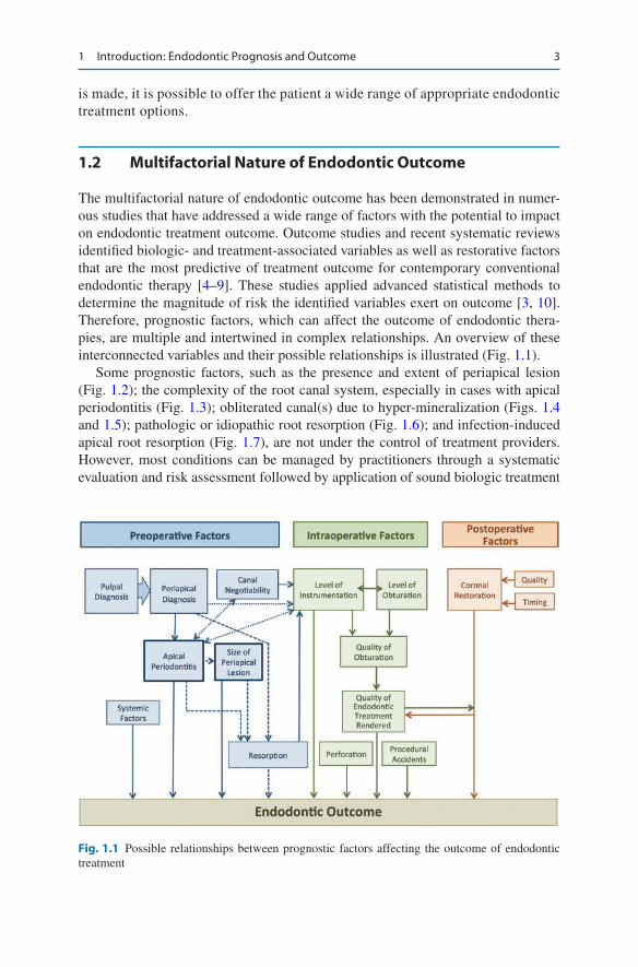
The multifactorial nature of endodontic outcome has been demonstrated in numer-ous studies that have addressed a wide range of factors with the potential to impact on endodontic treatment outcome. Outcome studies and recent systematic reviews identified biologic- and treatment-associated variables as well as restorative factors that are the most predictive of treatment outcome for contemporary conventional endodontic therapy [4–9]. These studies applied advanced statistical methods to determine the magnitude of risk the identified variables exert on outcome [3, 10]. Therefore, prognostic factors, which can affect the outcome of endodontic thera-pies, are multiple and intertwined in complex relationships. An overview of these interconnected variables and their possible relationships is illustrated (Fig. 1.1).
Some prognostic factors, such as the presence and extent of periapical lesion (Fig. 1.2); the complexity of the root canal system, especially in cases with apical periodontitis (Fig. 1.3); obliterated canal(s) due to hyper-mineralization (Figs. 1.4 and 1.5); pathologic or idiopathic root resorption (Fig. 1.6); and infection- induced apical root resorption (Fig. 1.7), are not under the control of treatment providers. However, most conditions can be managed by practitioners through a systematic evaluation and risk assessment followed by application of sound biologic treatment
Fig. 1.1 Possible relationships between prognostic factors affecting the outcome of endodontic treatment
1 Introduction: Endodontic Prognosis and Outcome
4
a b c
a b c
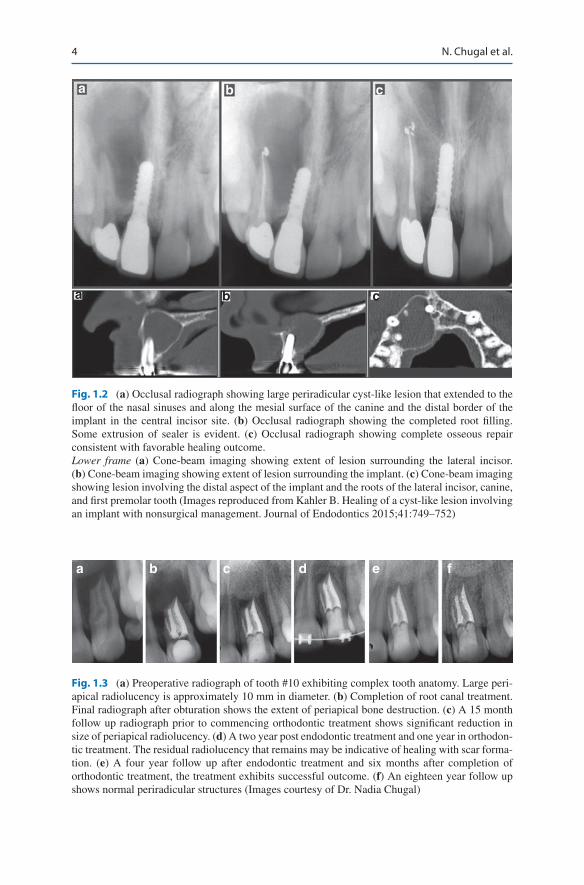
Fig. 1.2 (a) Occlusal radiograph showing large periradicular cyst-like lesion that extended to the floor of the nasal sinuses and along the mesial surface of the canine and the distal border of the implant in the central incisor site. (b) Occlusal radiograph showing the completed root filling. Some extrusion of sealer is evident. (c) Occlusal radiograph showing complete osseous repair consistent with favorable healing outcome. Lower frame (a) Cone-beam imaging showing extent of lesion surrounding the lateral incisor. (b) Cone-beam imaging showing extent of lesion surrounding the implant. (c) Cone-beam imaging showing lesion involving the distal aspect of the implant and the roots of the lateral incisor, canine, and first premolar tooth (Images reproduced from Kahler B. Healing of a cyst-like lesion involving an implant with nonsurgical management. Journal of Endodontics 2015;41:749–752)
a b c d e f
Fig. 1.3 (a) Preoperative radiograph of tooth #10 exhibiting complex tooth anatomy. Large peri-apical radiolucency is approximately 10 mm in diameter. (b) Completion of root canal treatment. Final radiograph after obturation shows the extent of periapical bone destruction. (c) A 15 month follow up radiograph prior to commencing orthodontic treatment shows significant reduction in size of periapical radiolucency. (d) A two year post endodontic treatment and one year in orthodon-tic treatment. The residual radiolucency that remains may be indicative of healing with scar forma-tion. (e) A four year follow up after endodontic treatment and six months after completion of orthodontic treatment, the treatment exhibits successful outcome. (f) An eighteen year follow up shows normal periradicular structures (Images courtesy of Dr. Nadia Chugal)
N. Chugal et al.

5
a b c
Fig. 1.4 (a) Radiograph of tooth #8 showing large periapical lucency and no canal is evident. (b) The tooth is root filled though the root filling is not centered in the root which has the potential to affect outcome. (c) At 18 month review the PA lucency is reduced in size and the tooth is asymptom-atic. This case is an example of a ‘functional outcome’ as the strict Strindberg criterion has not yet been met. However further healing with time is still possible (Images courtesy of Dr. Bill Kahler)
principles and technical expertise in order to achieve an optimal treatment outcome. Often, a complex-presenting condition of the tooth comprises multiple risk factors (Fig. 1.8).
Importantly, an understanding of these high-impact factors assists practitioner’s decision-making process about the appropriate treatment procedures. In addition, it also has practical implications related to treatment execution and preparation of armamentariums necessary to treat these preexisting conditions. For example, protocols may be different for immature vs. mature teeth, teeth with or without a periapical lesion, and for both nonsurgical and surgical management.
For clarity of analysis and comprehension, they can be grouped into three major categories: preoperative, intraoperative, and postoperative.
1.3 Preoperative Factors
An accurate assessment of the pulpal and periapical diagnosis is essential for an understanding of the major biological factors as this diagnosis reflects a change in the disease process and the extent of the infection into the periapical tissues [4, 6, 11, 12]. The literature is unequivocal that preoperative presence vs. absence of peri-radicular osteolysis is one major indicator of postoperative healing or failure [4, 10, 13, 14]. Consequently, teeth with a preoperative periapical lesion have a poorer outcome than teeth without a periapical lesion after nonsurgical root canal treatment [4, 7, 10, 14]. In addition, larger bone lesions show a significantly lower frequency of complete regeneration of the periapical bone than smaller lesions (4, 103). Therefore, when a periapical lesion is present, the smaller the lesion, the more favorable is the treatment prognosis [4, 10]. However, successful resolution of large
1 Introduction: Endodontic Prognosis and Outcome

6
a
b
c d
Fig. 1.5 (a) Radiograph of tooth #9 with a history of trauma. The root tip is blunted consistent with apical resorption. The canal appears to have undergone complete obliteration in the coronal half of the root. The canal in the apical half of the root is of an irregular shape and not centered in the root. (b) CBCT imaging revealed an irregular resorptive lesion in the apical half of the root. Therefore more complex imaging was advantageous as interpretation of conventional periapical radiography was suggestive of canal patency. Furthermore the extensive periapical radiolucency is revealed with erosion of the buccal and palatal cortical bone plates. After consultation with the patient it was decided that optimal treatment option was surgical management due to the prognos-tic considerations of calcified canal, resorptive defect in the apical third of the root and the exten-sive periapical radiolucency. (c) A radiograph taken after the surgical revision and placement of a MTA retrofill. (d) A radiograph at a 2 year review showing an intact lamina dura and periodontal ligament space around the root. The periapical radiolucency is consistent with a periapical scar and is a common observation following surgery when both cortical plates have been eroded (Images courtesy of Dr. Bill Kahler)
periapical radiolucencies is often achieved (Figs. 1.2 and 1.3), although the risk of future surgical treatment remains.
Teeth with a preoperative periapical lesion usually have a long-standing root canal infection compared to teeth without a periapical lesion. Therefore, these teeth have a well-established biofilm in the canal [15]. In addition, bacteria may also establish infection in some periapical lesions, resulting in an extraradicular infection [16].
N. Chugal et al.

7
a
b
c
Fig. 1.6 (a) Preoperative periapical radiograph of symptomatic first maxillary molar where crown was placed one month earlier. (b) CBCT images revealed a Heithersay Grade III invasive cervical lesion highly suggestive of pulpal involvement. (c) Radiograph showing completed root filling and resorptive lesion filled with mineral trioxide aggregate (Images courtesy of Dr. Bill Kahler)
a b c d e
Fig. 1.7 (a) Pre-treatment periapical radiograph showing periapical radiolucency around the mesial root of the mandibular first molar (white arrow). Note external resorption of the mesial root apex. (b–e) Sequential periapical radiographs over twenty four months follow up after completion of endodontic treatment show an increase in the radiodensity of the periapical bone, although a minimal area of rarefaction remains. Further resolution of the radiolucency with time is expected and at this stage can be considered as healing and a functional outcome as the tooth is asymptom-atic (Images courtesy of Dr. Nadia Chugal)
1 Introduction: Endodontic Prognosis and Outcome

8
Consequently, it would be more difficult to eliminate bacteria in the root canal system in teeth with than without a periapical lesion, thus affecting treatment outcome.
Medical conditions such as diabetes is one of the constitutive preoperative fac-tors, negatively affecting the success of endodontic treatment of teeth with apical periodontitis [17, 18]. This is in addition to the major effect of the presence and magnitude of the infection of root canal system and structural condition of the tooth in question. The existence of these factors is usually not under the control of the practitioner.
1.4 Intraoperative Factors
Practitioners through systematic and thorough preoperative evaluation and a well- executed clinical protocol can manage most intraoperative factors, such as level of instrumentation, quality of root canal obturation, and procedural mishaps. Over- instrumentation could introduce necrotic tissue and bacteria in the root canal into the periapical tissues [19, 20]. Under-instrumentation could leave bacteria in the apical few millimeters of the root canal [21].
The level of instrumentation of root canals is important for elimination of infec-tion and may not be the same for roots with a normal periapex or with apical peri-odontitis [22]. For teeth with apical periodontitis, it has been shown that one millimeter loss of working length is associated with 14% and 12% decrease in favorable outcome, respectively [10, 23].
In terms of underfilling, it should be distinguished between complete instrumenta-tion and underfilling and incomplete instrumentation and underfilling. The former has a better outcome than the latter because of elimination of intra-canal bacteria. Inadequate root canal obturation with voids may allow coronal leakage of oral bacte-ria to reach the periapical tissues [24, 25]. A separated instrument or root perforation may prevent complete chemomechanical debridement of the canal system apical to the separated instrument or perforation, thus preventing effective elimination of bac-teria in the root canal system and compromising the treatment outcome [26].
a b c d
Fig. 1.8 (a) Preoperative periapical and (b) bitewing radiographs shows large periapical radiolu-cencies associated with mesial and distal root apices, missed and untreated canals, fractured instru-ment and near perforation of pulpal floor. (c) Completion of root canal treatment. (d) Nine months follow up radiograph shows significant reduction in size of periapical lesion. Further healing with time is likely. Patient remained asymptomatic and the tooth was functional (Images courtesy of Dr. Nadia Chugal)
N. Chugal et al.

9
1.5 Postoperative Factors
Postoperative factors, such as timely placement and quality of coronal restoration of endodontically treated teeth, are under the control of the dentist and the patient. The importance of an adequate coronal restoration of endodontically treated teeth in relation to the success of root canal treatment has been demonstrated in many stud-ies [27–30]. For the best outcome, endodontically treated teeth should have both an adequate root canal treatment and adequate coronal restoration [30]. A permanent coronal restoration is critical for prevention of reinfection and further damage to the structural integrity of the tooth [3, 9, 27, 28, 31].
1.6 Effect of Root Canal Infection on Treatment Outcome
Maximizing successful outcomes for endodontic treatment rests on the elimination of microorganisms from the infected root canals [6, 11–14] and without bacterial inoculation of the periapical tissue [19, 20]. It must be emphasized that of all prog-nostic factors, the reduction and/or elimination of root canal infection is the key to the successful endodontic treatment outcome [32]. The effect of residual infection on treatment results was demonstrated in human and animal studies. A clinical study of the human teeth with apical periodontitis showed that negative bacterio-logic cultures before root filling resulted in 94% success rate of root canal therapy. In contrast, if bacteriologic cultures were positive, the success rate was reduced to 68% [13, 14]. An animal model study on monkeys showed that 79% of treated root canals had non-healed periapical lesions when bacteria remained after endodontic treatment, compared to 28% where no bacteria were found [33]. It was also reported that it is the presence of bacteria in the canal and not underfilling or overfilling that is the primary cause of persistent apical periodontitis of endodontically treated teeth [12]. Periapical lesions could heal even without placement of a root canal filling, if the root canal infection was effectively controlled and coronal leakage was pre-vented [34, 35]. Sometimes, even endodontically well-treated teeth could fail [36]. Therefore, prognostic factors have a profound effect on the control of root canal infection and subsequent treatment outcome.
1.7 Outcomes in Endodontic Therapy
Outcome is the consequence or the result of the treatment of disease, which is pro-foundly influenced by a multitude of prognostic factors. Outcome of endodontic therapy is usually assessed using radiographic and clinical examination. Radiographic examination is to detect the presence or absence of a periapical lesion and clinical examination for the presence or absence of symptoms/signs. Both con-ventional periapical radiography and cone beam computed tomography have been employed for radiographic examination in endodontics [37, 38]. Outcome assess-ment of endodontic therapy has evolved from Strindberg’s stringent criteria
1 Introduction: Endodontic Prognosis and Outcome

10
emphasizing the absence of clinical symptoms/signs and restoration of normal structure of the periapical tissues [4] to the patient-centered criteria focusing on absence of clinical symptoms/signs and survivability and functionality of endodon-tically treated teeth even with the presence of small and stable periapical lesions [39, 40]. However, the patient should be fully informed of the difference between disease and survival or function of a tooth. As pulpal and periapical pathosis is con-sidered a disease, then a tooth with persistent inflammatory periapical lesion after treatment, regardless of its size, should be considered as unsuccessful elimination of the disease. Therefore, complete elimination of disease still remains the ultimate goal of root canal treatment.
There is a wide range in reported success rates of endodontic therapy [4–6]. This can be attributed to variations in criteria for outcome measures, proportion of teeth of a given type in a study, length of follow-up period, distribution of preoperative diagnoses, interoperator and inter-evaluator variability, and endodontic treatment- associated factors [4, 5, 41–43]. These variations make it difficult to make a valid comparison between the findings of different studies.
Most prognostic factors in endodontic therapy can be managed by practitioners through careful evaluation of the risk factors and execution of appropriate treatment planning. Practitioners should always perform at the best standard of care to achieve the best treatment outcome [44]. It is paramount that both the patient and practitio-ner have a full understanding of the prognostic factors and the risks to subsequent outcome before commencement of root canal treatment.
Conclusion
To augment understanding and effective management of prognostic factors asso-ciated with optimal outcome of endodontic treatment, individual chapters of this book are dedicated to key facets of endodontic therapy. These include the range of essential topics, from accurate diagnosis of pulpal-periapical status to pathobiol-ogy of pulpal-periapical tissues. The appropriate treatment plan for the various stages of pulpal-periapical disease and meticulous treatment procedures to elimi-nate root canal infection and prevent reinfection are presented. At the end, the outcome assessment of the treatment and post-treatment sequelae is presented.
Acknowledgments We thank Dr. Sanjay Mallya and Brian Lozano for assistance with prepara-tion of the illustrations.
References
1. Friedman S. Treatment outcome and prognosis of endodontic therapy. In: Ørstavik D, Pitt Ford TR, editors. Essential endodontology. Oxford: Blackwell Science Ltd; 1998. p. 367–401. Chapter 15.
2. American Dental Association. Center for Evidence-Based Dentistry. http://ebd.ada.org/en/ 3. Chugal NM, Clive JM, Spångberg LSW. A prognostic model for assessment of the outcome of
endodontic treatment: effect of biologic and diagnostic variables. Oral Surg Oral Med Oral Pathol Oral Radiol Endod. 2001;91:342–52.
N. Chugal et al.

11
4. Strindberg LZ. The dependence of the results of pulp therapy on certain factors. Acta Odontol Scand. 1956;14:1–175.
5. Seltzer S, Bender IB, Smith J, Freedman I, Nazimov H. Endodontic failures – an analysis based on clinical, roentgenographic and histologic findings. Part II. Oral Surg Oral Med Oral Pathol. 1967;23:517–30.
6. Sjögren U, Hägglund B, Sundqvist G, Wing K. Factors affecting the long-term results of end-odontic treatment. J Endod. 1990;16:498–504.
7. Ng Y-L, Mann S, Rahbaran J, Lewsey J, Gulabivala K. Outcome of primary root canal treat-ment: systematic review of the literature – Part 2. Influence of clinical factors. Int Endod J. 2008;41:6–31.
8. Chugal N, Wang JK, Wang R, He X, Kang M, Li J, Zhou X, Shi W, Lux R. Molecular charac-terization of the microbial flora residing at the apical portion of infected root canals of human teeth. J Endod. 2011;37:159–64.
9. Ng Y-L, Mann V, Gulabivala K. A prospective study of the factors affecting outcomes of non-surgical root canal treatment: Part 2: tooth survival. Int Endod J. 2011;44:610–25.
10. Ng Y-L, Mann V, Gulabivala K. A prospective study of the factors affecting outcomes of non-surgical root canal treatment: Part 1: periapical health. Int Endod J. 2011;44:583–609.
11. Engström B, Härd af Segerstad L, Ramström G, Frostell G. Correlation of positive cultures with the prognosis for root canal treatment. Odontol Revy. 1964;15:257–70.
12. Lin LM, Skribner J, Gaengler P. Factors associated with endodontic treatment failures. J Endod. 1992;18:625–7.
13. Byström A, Happonen R-P, Sjögren U, Sundqvist G. Healing of periapical lesions of pulpless teeth after endodontic treatment with controlled asepsis. Endod Dent Traumatol. 1987;3:58–63.
14. Sjögren U, Figdor D, Persson S, Sundqvist G, Wing K. Influence of infection at the time of root filling on the outcome of endodontic treatment of teeth with apical periodontitis. Int Endod J. 1997;30:297–306.
15. Ricucci D, Siqueira Jr JK. Biofilm and apical periodontitis: study of prevalence and associa-tion with clinical and histopathologic findings. J Endod. 2010;36:1277–88.
16. Tronstad L, Barnett F, Riso K, Slot J. Extraradicular endodontic infections. Endod Dent Traumatol. 1987;3:86–90.
17. Fouad AF, Burlson J. The effect of diabetes mellitus on endodontic treatment outcome: data from an electronic patient record. J Am Dent Assoc. 2003;134:43–51.
18. Sequra-Egea JJ, Castellanos-Cosano L, Machca G, López-López J, Martín-González J, Velasco-Ortega E, Sánchez-Domínguez B, López-Frías FJ. Diabetes mellitus, periapical inflammation and endodontic treatment outcome. Med Oral Patol Oral Cir Bucal. 2012;17:e356–61.
19. Bergenholtz G, Lekholm U, Milthon R, Engström B. Influence of apical overinstrumentation and overfilling on re-treated root canals. J Endod. 1979;5:310–4.
20. Bergenholtz G, Lekholm U, Milthon R, Heden G, Ödesjö B, Engström B. Retreatment of endodontic fillings. Scand J Dent Res. 1979;87:217–24.
21. Baumgartner JC, Falkler Jr WA. Bacteria in the apical 5 mm of the infected root canals. J Endod. 1991;17:380–3.
22. Wu MK, Wesselink PR, Walton RE. Apical terminus location of root canal treatment proce-dures. Oral Surg Oral Med Oral Pathol Oral Radiol Endod. 2000;89:99–103.
23. Chugal N, Clive JM, Spångberg LSW. Endodontic infection: some biologic and treatment fac-tors associated with outcome. Oral Surg Oral Med Oral Pathol Oral Radiol Endod. 2003;96:81–90.
24. Boucher Y, Matossian L, Rillard F, Machtou P. Radiographic evaluation of the prevalence and technical quality of root canal treatment in a French subpopulation. Int Endod J. 2002;35:229–38.
25. Frisk F, Hugoson A, Hakeberg M. Technical quality of root fillings and periapical status in root filled teeth in Jonkoping, Sweden. Int Endod J. 2008;41:958–68.
26. Lin LM, Rosenberg PA, Lin J. Do procedural errors cause endodontic treatment failure? J Am Dent Assoc. 2005;136:187–93.
1 Introduction: Endodontic Prognosis and Outcome
12
27. Ray HA, Trope M. Periapical status of endodontically treated teeth in relation to the technical quality of the root canal filling and coronal restoration. Int Endod J. 1995;28:21–8.
28. Tronstad L, Asbjornsen K, Doving L, Pedersen I, Eriksen HM. Influence of coronal restoration on the periapical health of endodontically treated teeth. Endod Dent Traumatol. 2000;16:218–21.
29. Tavares PB, Bonte E, Boukpessi T, Siqueira Jr JF, Lasfargues JJ. Prevalence of apical peri-odontitis in root canal-treated teeth from an urban French population: influence of quality of root canal fillings and coronal restorations. J Endod. 2009;35:810–3.
30. Gillen BM, Looney SW, Gu L-S, Loushine BA, Weller RN, Loushine RJ, Pashley DH, Tay FR. Impact of the quality of coronal restoration versus the quality of root canal filling on suc-cess of root canal treatment: a systematic review and meta-analysis. J Endod. 2011;37:895–902.
31. Chugal N, Clive JM, Spångberg LSW. Endodontic treatment outcome: effect of the permanent restoration. Oral Surg Oral Med Oral Pathol Oral Radiol Endod. 2007;104:576–82.
32. Nair PN. Pathogenesis of apical periodontitis and the cause of endodontic failure. Crit Rev Oral Biol Med. 2004;15:348–81.
33. Fabricius L, Dahlin G, Sundqvist G, Happonen RP, Möller AJ. Influence of residual bacteria on periapical tissue healing after chemomechanical treatment and root filling of experimen-tally infected monkey teeth. Eur J Oral Sci. 2006;114:278–85.
34. Klevant FJ, Eggink CO. The effect of canal preparation on periapical disease. Int Endod J. 1983;16:68–75.
35. Sabeti MA, Nekofar M, Motahhary P, Ghandi M, Simon JH. Healing of apical periodontitis after endodontic treatment with and without obturation in dogs. J Endod. 2006;32:628–33.
36. Siqueira Jr JF. Aetiology of root canal treatment failure: why well-treated teeth can fail. Int Endod J. 2001;34:1–10.
37. Cotton TP, Geisler TM, Holden DT, Schwartz SA, Schindler WG. Endodontic applications of cone-beam volumetric tomography. J Endod. 2007;33:1121–32.
38. Ørstavik D, Kerekes K, Eriksen HM. The periapical index: a scoring system for radiographic assessment of apical periodontitis. Endod Dent Traumatol. 1986;2:20–34.
39. Friedman S. Prognosis of initial endodontic therapy. Endod Top. 2002;2:59–88. 40. Friedman S, Mor C. The success of endodontic therapy – healing and functionality. CDAJ.
2004;32:493–503. 41. Kerekes K, Tronstad L. Long-term results of endodontic treatment performed with a standard-
ized technique. J Endod. 1979;5:83–90. 42. Goldman M, Pearson AH, Darzenta N. Reliability of radiographic interpretation. Oral Surg
Oral Med Oral Pathol. 1974;38:287–93. 43. Reit C, Hollender L. Radiographic evaluation of endodontic therapy and the influence of
observer variation. Scand J Dent Res. 1983;91:205–12. 44. American Association of Endodontists. The standard of practice in contemporary endodontics.
Colleagues for Excellence Newsletter. 2014. http://www.aae.org/publications-and-research/endodontics-colleagues-for-excellence-newsletter/standard-of-practice-in-contemporary- -endodontics.aspx
N. Chugal et al.

13© Springer International Publishing Switzerland 2017N. Chugal, L.M. Lin (eds.), Endodontic Prognosis, DOI 10.1007/978-3-319-42412-5_2
Microbiology and Immunology of Endodontic Infections
Luis E. Chávez de Paz and Gunnar Dahlén
AbstractEndodontic infections are complex diseases associated with apical tissue inflam-mation that is determined by microbial, immunological, and environmental fac-tors. During the past years, the integration of research tools, including molecular techniques for identification, sophisticated in vitro modeling, and human micro-biome analysis, has provided additional insight in the understanding of endodon-tic infections. Recent studies suggest that the basis for infections associated to root canals of teeth is polymicrobial in nature and includes the emergence of microbial colonization in form of biofilms. Biofilms deep seated in areas that are difficult to reach by mechanical treatment will enhance microbial virulence, anti-biotic resistance, colonization potential, and resistance. Furthermore, with the advent of the human oral microbiome project, insights on the differences among oral microfloras in different individuals appear to have an important role in pro-gressing endodontic infections. This chapter discusses the current data regarding the role that microbial biofilms play in endodontic infections, as well as its place in the current knowledge of endodontic microbiology. The complex relations between the root canal microflora and the inflammatory response in apical peri-odontitis are also highlighted in this chapter, as well as their implications in regard to the diagnosis and clinical management of endodontic infections.
L.E. Chávez de Paz, DDS, MS, PhD (*) Department of Endodontics, Karolinska Institute, Stockholm, Swedene-mail: [email protected]
G. Dahlén, DDS, PhD Department of Oral Microbiology and Immunology, Institute of Odontology, Sahlgrenska Academy, University of Gothenburg, Box 450, Göteborg, SE 40530, Sweden
2

14
2.1 The Oral and Root Canal Environments
2.1.1 The Oral Ecosystem
The oral ecosystem comprises a group of sites including the tongue, mucosa, gingi-val sulcus, and tooth surfaces, each of which possesses unique ecological character-istics that foster the growth of different kinds of microorganisms. The general characteristics of the oral ecosystem include a constant temperature; the presence of various soft and hard tissue surfaces to adhere, colonize, and grow; and a variable supply of soluble nutrients. Specific collections of oral bacteria, also known as oral plaque, are associated to different sites in the oral ecosystem [1,2].
2.1.2 Microbial Colonization of Tooth Surfaces
The tooth surface is an ideal site for microbial colonization, as it comprises plenty of moisture, air, and the intervallic input of nutrients during food intake [3,4]. The microbial plaque associated to the tooth surfaces is divided into supragingival and subgingival, in reference to its location from the gingival margin. The microbial composition and differences of these two types of dental plaque are principally con-nected to ecological changes, pH, and nutritional factors. While the nutritional sources for supragingival plaque include dietary components, saliva, and gingival crevicular fluid, the subgingival plaque depends predominately on host-derived components of crevicular fluid, which has a composition similar to serum [5].
2.1.3 Oral Health and Disease
Health of the different structures in the oral cavity is dependent on the interplay between bacteria and their microenvironments, including the participation of immu-nological factors such as antigens, both humoral and cellular. This ecological bal-ance affecting oral health is of great complexity as it can also be influenced by external factors such as nutrition, habits, and the social lifestyle of each individual.
Marsh in 2003 proposed the ecological plaque hypothesis to clarify the changes in oral ecology that lead to the development of common oral diseases such as caries or periodontal disease. Caries and periodontitis occur as consequence of imbalances in the resident microflora resulting from enrichment within the microbial commu-nity of selected microorganisms that are associated with disease [6,7]. Excessive consumption of dietary fermentable carbohydrates will favor the overgrowth of highly fermentative aciduric organisms, e.g., Streptococcus mutans and Lactobacillus species. The acidified microenvironment produced by these organisms promotes the demineralization of the hydroxyapatite matrix of enamel, thus increasing the risk of dental caries. In the case of periodontal disease, lack of oral hygiene causes accu-mulation of dental biofilm in the subgingival sulcus, thus inducing a chronic inflam-matory condition. This inflammatory process will concomitantly lead to a change of the subgingival flora favoring the increase of anaerobic proteolytic Gram-negative
L.E. Chávez de Paz and G. Dahlén