assessment of neuroleptic-induced movement disorders in a naturalistic schizophrenia population
TRANSCRIPT

Department of PsychiatryUniversity of Helsinki
Finland
Assessment of
neuroleptic-induced movement disorders
in a naturalistic
schizophrenia population
Sven Janno
ACADEMIC DISSERTATION
To be publicly discussed, with the assent of the Medical Faculty of the University of Helsinki,
in the Psychiatric Centre Auditorium Christian Sibelius, Välskärinkatu 12, on June 16, 2006, at 12 noon.
Supervisors Professor Kristian Wahlbeck National Research and Development Centre for Welfare and Health (STAKES) Helsinki, Finland
Matti Holi Department of Psychiatry University of Helsinki Helsinki, Finland
Reviewers Professor Hannu Koponen Department of Psychiatry University of Kuopio Kuopio, Finland
Docent Seppo Kaakkola Department of Neurology Helsinki University Central Hospital Helsinki, Finland
Opponent Senior Lecturer Hannu Lauerma Mental Hospital for Prisoners Department of Psychiatry University of Turku Turku, Finland
© Sven Janno
ISBN 9949-13-566-4 (print)ISSN 9949-13-567-7 (PDF)Helsinki University Printing HouseHelsinki 2006

To Riina and our children

4
Contents
Abbreviations . . . . . . . . . . . . . . . . . . 6
1 List of original publications . . . . . . . . . . . . . . 7
2 Abstract . . . . . . . . . . . . . . . . . . . 8
3 Introduction . . . . . . . . . . . . . . . . . . 9
4 Review of the literature . . . . . . . . . . . . . . . 10
4.1 Antipsychotic drug treatment . . . . . . . . . . . . 10
4.1.1 History of antipsychotic treatment . . . . . . . . . 10
4.1.2 Mechanism of action . . . . . . . . . . . . . 11
4.1.3 Clinical effects . . . . . . . . . . . . . . 11
4.1.4 Adverse effects . . . . . . . . . . . . . . 11
4.1.5 Pharmacoepidemiology . . . . . . . . . . . . 12
4.2 Movement disorders . . . . . . . . . . . . . . 13
4.2.1 Classifi cation . . . . . . . . . . . . . . . 13
4.2.2 Epidemiology . . . . . . . . . . . . . . . 14
4.2.3 Movement disorders associated with mental disorders . . . . 14
4.3 Extrapyramidal adverse effects of neuroleptics . . . . . . . . 15
4.3.1 History . . . . . . . . . . . . . . . . 15
4.3.2 Aetiopathogenesis . . . . . . . . . . . . . 16
4.3.3 Typology . . . . . . . . . . . . . . . . 16
4.3.4 Current classifi cation . . . . . . . . . . . . . 17
4.3.5 Epidemiology of NIMD . . . . . . . . . . . . 18
4.3.6 Neuroleptic-induced akathisia . . . . . . . . . . 18
4.3.7 Neuroleptic-induced parkinsonism . . . . . . . . . 19
4.3.8 Neuroleptic-induced tardive dyskinesia . . . . . . . . 21
4.3.9 Pseudoakathisia . . . . . . . . . . . . . . 22
4.4 Measurement of neuroleptic-induced movement disorders . . . . . 22
4.4.1 Observational measurement . . . . . . . . . . . 23
4.4.2 Instrumental measurement . . . . . . . . . . . 26
5 Aims of the study . . . . . . . . . . . . . . . . 32
6 Subjects and Methods . . . . . . . . . . . . . . . 33
6.1 Subjects . . . . . . . . . . . . . . . . . 33
6.2 Methods . . . . . . . . . . . . . . . . . 33
6.2.1 Clinical evaluation . . . . . . . . . . . . . 33
6.2.2 Assessment scales . . . . . . . . . . . . . 34

5
6.2.3 Actometry . . . . . . . . . . . . . . . 34
6.2.4 Controlled rest activity . . . . . . . . . . . . 34
6.2.5 Actometric evaluation of patterns of NIMD . . . . . . . 35
6.2.6 Statistical analysis . . . . . . . . . . . . . 36
7 Results . . . . . . . . . . . . . . . . . . . 38
7.1 Participants . . . . . . . . . . . . . . . . . 38
7.2 Prevalence of NIMD with various diagnostic criteria (Study I) . . . . 38
7.3 Evaluation of BARS and actometry (Study II) . . . . . . . . . 40
7.4 Evaluation of SAS and actometry (Study III) . . . . . . . . . 40
7.4.1 Convergence of SAS and actometry to DSM-IV NIP diagnosis . . . 41
7.4.2 Convergence of SAS to quantitative actometry . . . . . . 41
7.4.3 NIP case fi nding by SAS . . . . . . . . . . . . 42
7.5 Evaluation of AIMS and quantitative actometry in measuring TD . . . 44
7.6 Patterns of NIMD in actometry (Study IV) . . . . . . . . . 44
7.7 Usefulness of self-rated question in screening NIMDs . . . . . . 47
8 Discussion . . . . . . . . . . . . . . . . . . 50
8.1 Prevalence of NIMD . . . . . . . . . . . . . . . 50
8.2 Validity of BARS . . . . . . . . . . . . . . . . 51
8.3 Validity of SAS . . . . . . . . . . . . . . . . 51
8.4 Lower limb controlled rest actometry in NIMD . . . . . . . . 52
8.4.1 Comparison of quantitative actometry and BARS . . . . . . 52
8.4.2 Comparison of quantitative actometry and SAS . . . . . . 53
8.4.3 Descriptive actometry in different NIMDs . . . . . . . . 53
8.5 Subjective question . . . . . . . . . . . . . . . 54
8.6 Methodological issues . . . . . . . . . . . . . . 54
9 Conclusions and future considerations . . . . . . . . . . . 56
9.1 Conclusions . . . . . . . . . . . . . . . . . 56
9.2 Clinical implications . . . . . . . . . . . . . . . 56
9.3 Implications for research . . . . . . . . . . . . . 56
10 Acknowledgements . . . . . . . . . . . . . . . . 57
11 References . . . . . . . . . . . . . . . . . . 58
12 Appendices . . . . . . . . . . . . . . . . . . 73

6
Abbreviations
AIMS Abnormal Involuntary Movement Scale ANOVA Analysis of variance AUC Area under curve BARS Barnes Akathisia Rating Scale CNS Central nervous system D2 Dopamine receptor, subtype 2 DDD Defi ned daily dose DSM-IV Diagnostic and Statistical Manual of Mental Disorders, 4th edition EMG Electromyography EP Extrapyramidal GABA γ-aminobutyric acid H Histamine HT Hydroxytryptamine ICC Intra-class correlation coeffi cient ICD-10 International Classifi cation of Diseases and Related Health Problems, 10th revision NIA Neuroleptic-induced akathisia NIMD Neuroleptic-induced movement disorder NIP Neuroleptic-induced parkinsonism NPV Negative predictive value PD Parkinson’s disease PPV Positive predictive value PsA Pseudoakathisia ROC Receiver operating characteristic SAS Simpson–Angus Scale SD Standard deviation TD Tardive dyskinesia UPDRS Unifi ed Parkinson’s Disease Rating Scale

7
1 List of original publications
This thesis is based on the following original publications, which are referred to in the text by Roman numerals I–IV:
I Janno S, Holi MM, Tuisku K, Wahlbeck K. Prevalence of neuroleptic-induced movement disorders in chronic schizophrenia inpatients. Am J Psychiatry 161: 160–163, 2004.
II Janno S, Holi MM, Tuisku K, Wahlbeck K. Actometry and Barnes Akathisia Rating Scale (BARS) in neuroleptic-induced akathisia. Eur Neuropsychopharmacol 15(1): 39–41, 2005.
III Janno S, Holi MM, Tuisku K, Wahlbeck K. Validity of Simpson–Angus Scale (SAS) in a naturalistic schizophrenia population. BMC Neurol 17; 5(1): 5, 2005.
IV Janno S, Holi MM, Tuisku K, Wahlbeck K. Neuroleptic-induced movement disorders in a naturalistic schizophrenia population: diagnostic value of actometric movement pat-terns. Psychiatry Res, (submitted).
These publications have been reprinted with the kind permission of their copyright holders.
In addition, some unpublished data have been included in this thesis.

8
2 Abstract
The prevalence and assessment of neuroleptic-induced movement disorders (NIMDs) in a naturalistic schizophrenia population that uses conventional neuroleptics were studied. We recruited 99 chronic schizophrenic institutionalized adult patients from a state nursing home in central Estonia. The total prevalence of NIMDs according to the diagnostic criteria of the Diagnostic and Statistical Manual of Mental Disorders, 4th edition (DSM-IV) was 61.6%, and 22.2% had more than one NIMD.
We explored the reliability and validity of different instruments for measuring these disorders. First, we compared DSM-IV with the established observer rating scales of Barnes Akathisia Rating Scale (BARS), Simpson–Angus Scale (SAS) (for neuroleptic-induced par-kinson ism, NIP) and Abnormal Involuntary Movement Scale (AIMS) (for tardive dyskinesia), all three of which have been used for diagnosing NIMD. We found a good overlap of cases for neuroleptic-induced akathisia (NIA) and tardive dyskinesia (TD) but somewhat poorer overlap for NIP, for which we suggest raising the commonly used threshold value of 0.3 to 0.65.
Second, we compared the established observer rating scales with an objective motor measurement, namely controlled rest lower limb activity measured by actometry. Actom-etry supported the validity of BARS and SAS, but it could not be used alone in this natural-istic population with several co-existing NIMDs. It could not differentiate the disorders from each other. Quantitative actometry may be useful in measuring changes in NIA and NIP severity, in situations where the diagnosis has been made using another method.
Third, after the relative failure of quantitative actometry to show diagnostic power in a naturalistic population, we explored descriptive ways of analysing actometric data, and demonstrated diagnostic power pooled NIA and pseudoakathisia (PsA) in our population.
A subjective question concerning movement problems was able to discriminate NIA patients from all other subjects. Answers to this question were not selective for other NIMDs.
Chronic schizophrenia populations are common worldwide, NIMD affected two-thirds of our study population. Prevention, diagnosis and treatment of NIMDs warrant more at-tention, especially in countries where typical antipsychotics are frequently used. Our study supported the validity and reliability of DSM-IV diagnostic criteria for NIMD in comparison with established rating scales and actometry. SAS can be used with minor modifi cations for screening purposes. Controlled rest lower limb actometry was not diagnostically spe-cifi c in our naturalistic population with several co-morbid NIMDs, but it may be sensitive in measuring changes in NIMDs.
9
3 Introduction
Schizophrenia is life-time disease that causes considerable suffering and economic bur-den to the individual and society, and necessitates long-term treatment, sometimes for decades (Murray & Lopez 1997, Csernansky & Schuchart 2002). The treatment of schizo-phrenia is based mainly on antipsychotic medication and various psychosocial treatments (Csernansky & Schuchart 2002). Traditionally, an obstacle to compliance with antipsychotic treatment has been the adverse effects, with movement disorders being particularly prob-lematic (Csernansky & Schuchart 2002). Neuroleptic-induced movement disorders (NIMDs) have been suggested to be caused by blockade of dopamine 2 (D2) receptors in the basal ganglia and to some extent in cortical structures (Cross et al. 1985). Nowadays, in most developed countries, atypical antipsychotics, which cause less blockade of D2 and also af-fect serotonergic receptors, are being used, and as they induce less NIMDs (Liebermann et al. 2003), these disorders may have become less common. In some parts of the world, however, even conventional antipsychotics are not available (World Health Organization 2005). The prevalence of NIMDs in non-developed economies has not been thoroughly studied by contemporary instruments.
NIMD has traditionally been assessed and diagnosed by clinical evaluation and by ob-server rating scales. Some established observer rating scales include the Barnes Akathisia Rating Scale (BARS) for neuroleptic-induced akathisia (NIA) (Barnes 1989), the Simpson–Angus Scale (SAS) for neuroleptic-induced parkinsonism (NIP) (Simpson & Angus 1970) and the Abnormal Involuntary Movement Scale (AIMS) for tardive dyskinesia (TD) (Guy 1976). These established instruments have not so far been studied against the current diagnostic criteria of NIMD.
These scales provide only a rough assessment, and they have been suggested to be insensitive to change (Caligiuri et al. 1997, Dean et al. 2004). Motor instrumental measure-ment, an objective way of quantifying movement, has been postulated be more sensitive to changes, and perhaps more sensitive to fi nd NIMD cases (Tuisku et al. 2000).
Actometry has been used in different NIMDs, as an objective measure of disordered movement, and is a promising tool for diagnosing and measuring the severity of NIA (Tuis ku et al. 1999). It has not however, been used in a naturalistic setting. Moreover, it has not been properly studied in NIP or TD. Descriptive actometry has rarely been investigated in patients with several NIMDs simultaneously.

10
4 Review of the literature
4.1 Antipsychotic drug treatment
Antipsychotics are drugs that specifi cally alleviate psychotic symptoms (i.e. not just by calming or tranquilizing the patient) (Deniker 1960). The fi rst of these drugs, chlor promaz-ine, started a new era in psychiatry in the last half of the twentieth century, as for the fi rst time psychotic symptoms could be managed by a drug, and many psychotic patients no longer required physical restraint or chronic hospitalization (Denham & Carrick 1961, Davis & Casper 1977). Different classes of antipsychotic drugs, categorized according to their structure or profi le of action on different neurotransmitters, exist. The blockage of dopamine receptors is a key feature common to all antipsychotics.
Antipsychotic drugs include dopamine receptor antagonists or typical (or conventional) antipsychotics (e.g. chlorpromazine, haloperidol), serotonin-dopamine antagonists or atypical antipsychotics (e.g. risperidone, clozapine) and dopamine partial agonists (e.g. aripiprazole) (Lieber mann 2004).
4.1.1 History of antipsychotic treatment
Before 1950, psychiatry treated schizophrenia with non-specifi c biological treatments such as insulin coma, electroconvulsive therapy, narco-analysis and psychosurgery; the drugs used were opioid derivatives, barbiturates and chlorals (Healy 1996, Caldwell 1978).
Henri Laborit, a surgeon from France, tried to fi nd a drug that acted centrally on the autonomous nerve system to prevent surgical shock and administered chlorpromazine in 1951. He identifi ed the psychotropic properties of chlorpromazine, as his patients did not lose consciousness; instead they had a tendency to drift off and become “disinterested” in their surroundings (Caldwell 1978).
Jean Sigwald, from the Val-de-Grace Hospital in France, started treating a psychotic fe-male patient with chlorpromazine on 28.12.1951 at doses of 25–50 mg, and the patient’s hallucinations lost their threatening character (Caldwell 1978).
Delay and Deniker prepared a series of reports on using chlorpromazine for psychotic patients beginning in March 1952 (Deniker 1960). They presented their results on the effective ness of chlorpromazine at a scientifi c meeting in May 1952. Usage of chlor promaz-ine rapidly spread over the world (Caldwell 1978). Several drugs similar to chlor promaz ine were synthesized within the next few years (Caldwell 1978). In 1958 Paul Janssen syn-thesized haloperidol, which had relatively selective D2 receptor actions and therefore better tolerability (Cunningham Owens 1999). The use of neuroleptics was associated with extra-pyramidal side-effects so frequently that these were seen as markers of effi cacy (Haase 1961). The introduction of clozapine, an antipsychotic with minimal motor adverse effects, in 1966 made this correlation questionable (Healy 1996).
Nevertheless, the clinical impact of introducing neuroleptics was dramatic, as for the fi rst time the number of patients in chronic mental hospitals began to drop, and more and more individuals became manageable as outpatients (Davis & Casper 1977).

11
4.1.2 Mechanism of action
The dopamine hypothesis has been offered as an explanation for the mechanism of action of antipsychotics (Carlsson & Lindqvist 1963). The simplest formulation of the dopamine hypothesis posits that schizophrenia results from excessive dopaminergic activity that af-fects two of the main dopamine pathways in the brain – the mesolimbic and mesocortical pathways – producing changes in brain activity. The theory evolved from two observations. First, the potency of dopamine receptor antagonist drugs to reduce psychotic symptoms is most closely correlated with the affi nity of these drugs to D2 receptors. The mechanism of therapeutic action for dopamine receptor antagonist drugs is hypothesized to be through D2 receptor antagonism, which prevents endogenous dopamine from activating the recep-tors (Carlsson & Lindqvist 1963). Second, drugs that increase dopaminergic activity, notably amphetamine, are psychotomimetic (Lieberman 1987).
Serotonin-dopamine antagonists or second-generation antipsychotics are antagonists of D2 receptors but have a diverse range of binding activities at other receptor types (Melt-zer et al 1989). Clozapine has variable affi nities for several neurotransmitter receptors, including high affi nity for serotonin 5-HT2A, 5-HT2C , 5-HT6 and 5-HT7, histamine H1, and muscarinic acetylcholine receptors, but moderate or low affi nity for various dopamine re-ceptors (Meltzer et al. 1989, Tarsy et al. 2002). Atypical antipsychotics with a low EP ad-verse effects risk, and clozapine in particular, have relatively low affi nity and occupation levels at D2 receptors and at least moderate affi nity for D4 receptors, which are present mainly in cortical and limbic regions, with especially low prevalence in the basal ganglia (Tarsy et al. 2002). The dopamine partial agonists have partial agonist activity at both D2 and 5-HT1A receptors (Liebermann 2004).
An antipsychotic will occupy dopamine receptor sites within an hour of a patient receiv-ing an adequate dose of the drug (Farde et al. 1992), but the full antipsychotic effect takes several weeks (Deniker 1960; Liebermann et al. 2003).
The precise mechanism of action of antipsychotics remains unclear.
4.1.3 Clinical effects
All typical neuroleptic agents have the ability to reduce positive psychotic symptoms after several weeks of treatment (Deniker 1960, Denham & Carrick 1961). Some atypical anti-psychotics have been shown to alleviate a greater variety of symptoms, resulting in more complete treatment response (Davis et al. 2003). Withdrawal of neuroleptic agents causes a relapse of psychosis in patients with schizophrenia, at the rate of approximately 10% per month, so that 50% or more will have relapsed by 6 months after discontinuation of neuroleptic agents. Consequently, long-term treatment with antipsychotics is indicated in schizophrenia for at least 1–2 years after alleviation of symptoms (Davis & Casper 1977, Csernansky & Schuchart 2002).
4.1.4 Adverse effects
The most common adverse effects of typical antipsychotics are extrapyramidal effects, which are described in detail in section 4.3.
Other side-effects are due to antihistaminic (weight gain) or alpha adrenergic (cardio-
12
vascular side-effects – hypotonia) blocking, and anticholinergic properties (tachycardia, dry mouth, blurred vision, constipation, exacerbation of narrow angle glaucoma, urinary retention) (Malhotra et al. 1993). Metabolic changes (increase of blood glucose, glyco-sylated haemoglobin, cholesterol, triglycerides) are associated more strongly with atypical anti psychotics (Lieberman et al. 2003, 2005).
The most common adverse effects associated with clozapine are sedation and hyper-salivation (Liebermann & Safferman 1992). Neuroleptics may decrease the seizure thresh-old (Woolley & Smith 2001).
Dermatological effects include skin rashes, pigmentation and photosensitivity. The most signifi cant neuroendocrine effect of antipsychotics is hyperprolactinaemia (Malhotra et al. 1993).
Possibly fatal side-effects include malignant neuroleptic syndrome (Nagamine et al. 2005), aplastic anaemia (remoxipride) (Laidlaw et al. 1993, Philpott et al. 1993), and agranulo cytosis with clozapine (Alvir et al. 1993, Idänpään–Heikkilä et al. 1997).
4.1.5 Pharmacoepidemiology
Economic resources differ between countries, resulting in different prescribing patterns.
Developed and developing economies
In 1989–1997 psychiatrists and other physicians in the United States prescribed olanza-pine or risperidone 40% of cases in which antipsychotics were prescribed. The amount of typical antipsychotics over time has not changed, but prescribing of atypicals had grown (Hermann et al. 2002). In 1997, novel antipsychotics were prescribed for almost half of all psychotic patients (47%) in the United States. Prescribing practices were reported to be infl uenced by both facility and patient characteristics (Owen et al. 2001).
The prescribing patterns are similar in Europe. In 1998 in Italy 55% of schizophrenia patients received typical antipsychotics (Magliano et al. 2004). Typical antipsychotics ac-counted for 44% of all antipsychotic prescriptions in the United Kingdom in 2003 (National Health Service 2003). According to data from the Finnish National Agency for Medicines (2002, 2005), 61% of antipsychotic DDD/1000 people in 2001 and 39% in 2004 were typical antipsychotics.
Typical antipsychotics comprised 72% of all prescribed antipsychotics in six East Asian countries (China, Hong Kong, Japan, Korea, Singapore and Taiwan) in 2001 (Chong et al. 2004).
These fi gures show that in the developed economies a transition to use of atypical antipsychotics has occurred, and today a majority of prescriptions for schizophrenia are atypicals. The data about developing countries is extremely scarce, but the majority of prescriptions are not atypical antipsychotics and in some parts of the world even typical antipsychotics are not available (World Health Organization 2005).
Transitional economies
According to data from the Bulgarian Drug Agency, 73% of antipsychotic DDD/1000 people in 2001 were typical antipsychotics (Bulgarian Drug Agency 2005). According to the Czech Republic State Institute for Drug Control, 67% of antipsychotic DDD/1000 people in 2001

13
and 57% in the year 2004 were typical antipsychotics (personal communication, 2005). According to the Latvian State Medicines Pricing and Reimbursement Agency (2005), 59% of antipsychotic DDD/1000 people in 2004 were typical antipsychotics.
Estonia
According to an Estonian study conducted in 2001–2002, 12.8% of schizophrenia patients received clozapine and 10.5% new atypicals (mainly risperidone); all the others received typical antipsychotics (Jaanson 2002). Data from the Estonian Health Insurance Fund (per-sonal communication 2005) indicate that 68% of patients with the diagnosis F20–F29 were prescribed typical antipsychotics (haloperidol, melperone, fl upenthixole, chlorpro-tixen, zuclopenthixol) and 32% atypical antipsychotics (clozapine, olanzapine, quetiapine, amisulpiride, risperidone) in 2004. According to the Estonian State Agency of Medicines, 80% of antipsychotic DDD/1000 people in 2004 were typical antipsychotics (Eesti Ravi-miamet 2005).
Taken together, these data show that in transitional economies the majority of anti-psychotic drug prescriptions are still for typical antipsychotics.
4.2 Movement disorders
Movement disorders are neurological motor disturbances characterized by abnormally in-creased motor activity or impaired back posture or by abnormally decreased motor func-tion, mobility or posture (Chouinard 2004).
4.2.1 Classification
Movement disorders due to basal ganglia diseases are classifi ed into different clusters by the International Classifi cation of Diseases, 10th revision (ICD-10): the majority are classi-fi ed as neurological diseases but some are classifi ed as mental and behavioural disorders (tic disorders including Gilles de la Tourette syndrome), some as endocrinological diseases (Wilson’s disease) and some as cardiological diseases (rheumatic chorea) (World Health Organization 1992). The DSM-IV defi ne a classifi cation for drug-induced movement dis-orders, which is presented in section 4.3.4.
The causes of movement disorders can be neurodegenerative, vascular, hereditary, in-fective, metabolic or drug-induced (Chouinard 2004, Alarcon & Gimenez-Roldan 2005).
Movement disorders can be divided into hypo- and hyperkinetic disorders (Litvan et al. 1998). The major features of akinetic-rigid syndromes are bradykinesia (small, slow movements), rigidity and tremor, often summarized as “parkinsonism” (Rice & Thomp-son 2001a). Hyperkinetic movements are characterized by involuntary movements and are classifi ed according to their different patterns into syndromes of chorea, ballism, tremor, dystonia, myoclonus and tics (Rice & Thompson 2001b).

14
4.2.2 Epidemiology
Approximately 80% of akinetic-rigid syndromes are due to Parkinson’s disease (PD) (Rice & Thompson 2001a). The prevalence of PD in Estonia is 152 per 100 000 individuals (Taba & Asser 2002). Published data indicate a PD prevalence of 108–257 per 100 000 in Europe (Campenhausen et al. 2005). Studies stating a higher prevalence include the parkinsonism syndrome; Hobson et al. (2005) found the prevalence of PD to be 105 and of parkinsonism to be 122 per 100 000. The prevalence of PD increases with age, reaching over 300 per 100 000 in the age group 60–69 years, over 400 per 100 000 in the age group 70–79 years and over 900 in the age group 80 years or older (Campenhausen et al. 2005). For another akinetic-rigid syndrome, progressive supranuclear palsy, the age-adjusted prevalence was 5 per 100 000 in Great Britain (Nath et al. 2001) and 6 per 100 000 in Japan (Kawashima et al. 2004).
The prevalence of essential tremor is 4.0% in the age group over 40 years (Dogu et al. 2003). For restless legs syndrome, the 12-month prevalence is reported to be 8.5% in a French adult population (Tison et al. 2005).
For Huntington’s disease, a hyperkinetic syndrome, the prevalence is reported to be 4–8 per 100 000 individuals (Harper 1992).
The prevalence of spontaneous dyskinesias in non-psychiatric patients was 0.8% be-tween the ages of 50 and 59 years, 6% between the ages of 60 and 69 years, and 7.8% between the ages of 70 and 79 years (Klawans & Barr 1982).
According to this data, the prevalence of movement disorders in the general population is moderately low.
4.2.3 Movement disorders associated with mental disorders
An increasing amount of neuropathology has been found underlying the psychiatric dis-orders classifi ed in the DSM-IV (American Psychiatric Association 2000). At one end of the continuum, this classifi cation includes primarily neurological diseases of an identifi ed organic aetiology with clear neurological and neurocognitive symptoms. At the other end of the continuum are disorders considered to be primarily psychogenic or functional (Beier 1997). Between the two extremes are a large number of neuropsychiatric disorders with some neurological signs and some evidence of organic neuropathology, but in which the pathophysiological mechanisms are far from clear.
Motor abnormalities have been studied in, for example, schizophrenia, autistic dis-orders, and attention defi cit hyperactivity disorder, all of which are considered neuro-developmental disorders (Weinberger 1995, Taylor 1999, American Psychiatric Association 2000, Tanguay 2000).
Minor motor abnormalities, also known as “soft signs”, are commonly encountered in each of these neurodevelopmental disorders (Jones & Prior 1985, Aronowitz et al. 1994, Flashman et al. 1996). While the clinical signifi cance of neurological soft signs is uncertain, they are widely regarded as indicators of non-specifi c brain damage (Kennard 1960). Soft signs include involuntary movements and abnormalities in gait, balance, laterality, integra-tive sensomotor functions and motor coordination (Krebs et al. 2000). The term soft sign has, however, been criticized for having blurred boundaries (Sanders & Keshavan 1998). By defi nition, soft signs are neurological abnormalities that are not readily localizable to a specifi c brain region, while “hard signs”, or “major neurological signs”, like refl ex asym-

15
metry, provide some indication of the underlying brain systems or regions affected (Sanders & Keshavan 1998).
Both soft and hard signs of nearly all functional domains are increased in schizophrenia, the subdomains of motor coordination and involuntary movements being the most promi-nent (Ismail et al. 1998). Soft signs were present in 23% of neuroleptic-naïve schizophrenia patients and in 46% of schizophrenia patients treated with neuroleptics but were absent in control group. However, neither AIMS nor SAS distinguished the neuroleptic-naïve group from the group treated with neuroleptics, which shows that the greater prevalence in the treated group was due to a medication effect (Gupta et al. 1995). At least mild parkinson-ism (SAS mean score higher than 0.1) was found in 17%, spontaneous mild dyskinesia in 1% and probable akathisia in 5% of neuroleptic-naïve patients (Chatterjee et al. 1995).
4.3 Extrapyramidal adverse effects of neuroleptics
Extrapyramidal adverse effects are the various movement disorders that arise from taking antipsychotic drugs.
Four different movement disorders, i.e. NIP, NIA, acute dystonia and TD, are the most prevalent (Jenner & Marsden 1982, Wirshing 2001, Sachdev 2005).
Extrapyramidal adverse effects are among the most important reasons for non-compli-ance with antipsychotic treatment, as they result in subjective suffering (van Putten 1974). They cause lowering of patients’ everyday function level, and social stigma (Kane et al. 1992, Krausz et al. 1999). Despite the increasing amount of atypical anti psychotics available, the extrapyramidal symptoms are still relevant in the treatment of psychosis because:
1 Globally, many patients continue to use conventional antipsychotics;2 Even atypical antipsychotics may cause extrapyramidal symptoms (Miller et al. 1998,
Tarsy et al. 2002);3 Clinicians have a poor ability to identify NIMDs, which vary between 10% and 59%
(Weiden et al. 1987, Hansen et al. 1992).
4.3.1 History
Almost immediately after introduction of neuroleptic drugs, reports have been made of extrapyramidal syndromes; NIP and NIA were reported by Steck in 1954 (Marsden & Jenner 1980), acute dystonic reactions by Delay and Deniker in 1957 (Deniker 1960) and TD which became recognized after months or years of treatment, by Sigwald in 1959 (Marsden & Jenner 1980).
The reported prevalence of parkinsonism, akathisia and dystonia has been steady for years (Ayd 1961, McCreadie et al. 1992, van Harten et al. 1996, Halliday et al. 2002). TD was not reported in the fi rst surveys (Deniker 1960, Ayd 1961), but Faurbye used the term tardive dyskinesia for the fi rst time and reported a prevalence of 26% in 1964 (Faurbye et al. 1964, Friedman 2004). The Nithsdale studies have shown a doubling of prevalence of TD from 1982 to 2002 (Halliday et al. 2002).
The understanding of neuroleptic-induced extrapyramidal symptoms has changed over the years. In the past, when all existing antipsychotics caused extrapyramidal syndromes,

16
extrapyramidal symptoms were considered a necessary step in achieving an adequate anti-psychotic effect (Denham & Carrick 1961, Haase 1961). After introduction of clozapine and other second generation (atypical) antipsychotics, these symptoms were considered an adverse effect of treatment, having no relationship with clinical effi cacy (Healy 1996).
4.3.2 Aetiopathogenesis
Cortical motor areas, cerebellum, basal ganglia and related subcortical nuclei, control nor-mal movement. The classic NIMDs are mediated primarily by the impact on D2 receptors located in the extrapyramidal system. Those agents with high D2 receptor affi nity are as-sociated with a greater risk of developing acute and late-onset movement disorders (Agnoli et al. 1983).
Such disorders are subserved primarily by the basal ganglia (Cross et al. 1985), which modulate motor activity through complex mechanisms, ultimately balancing inhibitory and stimulating impulses through dopamine and other neurotransmitters such as serotonin (5-hydroxytryptamine [5-HT]), acetylcholine, -aminobutyric acid (GABA) and glutamate. In this context, the optimal range of D2 blockade in drug-naïve patients may be very narrow. Kapur et al. (2000) have shown that typical antipsychotic D2 receptor occupancy in the corpus striatum that is greater than 78% is associated with increased risk of extrapyramidal side-effects and levels of less than 65% with suboptimal effi cacy. An important fi nding is that these levels can be achieved in fi rst-episode patients with haloperidol doses as low as 1–5 mg per day (Kapur et al. 2000).
Neuroleptic-induced parkinsonism (NIP) is an extrapyramidal syndrome which in asso-ciation with antipsychotic treatment is suggested to be caused by blockage of more than 80% of the dopamine receptors (D2) in the nigrostriatal pathway (basal ganglia) (Farde et al. 1992). TD is proposed to be caused by development of increased sensitivity to dopamine in the nigrostriatal system as a consequence of chronic blockade by antipsychotic drugs (Klawans & Rubovits 1972, Tarsy & Baldessarini 1973). Mesocortical D2 receptor blockade by antipsychotic drugs (Marsden 1980) and imbalance between the noradrenergic and the dopaminergic systems (Blaisdell 1994) have been suggested to cause neuroleptic-induced akathisia (NIA). The risk of developing extrapyramidal symptoms is smaller with atypical anti psychotics compared with traditional antipsychotics probably due to their relatively lower D2 receptor occupancy and commensurately higher 5-HT2 occupancy (Goldstein 2000) and their limbic selectivity (Arnt & Skarsfeldt 1998).
4.3.3 Typology
NIMDs, or extrapyramidal adverse effects, can be classifi ed into acute and tardive syn-dromes on the basis of the temporal relationship with neuroleptic use (Wirshing 2001), or on their characteristics (hyperkinetic or hypokinetic, sometimes referred to as positive or negative) (Sachdev 1995). Acute NIMDs include acute dystonia, parkinsonism, akathisia and neuroleptic malignant syndrome (Sachdev 1995, Wirshing 2001). Tardive syndromes are classifi ed by phenomenology (e.g. chorea, dystonia, tics) and can, unlike their acute counterparts, be irreversible (Wirshing 2001).
The predominant tardive syndrome is tardive dyskinesia, although several related syn-dromes have been described (Sachdev 2005). TD is a syndrome consisting of abnormal

17
involuntary movements, usually of the choreoathetoid type, sometimes stereotyped, princi-pally affecting the mouth and face, sometimes the limbs, and occasionally the trunk (Jeste & Wyatt 1982). For research and clinical purposes, Schooler and Kane (1982) suggested the following research criteria for diagnosing TD: (1) at least 3 months of cumulative expo-sure to neuroleptic medication, (2) the absence of other conditions that might cause the abnormal involuntary movements, and (3) movements of mild severity (score of 2 on the AIMS) in at least two discrete body parts or movements of moderate severity (score of 3 or more) in one body area. If these criteria are fulfi lled, a diagnosis of probable TD is made.
Dystonias most frequently occur during the fi rst fi ve days of treatment, though even a single dose of medication can induce a reaction (Ayd 1961). Acute dystonic reaction is a common adverse event characterized by involuntary muscular spasms that produce brief or sustained abnormal postures. These include oculogyric crisis, tongue protrusion, trismus, torticollis, laryngeal-pharyngeal constriction or bizarre positions of the limbs and trunk (Ayd 1961).
NIP may present with the classic triad of tremor, rigidity and bradykinesia, although only one symptom is necessary to establish the diagnosis (APA, DSM-IV). The tremor may have different manifestations and is often apparent at rest. Rigidity is commonly observed as a cogwheeling movement during passive fl exion-extension of the elbow or wrists. Brady-kinesia manifests as a decrease in spontaneous activity, a mask faces and a loss of accessory movements with a festinant gait (Deniker 1960). NIP is manifested mostly in upper limbs (Hassin-Baer et al. 2001). NIP is more symmetrical and involves gait and posture less com-monly than PD (Hassin-Baer et al. 2001).
NIA is characterized by an unpleasant (distressing) inner restlessness, an urge to move and the presence of restless movements (Sachdev 1995, Cunningham Owens 1999). NIA typically presents with restless movements, a coarse tremor and myoclonic jerking of the feet (Braude et al. 1983, Barnes & Braude 1985) NIA has marked and often distressing sub-jective component. Pseudoakathisia (PsA) has only motor component of disorder (Munetz & Cornes 1982, Barnes & Braude 1985, Havaki-Kontaxaki et al. 2000).
The principal features of neuroleptic malignant syndrome are hyperthermia, muscle rigid ity, alteration in consciousness and autonomic dysfunction. The syndrome is poten-tially fatal (Sachdev 2005).
4.3.4 Current classification
Unlike the ICD-10, DSM-IV classifi es medication-induced movement disorders (APA DSM-IV) into seven categories as follows:
NIP – Parkinsonian tremor, muscular rigidity or akinesia developing within a few weeks of starting or raising the dose of a neuroleptic medication (or after reducing a medication used to treat extrapyramidal symptoms).
Neuroleptic malignant syndrome – severe muscle rigidity, elevated temperature and other related fi ndings (e.g. diaphoresis, dysphagia, incontinence, changes in level of consciousness ranging from confusion to coma, mutism, elevated or labile blood pressure, elevated cre-atine phosphokinase) developing in association with the use of neuroleptic medication.
Neuroleptic-induced acute dystonia – Abnormal positioning or spasm of the muscles of the head, neck, limbs or trunk developing within a few days of starting or raising the dose of a neuroleptic medication (or after reducing a medication used to treat extrapyramidal symptoms).
18
Neuroleptic-induced acute akathisia – subjective complaints of restlessness accompa-nied by observed movements (e.g. fi dgety movements of the legs, rocking from foot to foot, pacing, inability to sit or stand still) developing within a few weeks of starting or raising the dose of a neuroleptic medication (or after reducing a medication used to treat extrapyramidal symptoms).
Neuroleptic-induced TD – involuntary choreiform, athetoid or rhythmic movements (lasting at least a few weeks) of the tongue, jaw or extremities developing in association with the use of neuroleptic medication for at least a few months (may be for a shorter period in elderly persons).
Medication-induced postural tremor – fi ne tremor occurring during attempts to main-tain a posture developing in association with the use of medication (e.g. lithium, anti-depressants, valproate).
Medication-induced movement disorder not otherwise specifi ed – this category is for medication-induced movement disorders that do not belong to any of the disorders listed above. Examples include (1) parkinsonism, acute akathisia, acute dystonia or dyskinetic movement associated with a medication other than a neuroleptic; (2) a presentation re-sembling neuroleptic malignant syndrome that is associated with a medication other than a neuroleptic; and (3) tardive dystonia.
4.3.5 Epidemiology of NIMD
The reported point prevalence for any NIMD in schizophrenia patient populations is 29–74% (Ayd 1961, McCreadie et al. 1992, van Harten et al. 1996, Muscettola et al. 1999, Modestin et al. 2000).
Van Harten and colleagues (1996) reported a prevalence in the higher end because they considered four NIMDs, including tardive dystonia (13%). The Nithsdale schizophrenia survey assessing three NIMDs (TD, NIP and NIA) found a point prevalence of 29% for TD, 27% for NIP, 18% for NIA, and 56% of schizophrenia patients had one or more NIMDs (McCreadie et al. 1992). After 11 years in the same region, the point prevalence of prob-able TD was 43%, of NIP 35% and NIA 15% for 136 patients (Halliday et al. 2002). Despite long-term use of clozapine in a Zurich hospital, 42% of inpatients suffered from at least one NIMD (Modestin et al. 2000).
Based on epidemiological data, NIMD seems to be a relevant problem in antipsychotic treatment, although the reported prevalence fi gures differ due to different study popula-tions and defi nitions.
4.3.6 Neuroleptic-induced akathisia
NIA, one of the most distressing adverse effects of antipsychotic treatment (Halstead et al. 1994), can decrease patient compliance, increase psychotic symptoms (van Putten 1974) and provoke aggression (Stubbs et al. 2000).
NIA patients tend to experience inner restlessness, and restless legs syndrome patients tend to experience leg paresthesias as an antecedent to motor restlessness (Walters et al. 1991). Restless legs symptoms are worse in the evening, while NIA symptoms are evident in the day time (Walters et al. 1991).
19
Epidemiology
NIA may appear within the fi rst few hours of antipsychotic exposure, but usually it takes days to weeks (Ayd 1961, Braude et al 1983).
The reported prevalence range for NIA is 9–35%, but the usual estimates are around 20% for patients receiving antipsychotic treatment (McCreadie et al. 1992, Halstead et al. 1994, Sachdev 1995, Van Harten et al. 1996, Cunningham Owens 1999, Muscettola et al 1999, Halliday et al 2002). A lower prevalence of NIA, 9.3%, was reported in a chronic inpatient setting (van Harten et al. 1996). A survey of 1559 in- and outpatients found an NIA prevalence 32% (Muscettola et al. 1999).
Socio-demographic variables (age, race, sex) do not appear to be signifi cant risk fac-tors for NIA (Braude et al. 1983, Sachdev & Kruk 1994, Sachdev 1995, Chong et al. 2003, Kim & Buyn 2003). Instead signifi cant risk factors are drug-related, including high-potency drugs (Ayd 1961), drug dose and rapid dosage escalation (Braude et al. 1983, Sachdev & Kruk 1994).
Treatment
NIA may occur less frequently if patients are adequately informed, carefully monitored and encouraged to report adverse effects.
The recommended interventions to treat NIA are dose reduction of antipsychotic medi-cation or switching to the drugs less likely to produce extrapyramidal symptoms (Taylor et al. 2001). When the intervention is not effective or proves impossible, either an anti-cholinergic drug or propranolol could be added to the treatment, but the former may be effective only in patients with comorbid drug-induced parkinsonian symptoms (Barnes & McPhillips 1999, Taylor et al. 2001). The evidence for a benefi t with central action beta blocker or anticholinergics is weak (Lima et al. 2004a, 2005b). Secondary options include benzodiazepines, cyproheptadine and clonidine (Taylor et al. 2001). Low-dose mianserine has also shown some effectiveness (Poyurovsky et al. 1999), and other serotonergic drugs may prove useful in the future (Poyurovsky & Weizman 2001).
Prognosis
Braude et al. (1983) found that improvement of NIA occurred in all ten patients whose neuro-leptic dose was reduced. Some patients experience withdrawal akathisia, which emerges after drug dose reduction (Barnes & Braude 1985). With maintenance anti psychotic treat-ment, akathisia can become a long-term problem (Barnes & Braude 1985). NIA can be uncovered by switching to atypical antipsychotics (Tuisku 2000).
4.3.7 Neuroleptic-induced parkinsonism
NIP can produce an inability to perform everyday tasks because of rigidity and disturbed movements, compromising the patient’s quality of life (Cunningham Owens 1999). Rigid-ity and dullness of thinking, sometimes called mental parkinsonism, have been described by patients in association with NIP (Hellewell 2002). NIP differs from Parkinsonian disease by more bilateral involvement with relative symmetry, and by affecting upper limbs more often than lower limbs (Hassin-Baer et al. 2001). NIP tends to be associated with the triad
20
of bradykinesia, tremor and rigidity, while PD more often involves gait and posture (Hassin-Baer et al. 2001).
Epidemiology
NIP may develop within a few days of drug treatment, with 50–75% of cases appearing by one month and 90% within three months (Ayd 1961).
The point prevalence range for NIP is 15%–36% (Ayd 1961, McCreadie et al. 1992, van Harten et al. 1996, Muscettola et al. 1999, Halliday et al. 2002). Ayd (1961) using clinical impression found the point prevalence of NIP in large (3775 patients) survey to be 15%. Modestin et al (2000) reported that point prevalence of NIP using UPDRS in in-patients using typicals and clozapine was 20%. Recent studies using rating scales have found the point prevalence in chronic in-patients to be 36% with UPDRS (Van Harten et al 1996) and 29% using a modifi ed version of SAS (Muscettola et al 1999). The Nithsdale population survey of 136 schizophrenia patients found that NIP was present as often in those receiving atypicals as in those receiving standard oral antipsychotics (29% and 27%, respectively) using SAS (Halliday et al. 2002).
Risk factors include use of higher doses of antipsychotics, high-potency typical anti-psychotics, age over 40 years (corresponding to the age of onset of idiopathic Parkinsonian disease) and a basal ganglia dysfunction (Marsden & Jenner 1980, Ayd 1961). There is an individual susceptibility to the development of NIP in response to a given dose of neuro-leptics (Jenner & Marsden 1982).
Treatment
The treatment strategies for NIP are reducing the dose of the neuroleptic or switching to a low-potency conventional neuroleptic or an atypical antipsychotic (Jenner & Marsden 1982, Holloman & Marder 1997, Wirshing 2001).
Anticholinergics have traditionally been used to treat parkinsonian symptoms, and they have been used prophylactically for many years (Jenner & Marsden 1982, Wirshing 2001). Anticholinergics should not, however, be given for more than three months, as longer pe-riods are neither necessary nor effective (Jenner & Marsden 1982). Compared with other available drugs, biperiden has a slightly higher affi nity for the muscarinic receptors that predominate in the central nervous system (Wirshing 2001). This means that biperiden is associated with fewer peripheral effects, although it still has the potential to cause confu-sion and memory disturbances (Wirshing 2001).
The dopaminergic agent amantadine has been shown in several clinical trials to be as effective as anticholinergics in treating NIP (Ananth et al. 1975). Advantages of amanta-dine over anticholinergics are that it may be better tolerated and that it does not adversely affect memory (Jenner & Marsden 1982).
Prognosis
Many patients respond to a reduction in neuroleptic dosage or a switch to a lower potency drug (Jenner & Marsden 1982).
The majority of patients are free of extrapyramidal signs within a few weeks of discon-tinuation of neuroleptic therapy, but in some cases signs may persist for weeks or months (Jenner & Marsden 1982).
21
4.3.8 Neuroleptic-induced tardive dyskinesia
TD is among the most serious adverse effects of long-term neuroleptic use in terms of its frequency, persistence, treatment resistance and overall impact on the well-being of pa-tients and their caregivers (Kane et al. 1992).
Epidemiology
TD appears after many months or even years of drug treatment (Jeste et al. 1995).The prevalence range for TD is 18–46% (Jeste & Wyatt 1981, Mukherjee et al. 1982,
Koshino et al. 1992, McCreadie et al. 1992, van Harten et al. 1996, Cunningham Owens 1999, Muscettola et al. 1999). Its prevalence has doubled over the last 20 years (Halliday et al. 2002).
Ageing is the main risk factor for TD (Mukherjee et al. 1982, Barnes et al. 1983, Jeste & Caligiuri 1993, Morgenstern & Glazer 1993). Other risk factors include female gender, mood disorders, diabetes mellitus and early extrapyramidal side-effects (Chouinard et al. 1986, Jeste & Caligiuri 1993, Sachdev 2004). High rates of TD have also been reported in neuroleptic-treated individuals with mental retardation (Wirshing 2001, Sachdev 2005).
Longitudinal follow-up studies suggest that the cumulative incidence of TD increases with duration of neuroleptic treatment, at a rate of about 3–5% per year for the fi rst sev-eral years, reaching a plateau of 20–25%. However, new cases continue to occur many years after drug initiation (Chouinard et al. 1986, Sachdev 2005).
Treatment
The primary strategy in the management of TD is preventative (Sachdev 2005). No effec-tive treatment is available, although several drugs have been tried based on preliminary understanding of its pathophysiology (Sachdev 2005).
The treatment of TD involves careful risk-benefi t analysis of the patient history of psy-chosis versus the disability caused by the movement disorder (Jeste and Wyatt 1982, Mal-hotra et al. 1993). Although neuroleptic cessation appears to be a fi rst-line recommenda-tion, no randomized controlled trials have been conducted to support this (McGrath & Soares-Weiser 2000). A number of pharmacological interventions proposed, including the use of non-neuroleptic catecholaminergic drugs, benzodiazepines, GABA agonists, cal-cium channel blockers and tocopherol (vitamin E), have subsequently been shown to be ineffective (Lyra da Silva et al. 2005, Umbrich & Soares 2003, Soares et al. 2001, Soares & McGrath 2001a, 2001b). No evidence has emerged to indicate that anticholinergic and cholinergic drugs relieve TD (Soares & McGrath 2000, Tammenmaa et al. 2004). Some-times transition to clozapine may alleviate TD symptoms (Louza & Bassitt 2005), but at least one case of clozapine-induced TD has been reported (Ertugrul & Demir 2005).
Prognosis
For most people, TD does not become progressively worse; if it does get worse, it gen-erally tends to show a fl uctuating course with some spontaneous remissions (Sachdev 2005). In a fi ve-year follow-up study by Bergen et al. (1989), at each examination only two-thirds of the subject pool was TD-positive. Of the patients, 24% showed a fl uctuating course, 45% were TD positive at most examinations and 31% in at least one examination.

22
Of these patients who were consistently TD-positive, 82% showed no overall signifi cant change in AIMS scores, 11% improved and 7% worsened (Bergen et al. 1989). Within 5–10 years, about 50% of patients demonstrate a 50% reduction in symptoms (Smith & Baldessarini 1980). The outcome is more favourable in the young and in those for whom drug treatment can be stopped (Smith & Baldessarini 1980). Improvement can be expected to continue for many years after neuroleptics have been terminated. The prognosis of withdrawal-emergent dyskinesia is more favourable, with over 75% showing improvement (Kane et al. 1992).
Jeste & Wyatt (1982) reported that TD remits in one-third of patients after three months of drug discontinuation and in 60% of patients after fi ve years.
4.3.9 Pseudoakathisia
PsA is not classifi ed in DSM-IV (APA 2004). This disorder has rhythmical movements, like NIA, but no subjective feeling of restlessness or distress (Munetz & Cornes 1982, Barnes & Braude 1985, Havaki-Kontaxaki et al. 2000). The overall prevalence of PsA has been reported to be 12.9% (van Harten et al. 1996), 4.8% in inpatients (Havaki-Kontaxaki et al. 2000) and 5% in outpatients (McCreadie et al. 1992). Munetz & Cornes (1982) have proposed that PsA is a progression of NIA to TD or a subtype of TD.
4.4 Measurement of neuroleptic-induced movement disorders
Methods of assessing human motor activity include systematic direct observations, self-report questionnaires, observer-based scales and instrumental measurement (Caligiuri 1994, Dale et al. 2002). Measuring movements and motor activity is common in clinical neurophysiology, neurology and sleep medicine, but it has also been utilized in psychiatry, geriatrics, orthopaedics, traumatology, physiatrics, occupational medicine and sports medi-cine (Tuisku 2002).
Adverse events can be monitored in several ways: awaiting spontaneous reports by the patient, inquiring generally about adverse events, systematic interview, using a checklist of symptoms, inquiring generally about health and inquiring specifi cally about target events (Schooler & Chengappa 2000).
In addition to the clinical evaluation, several specifi c rating scales have been developed to assess acute and late-developing extrapyramidal side-effects of antipsychotic medica-tions (Schooler & Chengappa 2000). Measures (e.g. scales) are designed to improve the reliability and validity of patient assessment over what might be accomplished in a standard clinical interview (Blacker & Endicott 2000). To be useful, observer-based rating scales must be reliable (i.e. have internal consistency, interrater reliability and test-retest reliability) and valid (i.e. accurate in representing the true event) (Blacker & Endicott 2000).
All motor extrapyramidal symptoms can be measured by objective instruments. Par-kinson ian symptoms have been studied more than the others, initially because of the clinical importance of primary Parkinson’s disease (Caligiuri 1994). The earliest objective measure for recording fi nger tremor to revolving drum appeared in the literature in 1889 by Peterson (Caligiuri & Tripp 2004). The earliest documented study in which an electro-mechanical transducer was used to record rigidity was reported in 1959 (Webster 1959).

23
4.4.1 Observational measurement
Observer-based rating scales for NIMD evaluation are designed to evaluate several NIMDs simultaneously (combined rating scales) or particularly one NIMD (specifi c rating scales).
Combined rating scales
Some rating scales are aimed to cover several different movement disorders included in NIMD measurement. The earliest examples from the beginning of the 1970s with 31 and 4 items (Kennedy et al. 1971, van Putten 1974) had no names and were not used widely (Cunning-Owens 1999). The Smith Scale (Bell & Smith 1978) with 15 items, for instance, has not been used, according to a Medline search, in later published studies. The most widely used scales, the Extrapyramidal Symptom Rating Scale (ESRS) (Chouinard et al. 1980) and the St. Hans Scale (Gerlach et al. 1993), with 26 and 21 items, respectively, are complex and diffi cult to use in day-to-day practice, but reportedly have good statistical properties (Cunningham-Owens 1999). Recently developed combined rating scales for NIMD are the Drug-Induced Extrapyramidal Symptoms Scale (DIEPSS) with 8 subscales and one total item (Inada et al. 2002, 2003; Kim et al. 2002) and the Schedule for the Assessment of Drug-Induced Movement Disorders (SADIMoD) with 8 subscales (Loonen et al. 2000).
Among the combined rating scales, one self-report has been developed, the Liverpool University Neuroleptic Side-Effect Rating Scale (LUNSERS) with 51 items (Day et al. 1995), which has demonstrated reasonable convergent validity with established NIA and NIP scales (Barnes Akathisia Rating Scale and Simpson–Angus Scale) (Jung et al. 2005).
Combined scales have been reported to be impractical in clinical use because of their exhaustiveness and complexity, and their usefulness therefore limited to the research set-ting (Cunningham Owens 1999). However, since signifi cant overlap exists between the different NIMDs, a combined approach may be more suitable than using specifi c rating scales for individual NIMDs.
Specific rating scales
Standardized rating scales have been developed for three NIMDs (NIA, NIP and TD), but not for acute dystonia, which can not be evaluated by such a formal assessment because of the wide spectrum of symptomatology, the rapid progression and the considerable dis-ability, which does not allow cross-sectional evaluation or patient participation (Cunning-ham Owens 1999).
Neuroleptic-induced akathisia
For evaluation of NIA, several scales has been used, e.g. Hillside Akathisia Scale (Fleisch-hacker et al. 1989), Barnes Akathisia Rating Scale (BARS) (Barnes 1989) and Prince Henry Hospital Akathisia Rating Scale (Sachdev 1994), BARS being the most established. Sev-eral epidemiological and medication studies have used BARS (McCreadie et al. 1992, van Harten et al. 1996, Halliday et al. 2002).
BARS is a four-item anchored scale (Barnes 1989). The fi rst three items assess objective and subjective characteristics of akathisia on a scale from 0 to 3. The fourth item, termed the global item, is measured on a scale from 0 to 5, with higher scores indicative of more
24
severe akathisia. Brief instructions are provided for raters regarding administration, but specifi c questions to be used in assessing subjective akathisia are not included (Barnes 1989).
The interrater reliability Cohen’s kappa values have been as high as 0.738 in objective items, 0.827 in subjective awareness items, 0.901 in subjective distress and 0.955 in global clinical assessment (Barnes 1989).
The scale has been widely used in recent phase III trials of new antipsychotics (Lieber-mann et al. 2003). Because these medications are hypothesized to reduce akathisia in comparison with typical antipsychotics, the studies provide opportunities to assess the validity of BARS (Schooler & Chengappa 2000). First-episode psychosis patients receiving olanzapine showed a statistically signifi cant mean reduction of 0.4 and patients receiving haloperidol showed a statistically signifi cant mean increase of 0.3 in the BARS score com-pared with baseline (Liebermann et al. 2003). An earlier study used BARS for screening purposes to fi nd NIA cases and actometry found difference in activity levels of NIA and non-NIA patients, which supports the validity of BARS (Poyurovski et al. 2000).
Neuroleptic-induced parkinsonism
The Unifi ed Parkinson’s Disease Rating Scale (UPDRS) was designed for assessment of treat-ment effi cacy of idiopathic PD, but has also been applied for assessment of NIP (Fahn et al. 1987). Several scales have been used to evaluate NIP, e.g. Mindham Scale (Mindham et al. 1972), Scale for Targeting Abnormal Kinetic Effects (Wojcik et al. 1980) and Simp-son–Angus Scale (SAS) (Simpson & Angus 1970), and modifi cations of the latter (Lehmann et al. 1970, Rifkin et al. 1978, Perenyi et al. 1984, Caligiuri et al. 1989).
SAS is most widely used in epidemiological and medication studies (McCreadie et al 1992, Halliday et al 2002, van Harten et al 1996, Liebermann et al 2003).
SAS contains ten items for assessing parkinsonian and related extrapyramidal side-effects, each scored from 0 to 4, with higher scores indicative of more severe symptoms (Simpson & Angus 1970). These original items are gait, arm dropping, shoulder shaking, elbow rigidity, wrist rigidity, leg pendulousness, head dropping, glabella tap, tremor and salivation. The mean score is obtained by adding all of scores and dividing by 10.
The mean interrater correlation coeffi cient between two raters was 0.87, with a range between 0.71 and 0.96, except for the salivation item, where it was between 0.16 and 1.0 (Simpson & Angus 1970). SAS has been criticized for its item choice (6 of 10 items concern rigidity) and the low mean interrater reliability coeffi cients for the gait, wrist rigidity, tremor and salivation items (Cunningham Owens 1999). The intraclass correlation coeffi cients (ICC) for wrist rigidity, tremor and salivation items were below 0.34 in a study conducted in elderly patients (Sweet et al. 1993).
SAS validity was obtained from a study involving two levels of haloperidol and placebo; the difference between the haloperidol group and the placebo group was statistically sig-nifi cant (Simpson & Angus 1970). A mean score of 0.3 was cited as the upper limit for patients without NIP or related extrapyramidal symptoms.
The scale has been widely used in recent phase III trials of new antipsychotics (Lieber-mann et al. 2003). Because these medications are hypothesized to have fewer extra-pyramidal side-effects than typical antipsychotics, studies of these medications provide opportunities to assess the validity of SAS (Schooler & Chengappa 2000). First-episode patients receiving olanzapine showed a statistically signifi cant mean reduction of 1.2, and

25
patients receiving haloperidol showed a statistically signifi cant mean increase of 0.6 in SAS score compared with baseline (Liebermann et al. 2003).
Neuroleptic-induced tardive dyskinesia
Abnormal Involuntary Movement Scale (AIMS) (Guy 1976, Munetz & Benjamin 1988) and the Rockland (Simpson) Tardive Dyskinesia Rating Scale (Simpson et al 1979) have been mostly used for evaluation of TD. Dyskinesia Identifi cation System: Condensed User Scale (DISCUS) was designed for developmentally based learning disabilities populations (Sprague & Kalachnik 1991).
AIMS was designed to record in detail the occurrence of dyskinesias in patients receiv-ing neuroleptic medication (Schooler & Chengappa 2000). AIMS is by far the most estab-lished scale for rating TD (Schooler & Chengappa 2000). It has been used in several epi-demio logical studies (McCreadie et al. 1992, van Harten et al. 1996, Halliday et al. 2002).
The AIMS is a 12-item anchored scale (Guy 1976). Items 1–7 assess specifi c involuntary movements of the orofacial region, the extremities and the trunk. Items 8–10 deal with global severity, as judged by the examiner, and the patients’ awareness of the movements and associated distress. Items 11 and 12 are yes–no items concerning problems with teeth and/or dentures because such problems can lead to a mistaken diagnosis of dyskinesia.
Each item is scored on a scale from 0 to 4, with higher scores indicative of more severe movements. The AIMS total score is a sum of items 1–7. Item 8 (severity of abnormal movements) can be used as an overall severity index. Specifi c instructions are provided for asking the patient certain questions and having him/her perform certain manoeuvres (Guy 1976). Score assignment is addressed well in an article by Munetz and Benjamin (1988).
Smith et al. (1979) assessed test–retest reliability, which range was from 0.12 to 0.75. Interrater reliability (Pearson correlation coeffi cients) in the same study ranged from 0.66 to 0.82 for individual body area items. The correlation for overall severity was 0.75 (Smith 1979). An interrater reliability ICC of 0.91 for the seven body areas was found when rating ten elderly patients with AIMS (Sweet et al. 1993). Satisfactory levels of test–retest con-sistency have been achieved for AIMS (Lane et al. 1985, Sweet et al. 1993). However, the interrater variability often exceeds intra-rater variability, this has been shown also for AIMS and SAS (Bergen et al. 1984, 1988, Tonelli et al. 2003).
In terms of content validity AIMS seems to cover the commonly observed clinical fea-tures that accompany TD (i.e. facial, oral, buccal, lingual, jaw and extremity movements) and the less common truncal movements. It does not cover rare or more severe movements, e.g. pharyngeal and respiratory movements or tardive dystonias (Schooler & Chengap pa 2000). Use of a threshold, such as the Schooler and Kane criteria (1982), permits construct validity to establish a probable diagnosis of TD associated with antipsychotics (Schooler & Chengappa 2000).
AIMS has been used to assess TD in trials of the newer antipsychotic drugs (Tollefson et al 1997). The ability of new medication to produce lower AIMS scores provides evidence of the validity of the scale (Schooler & Chengappa 2000). Tollefson and colleagues (1997) compared 707 patients treated with olanzapine for a median of 237 days with patients treated with haloperidol for a median of 203 days. Using the total of AIMS items 1–7 as their dependent variable, they found that scores were reduced by an average of 0.13 scale points in the olanzapine group and increased by an average of 0.36 scale points in the haloperidol group, a statistically signifi cant difference (F = 9.02, df = 1.898, p = 0.003).
26
Validity and reliability of rating scales
Rating scales bring reliability to psychiatric research, and thus have become a major means of psychiatric measurement. Monitoring for extrapyramidal side-effects is probably the most thoroughly developed area of adverse event measurement in psychiatry (Schooler & Chengappa 2000). Nevertheless, there are still some problems associated with observer ratings, such as variable reliability, non-linearity and poor sensitivity, which have been sug-gested to be overcome by instrumentation (Kane et al. 1992).
4.4.2 Instrumental measurement
Interest in using instrumental measurement in addition to clinical examination and ob-server-based rating scales has a long traditions. Instrumental measurement has been re-ported (1) to identify more EPS patients than observer-based ratings (Cortese et al. 2005), (2) to be more sensitive to subclinical motor changes, (3) to exhibit greater linearity with regard to severity (Lohr & Caligiuri 1992, Caligiuri et al. 1997, Dean et al. 2004), and (4) to need less training to achieve suffi cient interrater reliability and test–retest reliability (Cali giuri et al. 1997).
Multiple methods have been used to instrumentally measure NIMDs, including elec-tromyography (EMG), accelerometers, force and position transducers, ultrasound devices, digital movement analyses and videotape analyses (Lohr & Caligiuri 1992, Tuisku 2002).
An overview of studies in the fi eld of instrumental measurement of NIMDs is presented in Table 1.
Methods of instrumental measurement
Human motor activity can be measured directly as gross and fi ne three-dimensional move-ments by accelerometric methods and as electric activity of motor units by EMG and its ap-plications. EMG electrodes measure electrical signals emitted during muscle contractions.
Surface electrodes can detect activity from large superfi cial muscles, but more invasive needle or wire electrodes are needed to discriminate activity from smaller deep muscles. The disturbing effect of EMG electrodes on normal moving of subjects was studied in neuro logical child patients (Young et al. 1989), and as expected, the surface electrodes caused fewer disturbances.
Angular joint movements can be measured by electro-goniometers (e.g. strain gauges), which record the relative orientation of two bases connected by an elastic beam (Legnani et al. 2000). Position transducers show displacement and produce signals that can be mathematically differentiated to yield velocity and acceleration (Lohr & Caligiuri 1992, Cali-giuri et al. 1999).
Strain gauges are also components of a force transducers, which measures muscle rigidity (Caligiuri 1994), defi ned as the ratio of changing muscle force to changing muscle length. A force transducer is attached to the limb of the patient, and the external force applied by the examiner to displace the patient’s limb is transduced along with rotation (Caligiuri 1994). The basic technique in force transducers consists of having the patient exert pressure on a rigid beam instrumented with strain gauges. The patient is instructed to maintain a constant force under isometric conditions for a set time period (Caligiuri et al. 1991, Lohr & Caligiuri 1992).

27
Other indirect methods of measuring motor activity are, for example, ultrasonographic movement counters (May et al. 1983, Hoff et al. 1999) digital video camera movement analysis (Nilsson et al. 1996), photodetectors (May et al. 1983, Hoff et al. 1999), posturog-raphy (Bloem et al. 1998, Lanska 2001), pedometers (Dale et al. 2002) and static charge-sensitive beds (SCSB), which record electric potential changes in the mattress of the bed induced by body movements (Alihanka & Vaahtoranta 1979, Kronholm et al. 1993).
In addition to measuring body movements and motor activity of the muscles, other di-mensions of motor functions, such as central magnetic or electric potentials, corticospinal excitability and peripheral conduction velocity (Rossini & Mauguire 1990), are measured in neuropsychiatry.
Accelerometry
Actometry and actigraphy are used in the literature as synonyms for recording methods based on accelerometric sensors. Actometry (actigraphy), a direct instrumental method for measuring human motor activity both quantitatively and qualitatively, was developed from accelerometry. Accelerometric applications include both old mechanical and more modern
Table 1 Studies using instrumental assessment of NIMD.
Accelerometry EMG Force transducers
NIA Braude et al. 1984Gardos et al. 1992Rapoport et al. 1994Tuisku et al. 1999Tuisku et al. 2000Poyurovsky et al. 2000
Cunningham et al. 1996in polysomnography:Lipinski et al. 1991Walters et al.1991Nishimatsu et al. 1997
NIP Collins et al. 1979Rapoport et al. 1998Caligiuri & Tripp 2004
Bathien et al. 1984Rondot & Bathien 1986May et al. 1983
May et al. 1983Bartzokis et al. 1989Wirshing et al 1989Kern et al. 1991Caligiuri et al. 1991Caligiuri 1994Caligiuri et al. 1999Dean et al. 2004Cortese et al. 2005
TD Denney & Casey 1975Alpert et al. 1976Chien et al. 1977Fann et al. 1977Stafford & Fann 1977Nishikawa et al. 1986Tryon & Pologe 1987Sprague et al. 1993Adler et al. 1999
Jus et al. 1973Crayton et al. 1977Bathien et al. 1984Rondot & Bathien 1986Nishikawa et al. 1986May et al. 1983Yasufuku-Takano et al. 1995El-Mallakh et al. 2001
May et al. 1983Bartzokis et al. 1987Vrtunski et al. 1989Vrtunski et al. 1994Wirshing et al. 1989Wirshing et al. 1991Caligiuri et al. 1991Caligiuri et al. 1997Jeste et al.1995Adler et al. 1999Dean et al.2004Cortese et al. 2005

28
piezoelectric, computerized detectors, and they all react to acceleration signals produced by body movements. Accelerometers are small computerized movement detectors weigh-ing only a few grams; they do not signifi cantly obstruct movement, nor do they impose a signifi cant inertial load (Lohr & Caligiuri 1992, Tuisku et al. 1999, Caligiuri & Tripp 2004).
The modulation, integration and recording of the signal are varied, as is the output of the data, in different types of accelerometric methods and their commercial applications. In activity monitors (accelerometers), the three principal modes of data collection are known as “time above threshold”, “zero-crossing” and “digital integration”. In the fi rst mode, the monitor tracks the length of time that elapses from the point when acceleration ex-ceeds a threshold value until it falls below the threshold. In the second mode, the monitor detects the number of times that acceleration crosses the zero point within a certain time period. In digital integration, the amount of acceleration is recorded and sampled at a high rate, and these values are used to calculate the average activity level within a time window (Gorny & Allen 1999, Spiro & Spiro 2001). All accelerometric monitors measure locomotor activity quantitatively, and some also allow qualitative motion analysis. The smaller the time window, the higher the time resolution of the movement (Tuisku 2002).
An increasing amount of data from accelerometric studies in psychiatry has been col-lected, and activity monitoring has been suggested to be a valuable research tool for clin-icians in diagnosis and prediction of treatment response (Teicher 1995). The customary use of accelerometric activity monitoring is diurnal recording with a relatively long sampling window ranging between several minutes and one hour. In diurnal activity monitoring, the monitors typically are attached to the non-dominant wrist in a wristwatch manner.
However, movement disorders such as NIA manifest predominantly in the lower limbs. Gardos et al. (1992) have created an accelerometric method for quantifying lower limb activity in this disorder. Rapoport et al. (1998) reported that all 14 NIP patients had tremor in upper limbs and only three had tremor in lower limbs.
Actigraphy has proved to be useful as a sensitive, non-invasive tool for measuring the effect of antipsychotics on spontaneous motor activity (Kiang et al. 2003).
Instrumental assessment of NIA
NIA has been studied mostly by accelerometry (actometry, accelerometric applications) (Braude et al. 1984, Gardos et al. 1992, Rapoport et al. 1994, Tuisku et al. 1999, Poyu-rovsky et al. 2000, Tuisku et al. 2000) and by EMG (Cunningham et al. 1996), the latter being used mostly in polysomnographic studies (Lipinski et al. 1991, Walters et al. 1991, Nishimatsu et al. 1997).
Overall motor activity of eight NIA patients over a 24-hour period was measured by actometric monitor strapped to the non-dominant ankle, and NIA was found to have no relation ship with nocturnal activity and did not shorten the sleep period (Gardos et al. 1992). Diurnal motor activity analysis of wrist-worn accelerometer data of 16 NIA patients and 16 non-NIA patients revealed that NIA patients had a higher level of motor activity during two day-time intervals. This could be the result of a morning dose of neuroleptics. A comparable increase at night was not evident because patients were asleep (Poyurovsky et al. 2000). Quantifi ed movement activity measured by accelerometers on ankles and waist in standardized rest-activity (30 minutes) discriminated 10 pure NIA patients from themselves in remission and from 10 healthy controls with no overlap. Only activity of the non-dominant ankle (left ankle) was used for statistical comparison, since no signifi -
29
cant differences in laterality were found (Tuisku et al. 1999). This method was able to re-veal akathisia in a patient who had mostly subjective complaints and hypokinesia-masked akathisia (Tuisku et al. 2000).
The toe tremor was more informative than the fi nger tremor in distinguishing six akathi-sia patients from controls by actometry. Characteristic of NIA were low-frequency (less than 4 Hz) rhythmic toe movements (Braude et al. 1984).
The movement pattern of NIA has been studied by accelerometric methods, with a monitor being placed over the body segment with the involuntary movement (mostly a leg). The qualitative analysis of actometric movement patterns (Tuisku et al. 1999, Rapo-port et al. 1994) revealed characteristic patterns for NIA and PsA. In such a study 16 NIA patients manifested involuntary, intermittent, low frequency (less than 4 Hz) and rhythmic motor activity (Rapoport et al. 1994).
The movement patterns reported by accelerometric studies for NIA are consistent with those reported by EMG-derived from polysomnographic studies (Lipinski et al. 1991, Wal-ters et al. 1991): relatively irregular but rhythmic bursts of lower limb activity of 0.5–3 Hz. An EMG study used a marker for akathisia that was 10 s in duration and less than 4 Hz in the anterior tibialis tracings to evaluate 26 subjects (16 NIA patients) (Cunningham et al. 1996).
Instrumental assessment of NIP
NIP has been studied by EMG (May et al. 1983, Bathien et al. 1984, Rondot & Bathien 1987) and by force transducers (May et al. 1983, Bartzokis et al. 1989, Wirshing et al. 1989, Kern et al. 1991, Caligiuri et al. 1991, 1994, 1999, Dean et al. 2004, Cortese et al. 2005). In addition NIP tremor has been investigated by accelerometry (Collins et al. 1979, Rapoport et al. 1998, Caligiuri & Tripp 2004).
NIP tremor occurred in the range of 5–7 Hz when recorded by EMG in a pair of an tagon-ist muscles in studies with 8 and 12 NIP patients and was distinguishable from activity of TD (Bathien et al. 1984, Rondot & Bathien 1987).
Force and rotation transducers were useful in identifying asymmetries, measuring NIP hand tremor in the range of 4–6 Hz and rigidity in 21 patients with co-existing TD (Caligiuri et al. 1991).
Rigidity, bradykinesia and tremor were also assessed with wrist force and displacement transducer in a follow-up study with 56 older patients receiving a mean dose of 43 mg of chlorpromazine-equivalent antipsychotics, and a signifi cant increase in parkinsonism was found (Caligiuri et al. 1999).
Accelerometry revealed that the overall NIP tremor score by SAS and fi nger tremor frequencies were negatively and signifi cantly associated in 19 patients (Collins et al. 1979). PD, essential tremor and NIP tremor mean frequencies were similar for the resting tremor 6.0 Hz (SD 1.13). Tremor power (which was calculated by summing the acceleration ampli-tudes) had more variability, and the proportion of time that the hand movement met the criteria of tremor was 57% in NIP (Caligiuri & Tripp 2004).
Accelerometric recordings from 14 patients in frequencies between 4 and 7 Hz were helpful in differentiating neuroleptic-induced tremor from other NIMDs and psychogenic tremor, but their overall potential to distinguish neuroleptic-induced tremor from some other types of organic tremor was more limited (Rapoport et al. 1998).

30
Instrumental assessment of TD
TD has been studied instrumentally by EMG (Jus et al. 1973, Crayton et al. 1977, May et al. 1983, Bathien et al. 1984, Nishikawa et al. 1986, Rondot & Bathien 1987, Yasufuku-Takano et al 1995, El-Mallakh et al 2001), force transducers (May et al. 1983, Bartzokis et al. 1989, Vrtunski et al. 1989, 1994, Wirshing et al. 1989, 1991, Caligiuri et al. 1991, 1997, Jeste et al. 1995, Adler et al. 1999, Dean et al. 2004, Cortese et al. 2005), ultra-sound devices (Resek et al. 1981, Bartzokis et al. 1989, Kern et al. 1991, Wirshing et al. 1991), digital image processing (Buchel et al. 1995, Nilsson et al. 1996, Stanilla et al. 1996) and accelerometers (Denney & Casey 1975, Alpert et al. 1976, Chien et al. 1977, Fann et al. 1977, Stafford & Fann 1977, Nishikawa et al. 1986, Tryon & Pologe 1987, Sprague et al. 1993, Adler et al. 1999).
The fi rst EMG study, with more than 20 TD patients, revealed several patterns and reg-istered facial movements not visible in clinical observation (Jus et al. 1973).
Caligiuri group has done extensive TD research with force and position transducers. Force instability was used as an additional measurement to quantify hand and jaw dys-kinesia in a study of TD risk factors (Jeste et al. 1995). Force transducers are highly reliable across multiple study sites – the overall ICC from 45 patients was 0.995 (Caligiuri et al. 1997).
The accelerometer gave an abnormal movement frequency in one case of TD, 3.75 Hz, by spectral analysis measured between the fi ngers but not in the mouth (Denney & Casey 1975). Fann et al. (1977) described a method to record the acceleration profi les in TD with triaxial accelerometers on the wrist, ankles and chin. Patients were measured in a laboratory while at rest and while performing alternating paced arm movements between predefi ned targets. Results using these methods were not, however, reported (Fann et al 1977). Chien et al. (1977) described a signifi cant correlation between AIMS score and ac-celerometry for 9 patients with oral TD who completed the study out of the original 15 patients. An accelerometric study of 10 patients with TD and 8 controls revealed that TD patients had a lower peak for dyskinetic arm movements, and all patients were correctly classifi ed as to the presence or absence of TD (Tryon & Pologe 1987).
Bathien et al. (1984) described measuring by EMG sitting-position activity bursts of 20 ms or more. Three consecutive 20-s epochs were analysed. The movement pattern was described as one of three types of irregular bursts below 3 Hz: type I 1–3 Hz, type II < 1 Hz, type III great variability. The mean frequency of neuroleptic-induced tremor was 4.5 ± 1.1 Hz and the mean length 0.06 ± 0.01 s. TD was classifi ed into three types: types I and II were regular, with mean frequency of 1.6 ± 0.5 and 0.5 ± 0.2 Hz and a duration of 0.27 ± 0.06 and 1.27 ± 0.35 s, respectively, type III had great variability, but was irregular, with a frequency 0–3 Hz (Bathien et al. 1984). Almost all TD patients (15/16) had abnormal activity in their limbs (Bathien et al. 1984, Rondot & Bathien 1987).
Reliability of instrumental measurement
Reliability, or consistency, between measurements made under uniform conditions is a necessary attribute for any assessment tool (Caligiuri et al. 1997).
Laboratory studies have demonstrated relatively high test–retest reliability using instru-mental techniques. Isometric force procedures have yielded intra-patient reliability coeffi -cients of 0.85 (Caligiuri & Lohr 1990). Procedures that count movements, such as position sensors (Trzepacz and Webb 1987) or ultrasonic techniques (Resek et al. 1981, Bartzokis
31
et al. 1989) are highly reliable, with test–retest correlation coeffi cients greater than 0.90. Gattaz & Buchel (1993) reported an intra-subject reliability coeffi cient of 0.84, using an automated digital video image processing system that evaluated oral movements. In a multicentre (n = 9) study using force transducers to evaluate TD, an ICC 0.995 was found for all patients across sites (Caligiuri et al. 1997).
Truncal accelerometry has shown high absolute test–retest repeatability in healthy sub-jects – for walking tests ICC ranged from 0.79 to 0.94 for the three axes (Moe Nilsson 1998).
Digital movement analysis of orofacial TD gave internal reliability (by test–retest) cor-relation coeffi cients of 0.8–0.99, and the lowest coeffi cients particularly in Parkinsonian frequencies (3–6 Hz) were 0.8 and 0.84. In TD frequency bands, the lowest coeffi cient was 0.94 (Nilsson et al. 1996).
Digital movement analysis validity was estimated by correlation with AIMS; Pearson r coeffi cients were between 0.54 and 0.73 (Nilsson et al. 1996).
The accelerometric recordings demonstrated in all cases of akathisia a constant and regular wave form, frequency (below 4 Hz) and amplitude. Actometric fi ndings were re-producible, i.e. similar patterns appeared in several recordings of the same patient during the abnormal movements (Rapoport et al. 1994). Specifi city and sensitivity of instrumental measurement
An EMG study used a marker for akathisia that was 10 s duration and less than 4 Hz in the anterior tibialis tracings to evaluate 26 subjects (16 NIA patients); this method yielded a sensitivity of 69% and a specifi city 70% in detecting NIA (Cunningham et al. 1996).
Quantitative accelerometry had a trend towards positive correlation with the BARS global score (r = 0.56, p < 0.06), and NIA patients had a higher overall level of activity than patients without NIA (p < 0.01) (Poyurovsky et al. 2000). Actometric fi ndings had no over-lap in differentiating selected NIA patients from non-NIA patients (Tuisku 1999).
A study of wrist rigidity in 29 patients revealed that an objective rigidity score, repre-senting the degree to which motor activity increases muscular stiffness, correlated highly with clinical ratings of parkinsonian rigidity and demonstrated 89% specifi city and 82% sensitivity (Caligiuri 1994).

32
5 Aims of the study
NIMD in a chronic naturalistic schizophrenia inpatient population in Estonia was exam-ined. The prevalence of NIMD was estimated using different measuring methods, includ-ing clinical rating scales and actometric measurement. Detailed aims of the study were as follows:
I To assess the prevalence of NIMD in an Estonian naturalistic institutionalized schizo-phrenia inpatient population by using various diagnostic criteria.
II To evaluate the usefulness of a standardized actometric method (controlled rest activity measurement) and BARS in a clinical sample of unselected schizophrenia inpatients by comparing them with regard to case identifi cation and severity measurement of NIA.
III To evaluate the internal consistency of SAS and improve convergence between DSM-IV and SAS-based NIP case fi nding, and to determine how the SAS measures objective motor symptoms verifi ed by actometry.
IV To determine actometric patterns of NIMDs and PsA.
V To evaluate the usefulness of a single self-rated question in the diagnosis of NIMDs and PsA.
33
6 Subjects and Methods
6.1 Subjects
The subjects were 99 chronic schizophrenic institutionalized adult patients from a state nursing home in central Estonia. This typical Estonian nursing home had 354 inhabitants, 172 of whom were diagnosed with schizophrenia or schizoaffective disorder. Reasons for institutionalization were mostly a low level of functioning and a poor support network, the latter of which is important in Estonia as the social welfare system is weak.
Inclusion criteria were a DSM-IV diagnosis of schizophrenia or schizoaffective disorder (American Psychiatric Association 2000), stable antipsychotic medication (for at least one month) and an age of 18–65 years. Diagnosis was made using a semi-structured interview according to DSM-IV criteria for schizophrenia or schizoaffective disorder by a psychiatrist (S.J.) and available medical records. Of the schizophrenia patients, 68 failed to meet inclu-sion criteria: 63 due to old age and 5 due to not using antipsychotic medication. Exclusion criteria were severe somatic and neurological illness. One patient was excluded because of rheumatoid arthritis. In addition, there were 4 refusals.
After a complete description of the study to subjects, written informed consent was obtained. The Ethics Review Committee on Human Research of the University of Tartu ap-proved the study protocol. Data were collected from 29.10.2001 to 27.03.2002.
6.2 Methods
6.2.1 Clinical evaluation
A psychiatrist (S.J.) assessed all subjects to identify any of three NIMDs (NIA, NIP or TD) in accordance with DSM-IV. In this study, NIMD is defi ned as occurrence of at least one of the three DSM-IV neuroleptic-induced syndromes. Furthermore pseudoakathisia was diag-nosed using existing criteria (Barnes and Braude 1985, Rapoport et al. 1994).
The temporal connection between a NIMD and PsA with neuroleptic medication was established retrospectively by interview and medical records.
The psychiatrist (S.J.) observed all patients’ for abnormal movements in the sitting and standing position.
The psychiatrist (S.J.) posed one subjective question to all patients concerning problems with movement: “Do you have troubles with movements, and if so, does it disturb you?” The answer was allocated to one of four categories:
a) No.b) Yes, but it does not disturb me.c) Yes, and it disturbs me.d) Yes, and it is very diffi cult to cope with.
The psychiatrist (S.J.) evaluated postural tremor in patients’ outstretched hands. Rigidity was evaluated in upper limbs in the standing position. Rigidity in legs was evaluated with

34
patients sitting on a table. Rigidity in the neck was evaluated in a lying position on a couch. Gait and posture were evaluated when the patient walked in the corridor or in evaluation room.
6.2.2 Assessment scales
The Barnes Akathisia Rating Scale (BARS) (Barnes 1989) was used for assessment of clin-ical akathisia (and pseudoakathisia), the Simpson–Angus Scale (SAS) (Simpson & Angus 1970) for NIP, and the Abnormal Involuntary Movement Scale (AIMS) (Guy 1976) for TD to estimate prevalence and severity. For case fi nding, the threshold value for NIA was a BARS total score of 2 or more (scale range 0–5), and for NIP, SAS mean global score of 0.3 or more (scale range 0–4). TD cases were defi ned by AIMS according to the Schooler–Kane criteria, which require at least moderate dyskinetic movements in one body area or mild dyskinetic movements in two body areas (Schooler & Kane 1982).
6.2.3 Actometry
Ambulatory activity monitors of type PAM3 (IM-Systems, Baltimore, USA) were used to record subjects’ motor activity attached to both ankles. The mode of data collection in PAM3 is based on digital integration. These actometric monitors contain triaxial piezo-electric accelerometer sensors that react to acceleration rates above 0.1 g. The recorded acceleration signal is sampled as an activity count at a rate of 40 Hz, and the values for each sample are used to calculate the average activity counts within a chosen time window, generally 0.1 s. The time window is suffi ciently small to allow analysis of movements within the range of EP movement disorders. With the 0.1 s time window, a 1 g acceleration equals approximately 106 activity counts. The movement index for a chosen time period is the sum of average activity counts for each time window included in this period (Tuisku 2002).
6.2.4 Controlled rest activity
Thirty minutes seems to be the maximum time that akathisia patients are able to maintain the sitting position (Rapoport et al. 1994) and it is long enough for motor symptoms to manifest (Barnes 1989). Moreover, it is the usual length of a clinical interview, effective in keeping the patient’s attention focused on the discussion. The patient is not instructed to sit still during the interview to allow spontaneous, natural movements. However, it is customary for a co-operative patient to remain seated in a medical interview even in the presence of an urge to stand up and walk (Tuisku 2002). Most often, patients who are able and willing to give informed consent are also suffi ciently co-operative to follow this type of procedure (Tuisku 2002).
Controlled rest activity is recorded in a standardized setting, in which the subject is ob-served in a clinical assessment room. The solid, standard chair for subjects’ is equipped with a support for the back and hands, is located two metres from the interviewer, the clinician. The conversation with the subject is characterized by a low-stress, neutral atmosphere created by open questions and adhering to themes voluntarily discussed by the subject.
35
The interview opens with questions about the overall health record, including somatic, neurological and psychiatric history of symptoms, possible treatments, previous examina-tions and routine health check-ups. Next, questions about the subject’s activity rhythms are posed concerning daily activity, sleeping and physical training habits. Each subject is asked about possible movement disorder or hyperactivity symptoms. The subject is also encour-aged to talk about eating, drinking and smoking habits. Naturally, the subjects are aware of the actometric recording and the monitors attached to their bodies, but the idea of the interview is to focus their attention away from the monitors and from their own move-ments to allow as naturalistic a setting as possible while maintaining a standardized and controlled situation (Tuisku 2002).
Controlled rest-activity of lower limbs (the average of right- and left-ankle activity) is the main outcome parameter (Studies II and III, and unpublished data). The non-dominant side is typically chosen for actometric recording (Nagels et al. 1996), as we did in Study IV, using the data of left ankle movements. Purposeful movements, which can mask more pathological movement patterns, are lateralized to the dominant side by defi nition (Springer & Deutch 1993). In actometry, both a qualitative and a small quantitative differ-ence are present between the dominant and non-dominant upper limb activities (Nagels et al. 1996). However, data may be lost in unilateral analysis of motor activity, and defi nition of motor dominance can vary (Tuisku 2002).
Absolute movement indices are reported for controlled rest because it is a fi xed time period of 30 min (Tuisku 2002). Actometric movement index for 30 min of controlled rest activity was missing for one male patient due to non-cooperation. For this patient, we used the data for 20 min of controlled rest activity for descriptive analysis of movement patterns (Study IV).
6.2.5 Actometric evaluation of patterns of NIMD
Experienced raters of actometric data can evaluate raw actometric patterns quickly and intuitively (by the “picture” on the computer screen). We, however, wanted to develop a method that could be used by inexperienced raters. Five neuropsychiatrists developed con-sensus-based rating instructions and a data-collection form. Data extraction was taught during two hours to two raters blind to the clinical diagnoses of subjects and patterns of movement disorders.
At the pilot stage of training, data for 8 subjects outside the study with different NIMD patterns as well as for 2 normal subjects were used. After achieving an appropriate level of interrater reliability (ICC = 0.44–1.0, mean 0.82), the two raters evaluated all subjects’ actometric activity recordings. After the pilot stage of raters’ training, the data extraction form was slightly modifi ed (we added a second and a third dominant frequency).
Raters assessed the raw data for the existence of activity periods, the number of activity periods (activity for at least 10 s), the existence of rhythmic activity, the three most domin-ant frequencies and the highest acceleration peaks in activity periods (over 10 s). In the case of discrepant assessments, a consensus meeting, where raters reached a consensus for the frequencies of activity periods of 27 patients, was held after calculating interrater reliability.
Blindly extracted data by raters were evaluated to identify patterns for particular NIMDs, according to the DSM-IV diagnosis of NIMDs and the pseudoakathisia diagnosis (Barnes

36
and Braude 1985, Rapoport et al. 1994). We analysed the data for possible movement pat-terns of NIMDs and PsA. Answers to the subjective question were correlated with move-ment disorders diagnoses.
Raters achieved excellent interrater reliability in almost all parameters of movement patterns of actometric recordings, except in defi ning the minimal frequency of movement patterns. Interrater reliability coeffi cients are presented in Table 2.
6.2.6 Statistical analysis
In the fi rst study, we estimated the prevalence of NIMD using different approaches; a clin-ical diagnosis according to DSM-IV and standardized rating scales for movement disorders (AIMS, BARS, SAS) were applied.
Student’s independent sample t-tests and chi-square tests were used to compare sub-jects with and without NIMDs.
The correlation between the lower limb actometric activity index (mean activity of right and left ankles) and the BARS global score was analysed by Spearman correlation co-effi cients in Study II. Differences between the NIA and the non-NIA patient median values in these variables were analysed by the Mann–Whitney two-tailed U-test. The perform-ances of the two instruments in NIA case identifi cation were evaluated by receiver operat-ing characteristics (ROC) analyses (Murphy et al 1987). Validity coeffi cients (specifi city and sensitivity) for different thresholds were calculated, and the cut-off points were defi ned by optimal trade-off between sensitivity and specifi city.
Cronbach’s alpha was calculated to evaluate the internal consistency of SAS in Study III. The correlations between lower limb activity (the mean of right- and left-ankle move-ment indices) and individual item scores and mean SAS scores were analysed. Differences
Table 2 Interrater reliability coeffi cients for 99 inpatients with schizophrenia: kappa for categori-
cal and intraclass correlation coeffi cients (ICC) for continuous measures.
kappa
Presence of activity periods 0.905*
Presence of rhythmic activity 0.786*
ICC (95% CI)
Number of activity periods 0.967 (0.951–0.978)*
Dominant frequency 0.739 (0.624–0.822)*
Second most prevalent frequency 0.787 (0.672–0.864)*
Third most prevalent frequency 0.789 (0.672–0.867)*
Minimal frequency 0.351 (0.149–0.525)#
Maximal frequency 0.831 (0.750–0.887)*
Highest value of acceleration peaks 0.841 (0.768–0.893)*
* p < 0.001 # p = 0.001

37
between the NIP and non-NIP, as well as between the NIMD and the non-NIMD groups, in the SAS mean score and lower limb activity were examined. The performance of SAS mean score and individual item scores in case identifi cation was evaluated by ROC ana-lyses against the DSM-IV NIP diagnosis. Validity coeffi cients (specifi city, sensitivity, positive and negative predictive value [PPV and NPV, respectively]) for different mean SAS score thresholds were calculated. To explore the discriminatory power of individual single SAS items’ we performed ROC analyses for each item separately. We also explored the effect on the validity coeffi cients of merging the six rigidity items of SAS into a single item to de-emphasize the infl uence of rigidity on the mean SAS score. The Spearman test was used for correlation analysis and the Mann–Whitney two-tailed U-test for comparisons between two groups because of the non-normal distribution of the data.
The correlation between the lower limb actometric activity index (mean activity of right and left ankles) and the AIMS global score, was analysed by the Spearman correlation test (unpublished data). Performances of the two instruments in TD case identifi cation were evaluated by ROC analyses (Murphy et al. 1987). The area under the curve in ROC analyses range from 0.5 (no case-fi nding power) to 1.0 (optimal case-fi nding power). Validity co-effi cients (specifi city and sensitivity) for different thresholds were calculated, and the cut-off points were defi ned by optimal trade-off between sensitivity and specifi city.
The interrater reliability in Study IV was measured by kappa coeffi cients for categorical values and ICC (intra-class correlation coeffi cients) for continuous values to validate a new rating instrument lacking a “golden standard” (two-way ANOVA mixed model was used to calculate ICC in order to estimate the reliability of a single rating) (Fleiss 1981). One-way ANOVA was performed to determine whether a signifi cant ability to discriminate differ-ent qualities of movement patterns exists. Differences between movement disorders (NIA, NIP, TD and PsA) and non-movement disorders groups in frequencies of activity periods were analysed by Mann–Whitney two-tailed U-test for continuous variables (frequency, number of periods). Chi-square test was used for dichotomous variables (presence of ac-tivity periods, rhythmic activity). Fisher’s exact test for calculating signifi cance was used, if needed. The performances of qualities of movement patterns in case identifi cation were evaluated by ROC analyses.
Chi-square test was used for assessment of the self-rated question. The screening abil-ity of the self-rated question for different NIMDs and PsA was evaluated by ROC analysis.
The software used in all analyses was SPSS version 12.0 (SPSS Inc. Chicago, IL, USA).

38
7 Results
7.1 Participants
Of the 99 participants, 45 were male (45.5%) and 54 female (54.5%). Their mean age was 49.7 years (SD = 9.5). The mean documented length of continuous treatment in the hospital or nursing home was 13.6 years (SD = 9.0). Seventy-nine patients (79.8%) were receiving conventional antipsychotics and 20 (20.2%) clozapine (one was receiving cloza-pine combined with sulpiride). Sixteen patients (16.2%) were receiving combinations of typical antipsychotics (either predominantly low-dose [n = 10] or predominantly high-dose [n = 6] neuroleptic regimens), and 63 (63.6%) were receiving monotherapy (haloperidol: n = 29; zuclopenthixol: n = 28; perphenazine, chlorpromazine, or thioridazine: n = 6). No new atypical antipsychotics were used. The mean daily chlorpromazine equivalent dose (Bazire 2000) was 328 mg (SD = 221). Seven patients (7.1%) received benzodiazepines, 13 (13.1%) tricyclic antidepressants, 15 (15.2%) anticonvulsants and one (1.1%) lithium. Fourteen patients (14.2%) received the anticholinergic drug trihexyphenidyl. Seventy-one patients (71.7%) had paranoid, 7 (7.1%) disorganized, 9 (9.1%) catatonic, 2 (2.0%) re-sidual, and 5 (5.1%) simple schizophrenia, and a further 5 (5.1%) had a schizoaffective disorder. Cognitive level of patients varied, with only one patient fulfi lling the criteria for dementia.
Sixty-four patients (64.7%) were smokers.
7.2 Prevalence of NIMD with various diagnostic criteria (Study I)
The point prevalence of any NIMD according to DSM-IV criteria was 61.6%; 31.3% had NIA, 23.2% had NIP, and 32.3% had TD (see Figure 1 for co-morbidity).
The prevalence of NIMDs in the patients receiving clozapine was signifi cantly lower than in those receiving conventional antipsychotics (35.0% versus 68.4%) (χ2 = 7.51, df = 1, p = 0.006). The mean age of those with a NIMD (51.5 years [SD = 8.5]) was sig-nifi cantly higher than that of patients with no NIMD (46.9 years [SD = 10.4]) (t = –2.28, df = 97, p = 0.026). Among the NIMD categories, a signifi cant difference in mean age was found only in the NIP group (53.7 years [SD = 7.3] versus 48.5 years [SD = 9.8]; t = –2.77, df = 97, p = 0.008). Chi-square tests revealed no differences between patients with a NIMD and those without with respect to sex, smoking status and use of antidepressants, anti convulsants, benzodiazepines or anticholinergic drugs. Student’s independent sample t-tests also revealed no differences in length of institutionalization or antipsychotic dosage in chlorpromazine equivalents between patients with and those without a NIMD.
The prevalence of NIA according to global score on the Barnes scale was 27.3% (27 patients), which was 92% consistent with the prevalence according to DSM-IV criteria (i.e., of 99 patients classifi ed as having or not having akathisia per the Barnes scale, the DSM-IV criteria similarly identifi ed 91; sensitivity = 93% [N = 25 of 27 similarly classifi ed as having akathisia], specifi city = 92% [N = 66 of 72 similarly classifi ed as not having akathisia]). The prevalence of NIP according to mean score on the SAS was 72.7% (72 patients), which was 50.5% consistent with the prevalence according to DSM-IV criteria (i.e., of 99 patients

39
classifi ed as having or not having parkinsonism per the SAS, the DSM-IV criteria similarly identifi ed 50; sensitivity = 32% [N = 23 of 72 similarly classifi ed as having parkinsonism], specifi city = 100% [N = 27 of 27 similarly classifi ed as not having parkinsonism]). The prevalence of TD according to the Schooler–Kane criteria for the AIMS was 31.3% (31 patients), which was 99.0% consistent with the prevalence according to DSM-IV criteria (i.e., of 99 patients classifi ed as having or not having TD per the AIMS, the DSM-IV criteria similarly identifi ed 98; sensitivity = 100% [N = 31 of 31 similarly classifi ed as having TD], specifi city = 99% [N = 67 of 68 similarly classifi ed as not having TD]).
Figure 1 Prevalence of NIMD (NIA, NIP and TD) in a sample of 99 Estonian chronic schizophrenia
patients treated with conventional antipsychotics or clozapine was 61.6%
Without NIMD
38.4%
NIA
31.2%
10.1%
NIP 12.1%
23.2% 3.0%
6.1% TD
32.3%

40
7.3 Evaluation of BARS and actometry (Study II)
The BARS global score did not correlate signifi cantly with the lower limb activity index in either the total population (r = 0.189, p = 0.063) or the NIA subgroup (r = 0.159, p = 0.393). After controlling for the effect of presence or absence of NIP and/or TD DSM-IV diagnoses (post-hoc analysis of co-variance), a signifi cant correlation between BARS global score and the lower limb movement index was found (r = 0.265, p = 0.009). The median BARS global score for NIA patients was signifi cantly higher than that of non-NIA patients (2 and 0, respectively, U = −8.20, p < 0.001). The median lower limb movement index for NIA patients was also signifi cantly higher than that of non-NIA patients (19870 and 10119, respectively, U = −2.82, p = 0.005).
In ROC analysis, AUC for BARS global score was 0.971 (95% CI = 0.945–0.998), and for lower limb activity index 0.683 (95% CI = 0.578–0.787).
The validity coeffi cients and the optimal cut-off points of the methods are presented in Table 3.
Table 3 Validity coeffi cients and optimal cut-off points (bold text) of the Barnes Akathisia Rating
Scale (BARS) global score, and the lower limb activity index measured by actometry during a 30-
minute controlled rest in a clinical population of 98 chronic schizophrenia patients.
BARS Cut-off < 1 < 2 < 3 global
Sensitivity 1.00 0.81 0.48 score
Specifi city 0.78 0.97 1
PPV 0.67 0.93 1
NPV 1 0.92 0.81
Lower Cut-off < 6000 < 8000 < 10 000 < 12 000 < 14 000limb
Sensitivity 0.90 0.87 0.81 0.77 0.74activity
Specifi city 0.41 0.47 0.49 0.57 0.57index
PPV 0.45 0.43 0.42 0.45 0.43
NPV 0.87 0.89 0.84 0.85 0.81
7.4 Evaluation of SAS and actometry (Study III)
Only 10 of the 23 patients with NIP presented as pure NIP without co-morbidity of other motor disorders. Among patients with NIP, 10 had co-morbid NIA and 6 TD; three of the patients had all three. The SAS mean score correlated signifi cantly with age in our popula-tion (r = 0.203, p = 0.044).

41
7.4.1 Convergence of SAS and actometry to DSM-IV NIP diagnosis
The SAS mean score of DSM-IV NIP patients (1.24, SD = 0.44) was signifi cantly higher from that (0.56, SD = 0.33) of non-NIP patients (U = −6.90, p < 0.001). The mean scores of each single SAS item are presented in Table 4.
The mean scores of the “glabella tap” and “salivation” items for NIP patients were not signifi cantly higher than those for non-NIP patients. The SAS mean score for NIMD patients was signifi cantly higher from that for non-NIMD patients (U = −5.77, p < 0.001). The inter-nal consistency of SAS measured by Cronbach’s alpha was 0.79. Tremor was rated at least “2” – tremor of hand or arm occurring spasmodically – for 54.5% of NIP patients.
Actometric data was missing for one male patient due to non-cooperation. The median lower limb movement index for NIP patients was not signifi cantly higher than that for non-NIP patients (U = −0.46, p = 0.643). The median lower limb activity for NIMD patients was signifi cantly higher from that for non-NIMD patients (U = −2.66, p = 0.008).
7.4.2 Convergence of SAS to quantitative actometry
The SAS mean score did not correlate signifi cantly with actometric lower limb activity in either the whole population (r = 0.04, p = 0.717), the NIP group (r = −0.29, p = 0.192), the pure NIP subgroup (r = −0.21, p = 0.587) or the NIP subgroup with tremor (r = −0.16, p = 0.612).
Not even after a post-hoc analysis of co-variance in the whole population, where the ef-fect of NIA (BARS global score) and TD (AIMS severity score) were controlled for, could any
Table 4 Mean scores of Simpson–Angus Scale (SAS) items in the neuroleptic-induced par kinson-
ism (NIP) group and the non-NIP group.
NIP group Non-NIP groupSAS item Mean (SD) Mean (SD) U p
Gait 1.04 (0.93) 0.38 (0.52) 501 < 0.001
Arm dropping 1.43 (0.73) 0.59 (0.62) 368 < 0.001
Shoulder shaking 1.09 (0.60) 0.33 (0.47) 326 < 0.001
Elbow rigidity 1.83 (0.58) 0.47 (0.55) 127 < 0.001
Wrist rigidity 0.91 (0.85) 0.16 (0.40) 437 < 0.001
Leg pendulousness 0.91 (0.85) 0.28 (0.45) 476 < 0.001
Head dropping 1.48 (0.85) 0.66 (0.83) 442 < 0.001
Glabella tap 1.09 (1.04) 0.86 (1.07) 755 0.286
Tremor 1.78 (1.13) 1.14 (0.92) 603 0.018
Salivation 0.83 (0.78) 0.71 (0.58) 818 0.603
Mean of SAS items 1.24 (0.44) 0.56 (0.33) 138 < 0.001

42
signifi cant correlation between the SAS mean score and the lower limb movement index be found (r = 0.07, p = 0.494).
The tremor item of the SAS correlated signifi cantly with the lower limb movement in-dex in the whole population (r = 0.25, p = 0.013) but not in the NIP population (r = 0.26, p = 0.248) or in the pure NIP subgroup (r = 0.51, p = 0.160).
No correlation was evidenced between the hypokinesia item of the SAS and the lower limb movement index in the whole population (r = −0.07, p = 0.513) in either the NIP population (r = −0.24, p = 0.290) or the pure NIP subgroup (r = −0.37, p = 0.797).
No correlation was found between the mean of rigidity items of the SAS and the lower limb movement index in the whole actometry sample (r = −0.12, p = 0.256) in either the NIP population (r = −0.37, p = 0.090) or the pure NIP subgroup (r = −0.30, p = 0.426).
7.4.3 NIP case finding by SAS
The ROC curve for diagnostic performance of the SAS mean score is presented in Figure 2. Area under the curve (AUC) for the SAS mean score was 0.92 (95% CI = 0.87–0.97). AUC of the ROC curve for the SAS elbow rigidity item was 0.93 (95% CI = 0.86–1.0). AUC for the other items was less than 0.82. The validity coeffi cients of the SAS mean score are presented in Table 5.
1.00
0.75
0.50
0.25
0.000.00 0.25 0.50 0.75 1.00
ROC curve
1-specifi city
Sen
siti
vity
Figure 2 Receiver Operating Characteristic (ROC) curve for the mean SAS mean score against the
DSM-IV diagnosis of neuroleptic-induced parkinsonism (NIP).

43
The ROC-curve for screening performance of the SAS mean with a single averaged rigidity item was clearly inferior to the original SAS mean curve with an AUC of 0.80 (95% CI = 0.70–0.89).
Performances of the individual SAS items for NIP case fi nding are shown in Table 6.As the SAS elbow rigidity item had a case-fi nding power similar to the SAS mean score,
Table 5 Validity coeffi cients of the Simpson–Angus Scale (SAS) mean score at different cut-off
values. The optimal cut-off point range is presented in bold text.
SAS meancut-off 0.25 0.35 0.45 0.55 0.65 0.75 0.85 0.95 1.05 1.15
Sensitivity 1.0 1.0 1.0 1.0 1.0 0.96 0.87 0.78 0.70 0.52
Specifi city 0.17 0.36 0.45 0.49 0.62 0.74 0.86 0.86 0.89 0.93
Positive predictivevalue 0.27 0.32 0.35 0.37 0.44 0.96 0.65 0.64 0.67 0.71
Negative predictive value 1.0 1.0 1.0 1.0 1.0 0.98 0.96 0.93 0.91 0.87
Table 6 Area under the ROC curve of Simpson–Angus Scale (SAS) parameters against the DSM-IV
diagnosis of neuroleptic-induced parkinsonism.
SAS item Area under the ROC curve 95% Confi dence intervals
Gait 0.71 0.589–0.838
Arm dropping 0.79 0.690–0.889
Shoulder shaking 0.81 0.719–0.908
Elbow rigidity 0.93 0.855–1.000
Wrist rigidity 0.75 0.619–0.881
Leg pendulousness 0.73 0.603–0.853
Head dropping 0.75 0.636–0.858
Glabella tap 0.57 0.436–0.700
Tremor 0.66 0.530–0.781
Salivation 0.53 0.385–0.679
Mean of rigidity items 0.92 0.869–0.977
Mean of mean rigidity items and other SAS items 0.80 0.701–0.888
Mean of SAS items 0.92 0.869–0.973

44
we calculated an optimal cut-off point for this item. A cut-off threshold of 1.5 with a sensi-tivity of 0.83 and specifi city of 0.97, was superior to cut-off threshold of 0.5 with sensitivity of 0.96 and a specifi city of 0.55.
7.5 Evaluation of AIMS and quantitative actometry in measuring TD
Quantitative actometry failed to distinguish TD patients from non-TD patients.The AIMS global score did not correlate signifi cantly with the lower limb movement
index in either the total population (r = 0.13, p = 0.19) or the TD subgroup (r = 0.37, p = 0.03), or in TD subgroup without co-morbidity with (NIA, NIP and PsA) (r = 0.09, p = 0.81). AUC for the AIMS total score was 0.96 (95% CI = 0.92–1.01), and for the lower limb movement index 0.57 (95% CI = 0.45–0.69). Validity coeffi cients are presented in Table 7.
Table 7 Validity coeffi cients of the Abnormal Involuntary Movement Scale (AIMS) total score
and the lower limb movement index at different cut-off values. The optimal cut-off point range
is presented in bold text.
AIMS mean cut-off 0.5 1.5 2.5 3.5
Sensitivity 1.000 1.000 0.969 0.688
Specifi city 0.761 0.836 0.940 0.985
PPV 0.667 0.744 0.886 0.957
NPV 1.000 1.000 0.984 0.868
Lower limb activity 2000 4000 6000 8000
Sensitivity 0.938 0.875 0.750 0.688
Specifi city 0.075 0.269 0.343 0.373
PPV 0.326 0.364 0.353 0.344
NPV 0.714 0.818 0.742 0.714
7.6 Patterns of NIMD in actometry (Study IV)
Prevalence of PsA was 19.2%. The comorbidity with NIMD is shown on Figure 3.Pooled NIMD and PsA patients had more activity periods in actometric recordings than
patients without NIMD and PsA.More than 95% of NIA and PsA patients showed rhythmic activity in actometric record-
ings. NIMD, particularly PsA and NIA, patients had higher frequencies in rhythmic activity than patients without NIMD and PsA. NIMD or PsA, except TD, patients had higher median
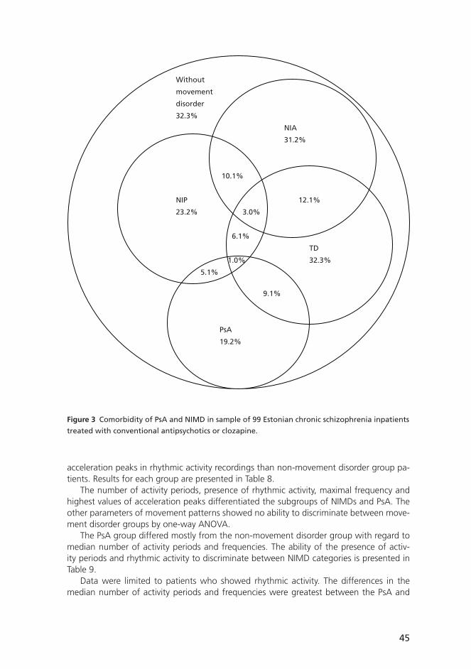
45
acceleration peaks in rhythmic activity recordings than non-movement disorder group pa-tients. Results for each group are presented in Table 8.
The number of activity periods, presence of rhythmic activity, maximal frequency and highest values of acceleration peaks differentiated the subgroups of NIMDs and PsA. The other parameters of movement patterns showed no ability to discriminate between move-ment disorder groups by one-way ANOVA.
The PsA group differed mostly from the non-movement disorder group with regard to median number of activity periods and frequencies. The ability of the presence of activ-ity periods and rhythmic activity to discriminate between NIMD categories is presented in Table 9.
Data were limited to patients who showed rhythmic activity. The differences in the median number of activity periods and frequencies were greatest between the PsA and
Figure 3 Comorbidity of PsA and NIMD in sample of 99 Estonian chronic schizophrenia inpatients
treated with conventional antipsychotics or clozapine.
Without
movement
disorder
32.3%
NIA
31.2%
10.1%
NIP 12.1%
23.2% 3.0%
6.1%
TD
1.0% 32.3%
5.1%
9.1%
PsA
19.2%

46
Tab
le 8
Neu
role
pti
c-in
du
ced
mo
vem
ent
dis
ord
er (
NIM
D)
pat
tern
s fo
r 99
inp
atie
nts
wit
h s
chiz
op
hre
nia
.
Pres
ence
of
acti
vity
p
erio
ds
n %
Med
ian
n
um
ber
o
f ac
tivi
ty
per
iod
sIQ
Pres
ence
of
rhyt
hm
ic
acti
vity
n %
Med
ian
d
om
inan
t fr
equ
ency
Hz
IQ
Med
ian
o
f se
con
d
freq
uen
cyH
zIQ
Med
ian
o
f th
ird
fr
equ
ency
Hz
IQ
Med
ian
of
min
imal
freq
uen
cyH
zIQ
Med
ian
of
max
imal
fr
equ
ency
Hz
IQ
Med
ian
h
igh
est
valu
e o
f ac
cele
rati
on
p
eaks
IQ
no
n-m
ove
men
t d
iso
rder
n =
32
25 78%
2 1.00
–7.6
324 75
%0.
500.
40–0
.59
0.43
0.40
–0.6
10.
550.
48–0
.78
0.33
0.30
–0.4
50.
650.
50–0
.98
106
57–
193
NIA
n =
31
31 100%
13 5.00
–22.
0030 97
%0.
580.
40–1
.01
0.60
0.45
–0.8
00.
700.
49–1
.01
0.38
0.30
–0.4
51.
150.
74–2
.18
177
81–
226
NIP
n =
23
20 87%
8 1.00
–14.
5016 70
%0.
750.
50–0
.95
0.73
0.54
–1.1
10.
930.
60–1
.09
0.40
0.35
–0.4
91.
050.
65–2
.26
216
140–
250
TD n =
32
30 94%
9.75
3.00
–16.
7529 91
%0.
550.
40–0
.80
0.55
0.45
–0.9
30.
650.
46–0
.95
0.40
0.30
–0.4
50.
950.
63–2
.03
125
75–
206
PsA
n =
19
18 95%
16 10.5
0–25
.50
18 95%
0.80
0.50
–1.5
30.
800.
49–1
.00
0.60
0.49
–1.0
40.
400.
30–0
.43
1.15
0.75
–2.5
812
7 7
9–20
8
All
pat
ien
tsn
= 9
988 89
%8 2.
00–1
7.50
83 84%
0.55
0.40
–0.8
00.
550.
40–0
.80
0.65
0.50
–0.9
80.
350.
30–0
.45
0.90
0.60
–1.4
512
9 8
0–21
3
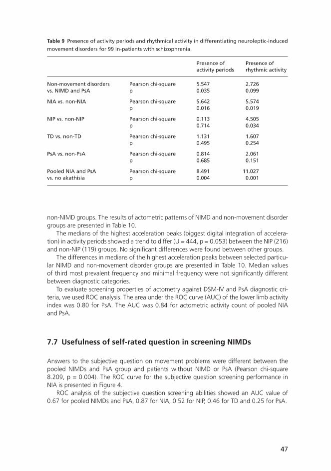
47
non-NIMD groups. The results of actometric patterns of NIMD and non-movement disorder groups are presented in Table 10.
The medians of the highest acceleration peaks (biggest digital integration of accelera-tion) in activity periods showed a trend to differ (U = 444, p = 0.053) between the NIP (216) and non-NIP (119) groups. No signifi cant differences were found between other groups.
The differences in medians of the highest acceleration peaks between selected particu-lar NIMD and non-movement disorder groups are presented in Table 10. Median values of third most prevalent frequency and minimal frequency were not signifi cantly different between diagnostic categories.
To evaluate screening properties of actometry against DSM-IV and PsA diagnostic cri-teria, we used ROC analysis. The area under the ROC curve (AUC) of the lower limb activity index was 0.80 for PsA. The AUC was 0.84 for actometric activity count of pooled NIA and PsA.
7.7 Usefulness of self-rated question in screening NIMDs
Answers to the subjective question on movement problems were different between the pooled NIMDs and PsA group and patients without NIMD or PsA (Pearson chi-square 8.209, p = 0.004). The ROC curve for the subjective question screening performance in NIA is presented in Figure 4.
ROC analysis of the subjective question screening abilities showed an AUC value of 0.67 for pooled NIMDs and PsA, 0.87 for NIA, 0.52 for NIP, 0.46 for TD and 0.25 for PsA.
Table 9 Presence of activity periods and rhythmical activity in differentiating neuroleptic-induced
movement disorders for 99 in-patients with schizophrenia.
Presence of Presence of activity periods rhythmic activity
Non-movement disorders Pearson chi-square 5.547 2.726vs. NIMD and PsA p 0.035 0.099
NIA vs. non-NIA Pearson chi-square 5.642 5.574 p 0.016 0.019
NIP vs. non-NIP Pearson chi-square 0.113 4.505 p 0.714 0.034
TD vs. non-TD Pearson chi-square 1.131 1.607 p 0.495 0.254
PsA vs. non-PsA Pearson chi-square 0.814 2.061 p 0.685 0.151
Pooled NIA and PsA Pearson chi-square 8.491 11.027vs. no akathisia p 0.004 0.001
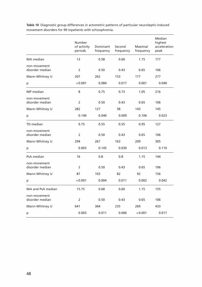
48
Table 10 Diagnostic group differences in actometric patterns of particular neuroleptic-induced
movement disorders for 99 inpatients with schizophrenia.
Median Number highest of activity Dominant Second Maximal acceleration periods frequency frequency frequency peak
NIA median 13 0.58 0.60 1.15 177
non-movement disorder median 2 0.50 0.43 0.65 106
Mann-Whitney U 207 262 153 177 277
p < 0.001 0.084 0.017 0.001 0.040
NIP median 8 0.75 0.73 1.05 216
non-movement disorder median 2 0.50 0.43 0.65 106
Mann-Whitney U 282 127 58 143 145
p 0.140 0.040 0.009 0.106 0.023
TD median 9.75 0.55 0.55 0.95 127
non-movement disorder median 2 0.50 0.43 0.65 106
Mann-Whitney U 294 267 163 209 305
p 0.003 0.145 0.030 0.013 0.110
PsA median 16 0.8 0.8 1.15 144
non-movement disorder median 2 0.50 0.43 0.65 106
Mann-Whitney U 87 103 82 92 156
p < 0.001 0.004 0.011 0.002 0.042
NIA and PsA median 15.75 0.68 0.60 1.15 155
non-movement disorder median 2 0.50 0.43 0.65 106
Mann-Whitney U 641 364 235 269 433
p 0.003 0.011 0.006 < 0.001 0.017

49
Sen
siti
vity
1-specifi city
1.0
0.8
0.6
0.4
0.2
0.00.0 0.2 0.4 0.6 0.8 1.0
Source of the Curve
lower limb activity index
subjective question
Reference line
Figure 4 Receiver Operating Characteristic (ROC) curve for answers to subjective question and
lower limb activity index against DSM-IV defi ned Neuroleptic-Induced Akathisia (NIA).
50
8 Discussion
We studied the prevalence of NIMDs in a naturalistic, globally relevant, schizophrenia in-patient sample that uses conventional neuroleptics and clozapine. We also explored the reliability and validity of different ways of measuring NIMDs. First, we compared the cur-rent diagnostic criteria (DSM-IV) with the most established observer rating scales (BARS, SAS, AIMS), which are also used in diagnosing these disorders. We found a good overlap in NIA and TD diagnostics, but somewhat poorer overlap in NIP, for which we suggested raising the commonly used threshold value. Second, we compared the established observer rating scales to objective motor measurement, the controlled rest lower limb activity meas-ured by actometry. Although quantitative actometry could not differentiate the disorders from each other, it did support the validity of BARS and NIP. However, it did not prove to be a suffi ciently valid instrument for use alone in a naturalistic population with co-morbid NIMDs. Still, actometry may be useful in measuring changes in NIA and NIP in situations where diagnoses have been made with another method. Third, after the failure of quanti-tative actometry to show diagnostic power, we explored more qualitative ways of analysing actometric data and demonstrated some diagnostic power in the case of pooled NIA and PsA data in our population.
8.1 Prevalence of NIMD
We found that nearly two-thirds of patients suffered from a NIMD despite relatively low antipsychotic doses and the use of anticholinergics. The clinical practice in the study population was fi rst to lower the antipsychotic dose and then, if necessary, to add anti-cholinergics.
To our knowledge, this is the fi rst prevalence study to use DSM-IV criteria for identifi ca-tion of NIMDs and one of a few studies identifying the three NIMDs simultaneously. Since the mean antipsychotic dose in the study population was relatively low, we suggest that the prevalence of NIMDs is probably higher at the more commonly recommended anti-psychotic doses.
We found a NIA DSM-IV prevalence of 31%. Previously reported NIA rates measured with BARS (Barnes 1989) are 9–11% in inpatients (van Harten et al. 1996, Modestin et al. 2000) and 15–18% in communities (McCreadie et al. 1992, Halliday et al. 2002). In long-term inpatients, 24% (Halstead et al. 1994) and in depot clinic outpatients 32%, are re-ported to have akathisia (Barnes & Braude 1985), which agrees with our fi nding that long-term patients have high prevalence of akathisia assessed with either DSM-IV or BARS.
We observed a NIP DSM-IV prevalence of 23%. Previously reported NIP prevalence rates measured by clinical impression (mild akinesia not included) are 15% (Ayd 1961) or with SAS 19% in large in- and outpatient surveys (Muscettola et al. 1999), and by UPDRS 20% in inpatients concomitantly using clozapine (Modestin et al. 2000). Higher rates (27–35%) are reported in surveys conducted in restricted areas using SAS (McCreadie at al. 1992, Hal-liday et al. 2002) and in inpatients (36%) using UPDRS (Van Harten et al. 1996), which are much lower than our estimate using the SAS instrument (73%), but are consistent with our estimate using DSM-IV criteria (23%) in long-term patients also receiving clozapine. One reason for the high NIP prevalence measured by SAS in our study could be our long-term
51
patients had more rigidity, which was emphasized in SAS measurement. Muscettola et al. (1999) used slightly modifi ed SAS with less rigidity items.
We found a TD DSM-IV prevalence of 32%. Previously reported TD prevalence rates are 29–32% in in- and outpatients (McCreadie et al. 1992, Mukherjee et al. 1982, Koshino et al. 1992) and 40–43% in long-term patients (Van Harten et al. 1996, Halliday et al. 2002), which agree with our estimate of 32% in long-term patients with a mean age of 50 years.
The prevalence rates of NIMDs defi ned by DSM-IV criteria and those obtained by rating scales were similar for NIA and TD but very different for parkinsonism. This could be due to the different focuses of DSM-IV and SAS criteria.
Despite the relatively low current antipsychotic dosage in our study, the prevalence of EPS was similar to that in previous studies. Thus the longer continuous treatment with antipsychotics, the potency of the antipsychotics and the older age of our patients made the prevalence here higher than in previous studies. Another important factor is that these chronic institutionalized patients had received much higher dosages of antipsychotics in the past.
8.2 Validity of BARS
BARS has been suggested to need objective validation by movement measurement (Barnes 1989). Objective validation by actometry is possible only for the objective BARS item. In our study, the convergence between actometry and BARS (the correlation and the ability to discriminate between NIA and non-NIA groups) provides some objective support for the validity of BARS. This methodological study was the fi rst to compare BARS and actometric lower limb controlled rest activity in NIA in a large naturalistic population with different NIMDs. While both BARS and actometric lower limb controlled rest activity measures differ-entiated the NIA group from the non-NIA group, BARS proved to be superior. Although an earlier report (Tuisku et al. 1999) suggests discriminant validity for quantitative actometry between pure NIA and non-NIA subjects, we were unable to distinguish NIA patients from the naturalistic sample. Tuisku et al. (1999) excluded NIP patients and used healthy people as controls. In our study, the confounding effect of other NIMDs with hyperactivity (PsA, parkinsonian tremor and TD) and possible subclinical forms of NIMD in patients prevented us from reaching a similar level of discriminant validity.
Actometric lower limb activity and the BARS global score correlated weakly but signifi -cantly. The correlation became statistically signifi cant only after excluding or controlling for other NIMDs. The instruments measure somewhat different constructs; actometry records only movements, while BARS includes subjective experience in its score.
8.3 Validity of SAS
According to ROC analysis, SAS had good case-fi nding properties, converging with the DSM-IV NIP diagnosis. In our population, the commonly used cut-off threshold of SAS mean score 0.3 was inappropriate: according to our results, the optimal cut-off point should be between 0.65 and 0.95, depending on the emphasis in the trade-off between sensitivity

52
and specifi city. We therefore suggest that the cut-off value for screening of NIP should be 0.65, whereby specifi city is doubled without losing any sensitivity. For diagnostic purposes a combination of high specifi city and high positive predictive value (PPV) is reached at cut-off of 0.75 (Naarding et al. 2002).
The internal consistency of SAS was satisfactory, which suggests at least moderate reliability of the scale and is consistent with an earlier report (Loonen et al. 2000). We compared SAS with DSM-IV to assess the former’s discriminant validity and to evaluate its ability to detect NIP cases.
The comparison with objective movement assessment aimed to estimate the concur-rent validity of SAS in NIMD diagnosing. As expected, the SAS had discriminant validity for a clinical diagnosis of NIP. The SAS mean score discriminated NIMD patients well from those without NIMD, and more specifi cally, also NIP patients from other patients. Actom-etry discriminated NIMD patients from non-NIMD patients, but did not identify DSM-IV NIP patients. To investigate criticism about the overrepresentation of rigidity items (Cunning-ham Owens 1999, Loonen et al. 2000), we averaged the six rigidity items to form one item. This procedure worsened the NIP case detection capacity of SAS. Using the single elbow rigidity item for case detection yielded the same (or slightly better) case detection capacity than the SAS mean score. This fi nding supports the use of elbow rigidity testing when as-sessing parkinsonism in everyday practice, as the cut-off value of 0.5 has a good sensitivity and specifi city for DSM-IV NIP. Rating “0” means “normal” and “1” means “slight stiffness and resistance”.
In conclusion, SAS seems to be a reliable and valid instrument. It performs similarly to DSM-IV in NIP case detection. The optimal SAS mean score cut-off value in a naturalistic population of neuroleptic-treated schizophrenia patients is, however, higher than the com-monly used 0.3. We suggest that the new cut-off value for screening of NIP could be 0.65, whereby specifi city is doubled without losing sensitivity. Pooling the SAS rigidity items did not improve the performance of the scale.
8.4 Lower limb controlled rest actometry in NIMD
We failed to detect subclinical movement disorders because we used DSM-IV (American Psychiatric Association 2000) and PsA (Rapoport et al. 1984, Barnes & Braude 1985) diag-nostic criteria as the golden standards. Differences between Parkinsonian disease sub clinical resting tremor from controls in terms of amplitude fl uctuation, frequency dispersion, har-monic index and proportional power in 4.4–6 Hz have, however, been reported by Beuter et al. (2005) using laser system measuring displacement.
8.4.1 Comparison of quantitative actometry and BARS
Both measures differentiated NIA from non-NIA group, but BARS proved to be superior to actometry in fi nding NIA cases. Although an earlier report has suggested discriminant validity for quantitative actometry between pure NIA and non-NIA subjects (Tuisku et al. 1999), we were unable to accurately identify NIA patients in the naturalistic sample. This may be due to the confounding effect of other NIMDs (PsA, parkinsonian tremor and TD) manifesting with hyperactivity. With cut-off 12000 the sensitivity of actometry was 0.9 in
53
fi nding akathisia cases. Asking about subjective symptoms (e.g., BARS awareness item) is still needed to rule out other NIMDs in a naturalistic population. Minor changes may be diffi cult to detect with BARS as compared with actometry. Quantitative lower limb activity index has proved to be a sensitive follow-up tool (Tuisku et al. 2002).
The convergence between actometry and BARS provides some objective support for the validity of BARS.
8.4.2 Comparison of quantitative actometry and SAS
We found that neither SAS mean score nor hypokinesia observed during the SAS gait item correlated with actometric lower limb activity. There are a few explanations for this: Firstly, actometry measures only the productive motor dimension of parkinsonian symptoms, while SAS also takes into account rigidity, gait, salivation and glabella tap, with a clear emphasis on rigidity. The lack of correlation with actometric fi ndings in the NIP subgroup indicates that tremor may not be the core feature of NIP. This is also supported by the small AUC for the SAS tremor item. Our fi nding is consistent with previous studies, which have reported tremor in 60% (Ayd 1961) and 44% (Hassin-Baer et al. 2001) of NIP patients. Secondly, we used lower limb actometry, although the clinical assessment by SAS and DSM-IV considers predominantly upper limbs. NIP may be more symptomatic in the upper limbs (Hassin-Baer et al. 2001), and upper limb disturbances may have infl uenced our SAS and DSM-IV assess-ments more than lower limb disturbances. Our fi ndings indicate that lower limb actometry is not suitable for diagnosing NIP. Thirdly, diurnal naturalistic actometry may have more power than controlled rest activity window in detecting hypokinesia.
8.4.3 Descriptive actometry in different NIMDs
Actometry in NIA and PsA
Our fi ndings of actometric patterns of NIMDs and PsA agree with those of a previous accelero metric study (Rapoport et al. 1994), reporting no differences between clinical ob-servations and accelerometric recordings of NIA and PsA.
Sachdev (1995a) has proposed that PsA is not a subtype of NIA. Differences in median values of actometric count for the PsA group and the non-movement disorder group in our study validates the diagnosis of PsA. These patients have higher frequency movements than other patients, but are unaware of them (according to answers to our subjective question). They have diffi culties in noticing their disturbance, which clearly distinguishes this disorder from NIA.
Actometry in NIP
The parkinsonian tremor shows rhythmic activity in accelerometric recordings, discriminat-ing NIP patients from non-movement disorder patients. NIP patients had higher peaks of accelerations in activity periods than non-NIP patients. Comparing the NIP group with the non-movement disorder group made this fi nding more signifi cant. In our descriptive analysis, the frequencies of rhythmic activity in NIP were much lower (median below 1 Hz) than previously reported (above 4 Hz) (Bathien et al. 1984, Rapoport et al. 1998, Caligiuri

54
and Tripp 2004). One explanation may be that other NIMDs and PsA confound the results. Another possible explanation is that by measuring lower limb activity in the sitting posi-tion we could not detect high-frequency NIP tremor. The latter is characteristic for upper limbs – all fourteen NIP patients had tremor in the upper limbs and three had tremor in accelero metric recordings of the lower limbs (Rapoport 1998), and another accelerometric study also measured tremor from the upper limbs (Caligiuri and Tripp 2004). There may have been a high-frequency tremor (close to 10 Hz) in our sample that we were not able to detect with our method due to limitations in the time window of our actometers.
Actometry in TD
Recording of movements of the ankle gave little information for detecting TD, despite the movement pattern of TD having more activity periods, more rhythmic activity, and slightly higher frequencies than non-movement disorder patterns. This is explained by TD affect-ing other body segments than ankles, mostly the orofacial region and sometimes the legs. Another explanation could be that the non-movement disorder group included subclinical NIMD patients, who went undetected using DSM-IV criteria for NIMD. The second expla-nation is supported by the following fi ndings: (1) up to 75% of non-movement disorder group patients by DSM-IV or PsA criteria showed rhythmic activity that might be related to movement disorders, seldom normal activity during rest; and (2) non-movement disorder patients had less activity periods in recordings, but when they had them, the frequencies were quite similar to those of NIMD (mostly TD) patients.
8.5 Subjective question
Almost all NIA patients have reported subjective complaints of movement problems in the lower limbs in previous studies (Braude et al. 1983, Sachdev & Kruk 1994). The subjective question on movement problems in our study was able to distinguish NIA patients from all other subjects. Answers to this question were highly selective for NIA cases, but not for other NIMDs or PsA.
8.6 Methodological issues
A strength of our study is the naturalistic non-selected inpatient sample. The sample was gathered from the largest nursing home in Estonia; approximately 13% of all institutional-ized psychiatric patients in Estonia are treated there. The sample is representative of schizo-phrenia and schizoaffective patients in the nursing home since 91% of all patients with these diagnoses aged 18–65 years participated. Medication of the sample represents typi-cal medication available in state institutions for schizophrenia patients in Estonia (Jaanson 2002). We can therefore generalize our results to other chronic schizophrenia inpatients, treated with mostly typical antipsychotics.
We achieved excellent interrater reliability in descriptive evaluation of actometric data with a relatively short training of inexperienced raters with no previous actometric rating skills (Study IV).

55
We used 30-min controlled rest activity recorded with actometry because Tuisku et al. (1999) reported signifi cant differences in controlled rest activity of NIA patients compared with controls. A longer recording time (14.5 hours) did not show signifi cant differences between these groups (Tuisku et al. 1999) and would be impractical in our study design.
We used several assessment methods for NIMDs simultaneously, which allowed us to compare the diagnostic criteria of rating scales and an objective assessment method, ac-tometry.
The main limitations of this study are connected to the nature of NIMD: (1) co-occur-rence of spontaneous movement disorders, commonly detected in schizophrenia popula-tions (Chatterjee et al. 1995), could not be excluded, and (2) the DSM-IV diagnoses of NIMDs in this study, as in clinical settings in general, are partially based on retrospective information.
The co-morbid movement disorders that exist in this real-life sample may have partially overlapping characteristics. The differentiating ability of actometry was not high for indi-vidual NIMDs.
Another limitation of the study is that for practical reasons the clinical diagnosis of NIMD or PsA, the ratings for scales and the subjective question was all assessed by the same clinician, which could result in judgement bias. Furthermore, as there was only one rater for the scales, a cross-scale contamination might have occurred.
DSM-IV was used as the standard in this study; however, little data are available on the validity of the NIMD criteria of DSM-IV. A better golden standard would probably have been an expert-consensus diagnosis. Another limitation here was that subclinical move-ment disorders went undetected since we used DSM-IV (American Psychiatric Association 2000) and PsA (Rapoport et al. 1984, Barnes and Braude 1985) diagnostic criteria as a gold standard.
This study was limited to a few aspects of the utility/validity of SAS: internal consistency, convergence to DSM-IV NIP diagnosis and convergence to objectively measured motor activ ity. Many aspects of the scale’s reliability (e.g. test-retest and interrater reliability) and validity (e.g. construct) were not evaluated. With age, the prevalence of spontaneous NIMDs is known to rise. While our material did not allow a thorough examination of the issue, age did correlate with SAS mean score.
The limitation of actometric data collection was the recording of only ankle activity; recordings from the wrist and legs could give more information, especially in case of NIP, which is manifested more in the upper limbs (Hassin-Baer et al. 2001).

56
9 Conclusions and future considerations
9.1 Conclusions
Chronic schizophrenia populations, similar to our study population, are common world-wide. NIMD affected two-thirds of our study population. Prevention, diagnosis and treat-ment of NIMDs warrant more attention, especially in countries, where typical antipsychotics are frequently used. Our study supported the validity and reliability of DSM-IV diagnostic criteria for NIMD in comparison with established rating scales and actometry. BARS and SAS are valid instruments in diagnosing NIA and NIP, respectively. However, commonly ap-plied SAS threshold for diagnosing NIP should be raised from 0.3 to 0.65. Controlled rest lower limb actometry did not achieve suffi cient specifi city in our population with several co-morbid NIMDs and PsA, but it may be sensitive in measuring changes in NIMDs or PsA. The subjective question on movement disturbances was highly specifi c and cost-effective in detecting NIA, but not other NIMDs or PsA.
9.2 Clinical implications
Nearly two-thirds of institutionalized schizophrenia patients were shown to suffer from adverse effects of conventional antipsychotics. Since the costs of switching to atypical antipsychotics are too high for many countries, other ways of coping with NIMDs must be explored, such as lowering antipsychotic dosage and using clozapine or anticholinergics.
Our subjective question was specifi c for NIA cases, but not for other NIMDs or PsA, probably because of the subjective component of NIA. Posing such a subjective question is a cost-effective way of screening for NIA. Careful questioning of patients is useful method of diagnosing NIA in clinical setting. The use of elbow rigidity testing has good sensitivity and specifi city for DSM-IV NIP. Actometry does not offered any advantages in diagnosing TD in comparison with structured clinical observation using AIMS.
9.3 Implications for research
Actometric lower limb recording and analysis are moderately time-consuming but use-ful for differentiating pooled NIA and PsA patients from other patients in a non-selected populations, but do not discriminating between these two subgroups. Exact recording of a patient’s present state is an advantage of actometry. Because our study was cross-sectional, we were unable to show changes in movement activity index or patterns after changes in risk factors (e.g. dosage, antipsychotic type and time course). Future research should con-centrate on whether actometry is sensitive to change, especially in comparison with rating scales. The fi rst studies have been done already (Adler et al. 1999, Tuisku et al. 1999).
The epidemiology of NIMDs and PsA should be studied in a naturalistic schizo phrenia population using mostly atypical antipsychotics to estimate their prevalence and inci-dence.

57
10 Acknowledgements
I am indebted to all Võisiku Nursing Home patients who participated in this research.My fi rst contact with psychiatric research was in attending in 1998 a Finnish-Estonian
psychiatric research course organized by Riittakerttu Kaltiala-Heino, Matti Holi and Markku Eronen, among others.
Matti Holi, a teacher at the course and my fi rst supervisor, gave me the idea of con-ducting research in NIMD and helped with the study design and analyses. My fi rst three studies were carried out in close co-operation with him and he continued to mentor me afterwards. Without his support, I would not have gotten nearly as far as I have.
Kristian Wahlbeck, my second supervisor, always found time to answer my questions and stressed the importance of straightforward, honest scientifi c research. His support was crucial to my fi nishing this dissertation.
Katinka Tuisku, a student at the course and my third supervisor, guided me in the meth-odology of actometry, helped me in analysing and reporting results, and generously shared her knowledge in the fi eld of movement disorders.
I am indebted to Björn Appelberg for his encouraging feedback of this manuscript.I thank reviewers of this dissertation for constructive comments. Neurologist Seppo
Kaakkola for his up-to-date neurological viewpoints. Psychopharmacologist Hannu Kopo-nen for accurate critical remarks.
I am indebted to Carol Ann Pelli for editing the language of this manuscript.I thank Birgit Aumeste and Andres Valk for rating the actometric data.My colleagues – Ene Ojassalu, Peeter Ott, Veiko Vasar, Katrin Orav, Peeter Jaanson, Pille
Taba, Jakov Shlik – are warmly acknowledged for their valuable contribution to this work.I am indepted to scientifi c librarian Eeva-Liisa Aatola for her patience with my numer-
ous demands.My heartfelt thanks are due to my parents for their love and encouragement.Last, but not least, I thank Riina, Katri-Helena and Marie-Johanna for their understand-
ing and support.Financial support from Finska Läkaresällskapet and the Eli Lilly Estonian Affi liate is grate-
fully acknowledged.
58
11 References
Adler LA, Rotrosen J, Edson R, Lavori P, Lohr J, Hitzemann R, Raisch D, Caligiuri M, Tracy K. Vitamin E treatment for tardive dyskinesia. Veterans Affairs Cooperative Study #394 Study Group. Arch Gen Psychiatry 1999; 56(9): 836–41.
Agnoli A, Ruggieri S, Del Roscio S, Baldassarre M, Bocola V, Denaro A. Abnormal involuntary movements: a study of dopaminergic receptor interaction. Adv Neurol 1983; 37: 305–12.
Alarcon F, Gimenez-Roldan S. Systemic diseases that cause movement disorders. Parkinsonism Relat Disord 2005; 11: 1–18.
Alihanka J, Vaahtoranta K. A static charge sensitive bed. A new method for recording body movements during sleep. Electroencephalogr Clin Neurophysiol 1979; 46(6): 731–4.
Alpert M, Diamond F, Friedhoff AJ. Tremographic studies in tardive dyskinesia. Psychopharmacol Bull 1976; 12: 5–7.
Alvir JM, Lieberman JA, Safferman AZ, Schwimmer JL, Schaaf JA. Clozapine-induced agranulo-cytosis. Incidence and risk factors in the United States. N Engl J Med 1993; 329(3): 162–7.
American Psychiatric Association. Diagnostic and Statistical Manual of Mental Disorders, 4th ed. American Psychiatric Press, Washington DC, 2000.
Ananth J, Sangani H, Noonan JP. Amantadine in drug-induced extrapyramidal signs: a compara-tive study. Int J Clin Pharmacol Biopharm 1975; 11(4): 323–6.
Arnt J, Skarsfeldt T. Do novel antipsychotics have similar pharmacological characteristics? A review of the evidence. Neuropsychopharmacology 1998; 18(2): 63–101.
Aronowitz B, Liebowitz M, Hollander E, Fazzini E, Durlach-Misteli C, Frenkel M, Mosovich S, Garfi nkel R, Saoud J, DelBene D, et al. Neuropsychiatric and neuropsychological fi ndings in conduct disorder and attention-defi cit hyperactivity disorder. J Neuropsychiatry Clin Neurosci 1994; 6(3): 245–9.
Ayd FJ. A survey of drug-induced extrapyramidal reactions. JAMA. 1961; 175: 1054–60.
Barnes TR, Kidger T, Gore SM. Tardive dyskinesia: a 3-year follow-up study. Psychol Med 1983; 13(1): 71–81.
Barnes TRE, Braude WM. Akathisia variants and tardive dyskinesia. Arch Gen Psychiatry 1985; 42: 874–8.
Barnes TRE. A rating scale for drug-induced akathisia. Br J Psychiatry 1989; 154: 672–6.
Barnes TR, McPhillips MA. Critical analysis and comparison of the side-effect and safety profi les of the new antipsychotics. Br J Psychiatry 1999; (38S): 34–43.
Bartzokis G, Wirshing WC, Hill MA, Cummings JL, Altshuler L, May PR. Comparison of electro-mechanical measures and observer ratings of tardive dyskinesia. Psychiatry Res 1989; 27(2): 193–8.
Bazire S. Psychotropic drug directory, Dinton: Quay Books; 2000
Bathien N, Koutlidis RM, Rondot P. EMG patterns in abnormal involuntary movements induced by neuroleptics. J Neurol Neurosurg Psychiatry 1984; 47(9): 1002–8.

59
Beier H. Psykiatri och neurologi måste samarbeta. Motoriska symptom centrala vid psykiatrisk sjukdom. Läkartidningen 1997; 94(36): 3057–61.
Bell RC, Smith RC. Tardive dyskinesia: characterization and prevalence in a statewide system. J Clin Psychiatry. 1978 Jan; 39(1): 39–47.
Bergen JA, Griffi ths DA, Rey JM, Beumont JV. Tardive dyskinesia: fl uctuating patient or fl uctuat-ing rater. Br J Psychiatry 1984; 144: 498–502.
Bergen JA, Carter NB, Craig J, Macfarlane D, Smith EF, Beumont PJ. AIMS ratings--repeatability. Br J Psychiatry 1988; 152: 670–3.
Bergen JA, Eyland EA, Campbell JA, Jenkins P, Kellehear K, Richards A, Beumont PJ. The course of tardive dyskinesia in patients on long-term neuroleptics. Br J Psychiatry 1989; 154: 523–8.
Beuter A, Barbo E, Rigal R, Blanchet PJ. Characterization of subclinical tremor in Parkinson’s disease. Mov Disord. 2005; 20(8): 945–50.
Blacker D, Endicott J. Psychometric Properties: Concepts of Reliability and Validity. In: Ed Rush AJ et al. Handbook of psychiatric measures. Washington, DC: American Psychiatric Association 2000: pp.7–14.
Blaisdell GD. Akathisia: a comprehensive review and treatment summary. Pharmacopsychiatry 1994; 27: 139–46.
Bloem BR, Beckley DJ, van Hilten BJ, Roos RA. Clinimetrics of postural instability in Parkinson’s disease. J Neurol 1998; 245(10): 669–673.
Braude WM, Barnes TR, Gore SM. Clinical characteristics of akathisia. A systematic investigation of acute psychiatric inpatient admissions. Br J Psychiatry 1983; 143: 139–50.
Braude WM, Charles IP, Barnes TR. Coarse, jerky foot tremor: tremographic investigation of an objective sign of acute akathisia. Psychopharmacology(Berl) 1984; 82(1–2): 95–101.
Buchel C, de Leon J, Simpson GM, Gattaz WF. Oral tardive dyskinesia: validation of a measuring device using digital image processing. Psychopharmacology (Berl) 1995; 117(2): 162–5.
Bulgarian Drug Agency. Lekarstvena konsumacia v Republika Blgaria, online report. Accessed 26/10/2005. http: //www.bda.bg/web_bul/main.htm
Caldwell AE. History of psychopharmacology. In Principles of psychopharmacology. Eds: Clark WC, Giudice J, New York: Academic Press, 1978.
Caligiuri MP, Bracha HS, Lohr JB. Asymmetry of neuroleptic-induced rigidity: development of quantitative methods and clinical correlates. Psychiatry Res 1989; 30(3): 275–84.
Caligiuri MP, Lohr JB. Fine force instability: a quantitative measure of neuroleptic-induced dys-kinesia in the hand. J Neuropsychiatry Clin Neurosci 1990; 2(4): 395–8.
Caligiuri MP, Lohr JB, Bracha HS, Jeste DV. Clinical and instrumental assessment of neurolep-tic-induced parkinsonism in patients with tardive dyskinesia. Biol Psychiatry 1991; 29(2): 139–48.
Caligiuri MP, Lohr JB, Jeste DV. Parkinsonism in neuroleptic-naive schizophrenic patients. Am J Psychiatry 1993; 150(9): 1343–8.

60
Caligiuri MP. Portable device for quantifying Parkinsonian wrist rigidity. Mov Disord 1994; 9(1): 57–63.
Caligiuri MP, Lohr JB, Rotrosen J, Adler L, Lavori P, Edson R, Tracy K. Reliability of an instru-mental assessment of tardive dyskinesia: results from VA Cooperative Study #394. Psychopharmacology(Berl) 1997; 132(1): 61–6.
Caligiuri MP, Lacro JP, Jeste DV. Incidence and predictors of drug-induced parkinsonism in older psychiatric patients treated with very low doses of neuroleptics. J Clin Psychopharmacol 1999; 19(4): 322–8.
Caligiuri MP, Tripp RM. A portable hand-held device for quantifying and standardizing tremor assessment. J Med Eng Technol 2004; 28(6): 254–62.
von Campenhausen S, Bornschein B, Wick R, Botzel K, Sampaio C, Poewe W, Oertel W, Siebert U, Berger K, Dodel R. Prevalence and incidence of Parkinson’s disease in Europe. Eur Neuro-psychopharmacol 2005; 15(4): 473–90.
Carlsson A, Lindqvist M. Effect of chlorpromazine or haloperidol on formation of 3-methoxy-tyramine and normetanephrine in mouse brain. Acta Pharmacol Toxicol (Copenh) 1963; 20: 140–4.
Chatterjee A, Chakos M, Koreen A, Geisler S, Sheitman B, Woerner M, Kane JM, Alvir J, Lieber-man JA. Prevalence and clinical correlates of extrapyramidal signs and spontaneous dys-kinesia in nevermedicated schizophrenic patients. Am J Psychiatry 1995; 152(12): 1724–9.
Chien CP, Jung K, Ross-Townsend A, Stearns B. The measurement of persistent dyskinesia by piezoelectric recording and clinical rating scales. Psychopharmacol Bull 1977; 13(3): 34–6.
Chong MY, Tan CH, Fujii S, Yang SY, Ungvari GS, Si T, Chung EK, Sim K, Tsang HY, Shinfuku N. Antipsychotic drug prescription for schizophrenia in East Asia: rationale for change. Psy-chiatry Clin Neurosci 2004; 58: 61–7.
Chong SA, Mythily, Remington G. Clinical characteristics and associated factors in antipsychotic-induced akathisia of Asian patients with schizophrenia. Schizophr Res 2003; 59(1): 67–71.
Chouinard G, Ross-Chouinard A, Annable L, Jones B. The extrapyramidal symptom rating scale. Can J Neurol Sci 1980; 7: 233.
Chouinard G, Annable L, Mercier P, Ross-Chouinard A. A fi ve year follow-up study of tardive dyskinesia. Psychopharmacol Bull 1986; 22: 259–63.
Chouinard G. New nomenclature for drug-induced movement disorders including tardive dys-kinesia. J Clin Psychiatry 2004; 65(S9): 9–15.
Cohen J. A coeffi cient of agreement for nominal scales. Educational and Psychological Measure-ment 1960; 20: 37–46.
Collins P, Lee I, Tyrer P. Finger tremor and extrapyramidal side effects of neuroleptic drugs. Br J Psychiatry 1979; 134: 488–93.
Cortese L, Caligiuri MP, Malla AK, Manchanda R, Takhar J, Haricharan R. Relationship of neuro-motor disturbances to psychosis symptoms in fi rst-episode neuroleptic-naive schizophrenia patients. Schizophr Res 2005; 75(1): 65–75.
61
Crayton JW, Smith RC, Klass D, Chang S, Ericksen SE. Electrophysiological (H-refl ex) studies of patients with tardive dyskinesia. Am J Psychiatry 1977; 134(7): 775–81.
Cronbach LJ: Coeffi cient alpha and the internal structure of tests. Psychometrika 1951; 16: 297–334.
Cross AJ, Crow TJ, Ferrier IN, Johnson JA, Johnstone EC, Owen F, Owens DG, Poulter M. Chemi-cal and structural changes in the brain in patients with movement disorder. Psychopharma-cology 1985; 2: 104–10.
Csernansky JG, Schuchart EK. Relapse and rehospitalisation rates in patients with schizophrenia: effects of second generation antipsychotics. CNS Drugs 2002; 16(7): 473–84.
Cunningham SL, Winkelman JW, Dorsey CM, Lukas SE, Richardson GS, Sholar MB, Hunt A. An electromyographic marker for neuroleptic-induced akathisia: preliminary measures of sen-sitivity and specifi city. J Clin Neuropharmacol 1996; 19: 321–332.
Cunningham Owens DG. A guide to the extrapyramidal side-effects of antipsychotic drugs. Cambridge: Cambridge University Press, 1999.
Dale D, Welk GJ, Matthews CE. Methods for assessing physical activity and challenges for re-search. In: Ed: Welk GJ Physical activity assessments for health-related research. Cham-paign: Human Kinetics Publishers, U.S.A., 2002.
Davis JM, Casper R Antipsychotic drugs: clinical pharmacology and therapeutic use. Drugs 1977; 14: 260–82.
Davis JM, Chen N, Glick ID. A meta-analysis of the effi cacy of second-generation antipsychotics. Arch Gen Psychiatry 2003; 60: 553–64.
Day J, Bentall RP, Wood G, Dewey M. A Self-rating scale for measuring neuroleptic side-effects: Validation in a group of schizophrenic patients. Br J Psychiatry 1995; 166(5): 650–653.
Dean CE, Russell JM, Kuskowski MA, Caligiuri MP, Nugent SM. Clinical rating scales and in-struments: how do they compare in assessing abnormal, involuntary movements? J Clin Psychopharmacol 2004; 24(3): 298–304.
Denham J, Carrick DJ. Therapeutic value of thioproperazine and the importance of the associ-ated neurological disturbances. J Ment Sci 1961; 107: 326–45.
Deniker P. Experimental neurological syndromes and the new drug therapies in psychiatry. Compr Psychiatry 1960; 1: 92–102.
Denney D, Casey D. An objective method for measuring dyskinetic movements in tardive dys-kinesia. Electroencephalogr Clin Neurophysiol 1975; 38(6): 645–6.
Dogu O, Sevim S, Camdeviren H, Sasmaz T, Bugdayci R, Aral M, Keleagasi H, Un S, Louis ED. Prevalence of essential tremor: door-to-door neurologic exams in Mersin Province, Turkey. Neurology. 2003; 61(12): 1804–6.
Eesti Ravimiamet. Eesti ravimistatistika aastaraamat. 2005, online report. Accessed 25/10/2005. http: //www.sam.ee/orb.aw/class=fi le/action=preview/id=5471/Ravimistatistika+aastaraamat+2004.pdf

62
El-Mallakh RS, Pant B, Caudill R, Bacani-Oropilla T. Does peripheral neuropathy allow for the clinical expression of tardive dyskinesia by unmasking central nervous system changes? Med Hypotheses 2001; 57(2): 210–5.
Ertugrul A, Demir B. Clozapine-induced tardive dyskinesia: a case report. Prog Neuro psycho-pharmacol Biol Psychiatry. 2005; 29(4): 633–5.
Fahn S, Elton RL, & Members of the UPDRS Development Committee. Unifi ed Parkinson’s Dis-ease Rating Scale. In Recent Developments in Parskinson’s disease. Eds. Fahn S, Marsden CD, Goldstein M, Calne DB. New Jersey: MacMillan, 1987.
Fann WE, Stafford JR, Malone RL, Frost JD, Richman BW. Clinical research techniques in tardive dyskinesia. Am J Psychiatry 1977; 134: 759–62.
Farde L, Nordström AL, Wiesel FA, Pauli S, Halldin C, Sedvall G. Positron emission tomographic analysis of central D1 and D2 dopamine receptor occupancy in patients treated with classi-cal neuroleptics and clozapine. Arch Gen Psychiatry 1992; 49(7): 538–44.
Faurbye A, Rasch PJ, Petersen PB, Brandborg G, Pakkenberg H. Neurological symptoms in pharmaco therapy of psychoses. Acta Psychiatr Scand 1964; 40: 10–27.
Flashman LA, Flaum M, Gupta S, Andreasen NC. Soft signs and neuropsychological perform-ance in schizophrenia. Am J Psychiatry 1996; 153(4): 526–32.
National Agency for Medicines and Social Insurance Institution. Finnish Statistics on Medicines. Helsinki 2002.
National Agency for Medicines and Social Insurance Institution. Finnish Statistics on Medicines. Helsinki 2005.
Fleischhacker WW, Bergmann KJ, Perovich R, Pestreich LK, Borenstein M, Lieberman JA, Kane JM. The Hillside Akathisia Scale: a new rating instrument for neuroleptic-induced akathisia. Psychopharmacol Bull 1989; 25(2): 222–6.
Fleiss JL. Statistical Methods for Rates and Proportions (2: nd edition). New York: John Wiley & Sons. 1981: p.218.
Friedmann JH. Historical perspective on movement disorders. J Clin Psychiatry 2004; 65(S9): 3–8.
Gardos G, Teicher MH, Lipinski JF, Matthews JD, Morrison L, Conley C, Cole JO. Quantitative assessment of psychomotor activity in patients with neuroleptic-induced akathisia. Prog Neuro-Psychopharmacol Biol Psychiatry 1992; 16(1): 27–37.
Gattaz WF, Buchel C. Assessment of tardive dyskinesia by means of digital image processing. Psychopharmacology (Berl) 1993; 111(3): 278–84.
Gerlach J, Korsgaard S, Clemmesen P, Lauersen AM, Magelund G, Noring U, Povlsen UJ, Bech P, Casey DE. The St. Hans Rating Scale for extrapyramidal syndromes: reliability and validity. Acta Psychiatr Scand 1993; 87(4): 244–52.
Goldstein JM. The new generation of antipsychotic drugs: how atypical are they? Int J Neuro-psychopharmacol 2000; 3(4): 339–49.
Gorny SW, Allen RP. What is an activity count?: A comparison of different methodologies used in wrist actigraphy. Sleep 1999; 22(S1): S52.

63
Gupta S, Andreasen NC, Arndt S, Flaum M, Schultz SK, Hubbard WC, Smith M. Neurological soft signs in neuroleptic-naïve and neuroleptic-treated schizophrenic patients and in normal comparison subjects. Am J Psychiatry 1995; 152(2): 191–6.
Guy W. ECDEU Assessment manual for psychopharmacology. Washington DC: US Government Printing Offi ce, 1976: 534–7.
Haase HJ. The purely neuroleptic effects and its relation to the “neuroleptic treshold”. Acta Psych Belgica 1978; 78(1): 19–36.
Halliday J, Farrington S, Macdonald S, Mac Ewan T, Sharkey V, McCreadie R. Nithsdale Schizo-phrenia surveys 23: movement disorders 20 year review. Br J Psychiatry. 2002; 181: 422–7.
Halstead SM, Barnes TR, Speller JC. Akathisia: prevalence and associated dysphoria in an in-patient population with chronic schizophrenia. Br J Psychiatry 1994; 164(2): 177–83.
Hansen TE, Brown WL, Weigel RM, Casey DE. Underrecognition of tardive dyskinesia and drug-induced parkinsonism by psychiatric residents. Gen Hospital Psychiatry 1992; 14(5): 340–344.
Harper PS. The epidemiology of Huntington’s disease. Hum Genet 1992; 89(4): 365–76.
Hassin-Baer S, Sirota P, Korczyn AD, Treves TA, Epstein B, Shabtai H, Martin T, Litvinjuk Y, Giladi N. Clinical characteristics of neuroleptic-induced parkinsonism. J Neural Transm 2001; 108: 1299–1308.
Havaki-Kontaxaki BJ, Kontaxakis VP, Christodoulou GN. Prevalence and characteristics of pa-tients with pseudoakathisia. Eur Neuropsychopharmacol 2000; 10(5): 333–6.
Hellewell JS. Patients’ subjective experiences of antipsychotics: clinical relevance. CNS Drugs. 2002; 16(7): 457–71.
Healy D. The psychopharmacologists. London: Chapman and Hall, 1996.
Hermann RC, Yang D, Ettner SL, et al. Prescription of antipsychotic drugs by offi ce-based physi-cians in th United States, 1989–1997. Psychiatr Serv 2002; 53: 425–30.
Hobson P, Gallacher J, Meara J. Cross-sectional survey of Parkinson’s disease and parkinsonism in a rural area of the United Kingdom. Mov Disord 2005; 20: 995–8.
Hoff JI, van Hilten BJ, Roos RA. A review of the assessment of dyskinesias. Mov Disord 1999; 14(5): 737–743.
Holloman LC, Marder SR. Management of acute extrapyramidal effects induced by anti psychotic drugs. Am J Health Syst Pharm 1997; 54(21): 2461–77.
Idänpään-Heikkilä J, Alhava E, Olkinuora M, Palva IP. Agranulocytosis during treatment with chlozapine. Eur J Clin Pharmacol 1977; 11(3): 193–8.
Inada T, Yagi G, Miura S. Extrapyramidal symptom profi les in Japanese patients with schizo-phrenia treated with olanzapine or haloperidol. Schizophr Res. 2002; 57(2–3): 227–38.
Inada T, Beasley CM, Tanaka Y, Walker DJ. Extrapyramidal symptom profi les assessed with the Drug-Induced Extrapyramidal Symptom Scale: comparison with Western scales in the clini-cal double-blind studies of schizophrenic patients treated with either olanzapine or halo-peridol. Int Clin Psychopharmacol 2003; 18(1): 39–48.
64
Ismail B, Cantor-Graae E, McNeil T. Neurological abnormalities in schizophrenic patients and their siblings. Am J Psychiatry 1998; 155(1): 84–9.
Jaanson P. Skisofreenia haiglaväline ravi. Uurimus ku”simustiku pohjal (Description of the treat-ment of schizophrenia spectrum psychosis outpatients using a questionnaire). Eesti Arst 2002; 81: 333–7.
Jenner P, Marsden CD. Antiparkinsonian and antidyskinetic drugs. In Ed: Tyrer PJ. Drugs in psy-chiatric practice., London. Butterworths, 1982.
Jeste DV, Wyatt RJ. Changing epidemiology of tardive dyskinesia: an overview. Am J Psychiatry 1981; 138: 297–309.
Jeste DV, Wyatt RJ. Therapeutic strategies against tardive dyskinesia. Two decades of experience. Arch Gen Psychiatry. 1982; 39: 803–16.
Jeste DV, Caligiuri MP. Tardive dyskinesia. Schizophr Bull 1993; 19(2): 303–15.
Jeste DV, Caligiuri MP, Paulsen JS, Heaton RK, Lacro JP, Harris MJ, Bailey A, Fell RL, McAdams LA. Risk of tardive dyskinesia in older patients: a prospective longitudinal study of 266 outpa-tients. Arch Gen Psychiatry 1995; 52: 756–65.
Jones V, Prior M. Motor imitation abilities and neurological signs in autistic children. J Autism Dev Disord 1985; 15(1): 37–46.
Jung HY, Kim JH, Ahn YM, Kim SC, Hwang SS, Kim YS. Liverpool University Neuroleptic Side-Effect Rating Scale (LUNSERS) as a subjective measure of drug-induced parkinsonism and akathisia. Hum Psychopharmacol 2005; 20(1): 41–5.
Jus K, Jus A, Villeneuve A. Polygraphic profi le of oral tardive dyskinesia and of rabbit syndrome: for quantitative and qualitative evaluation. Dis Nerv Syst 1973; 34(1): 27–32.
Kane JM, Jeste DV, Barnes TRE et al. Tardive dyskinesia: A task force report of the American Psychiatric Association. Washington, DC: American Psychiatric Association, 1992.
Kapur S, Zipursky R, Jones C, Remington G, Houle S. Relationship between dopamine D2 oc-cupancy, clinical response, and side effects: a double-blind PET study of fi rst-episode schizo-phrenia. Am J Psychiatry 2000; 157: 514–20.
Kawashima M, Miyake M, Kusumi M, Adachi Y, Nakashima K. Prevalence of progressive supra-nuclear palsy in Yonago, Japan. Mov Disord 2004; 19(10): 1239–40.
Kennard MA. Value of equivocal signs in neurologic diagnosis. Neurology 1960: 10: 753–764.
Kennedy PF, Hershon HI, McGuire RJ. Extrapyramidal disorders after prolonged phenothiazine therapy. Br J Psychiatry 1971; 118(546): 509–18.
Kern RS, Green MF, Satz P, Wirshing WC. Patterns of manual dominance in patients with neuro-leptic-induced movement disorders. Biol Psychiatry 1991; 30(5): 483–92.
Kiang M, Daskalakis ZJ, Christensen BK, Remington G, Kapur S. Actigraphic measurement of the effects of single-dose haloperidol and olanzapine on spontaneous motor activity in normal subjects. J Psychiatry Neurosci 2003; 28(4): 293–9.
Kim JH, Jung HY, Kang UG, Jeong SH, Ahn YM, Byun HJ, Ha KS, Kim YS. Metric characteristics of the drug-induced extrapyramidal symptoms scale (DIEPSS): a practical combined rating scale for drug-induced movement disorders. Mov Disord 2002; 17(6): 1354–9.

65
Kim JH, Byun HJ. Prevalence and characteristics of subjective akathisia, objective akathisia, and mixed akathisia in chronic schizophrenic subjects. Clin Neuropharmacol 2003; 26(6): 312–6.
Klawans HL, Rubovits R. An experimental model of tardive dyskinesia. J Neural Transm 1972; 33(3): 235–46.
Klawans HL, Barr A. Prevalence of spontaneous lingual-facial-buccal dyskinesias in the elderly. Neurology 1982; 32(5): 558–9.
Koshino Y, Madokoro S, Ito T, Horie T, Mukai M, Isaki K. A survey of tardive dyskinesia in psychi-atric inpatients in Japan. Clin Neuropharmacol 1992; 15(1): 34–43.
Krebs MO, Gut-Fayand A, Bourdel MC, Dischamp J, Olie JP. Validation and factorial structure of a standardized neurological examination assessing neurological soft signs in schizophrenia. Schizophrenia Res 2000; 45: 245–260.
Kronholm E, Alanen E, Hyyppa MT. Nocturnal motor activity in a community sample. Sleep 1993; 16(6): 565–71.
Laidlaw ST, Snowden JA, Brown MJ. Aplastic anaemia and remoxipride. Lancet 1993; 342: 1245.
Lane RD, Glazer WM, Hansen TE, Berman WH, Kramer SI. Assessment of tardive dyskinesia using the Abnormal Involuntary Movement Scale. J Nerv Ment Dis 1985; 173(6): 353–7.
Lanska DJ. Nineteenth-century contributions to the mechanical recording of postural sway. Arch Neurology 2001; 58(7): 1147–50.
Latvian State Medicines Pricing and Reimbursement Agency (Zalu Cenu Valsts Agentura). 2004. gada kompensejamo zalu paterina statistika. Online report. Accessed 15/11/2005. http: //www.zca.gov.lv/docs/new2005/doc21-001.pdf
Legnani G, Zappa B, Casolo F, Adamini R, Magnani PL. A model of an electro-goniometer and its calibration for biomedical applications. Med Engineering Physics 2000; 22(10): 711–22.
Lehmann HE, Ban TA, Saxena BM. A survey of extrapyramidal manifestations in the inpatient population of a psychiatric hospital. Laval Med 1970; 41(7): 909–16.
Liebermann JA, Kane JM, Alvir J. Provocative tests with psychostimulant drugs in schizophrenia. Psychopharmacology (Berl) 1987; 91(4): 415–33.
Lieberman JA, Safferman AZ. Clinical profi le of clozapine: adverse reactions and agranulo cytosis. Psychiatr Q 1992; 63(1): 51–70.
Lieberman JA, Tollefson G, Tohen M, Green AI, Gur RE, Kahn R, McEvoy J, Perkins D, Sharma T, Zipursky R, Wei H, Hamer RM, HGDH Study Group. Comparative effi cacy and safety of atypical and conventional antipsychotic drugs in fi rst-episode psychosis: a randomized, dou-ble-blind trial of olanzapine versus haloperidol. Am J Psychiatry 2003; 160(8): 1396–404.
Lieberman JA. Dopamine partial agonists: a new class of antipsychotic. CNS Drugs 2004; 18(4): 251–67.
Lieberman JA, Stroup TS, McEvoy JP, Swartz MS, Rosenheck RA, Perkins DO, Keefe RS, Davis SM, Davis CE, Lebowitz BD, Severe J, Hsiao JK. Effectiveness of antipsychotic drugs in pa-tients with chronic schizophrenia. N Engl J Med 2005; 353(12): 1209–23.

66
Lima AR, Bacalcthuk J, Barnes TR, Soares-Weiser K. Central action beta-blockers versus pla-cebo for neuroleptic-induced acute akathisia. Cochrane Database Syst Rev. 2004; (4): CD001946.
Lima AR, Weiser KV, Bacaltchuk J, Barnes TR. Anticholinergics for neuroleptic-induced acute akathisia. Cochrane Database Syst Rev 2004; (1): CD003727.
Lipinski JF, Hudson JI, Cunningham SL, Aizley HG, Keck PE, Mallya G, Aranow RB, Lukas SE. Polysomnographic characteristics of neuroleptic-induced akathisia. Clin Neuropharmacol 1991; 14(5): 413–9.
Litvan I, Paulsen JS, Mega MS, Cummings JL. Neuropsychiatric assessment of patients with hyper kinetic and hypokinetic movement disorders. Arch Neurol 1998; 55(10): 1313–9.
Lohr JB, Caligiuri MP. Quantitative instrumental measurement of tardive dyskinesia: a review. Neuropsychopharmacology 1992; 6(4): 231–9.
Loonen AJM, Doorschot CH, van Hemert DA, Oostelbos MCJM, Sijben AES. The Schedule for the assessment of drug-induced movement disorders (SADIMoD): test-retest reliability and concurrent validity. Int J Neuropsychopharmacol 2000; 3: 285–96.
Louza MR, Bassitt DP. Maintenance treatment of severe tardive dyskinesia with clozapine: 5 years’ follow-up. J Clin Psychopharmacol 2005; 25(2): 180–2.
Lyra da Silva JP, Soares-Weiser KV, McGrath JJ. Non-neuroleptic catecholaminergic drugs for neuroleptic-induced tardive dyskinesia. Cochrane Database Syst Rev 2005; (4): CD00429.
Magliano L, Fiorillo A, Guarneri M, Marasco C, De Rosa C, Malangone C, Maj M, National Mental Health Project Working Group. Prescription of psychotropic drugs to patients with schizophrenia: an Italian national survey. Eur J Clin Pharmacol 2004; 60(7): 513–22.
Malhotra AK, Litman RE, Pickar D. Adverse effects of antipsychotic drugs. Drug Saf 1993; 9(6): 429–36.
Marsden CD, Jenner P. The pathophysiology of extrapyramidal side-effects of neuroleptic drugs. Psychol Med 1980; 10: 55–72.
May PRA, Lee MA, Bacon RC. Quantitative assessment of neuroleptic-induced extrapyramidal symptoms: Clinical and nonclinical approaches. Clin Neuropharmacol 1983; 6(S1): 35–51.
McGrath JJ, Soares-Weiser KV. Neuroleptic reduction and/or cessation and neuroleptics as spe-cifi c treatments for tardive dyskinesia. Cochrane Database Syst Rev. 2000; (2): CD000459.
McCreadie RG, Robertson LJ, Wiles DH. The Nithsdale schizophrenia surveys. IX: Akathisia, parkinsonism, tardive dyskinesia and plasma neuroleptic levels. Br J Psychiatry 1992; 160: 793–9.
Meltzer Hy, Matsubara S, Lee JC. Classifi cation of typical and atypical antipsychotic drugs on the basis of dopamine D-1, D-2 and serotonin2 pKi values. J Pharmacol Exp Ther 1989; 251(1): 238–46.
Miller CH, Mohr F, Umbricht D, Woerner M, Fleischhacker WW, Lieberman JA. The prevalence of acute extrapyramidal signs and symptoms in patients treated with clozapine, risperidone, and conventional antipsychotics. J Clin Psychiatry 1998; 59(2): 69–75.
67
Mindham RH, GaindR, Anstee BH, Rimmer L. Comparison of amantadine, orphenadrine, and placebo in the control of phenothiazine-induced Parkinsonism. Psychol Med 1972; 2(4): 406–13.
Modestin J, Stephan PL, Erni T, Umari T. Prevalence of extrapyramidal syndromes in psychiatric inpatients and the relationship of clozapine treatment to tardive dyskinesia. Schizophr Res 2000; 42(3): 223–30.
Morgenstern H, Glazer WM. Identifying risk factors for tardive dyskinesia among long-term outpatients maintained with neuroleptic medications. Results of the Yale Tardive Dyskinesia Study. Arch Gen Psychiatry 1993; 50(9): 723–33.
Mukherjee S, Rosen AM, Cardenas C, Varia V, Olarte S. Tardive dyskinesia in psychiatric out-patients: a study of prevalence and association with demographic, clinical, and drug history variables. Arch Gen Psychiatry 1982; 39(4): 466–9.
Munetz MR, Cornes CL. Akathisia, pseudoakathisia and tardive dyskinesia: clinical examples. Compr Psychiatry 1982; 23(4): 345–52.
Munetz MR, Benjamin S. How to examine patients using the Abnormal Involuntary Movement Scale. Hosp Community Psychiatry 1988; 39(11): 1172–7.
Murphy JM, Berwick DM, Weinstein MC, Borus JF, Budman SH, Klerman GL. Performance of screening and diagnostic tests: application of receiver operating characteristic analysis. Arch Gen Psychiatry 1987; 44: 550–555.
Murray CJ, Lopez A. Global mortality, disability, and the contribution of risk factors: Global Bur-den of Disease Study. Lancet 1997; 349(9063): 1436–42.
Muscettola G, Barbato G, Pampallona S, Casiello M, Bollini P. Extrapyramidal syndromes in neuro-leptic-treated patients: prevalence, risk factors, and association with tardive dys kinesia. J Clin Psychopharmacol 1999; 19(3): 203–8.
Naarding P, Leentjens AF, van Kooten F, Verhey FR. Disease-specifi c properties of the Rating Scale for Depression in patients with stroke, Alzheimer’s dementia, and Parkinson’s disease. J Neuropsychiatry Clin Neurosci 2002; 14(3): 329–34.
Nagamine M, Yoshino A, Sakurai Y, Sanga M, Takahashi R, Nomura S. Exacerbating factors in neuroleptic malignant syndrome: comparisons between cases with death, sequelae, and full recovery. J Clin Psychopharmacol 2005; 25: 499–501.
Nagels G, Marion P, Pickut BA, Timmermans L, De Deyn PP. Actigraphic evaluation of handed-ness. Electroencephalography Clin Neurophysiology 1996; 101(3): 226–32.
Nath U, Ben-Shlomo Y, Thompson RG, Morris HR, Wood NW, Lees AJ, Burn DJ. The prevalence of progressive supranuclear palsy (Steele-Richardson-Olszewski syndrome) in the UK. Brain 2001; 124(Pt 7): 1438–49.
National Health Service. Prescription Pricing Authority: NHS 2003. Accessed 26/10/2005. http: //www.ppa.org.uk/index.htm.
Nilsson FM, Hansen BL, Buchel C, Gattaz WF, Gerlach J. Digital movement analysis, a new method of measuring tardive dyskinesia and drug-induced parkinsonian tremor: accept-ability, reliability and validity. Eur Arch Psychiatry Clin Neurosci 1996; 246(2): 71–77.

68
Nishikawa T, Tanaka M, Tsuda A, Kuwahara H, Koga I, Uchida Y. Effect of ceruletide on tardive dyskinesia: a pilot study of quantitative computer analyses on electromyogram and micro-vibration. Psychopharmacology (Berl) 1986; 90(1): 5–8.
Nishimatsu O, Horiguchi J, Inami Y, Sukegawa T, Sasaki A. Periodic limb movement disorder in neuroleptic-induced akathisia. Kobe J Med Sci 1997; 43(5): 169–77.
Owen RR, Feng W, Thrush CR, et al. Variations in prescribing practices for novel antipsychotic agents among Veteran’s Affairs hospitals. Psyciatr Serv 2001; 52: 1523–5.
Philpott NJ, Marsh JC, Gordon-Smith EC, Bolton JS. Aplastic anaemia and remoxipride. Lancet 1993; 342: 1244–5.
Perenyi A, Bagdy G, Arato M, Frecska E. Biochemical markers in the study of clinical effects and extrapyramidal side effects of neuroleptics. Psychiatry Res 1984; 13(2): 119–27.
Poyurovsky M, Shardorodsky M, Fuchs C, Schneidman M, Weizman A. Treatment of neuro-lepticinduced akathisia with the 5-HT2 antagonist mianserin. Br J Psychiatry 1999; 174: 238–42.
Poyurovsky M, Nave R, Epstein R, Tzischinsky O, Schneidman M, Barnes TRE, Weizman A, La-vie P. Actigraphic monitoring (actigraphy) of circadian locomotor activity in schizophrenic patients with acute neuroleptic-induced akathisia. Eur Neuropsychopharmacol 2000; 10: 171–176.
Poyurovsky M, Weizman A. Serotonin-based pharmacotherapy for acute neuroleptic-induced akathisia: a new approach to an old problem. Br J Psychiatry 2001; 179: 4–8.
Rapoport A, Stein D, Grinshpoon A, Elizur A. Akathisia and pseudoakathisia: Clinical observa-tions and accelerometric recordings. J Clin Psychiatry 1994; 55(11): 473–477.
Rapoport A, Stein D, Shamir E, Schwartz M, Levine J, Elizur A, Weizman A. Clinico-tremorgraphic features of neuroleptic-induced tremor. Int Clin Psychopharmacol 1998; 13(3): 115–20.
Resek G, Haines J, Sainsbury P. An ultrasound technique for the measurement of tardive dys-kinesia. Br J Psychiatry 1981; 138: 474–8.
Rice JE, Thompson PD. 5: Movement disorders I: parkinsonism and the akinetic-rigid syndromes. Med J Aust 2001; 174(7): 357–63.a
Rice JE, Thompson PD. 6: Movement disorders II: the hyperkinetic disorders. Med J Aust 2001; 174(8): 413–9.b
Rifkin A, Quitkin F, Kane J, Struve F, Klein DF. Are prophylactic antiparkinson drugs necessary? A controlled study of procyclidine withdrawal. Arch Gen Psychiatry 1978; 35(4): 483–9.
Rondot P, Bathien N. Movement disorders in patients with coexistent neuroleptic-induced tremor and tardive dyskinesia: EMG and pharmacological study. Adv Neurol 1987; 45: 361–6.
Rossini PM, Mauguire F. New trends and advanced techniques in clinical neurophysiology. Am-sterdam. Elsevier, 1990.
Sachdev P, Kruk J. Clinical characteristics and predisposing factors in acute drug-induced aka-thisia. Arch Gen Psychiatry 1994; 51(12): 963–74.
Sachdev P. Research diagnostic criteria for drug-induced akathisia: conceptualization, rationale and proposal. Psychopharmacology (Berl) 1994; 114: 181–6.
69
Sachdev P. The development of the concept of akathisia: a historical overview. Schizophr Res 1995a; 16: 33–45.
Sachdev P. The epidemiology of drug-induced akathisia: Part 1. Acute akathisia. Schizophr Bull 1995b; 21(3): 431–61.
Sachdev P. Early extrapyramidal side-effects as risk factors for later tardive dyskinesia: a prospec-tive study. Aust N Z J Psychiatry 2004; 38(6): 445–9.
Sachdev P. Neuroleptic-induced movement disorders: an overview. Psychiatr Clin North Am 2005; 28(1): 255–74.
Sanders RD, Keshavan MS. The neurologic examination in adult psychiatry. From soft signs to hard science. J Neuropsychiatry Clin Neurosci 1998; 10: 395–404.
Schooler NR, Kane JM. Research diagnoses for tardive dyskinesia. Arch Gen Psychiatry 1982; 39(4): 486–7.
Schooler NR, Chengappa KNR. Adverse effect measures. In: Eds Rush AJ et al. Handbook of psy-chiatric measures. Washington, DC: American Psychiatric Association 2000: pp.151–68.
Simpson GM, Angus JWS. A rating scale for extrapyramidal side effects. Acta Psych Scand 1970; 212S: 11–9.
Simpson GM, Lee JH, Zoubok B, Gardos G. A rating scale for tardive dyskinesia. Psycho-pharmacology (Berl) 1979; 64(2): 171–9.
Smith JM, Kucharski LT, Oswald WT, Waterman LJ. A systematic investigation of TD in inpatients. Am J Psychiatry 1979; 136(7): 918–22.
Smith JM, Baldessarini RJ. Changes in prevalence, severity, and recovery in tardive dyskinesia with age. Arch Gen Psychiatry 1980; 37(12): 1368–73.
Smith RC, Kadewari RP, Rosenberger JR, Bhattacharyya A. Nonresponding schizophrenia: dif-ferentiation by neurological soft signs and neuropsychological tests. Schizophr Bull 1999; 25(4): 813–25.
Soares KV, McGrath JJ. Anticholinergic medication for neuroleptic-induced tardive dyskinesia. Cochrane Database Syst Rev 2000; (2): CD000204.
Soares KV, McGrath JJ, Deeks JJ. Gamma-aminobutyric acid agonists for neuroleptic-induced tardive dyskinesia. Cochrane Database Syst Rev. 2001; (2): CD000203.
Soares KV, McGrath JJ. Calcium channel blockers for neuroleptic-induced tardive dyskinesia. Cochrane Database Syst Rev. 2001; (1): CD000206.a
Soares KV, McGrath JJ. Vitamin E for neuroleptic-induced tardive dyskinesia. Cochrane Database Syst Rev. 2001; (4): CD000209.b
Spiro SW, Spiro JR. Comparing different methodologies used in wrist actigraphy. Sleep Re-view 2001, online article. Accessed 18/9/2005. www.sleepreviewmag.com/Articles.ASP?articleid=S0107F04.
Sprague RL, Kalachnik JE. Reliability, validity, and a total score cutoff for the dyskinesia iden-tifi cation system: condensed user scale (DISCUS) with mentally ill and mentally retarded populations. Psychopharmacol Bull 1991; 27(1): 51–8.
70
Sprague RL, Korach MS, van Emmerick RE, Newell KM. Correlations between kinematic and rating scale measures of tardive dyskinesia in a developmentally disabled population. J Nerv Ment Dis 1993; 181(1): 42–7.
Springer SP, Deutch G. Left brain, right brain. New York: W.H. Freeman and Co, 1993.
Stafford JR, Fann WE. Deanol acetamidobenzoate (Deaner) in tardive dyskinesia. Dis Nerv Syst 1977; 38(12 Pt 2): 3–6.
Stanilla JK, Buchel C, Alarcon J, de Leon J, Simpson GM. Diurnal and weekly variation of tardive dyskinesia measured by digital image processing. Psychopharmacology (Berl) 1996; 124(4): 373–6.
Stubbs JH, Hutchins DA, Mountjoy CQ. Relationship of akathisia to aggressive and self-injurious behaviour: A prevalence study in a UK tertiary referral centre. Int J Psychiatry Clin Pract 2000; 4: 319–25.
Sweet RA, DeSensi EG, Zubenko GS. Reliability and applicability of movement disorder rating scales in the elderly. J Neuropsychiatry Clin Neurosci 1993; 5(1): 56–60.
Taba P, Asser T. Prevalence of Parkinson’s disease in Estonia. Acta Neurol Scand 2002; 106(5): 276–81.
Tammenmaa IA, Sailas E, McGrath JJ, Soares-Weiser K, Wahlbeck K. Systematic review of cholin-ergic drugs for neuroleptic-induced tardive dyskinesia: a meta-analysis of randomized con-trolled trials. Prog Neuropsychopharmacol Biol Psychiatry 2004; 28(7): 1099–107.
Tanguay PE. Pervasive developmental disorders: A 10-year review. J Am Acad Child Adolesc Psychiatry 2000; 39(9): 1079–95.
Tarsy D, Baldessarini RJ. Pharmacologically induced behavioural supersensitivity to apomorphine. Nat New Biol 1973; 245(148): 262–3.
Tarsy D, Baldessarini RJ, Tarazi FI. Effects of newer antipsychotics on extrapyramidal function. CNS Drugs 2002; 16(1): 23–24.
Taylor E. Developmental neuropsychopathology of attention defi cit and impulsiveness. Develop-ment Psychopathology 1999; 11(3): 607–28.
Taylor D, McConnell H, Duncan-McConnell D, Kerwin R. The Maudsley 2001: Prescribing guide-lines, 6th ed. London: Martin Dunitz Ltd, 2001.
Teicher MH. Actigraphy and motion analysis: new tools for psychiatry. Harvard Rev Psychiatry 1995; 3(1): 18–35.
Tison F, Crochard A, Leger D, Bouee S, Lainey E, El Hasnaoui A. Epidemiology of restless legs syn-drome in French adults: a nationwide survey: the INSTANT Study. Neurology. 2005; 65(2): 239–46.
Tollefson GD, Beasley CM, Tamura RN, Tran PV, Potvin JH. Blind, controlled, long-term study of the comparative incidence of treatment-emergent tardive dyskinesia with olanzapine or haloperidol. Am J Psychiatry 1997; 154(9): 1248–54.
Tonelli H, Tonelli D, Poiani GR, Vital MA, Andreatini R. Reliability and clinical utility of a Portu-guese version of the Abnormal Involuntary Movements Scale (AIMS) for tardive dyskinesia in Brazilian patients. Braz J Med Biol Res. 2003; 36(4): 511–4.

71
Trzepacz PT, Webb M. The choreometer: an objective test of chorea during voluntary move-ments. Biol Psychiatry 1987; 22(6): 771–6.
Tryon WW, Pologe B. Accelerometric assessment of tardive dyskinesia. Am J Psychiatry 1987; 144(12): 1584–7.
Tuisku K, Lauerma H, Holi M, Markkula J, Rimon R. Measuring neuroleptic-induced akathisia by three-channel actometry. Schizophr Res 1999; 40(2): 105–10.
Tuisku K, Lauerma H, Holi MM, Honkonen T, Rimon R. Akathisia masked by hypokinesia. Pharmaco psychiatry 2000; 33(4): 147–9.
Tuisku K. Motor activity measured by actometry in neuropsychiatric disorders. Academic disser-tation. Helsinki: Helsinki University Press, 2002.
Umbrich P, Soares KV. Benzodiazepines for neuroleptic-induced tardive dyskinesia. Cochrane Database Syst Rev. 2003; (2): CD000205.
Van Harten PN, Matroos GE, Hoek HW, Kahn RS. The prevalence of tardive dystonia, tardive dyskinesia, parkinsonism and akathisia The Curacao Extrapyramidal Syndromes Study: I. Schizophr Res. 1996; 19(2–3): 195–203.
Van Putten T. Why do schizophrenic patients refuse to take their drugs? Arch Gen Psychiatry 1974; 31: 67–72.
Vrtunski PB, Simpson DM, Meltzer HY. Voluntary movement dysfunction in schizophrenics. Biol Psychiatry 1989; 25(5): 529–39.
Vrtunski PB, Alphs LD, Meltzer HY. Motor mechanisms in schizophrenic patients with tardive dyskinesia. Psychiatry Res 1994; 54(1): 71–86.
Walters AS, Hening W, Rubinstein M, Chokroverty S. Clinical and polysomnographic compari-son of neuroleptic-induced akathisia and the idiopathic restless legs syndrome. Sleep 1991; 14(4): 339–45.
Webster DD. A method of measuring the dynamic characteristics of muscle rigidity, strength, and tremor in the upper extremity. IRE Trans Med Electron 1959; 6: 159–64.
Weiden PJ, Mann JJ, Hass G, Mattson M, Frances A. Clinical nonrecognition of neuroleptic-induced movement disorders: a cautionary study. Am J Psychiatry 1987; 144: 1148–1153.
Weinberger DR. From neuropathology to neurodevelopment. Lancet 1995; 346: 552–7.
Wirshing WC, Freidenberg DL, Cummings JL, Bartzokis G. Effects of anticholinergic agents on patients with tardive dyskinesia and concomitant drug-induced parkinsonism. J Clin Psycho-pharmacol 1989; 9(6): 407–11.
Wirshing WC, Cummings JL, Dencker SJ, May PR. Electromechanical characteristics of tardive dyskinesia. J Neuropsychiatry Clin Neurosci 1991; 3(1): 10–7.
Wirshing WC. Movement disorders associated with neuroleptic treatment. J Clin Psychiatry 2001; 62 Suppl 21: 15–8.
Wojcik JD, Gelenberg Aj, LaBrie RA, Mieske M. Prevalence of tardive dyskinesia in an outpatient population. Compr Psychiatry 1980; 21(5): 370–80.
72
Woolley J, Smith S. Lowered seizure threshold on olanzapine. Br J Psychiatry 2001; 178: 85–6.
World Health Organization. International classifi cation of diseases, tenth revision (ICD-10). Gen-eva: WHO, 1992.
World Health Organization. Mental Health Atlas (online). 2005 (cited 2005 Nov 13). Available from: URL: http: //www.who.int/mental_health/evidence/atlas/
Yasufuku-Takano J, Sakurai M, Kanazawa I, Nagaoka M. Successful treatment of intractable tar-dive dyskinesia with botulinum toxin. J Neurol Neurosurg Psychiatry 1995; 58(4): 511–2.
Young CC, Rose SE, Biden EN, Wyatt MP, Sutherland DH. The effect of surface and internal electrodes on the gait of children with cerebral palsy, spastic diplegic type. J Orthop Res 1989; 7(5): 732–7.