association of pplo infection and antibody response in rats and
TRANSCRIPT
J. Hyg., Camb. (1961), 59, 401 401Printed in Great Britain
Association of PPLO infection and antibody responsein rats and mice
BY RUTH M. LEMCKEThe Lister Institute of Preventive Medicine, London, S.W 1
(Received 26 April 1961)
INTRODUCTION
Although naturally occurring infections with pleuropneumonia-llke organisms(PPLO), such as bronchopneumonia and polyarthritis in rats and pneumonia andcatarrh in mice, have been studied extensively (Klieneberger & Steabben, 1937,1940; Nelson, 1937 a, b, c; Findlay, Mackenzie, MacCallum & Klieneberger, 1939;Woglom & Warren, 1938; Collier, 1939; Edward, 1940, 1947; Sabin, 1941; Preston,1942), the production of specific antibodies as a result of these infections has notbeen systematically investigated.
In recent years there has been increasing interest in PPLO infections of thehuman urogenital tract, in particular in non-gonococcal urethritis. Severalworkers (Melen & Gotthardson, 1955; Stokes, 1955; Card, 1959; Klieneberger-Nobel, 1959) obtained cultural and serological results suggesting that PPLO werethe infecting agents. However, the causal association of PPLO in the tissues andantibodies to PPLO in the blood has been questioned, especially in non-gonococcalurethritis (Freundt, 1953; Nicol & Edward, 1953).
It was therefore decided to study the immunological responses of rats and miceto PPLO infections to provide not only further information about the rat andmouse diseases but also to throw some light on the significance ofPPLO antibodiesfound in man.
PPLO strains MATERIAL AND METHODS
A total of fifteen strains of PPLO were used, four obtained from rats, ten frommice and one from a human patient. The following table gives the designations andsources of the strains.
Strains Kon and R 107 were isolated from the lungs of rats with broncho-pneumonia; Jasmin, a rat polyarthritis strain, was isolated from tumour materialsent by Dr G. Jasmin of Montreal and strain R 38 was isolated from pus in thenose of a rat with catarrh. Strain R 38 is serologically distinct from the lung strainsbut related to Jasmin, although complement fixation tests indicate that Jasminhas antigens not present in R 38. Nine of the mouse strains were newly isolatedfrom mice belonging to two different stocks; further details about their isolationare given later. The tenth strain, DGE had been in culture for many years.Strain H 34 was obtained from the genital tract of a human female.
RUTH M. LEMCKE
MediaThe solid medium was a heart infusion peptone agar base (1.5% agar) containing
Oxoid Yeast extract (0.5 %), desoxyribonucleic acid (20 ,ug./ml.), inactivatedpooled human serum (20 %) and penicillin (50 units/ml.). This was similar to themedium described by Klleneberger-Nobel (1959) for the isolation of human genitalPPLO, but the basal agar was less rich and staphylococcal filtrate was omitted.The addition of thallium acetate (1/4000) was useful in preventing the growth ofcontaminating bacteria in cultures made from mice. The liquid medium consistedof a tryptic-digest horse meat broth with the same supplements. All the strainsgrew better aerobically than anaerobically, so plates were incubated aerobicallyat 370 in a moist atmosphere and observed for 12-14 days before a negative resultwas recorded. Broth cultures were shaken during incubation.
Table 1. Strains of PPLODesignation SourceKon Rat lungR107 Rat lungJasmin Rat tumourR38 Rat noseM I Mouse lungM 2 Mouse lungMB Mouse brainPeter Mouse brain68NP Mouse nasopharynx72L Mouse lung |73B Mouse brain Stock 'B' miceKSA Mouse brain79B Mouse brainDGE Mouse lung (originally isolated by
Dr D. G. ff. Edward)H34 Human genital tract
AnimalsThe albino rats used belonged to a stock derived from breeding animals supplied
by the Chester Beatty Research Institute. Newborn rats were removed aseptic-ally by Caesarean section from two females on the point of delivery. Young ratsless than 3 months old were infected with an inoculum prepared as follows.A piece of agar approximately 1 cm. square from a 24 or 48 hr. plate culture ofPPLO was macerated in a sterile mortar and taken up in 1-2 ml. of liquid medium.Infected material such as pus or necrotic tissue from abscesses, was ground up inthe same way and 0 5 ml. samples ofsuch suspensions were injected subcutaneouslyat one side of the abdomen. Rats were killed with coal gas and blood was removedfrom the heart with a fine capillary pipette. Lesions and other tissues were excisedaseptically, cut up finely and inoculated into appropriate media. Samples fromthe nose and nasopharynx were obtained after swabbing the muzzle with alcoholand removing the external nares; a sterile platinum wire was inserted first throughthe nares and then through the back of the buccal cavity behind the soft palateafter cutting back through the angle of the jaw.
402

PPLO infection in rats and mice
The two mouse stocks, A and B, were commercially available stocks of albinomice. Inoculation was either intracerebral or intranasal, both under ether anaes-thetic. For the intracerebral, 0-03 ml. of a 24 hr. agar culture macerated in brothwas injected; in one experiment the same amount of PPLO-infected brain tissuemacerated in broth was inoculated. For the intranasal, 003-0 05 ml. of eithermacerated agar culture or lung suspension was instilled. Mice were killed withether at various intervals after inoculation and blood was taken from the axillaafter cutting the brachial vessels. The blood from two mice was usually pooled.However, when the lesions were obvious and the individual serum titre was ofparticular interest, individual samples were kept separate, though sometimes,even in these cases, the pooling of two samples was necessary to provide enoughserum for the full tests. Swabs were taken from the nose and nasopharynx in thesame way as from the rats, and tissues required for culture were removed andtreated in the same way as material from rats.
Serological testsThe clotted blood samples were held at 4° C. overnight, the serum separated and
stored at - 25° C. without preservative. Antibody against antigens of variousPPLO strains was estimated by the complement fixation test (CFT) of Card (1959).Sera were tested at 2-fold dilutions between 1/10 and 1/5120. At first both 'fresh'(F) and 'boiled' (B) antigens (Card, 1959) were used, but since rat sera reactedequally well with both F and B antigens only F antigen was used in the laterexperiments and F titres are quoted throughout. As controls, known positive andnegative sera were included in each test. Rat sera were titrated against Kon,Jasmin, R38 and H34 antigens. Mouse sera were tested in the same way usingF antigens of Peter, KSA and H 34.For identification of PPLO isolates, the CFT was carried out with F antigens
and rabbit antisera prepared against known strains. Only F antigens were usedbecause B antigens gave lower titres than F with rabbit antisera and the reactionswith B antigens were less specific. Homologous antigens were titrated along withthe unknown antigens and all were simultaneously titrated against pre-inoculationrabbit serum as a control.
Titres are cited as the reciprocal of the serum dilution and sera not reacting ata dilution of 10 ( < 10) are regarded as negative for antibody.
RESULTS
The naturally occurring lung infection in rats
The incidence of PPLO infection and the serum antibody in rats of differentages was determined from the examination of twelve newborn from two mothers,twenty-six 4-8-week-old and twenty-eight 8-20-month-old rats. Cultures inliquid and solid medium from the twelve newborn rats yielded no PPLO althoughboth mothers had PPLO in the lung and nasopharynx and serum antibody againstthe rat lung strain Kon. It would seem that newborn rats are not infected eitherin utero or immediately post-partum.
403
RUTH M. LEMCKE
Five of the twenty-six 4-8-week-old rats had PPLO in the lung although nonehad any macroscopically visible lesions. Nasopharyngeal swabs were taken fromonly seven rats, but from five of these PPLO were cultured. These results suggestthat although very few young rats have PPLO in the lung, nearly all of themalready have the organism in the nasal passages.
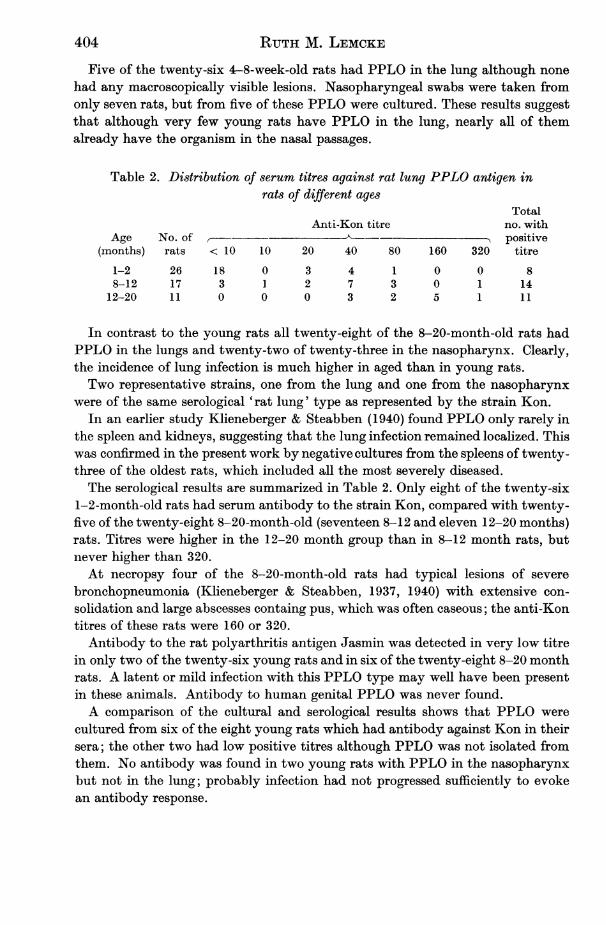
Table 2. Distribution of serum titres against rat lung PPLO antigen inrats of different ages
TotalAnti-Kon titre no. with
Age No. of positive(months) rats < 10 10 20 40 80 160 320 titre
1-2 26 18 0 3 4 1 0 0 88-12 17 3 1 2 7 3 0 1 14
12-20 11 0 0 0 3 2 5 1 11
In contrast to the young rats all twenty-eight of the 8-20-month-old rats hadPPLO in the lungs and twenty-two of twenty-three in the nasopharynx. Clearly,the incidence of lung infection is much higher in aged than in young rats.Two representative strains, one from the lung and one from the nasopharynx
were of the same serological 'rat lung' type as represented by the strain Kon.In an earlier study Klieneberger & Steabben (1940) found PPLO only rarely in
the spleen and kidneys, suggesting that the lung infection remained localized. Thiswas confirmed in the present work by negative cultures from the spleens of twenty-three of the oldest rats, which included all the most severely diseased.The serological results are summarized in Table 2. Only eight of the twenty-six
1-2-month-old rats had serum antibody to the strain Kon, compared with twenty-five of the twenty-eight 8-20-month-old (seventeen 8-12 and eleven 12-20 months)rats. Titres were higher in the 12-20 month group than in 8-12 month rats, butnever higher than 320.At necropsy four of the 8-20-month-old rats had typical lesions of severe
bronchopneumonia (Klieneberger & Steabben, 1937, 1940) with extensive con-solidation and large abscesses containg pus, which was often caseous; the anti-Kontitres of these rats were 160 or 320.Antibody to the rat polyarthritis antigen Jasmin was detected in very low titre
in only two of the twenty-six young rats and in six of the twenty-eight 8-20 monthrats. A latent or mild infection with this PPLO type may well have been presentin these animals. Antibody to human genital PPLO was never found.A comparison of the cultural and serological results shows that PPLO were
cultured from six of the eight young rats which had antibody against Kon in theirsera; the other two had low positive titres although PPLO was not isolated fromthem. No antibody was found in two young rats with PPLO in the nasopharynxbut not in the lung; probably infection had not progressed sufficiently to evokean antibody response.
404

PPLO infection in rats and mice
Experimental infections in ratsKlieneberger-Nobel (1960) investigated two rat polyarthritis strains, Jasmin
and Baxter. Subcutaneous inocula induced, at the site of inoculation, an encap-sulated abscess which increased in size during the first 3-4 weeks and usuallyregressed slowly. Antibody titres rose to 2560 in the first 4-5 weeks after inocu-lation, and then declined gradually during the next 18 weeks. The titre in youngcontrol rats never reached 10. She also found that inocula of human genital PPLOfailed to produce much reaction. At most, a small lump developed at the inocula-tion site, and, although PPLO remained alive in the lesion for about a fortnight,no appreciable antibody was produced.
This work was extended, using the freshly isolated rat lung strain R 107 and thenasal strain R38.
Unlike Jasmin and Baxter, R 107 produced very little on subcutaneous inocula-tion into young rats examined 2-14 weeks later. Small encapsulated, PPLO-containing abscesses arose, but regressed within 6-8 weeks. The spleens were sterileand titres did not exceed 80. These results suggest that the rat lung strain was notvery virulent even when freshly isolated, and produced infection only at the site ofinoculation, just as the natural infection is usually restricted to the nasopharynxand lungs.
Table 3. Cultural and serological results from rats inoculated with rat strain R 38
Duration Culture fromof , Titres
Rat experiment Lymphno. (days) Abscess Spleen nodes Lung Anti-R 38 Anti-Kon
1 8 + + NT + <10 1602 12 + + + + <10 403 12 + + + + 10 404 31 +t - NT + 10 805 31 -* t NT + 320 1606 42 * NT + 320 407 42 + - NT + 40 808 49 +t+ - NT + 1 160 160
* Abscess regressed before necropsy.t Culture tested by CFT and found to be type R 38.1 Culture tested by CFT and found to be type Kon.NT, not tested.
The results of inoculating strain R 38 are given in Table 3. In all of eight ratsabscesses were produced and regressed within 4-6 weeks in two of them. PPLOwere isolated from the spleen or enlarged axillary or inguinal lymph nodes of fourof the rats, indicating that at some stage the infection was systemic. In thisrespect R 38 resembled the polyarthritis strains, but high serum titres like thoseaccompanying polyarthritis infections were not found. The highest titre observedwas 320 in the two rats in which the abscesses had regressed. Thus, strain R 38seemed less virulent than the polyarthritis PPLO. The results of CF tests suggestthat Jasmin and R 38 are related but that Jasmin has antigens not possessed by
405

RUTH M. LEMCKE
R 38; the difference in virulence between the two strains may be associated withthis antigenic difference.Although these rats were only 2-3 months old, PPLO were found in the lung
and antibodies to Kon were present in the sera of all eight. Three isolates fromthe lung were typed (see Table 3) and found to be indistinguishable from Kon.Two isolates from abscesses and one from the spleen were also typed to check thatinfections were not caused by endogenous PPLO; these were identical with theinoculated strain R 38 and distinct from the rat lung type. One abscess containedthe rat lung type as well as the R 38 type, suggesting that PPLO which are normallylocalized in the lung can under some conditions spread to a site of trauma remotefrom their usual habitat.
Naturally occurring infections in miceA natural infection of Stock A mice was discovered when brain suspensions from
two series of mice, one used for the intracerebral passage of trachoma virus, theother a corresponding control series in which brain suspensions alonewerepassaged,proved to be heavily infected with PPLO. The representative strain isolated wasdesignated 'MB'. Other untreated mice selected at random yielded three morestrains, M I and M 2 from the lungs of two mice and Peter from the brain of a third.The four strains were typed serologically using rabbit sera prepared against MIand MB. Five antigens from these four strains were tested because M2 producedtwo colonial variants, and all five reacted to the same titre or to within 25 % of it.The strain DGE obtained from Dr Edward many years ago was also closely related.In view of this and because strain Peter behaved characteristically in mice, asshown in the next section, the type of PPLO to which these strains belong hasbeen designated 'mouse lung'. It is very closely related serologically to the ratlung PPLO represented by the strain Kon. In CF tests with MI, MB and Konantisera it was not possible to distingusish the mouse lung from the rat lung PPLO.Mouse lung PPLO usually had a very characteristic colony on primary culture.It was coarsely granular and, except when very young, was dark brown. Thepigment obscures the typical PPLO colony structure with its dense centre and moretransparent peripheral zone, which is seen only in young colonies. In later sub-cultures the pigment tends to be lost. The colony resembles that of the rat lungstrain in which also the central zone is not clearly differentiated from the peri-pheral area, although it has more pigment than the rat lung strain on primaryculture. When first isolated the growth of both strains in liquid media is finelygranular, just visible with a hand lens by transmitted light. After several sub-cultures, the growth is more evenly turbid.Four-month-old Stock B mice were originally obtained for the production of
ascitic fluid by the intraperitoneal inoculation of Krebs ascites tumour cells. Whenkilled to harvest the ascites cells and fluid some mice proved to be infected withPPLO. Of eleven mice, two had PPLO in the brain and three in the lung, althoughthere were no obvious signs of disease. The colony of one brain isolate, KSA,differed from the mouse lung type in having a small discrete central zone clearlydemarcated from the periphery. It was also distinct serologically from the mouse
406

PPLO infection in rats and mice4
lung type. In CF tests with antisera prepared against M 1 and MB the titre withKSA was 80 compared with homologous titres of 5120 and 1280 for M1 and MBrespectively.As the incidence of lung infection and the amounts of serum antibody in rats
had been found to increase with the age of the animals, it was thought that thismight also happen in mice. Accordingly the presence of PPLO and PPLO anti-bodies in mice of different ages were investigated; the results are given in Table 4.
Table 4. PPLO and antibody found in untreated mice of different ages
No. ofmice No. of PPLO-positivewith cultures from Titre
Age of No. of PPLO- againstMouse mice mice positive Naso- strainStock (weeks) examined cultures pharynx Lung Brain Peter
A 3-4 16 0 0 0 0 < 107-8 16 0 0 0 0 < 1012 5 4 NT 4 NT 160
B 4 16 6 6 3 4 < 10-1016 6 4 4 1 1 40-80
24-32 17 17 15 1 7 80-32014 1* 1 1 1 0 640
* Developed pneumonia spontaneously. NT, not tested.
The sixteen 3-4-week-old Stock A mice were taken from a fresh consignment andwere found to be negative for PPLO both culturally and serologically. Four weekslater another sixteen from the same consignment, now 7-8 weeks old, were againnegative. However, four of 5-12-week-old mice from another consignment ofStock A had PPLO in the lung and a corresponding titre of 160. There appearedto be a difference in the incidence ofPPLO infection between different consignmentsof this stock. Whereas the consignment from which the 3-4- and 7-8-week-oldmice were taken seemed entirely free from PPLO, others were clearly infected,e.g. those from which the strains MB, Ml, M2 and Peter were obtained and those12 weeks old. It is probable that some consignments of 3-4-week-old mice werealready lightly infected when received from the dealer and that PPLO proliferatedand antibodies were produced as the mice aged. The other possibility is that allthe mice were at first free from PPLO but that some acquired the organisms laterfrom other mice or rats on the premises. However, mice were never placed indirect contact with rats or mice of other stocks, and Nelson (1937a) found thatdirect contact was necessary for the transmission of PPLO respiratory infection inrodents. Whichever possibility is correct, it is important to note that a detectablePPLO infection in Stock A mice was accompanied by the presence of serum anti-body against the mouse lung type.
Stock B mice of different ages were also examined (Table 4). Of sixteen mice4 weeks old, six were infected with PPLO but had little or no antibody. At16 weeks old, most of them were infected, PPLO being present in, e.g. four out of
407
RUTH M. LEMCKE
six, with titres against lung PPLO of 40-80, although no lesions were visible.Mice 24-32 weeks old were all infected, with titres of 80 to 320. One 24-week-oldmouse spontaneously developed active pneumonia with consolidation of the lung;a heavy growth of PPLO was obtained from the lung and the titre was 640. Ascan be seen from Table 4, of forty mice of different ages, twenty-six had PPLOin the nasopharynx, six in the lung and twelve in the brain. Sometimes PPLOwere present in both nasopharynx and lung or in nasopharynx and brain; morerarely all three organs were infected. In every age group more mice carried PPLOin the nasopharynx than in the brain or lungs. Since strains 72L, 73B and 68NP,isolated respectively from the lung, brain and nasopharynx of three mice, wereserologically identical and indistinguishable from others of the mouse lung type,the preponderance of positive cultures from the nasopharynx suggests that this isthe primary site of infection and other organs are secondarily invaded. Edward(1947) found that a primary PPLO infection of the upper respiratory tract in micecould spread to the lung and cause pneumonia. The results show that PPLO arealready present in some 4-week-old Stock B mice when they are received from thedealer. The incidence of PPLO infection and the amount of serum antibody bothincrease with the age of the mice.
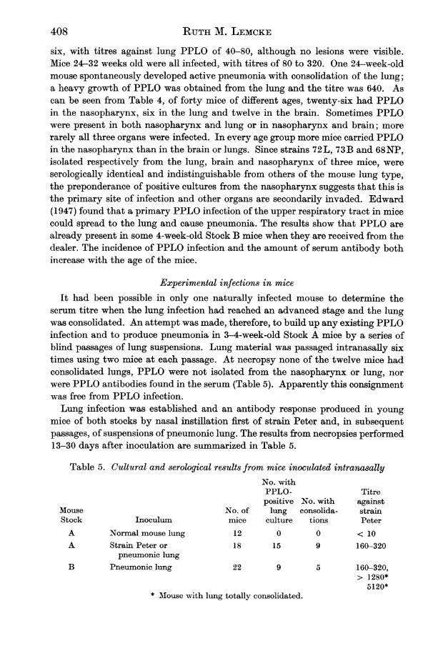
Experimental infections in miceIt had been possible in only one naturally infected mouse to determine the
serum titre when the lung infection had reached an advanced stage and the lungwas consolidated. An attempt was made, therefore, to build up any existing PPLOinfection and to produce pneumonia in 3-4-week-old Stock A mice by a series ofblind passages of lung suspensions. Lung material was passaged intranasally sixtimes using two mice at each passage. At necropsy none of the twelve mice hadconsolidated lungs, PPLO were not isolated from the nasopharynx or lung, norwere PPLO antibodies found in the serum (Table 5). Apparently this consignmentwas free from PPLO infection.Lung infection was established and an antibody response produced in young
mice of both stocks by nasal instillation first of strain Peter and, in subsequentpassages, of suspensions of pneumonic lung. The results from necropsies performed13-30 days after inoculation are summarized in Table 5.
Table 5. Cultural and serological results from mice inoculated intranasallyNo. withPPLO- Titrepositive No. with against
Mouse No. of lung consolida- strainStock Inoculum mice culture tions PeterA Normal mouse lung 12 0 0 < 10A Strain Peter or 18 15 9 160-320
pneumonic lungB Pneumonic lung 22 9 5 160-320,
> 1280*5120*
* Mouse with lung totally consolidated.
408
PPLO infection in rats and mice
Of eighteen Stock A mice inoculated with PPLO or infected lung, fifteen hadthe organisms in the lung and nine partial hepatization of the lung. In all of themthe titre against the infecting strain was 160-320. In comparison with Stock A,fewer of the Stock B mice developed a lung infection; PPLO were isolated fromthe lungs of nine, and five of these had consolidations; in two mice the whole lungappeared hepatized. These two most severely affected animals had titres > 1280and 5120. In the less severely infected mice the titres were 160-320-the same levelas in the infected Stock A mice.PPLO were not isolated from the lungs of all the mice inoculated with PPLO
although all had positive serum titres. This failure may be due in part to thegrowth on four plates, seeded with lung material, of bacteria that inhibit PPLO.The PPLO-negative lung cultures in the remaining inoculated mice were notcontaminated in this way so that the infection was probably confined to the naso-pharynx although it had given rise to detectable antibody. As PPLO, when presentin the upper respiratory tract can produce a catarrhal condition in mice (Edward,1947) such an antibody response is feasible. However, it was only in mice withextensive pneumonia affecting the whole lung that the highest titres were found.
Mice which developed pneumonia became very thin and usually made a charac-teristic chattering noise, but some remained lively even when large parts of thelung were consolidated. Edward (1940) and Nelson (1937a) both reported a lowdeath rate in mice with PPLO pneumonia. Nelson (1937a) never observedabscesses in infected mice, but the lungs of two animals in the present series con-tained yellow nodules which exuded sticky pus when cut.
In only two mice did conjunctivitis accompany the pneumonia, but PPLO werenot isolated from the eyes. Nelson (1950) reported on a PPLO conjunctivitis whichsometimes accompanied PPLO catarrhal infections in mice, but in the presentinvestigations the incidence was too low to determine the aetiology.The strain Peter was also injected intracerebrally into six 3-4-week-old Stock A
mice. No signs of neural damage were observed, but at necropsy 9 days afterinjection small abcesses were present on the skull of two of the mice at the pointof needle entry and PPLO were isolated from the brains of all the mice as well asfrom the abscesses. Abscess material re-inoculated intracerebrally into other miceproduced no lesions and PPLO were not found in the brains 21 days later. ThisPPLO serotype was apparently not neurotoxic. These results agree with those ofEdward (1940) who found that intracerebral inoculation of mouse lung PPLO waswithout effect.
In contrast, strain KSA damaged the brain when introduced intracerebrally;some mice died within 24 hr. and the rest within 72 hr. Of eight mice thus inocu-lated, three exhibited the characteristic 'rolling' described by Findlay, Kliene-berger, MacCallum & Mackenzie (1938). It was not possible to obtain blood fromany of these mice for CFT. Another brain strain, 79 B, with a colonial form distinctfrom that of the mouse lung PPLO and more like that of KSA, was isolated fromuntreated Stock B mice and inoculated intracerebrally into eight mice; five diedwithin 72 hr., one exhibiting 'rolling'. Two of the three survivors developed apronounced hydrocephalus and the characteristic excitability associated with
409
RUTH M. LEMCKE
' rolling disease'. At necropsy 26 days after inoculation, PPLO were still present inthe brain of all three mice; the serum titre of the two with hydrocephalus was 40and of the third 20 against strain KSA but < 10 against strain Peter. Eightcontrol mice given sterile agar emulsion remained healthy throughout and nolesions were observed at necropsy.
These results confirm the conclusion from CF tests that KSA is distinct from themore frequently isolated mouse lung PPLO, and suggest that it is the same as theL5 of Findlay et al. (1938) and the Type A of Sabin (1941), the causative agent ofrolling disease. The serological characters of strain 79B were not determined, butthe symptoms it produced, associated with antibodies to KSA, which appearedonly in mice infected with strain 79 B, suggest that it is very similar to KSA and tothe original L5 strain.
DISCUSSIONCultural results from the study of rat bronchiectasis suggest that newborn rats
are free from PPLO infection, but that they become infected in the first weeks oflife, presumably from the mothers. In young rats the infection is usually confinedto the nasopharynx and at this stage serum antibody is either absent or detectableonly at low titre. As the rats age the lungs are invaded and eventually the lesionsof bronchopneumonia may develop. Higher serum titres are also found as therats age, the highest being associated with severe bronchopneumonia. Thus thechanges in serum antibody levels demonstrated by CFT reflect the progressiveincrease of a chronic infection in rats.
There is rarely any cultural evidence that the infection becomes systemic inthe chronic form of rat bronchiectasis or when the rat lung PPLO is inoculatedsubcutaneously. This is in contrast to infections with rat polyarthritis and relatedstrains such as R 38 which are generalized at some stage, as shown by the presenceof the PPLO in the lymph nodes, spleen and blood. The comparatively low serumtitres, not exceeding 320, which accompany bronchiectasis also suggest a morelocalized infection. In contrast Klieneberger-Nobel (1960) found serum titres of2560 in polyarthritis infections.However, the isolation of both rat lung and R38 strains from a subcutaneous
abscess produced by inoculating R 38, indicates that under certain conditions thelung strain can be disseminated to other parts of the body. This is reminiscent ofa report by Stokes (1955) on a patient who developed a post-operative empyemacaused by PPLO. There was pyrexia lasting some days and a serum titre of 480.The PPLO responsible appeared to be of the human genital type and must presum-ably have been transferred to the pleural cavity via the blood stream from thegenital tract.In view of the fact that seven representative mouse strains from both stocks were
serologically identical with a strain isolated by Dr Edward, the mice probablyhad the same type of respiratory infection as described by Edward (1940, 1947).The lung serotype must be distinct from the PPLO isolated from induced pneu-monia by Sullivan & Dienes (1939), since Sabin (1941) reported that these werethe same as his strain A, which was closely related to the L 5 rolling disease strain.
410

PPLO infection in rats and mice
The relationship of the mouse lung serotype to strains found by Nelson (1937a,b, c) in catarrhal infections and pneumonia of mice and to other isolated by Sabin(1941) from the brain, nose and lung of mice cannot now be established.In mice, just as in rats, the incidence of infection with lung PPLO increased
with the age of the animals; this was shown both by the increasing frequency ofisolation of PPLO and higher serum titres. Although the rat and mouse lungPPLO are related serologically and the primary site of infection in both rats andmice seems to be the nasopharynx, a nasal infection in mice is not always followedby infection of the lung, as inevitably happens in rats. Involvement of the lungin mice with a nasal infection occurs much less regularly than in rats.
In both rats and mice there was a high correlation between the presence ofPPLO demonstrable by culture and the occurrence in the blood of antibodiesspecific to the infecting strain. The level of the serum titre was related to theseverity of the infection in both species.The specificity of the antibody detected by CFT is evident from the results.
Antibody to human genital PPLO was never found in rats or mice. Moreover,when an experimental infection with strain R 38 was superimposed on an existinglung infection in rats, antibodies to both R 38 and rat lung PPLO could be demon-strated. Whereas antibody to the more frequently isolatedlungPPLO wasrepeatedlyfound in mice, antibody to strain KSA was present only in those intracerebrallyinoculated with a related mouse brain strain.
These experiments demonstrate the immune response of rats and mice to variousrat- and mouse-pathogenic PPLO which naturally infect them. Specific serumtitres in these animals reflect the presence and severity of PPLO infection. Asthe PPLO are a closely related group of organisms, it seems permissible to regardthese rat and mouse diseases as immunologically analogous to PPLO infections inother species.
If, then, antibody response in rats and mice signify infection it seems likelythat antibody against human genital PPLO found in man is also indicative of pastor present PPLO infection. It is true that high PPLO titres have been reportedin human patients only infrequently, but when such titres occur it is usually whenthere has been an acute generalized infection (see e.g. Stokes, 1955). In contrast,the titres recorded by Card (1959) in patients with non-gonococcal urethritis seldomexceeded 80. More recently the author has examined sera from women withsalpingitis; again the titres were usually low. However, these genital tract infection,like the rat lung disease tends to remain localized so that a strong antibody responseis not to be expected.
SUMMARY
By means of a complement-fixation test of the sera of laboratory rats and mice,the immunological response of these animals to both naturally occurring andinduced PPLO infections was determined, and the presence and extent of infectionin the animals determined by culture.PPLO antibodies specific for the infecting strain were demonstrable in rats and
mice from which PPLO were isolated.
411
27 Hyg. 59, 4
RUTH M. LEMCKE
The amount of serum antibody rises with the extent and severity of the in-fection. Thus, young rats with PPLO infections confined to the nasopharynx hadlittle or no antibody whereas the oldest rats with consolidated lungs had thehighest titres. In mice too, the sera of those with pneumonia had the highesttitres.The comparatively low titres found in rat bronchiectasis together with the
failure to isolate PPLO from the spleen and other organs, suggest that the chronicform of the disease remains localized. This is in contrast to infections with ratpolyarthritis and related PPLO in which the organisms can be isolated from thelymph nodes and other organs and in which antibody is present in high titre.
In view of the high degree of correlation between the presence of antibodies toPPLO in the blood and the presence of PPLO in the tissue of rats and mice, it issuggested that specific antibody found in man is a significant indicator of PPLOinfection.
REFERENCES
CARD, D. H. (1959). Brit. J. vener. Di8. 35, 27.COLLIER, WV. A. (1939). J. Path. Bact. 48, 579.EDWARD, D. G. FF. (1940). J. Path. Bact. 50, 409.EDWARD, D. G. FF. (1947). J. Path. Bact. 59, 209.FREUNDT, E. A. (1953). Acta path. microbiol. 8cand. 32, 468.FINDLAY, G. M., KLiENEBERGER, E., MACCALLUM, F. 0. & MACKENZIE, R. D. (1938). Lancet,
ii, 1511.FINDLAY, G. M., MACKENZIE, R. D., MACCALT UM, F. 0. & K.LIENEBERGER, E. (1939). Larcet,
ii, 7.KLIENEBERGER, E. & STEABBEN, D. B. (1937). J. Hyg., Camb., 37, 143.KLIENEBERGER, E. & STEABBEN, D. B. (1940). J. Hyg., Camb., 40, 223.KLIENEBERGER-NOBEL, E. (1959). Brit. med. J. i, 19.KLIENEBERGER-NOBEL, E. (1960). Ann. N.Y. Acad. Sci. 79, 615.MEL*N, B. & GOTTHARDSON, A. (1955). Acta path. microbiol. secand. 37, 196.NELSON, J. B. (1937a). J. exp. Med. 65, 833.NELSON, J. B. (1937b). J. exp. Med. 65, 843.NELSON, J. B. (1937c). J. exp. Med. 65, 851.NELSON, J. B. (1950). J. exp. Med. 92, 309.NIcOL, C. S. & EDWARD, D. G. FF. (1953). Brit. J. vener. Die. 29, 141.PRESTON, W. S. (1942). J. infect. Dis. 70, 180.SABIN, A. (1941). Bact. Rev. 5, 1.STOKES, E. J. (1955). Lancet, i, 276.SuLLIvAN, E. R. & DIENES, L. (1939). Proc. SQc. exp. Biol., N.Y., 41, 620.WOGLOM, WV. H. & WARREN, J. (1938). J. exp. Med. 68, 513.
412