molpharm.aspetjournals.org at aspet journals on...
TRANSCRIPT
MOL #111369
1
Title page
Title:
GPR40-mediated Gα12 activation by allosteric full agonists highly efficacious at potentiating
glucose-stimulated insulin secretion in human islets
Authors:
Marie-Laure Rives, Brian Rady, Nadia Swanson, Shuyuan Zhao, Jenson Qi, Eric Arnoult, Ivona
Bakaj, Arturo Mancini, Billy Breton, S. Paul Lee, Mark R. Player and Alessandro Pocai
Affiliations:
Molecular and Cellular Pharmacology, Janssen Research & Development, LLC, La Jolla, CA
92121, USA (MLR, NS); Cardiovascular and Metabolism, Janssen Research & Development,
LLC, Welsh and McKean Roads, Spring House, PA 19477-0776, USA (BR, SZ, JQ, IB, SPL,
MRP and AP); Computational Chemistry, Janssen Research & Development, LLC, Welsh and
McKean Roads, Spring House, PA 19477-0776, USA (EA) and Domain Therapeutics NA Inc,
7171 Frédérick Banting, Montreal, H4S 1Z9, Quebec, Canada (AM and BB).
This article has not been copyedited and formatted. The final version may differ from this version.Molecular Pharmacology Fast Forward. Published on March 23, 2018 as DOI: 10.1124/mol.117.111369
at ASPE
T Journals on June 26, 2018
molpharm
.aspetjournals.orgD
ownloaded from
MOL #111369
2
Running title page
a/ Running title: GPR40 full agonists induce coupling to Gα12
b/ Address correspondence to: Dr. Marie-Laure Rives, Molecular and Cellular Pharmacology
Janssen, Pharmaceutical companies of Johnson & Johnson, 3210 Merryfield Road, San Diego, CA
92121, Phone: 858-320-3494
Email: [email protected]
c/ Number of text pages: 40
Number of tables: 1
Number of figures: 7
Number of references: 54
Number of words in abstract: 245
Number of words in introduction: 618
Number of words in discussion: 1493
d) List of nonstandard abbreviations: GSIS, Glucose-stimulated insulin secretion; GLP-1,
Glucagon-like peptide 1; T2DM, Type-2 Diabetes mellitus; IP1, Inositol-1-phosphate; IP3, D-
myo-inositol 1,4,5-triphosphate; PKD, Protein kinase D; GPCR, G protein-coupled receptor; FBS,
Fetal bovine serum; PAM, Positive Allosteric Modulator
This article has not been copyedited and formatted. The final version may differ from this version.Molecular Pharmacology Fast Forward. Published on March 23, 2018 as DOI: 10.1124/mol.117.111369
at ASPE
T Journals on June 26, 2018
molpharm
.aspetjournals.orgD
ownloaded from
MOL #111369
3
ABSTRACT:
GPR40 is a clinically validated molecular target for the treatment of diabetes. Many GPR40
agonists have been identified to date, with the partial agonist fasiglifam (TAK-875) reaching phase
III clinical trials before its development was terminated due to off-target liver toxicity. Since then,
attention has shifted toward the development of full agonists that exhibit superior efficacy in
preclinical models. Full agonists bind to a distinct binding site, suggesting conformational
plasticity and a potential for biased agonism. Indeed, it has been suggested that alternative
pharmacology may be required for meaningful efficacy. In this study, we described the discovery
and characterization of Compound A, a newly identified GPR40 allosteric full agonist highly
efficacious in human islets at potentiating glucose-stimulated insulin secretion. We compared
Compound A-induced GPR40 activity to that induced by both fasiglifam and AM-1638, another
allosteric full agonist previously reported to be highly efficacious in preclinical models, at a panel
of G proteins. Compound A was a full agonist at both the Gαq and Gαi2 pathways and in contrast
to fasiglifam, Compound A also induced Gα12 coupling. Compound A and AM-1638 displayed
similar activity at all pathways tested. The Gα12/Gα13-mediated signaling pathway has been linked
to protein kinase D activation as well as actin remodeling, well known to contribute to the release
of insulin vesicles. Our data suggest that the pharmacology of GPR40 is complex and that
Gα12/Gα13-mediated signaling, which may contribute to GPR40 agonists therapeutic efficacy, is
a specific property of GPR40 allosteric full agonists.
This article has not been copyedited and formatted. The final version may differ from this version.Molecular Pharmacology Fast Forward. Published on March 23, 2018 as DOI: 10.1124/mol.117.111369
at ASPE
T Journals on June 26, 2018
molpharm
.aspetjournals.orgD
ownloaded from
MOL #111369
4
Introduction:
Activation of the free fatty acid receptor 1 (FFA1), also known as GPR40, potentiates glucose-
stimulated insulin secretion (GSIS) from pancreatic β-cells and stimulates the release of incretins,
such as glucagon-like peptide 1 (GLP-1) from enteroendocrine cells (Briscoe et al., 2006; Briscoe
et al., 2003; Edfalk et al., 2008; Hardy et al., 2005; Itoh et al., 2003; Latour et al., 2007; Luo et
al., 2012; Mancini et al., 2013; Shapiro et al., 2005; Stoddart et al., 2008; Tomita et al., 2005;
Yonezawa et al., 2004). GLP-1 further promotes GSIS and also decreases hepatic
gluconeogenesis, inhibits glucagon secretion, reduces body weight, and improves insulin
sensitivity (Baggio et al., 2007; Gorski et al., 2017; Holst, 2007; Pocai, 2012). Thus, the dual
mechanisms of GPR40 in pancreatic β-cells as well as in enteroendocrine cells provide
considerable rationale for the development of GPR40 agonists for the treatment of type-2 diabetes
mellitus (T2DM), with a potential for weight management.
A number of potent, synthetic GPR40 agonists have been reported and a GPR40 partial agonist,
fasiglifam from Takeda, advanced as far as phase III clinical trials (Kaku et al., 2015). In a phase
II study in T2DM, fasiglifam induced a similar glucose-lowering effect (HbA1c: ca.1%) to that of
glimepiride (Burant et al., 2012; Leifke et al., 2012). In spite of similar promising results in Phase
III, fasiglifam was withdrawn from development due to drug-induced liver injury (DILI)
(Hedrington et al., 2014; Otieno et al., 2017).
Since then, numerous full agonists with superior efficacy both in vitro and in vivo compared to
fasiglifam have been reported (Defossa et al., 2014; Li et al., 2016). Interestingly, these full
agonists bind to a recently identified binding site, distinct from previously predicted pockets, and
This article has not been copyedited and formatted. The final version may differ from this version.Molecular Pharmacology Fast Forward. Published on March 23, 2018 as DOI: 10.1124/mol.117.111369
at ASPE
T Journals on June 26, 2018
molpharm
.aspetjournals.orgD
ownloaded from
MOL #111369
5
different from that of endogenous fatty acids and of fasiglifam or other partial agonists (Defossa
et al., 2014; Hauge et al., 2015; Lin et al., 2012; Lu et al., 2017; Srivastava et al., 2014). The
presence of multiple binding sites suggests conformational plasticity, highlighting a potential for
biased agonism (Costa-Neto et al., 2016; Kenakin et al., 2013; Kenakin et al., 2012; Rankovic et
al., 2016). GPR40 is mostly known to couple to the heterotrimeric G protein Gαq/11 (Shapiro et
al., 2005). However, it has also been shown that GPR40 could couple to other pathways in a ligand-
dependent manner and that only allosteric full agonists able to induce the activation of such
alternative pathways, such as the Gαs/cAMP pathway, could trigger maximal efficacy in
preclinical models (Defossa et al., 2014; Hauge et al., 2015; Lin et al., 2012). GPR40 has also
been shown to couple to Gαi/o and to arrestin (Mancini et al., 2015; Schroder et al., 2011), and
arrestin recruitment has been shown to contribute to GPR40-mediated GSIS (Mancini et al., 2015).
Through a rational design approach, we have identified a new hGPR40 full agonist at the
Gαq/IP1/calcium pathway fully efficacious at enhancing GSIS in human islets. We compared
Compound A-induced GPR40 activity at a panel of G proteins and to that induced by both
fasiglifam as well as AM-1638, previously reported as a highly efficacious hGPR40 allosteric full
agonist (Hauge et al., 2015; Li et al., 2016). Our data indicated that Compound A and AM-1638
were both hGPR40 allosteric full agonists, not only at the Gαq pathway but also at Gαi2, with no
to very weak efficacy at the Gαs/cAMP pathway. Interestingly, in contrast to fasiglifam and α-
linolenic acid, Compound A and AM-1638 strongly engaged the Gα12 protein. Our data suggest
that the pharmacology of GPR40 is complex and that Gα12/Gα13-mediated signaling, which may
contribute to the release of vesicles possibly via protein kinase D (PKD) activation and actin
This article has not been copyedited and formatted. The final version may differ from this version.Molecular Pharmacology Fast Forward. Published on March 23, 2018 as DOI: 10.1124/mol.117.111369
at ASPE
T Journals on June 26, 2018
molpharm
.aspetjournals.orgD
ownloaded from

MOL #111369
6
remodeling, is a specific property of the GPR40 allosteric full agonists Compound A and AM-
1638.
This article has not been copyedited and formatted. The final version may differ from this version.Molecular Pharmacology Fast Forward. Published on March 23, 2018 as DOI: 10.1124/mol.117.111369
at ASPE
T Journals on June 26, 2018
molpharm
.aspetjournals.orgD
ownloaded from
MOL #111369
7
Materials and Methods
Cell lines and cell culture
The hGPR40 low-expressing stable CHO-K1 cell line used in this study was purchased from
Multispan, Inc (Cat # C1101-1A). The receptor density in this cell line was evaluated by whole
cell radioligand saturation binding at 47,112 ± 5,088 receptors per cell, which was comparable to
the GPR40 density in a rat insulinoma β-cell line INS-1 832/13 (41,519 ± 9,516 receptors per cell).
Cells were maintained in DMEM/F-12 supplemented with 10% FBS, 1% penicillin/streptomycin
and 10 µg/ml puromycin and incubated at 37 °C with 5% CO2.
IP1 HTRF assay
The day before the assay, hGPR40-expressing CHO-K1 cells were plated overnight in 384-well
plates (4,000 cells per well) in complete media, with or without 100 ng/mL Pertussis Toxin (Tocris,
Cat # 3097). The following day, the culture media was replaced with assay buffer containing HBSS
with calcium and magnesium, 20 mM HEPES and 0.1% Fatty acid free BSA, pH 7.4. Compounds
were then added and incubated with cells at 37 °C for 90 min. Analytes were detected according
to the manufacturer’s protocol (CisBio IPone Tb kit, Cat # 62IPAPEC). Data presented are
representative of at least three independent experiments performed in quadruplicate for each
compound. Data are represented as averages ± S.D.
Calcium measurements
The day before the assay, hGPR40-expressing CHO-K1 cells were plated overnight in 384-well
plates (20,000 cells per well) in complete media. The following day, the culture media was
replaced with 25 µL of assay buffer containing HBSS with calcium and magnesium, 20 mM
This article has not been copyedited and formatted. The final version may differ from this version.Molecular Pharmacology Fast Forward. Published on March 23, 2018 as DOI: 10.1124/mol.117.111369
at ASPE
T Journals on June 26, 2018
molpharm
.aspetjournals.orgD
ownloaded from

MOL #111369
8
HEPES and 0.1% Fatty acid free BSA, pH 7.4, and starved for 1 h at 37 °C. Calcium-sensitive
fluorescent dye (Fluo 6, Molecular Devices, Cat # R8190) was then added in 25 µL assay buffer
and the cells incubated for another hour at 37 °C protected from light. Plates were read on the
FLIPR Tetra (Molecular Devices) measuring emission at 515-575 nm caused by excitation at 470-
495 nm before and up to 8 min after addition of 12.5 µL of 5X agonist solution (prepared in assay
buffer). The concentration response curves were constructed based on the maximal responses over
baseline obtained for different concentrations of each compound. Data presented are representative
of three independent experiments performed in quadruplicate for each compound. Data are
represented as averages ± S.E.M.
cAMP HTRF measurements
The day before the assay, hGPR40-expressing CHO-K1 cells were plated overnight in 384-well
plates (20,000 cells per well) in complete media. The following day, the culture media was
replaced with assay buffer containing HBSS with calcium and magnesium, 20 mM HEPES and
0.1% Fatty acid free BSA, pH 7.4, and starved for 1 h at 37 °C. The assay buffer was then replaced
with fresh assay buffer containing 500 µM IBMX, and compounds were added in assay buffer (no
IBMX) for 30 min. Analytes were detected according to the manufacturer’s protocol (CisBio
cAMP Dynamic kit kit, Cat # 62AM4PEC). Fluorescence was read with a PHERAstar plate reader
using an excitation of 337 nm and emissions of 620 and 665 nm. Raw data were converted to nM
cAMP by interpolation from a cAMP standard curve. Emax and EC50 determinations were made
from an agonist-response curve analyzed with a curve fitting program using a 4-parameter logistic
dose response equation in Graphpad Prism 7.0. Data presented are representative of three
This article has not been copyedited and formatted. The final version may differ from this version.Molecular Pharmacology Fast Forward. Published on March 23, 2018 as DOI: 10.1124/mol.117.111369
at ASPE
T Journals on June 26, 2018
molpharm
.aspetjournals.orgD
ownloaded from

MOL #111369
9
independent experiments performed in quadruplicate for each compound. Data are represented as
averages ± S.D.
DiscoveRx arrestin recruitment
The ability of hGPR40 to recruit β-arrestin-2 was determined using the DiscoveRx PathHunter
technology (DiscoveRx) that involves enzyme complementation of fusion-tagged receptor along
with an arrestin recruitment modulating sequence and β-arrestin-2 proteins. DLD1 cells expressing
hGPR40 (DiscoveRx) were seeded in Cell Plating Media 2 (DiscoveRx, Cat # 93-0563R2A) at a
density of 15,000 cells/well in 384-well black, clear-bottom plates. The following day, the culture
media was replaced with assay buffer containing HBSS with calcium and magnesium, 20 mM
HEPES and 0.1% Fatty acid free BSA, pH 7.4, and starved for 1 h at 37 °C. The cells were then
treated with multiple concentrations of agonists in PBS and incubated at 37 °C for 60 minutes.
DiscoveRx reagent was then added to the cells according to the manufacturer’s recommendations
followed by 1 h incubation at room temperature and luminescence was measured on a PHERAstar
reader. Data presented are representative of three independent experiments performed in triplicate
for each compound. Data are represented as averages ± S.D.
Bioluminescence resonance energy transfer (BRET)-based biosensor assays (BioSensAll®)
BioSensAll® biosensor assays were conducted at Domain Therapeutics NA Inc. (Montreal, QC,
Canada). Assays were performed in HEK-293T cells, cultured in Dulbecco's Modified Eagle
Medium (DMEM) (Wisent, Cat # 319-015-CL) supplemented with 1% penicillin-streptomycin
(Wisent, Cat # 450-201-EL) and 10% fetal bovine serum (Wisent, Cat # 090150) and maintained
at 37 °C with 5% CO2. All biosensor-coding plasmids and related information are the property of
This article has not been copyedited and formatted. The final version may differ from this version.Molecular Pharmacology Fast Forward. Published on March 23, 2018 as DOI: 10.1124/mol.117.111369
at ASPE
T Journals on June 26, 2018
molpharm
.aspetjournals.orgD
ownloaded from
MOL #111369
10
Domain Therapeutics NA Inc.: GAPL-Gs (Cat # DTNA A27), GAPL-Gq (Cat # DTNA A34),
GAPL-G11 (Cat # DTNA A35), GAPL-Gi2 (Cat # DTNA A29), GAPL-GoB (Cat # DTNA A32),
GAPL-Gz (Cat # DTNA A33) and GAPL-G12 (Cat # DTNA A38). Transfections were performed
using 25-kDa linear PEI (Polysciences, Warrington, PA) at a 3:1 μl of PEI/μg of DNA ratio.
Briefly, DNA and PEI were diluted separately in 150 mM NaCl, mixed and then incubated for at
least 20 minutes at room temperature (note: total amount of DNA transfected was adjusted to a
final quantity of 2 μg with salmon sperm DNA (Invitrogen)). During the 20-minute incubation,
HEK-293T cells were detached, counted and re-suspended into cell culture medium to a final
density of 350,000 cells/mL. At the end of the 20-minute incubation, DNA/PEI complexes were
added to the cells followed by gentle mixing. Cells were subsequently distributed in cell culture-
treated 96-well plates (White Opaque 96-well Microplates, Greiner, Cat # 655) at a density of 35,
000 cells per well (i.e., 100 µL of cell suspension per well) and incubated at 37 °C for 48 h. For
PTX treatment, 24 hours after transfection and the day before the assay, the medium was replaced
by fresh medium containing 100 ng/mL Pertussis Toxin (Tocris, Cat # 3097). 48 hours after the
transfection, the transfection medium was removed and cells were washed once with 100 µL of
Tyrode-HEPES buffer (Sigma, Cat # T2145-H9136) per well. Wash buffer was then replaced with
100 µL of fresh Tyrode-HEPES buffer per well and plates were incubated for 60 min at room
temperature. At the end of this equilibration period, 10 µL of 20 µM e-Coelenterazine Prolume
Purple (Methoxy e-CTZ; Nanolight, Cat # 369) was added to each well followed immediately by
the addition of increasing test compound concentrations. For PAM mode experiments, 10 minutes
after the addition of increasing concentrations of test compound, an EC20 of α-linolenic acid (1
µM) was added to the cells. Cells were incubated at room temperature for 10 minutes and BRET
readings subsequently collected with a 0.4 sec integration time on a Synergy NEO plate reader
This article has not been copyedited and formatted. The final version may differ from this version.Molecular Pharmacology Fast Forward. Published on March 23, 2018 as DOI: 10.1124/mol.117.111369
at ASPE
T Journals on June 26, 2018
molpharm
.aspetjournals.orgD
ownloaded from

MOL #111369
11
(BioTek Instruments, Inc., USA; filters: 400nm/70nm and 515nm/20nm, donor and acceptor filters
respectively). The BRET signal was calculated as the ratio of acceptor emission to donor emission.
Data from at least three independent experiments for each compound and performed in duplicates
were combined and symbols presented are the mean ± S.E.M.
Note that due to the nature of the sensors, apart from the GAPL-Gs sensor whose activation leads
to a decrease in the BRET signal, activation of the other sensors leads to an increase in the BRET
signal.
To calculate the bias factor between some of those pathways for our compounds, we used the
Black–Leff operational model to fit the agonist concentration ([A])–response curves as follows
(Kenakin et al., 2012):
where the maximal response of the system is given by Em, while n is the “transducer slope” for the
function linking agonist concentration to measured response and the parameters KA and τ as the
equilibrium constants governing the “reaction”. Log (τ/KA) is defined as the transduction
coefficient and using α-linolenic acid as a reference, we calculated the relative efficiency of our
compounds at relevant pathways [Δlog(τ/KA)]. The bias factor [ΔΔlog(τ/KA) or log bias] between
pathways j1 and j2 can be calculated as follows:
where
This article has not been copyedited and formatted. The final version may differ from this version.Molecular Pharmacology Fast Forward. Published on March 23, 2018 as DOI: 10.1124/mol.117.111369
at ASPE
T Journals on June 26, 2018
molpharm
.aspetjournals.orgD
ownloaded from

MOL #111369
12
Molecular modeling
Molecular modeling and docking has been performed using the recent co-crystal structure of
hGPR40 in complex with MK-8666 and AgoPAM AP8 (PDB code 5TZY) (Lu et al., 2017). MOE
(Molecular Operating Environment, 2015.1001, Chemical Computing Group Inc., 1010
Sherbooke St. West, Suite #910, Montreal, QC, Canada) was used for loop modeling, energy
minimization (AMBER10:EHT forcefield and Born solvation model) and rescoring of the docking
poses. Glide was used for molecular docking of Compound A (Small-Molecule Drug Discovery
Suite 2016-3: Glide, version 6.9, Schrödinger, LLC). The docking poses generated with Glide-XP
were rescored using the GBVI/WSA dG scoring function available in MOE (Corbeil et al., 2012).
The top ranked pose of Compound A was imported, with the hGPR40 protein structure, into a
Pymol session to create all pictures (The PyMOL Molecular Graphics System, Version 1.8
Schrödinger, LLC).
Radioligand binding experiment
Membranes were prepared as follows. Cells stably expressing hGPR40 were harvested by
centrifugation (10 min at 5,000 g). The pellet was resuspended in lysis buffer [10 mM Tris-HCl,
pH 7.4, 137 mM NaCl, and Complete protease inhibitor cocktail (Roche, Cat # 11873580001: 1
tablet per 40 mL)], and lysed using 30 strokes with a Dounce homogenizer on ice. The homogenate
was centrifuged at 4 °C (10 min at 900 g). The supernatant was centrifuged at 4 °C for 60 min at
100,000 g. The resulting pellet was resuspended in wash buffer [10 mM Tris-HCl, pH 7.4, 1 M
NaCl, and Complete protease inhibitor cocktail (1 tablet per 40 mL)]. The homogenate was
This article has not been copyedited and formatted. The final version may differ from this version.Molecular Pharmacology Fast Forward. Published on March 23, 2018 as DOI: 10.1124/mol.117.111369
at ASPE
T Journals on June 26, 2018
molpharm
.aspetjournals.orgD
ownloaded from
MOL #111369
13
centrifuged at 4 °C for 30 min at 100,000 g. Membranes were resuspended at 10 mg/mL protein
in 10 mM Tris-HCl, pH 7.4, and 137 mM NaCl.
Test compounds were serially diluted in binding buffer (PBS + 0.1% fatty acid-free BSA). Each
well of the 96-well assay plate contained diluted test compounds, 50 nM [3H]-Compound A or 10
nM [3H]-AM-1638, and 10 µg/well hGPR40 membrane suspension in a total volume of 100 µL.
The binding reaction was allowed to equilibrate for 60 minutes at room temperature with shaking.
Binding assays were terminated using a Harvester Filtermate 96 (PerkinElmer). Bound and free
radioligands were separated by collecting the membrane-bound fraction onto GF/B filterplates
impregnated with PEI 0.5% and pre-wetted with binding buffer. Filterplates were washed 4 times
with ice-cold binding buffer and dried for 2 hours. Microscint O (50 µL) was added to each well
and radioactivity was counted using the Topcount (PerkinElmer). Nonspecific binding was
determined using 10 µM cold Compound A or AM-1638. Data analysis was performed using
GraphPad Prism 7.0 (GraphPad Software, Inc., La Jolla, CA, 92037, USA). Data presented are
representative of three independent experiments performed in triplicate for each compound. Data
are represented as averages ± S.D.
For receptor densities evaluation, whole cell saturation binding experiments were performed
according to what has been previously described (Jin et al., 2009), using a binding buffer composed
of DMEM, 25 mM HEPES and 0.1 % fatty acid-free BSA (pH 7.4). For receptor number
determination, a normalization sample (Norm) was used and the receptor density was calculated
as follows:
Number of receptors per cell = 6.022*10^23 * Bmax * number of mol radioligand used for NORM
Number of cells * CPM of NORM
Insulin secretion in human islets
This article has not been copyedited and formatted. The final version may differ from this version.Molecular Pharmacology Fast Forward. Published on March 23, 2018 as DOI: 10.1124/mol.117.111369
at ASPE
T Journals on June 26, 2018
molpharm
.aspetjournals.orgD
ownloaded from
MOL #111369
14
Human islets were dispersed with Accutase (Thermo Fisher Scientific, Cat # A1110501) for 10
minutes at 37 °C. 20,000 cells per well were plated in V-bottom 96-well plates and cultured
overnight in complete medium containing: CMRL Media (Thermo Fisher Scientific, Cat # 11530-
037), 10 mM Niacinamide, [1 mg/mL, 0.55 mg/mL, 0.67 ug/mL] ITS (Thermo Fisher Scientific,
Cat # 41400045), 16.7 mM Zinc Sulfate, 5 mM Sodium Pyruvate, 2 mM Glutamax (Thermo Fisher
Scientific, Cat # 35050-061), 25 mM HEPES and 10% FBS. The next day, medium was replaced
with assay buffer (Krebs Ringer) and cells were pre-incubated in 2 mM glucose for 1 hour. Next,
the indicated concentrations of compounds were added in either 2 mM or 12 mM glucose and the
cells were incubated at 37 °C for 1 hour. The supernatant was then collected and tested for insulin
using the CioBio HTRF Insulin assay kit (Cat # 62INSPEC). Data are represented as averages ±
S.E.M. from 3 different islets. Donors are different between graphs A and B. Statistical
significance was determined by one-way ANOVA with Dunnett post hoc analysis using GraphPad
Prism 7.0 (GraphPad Software, Inc., La Jolla, CA, 92037, USA).
Statistics
All data are expressed as the mean ± SEM or SD as indicated in the Methods and Figures’ legends
of the indicated number of experiments. Statistical significance was determined by one-way
ANOVA with Dunnett post hoc analysis using GraphPad Prism 7.0 (GraphPad Software, Inc., La
Jolla, CA, 92037, USA).
Materials
The synthesis of Compound A is summarized in the Supplemental Methods. Forskolin and IBMX
were obtained from Tocris Bioscience (Bristol, UK). DMEM/F12 (Cat # 11320033) and
This article has not been copyedited and formatted. The final version may differ from this version.Molecular Pharmacology Fast Forward. Published on March 23, 2018 as DOI: 10.1124/mol.117.111369
at ASPE
T Journals on June 26, 2018
molpharm
.aspetjournals.orgD
ownloaded from
MOL #111369
15
DMEM/High Glucose media (Cat # 11965175), penicillin/streptomycin (Cat # 15140122), L-
Glutamine (Cat # 25030081), G418 (Cat # 10131027) and Hygromycin (Cat # 10687010) were
purchased from Thermo Fisher Scientific (MA, USA). FBS (Hyclone Cat # SH30070.03) was
purchased from GE Healthcare (IL, USA). HBSS (Cat # 21-023-CV) and HEPES (Cat # 25-060-
CI) were purchased from CellGro (VA, USA). Fatty acid free BSA (Cat # A9205) was purchased
from Sigma-Aldrich (St. Louis, MO, USA). Human islets were obtained from healthy donors
through Prodo (Prodo Laboratories Inc, CA, USA, 92656).
This article has not been copyedited and formatted. The final version may differ from this version.Molecular Pharmacology Fast Forward. Published on March 23, 2018 as DOI: 10.1124/mol.117.111369
at ASPE
T Journals on June 26, 2018
molpharm
.aspetjournals.orgD
ownloaded from

MOL #111369
16
Results
Identification and characterization of a new hGPR40 full agonist
Compound A is a full agonist at the IP1/calcium pathway
Multiple series of hGPR40 agonists were rationally designed based on existing structures and
evaluated in a calcium assay using a hGPR40 low-expressing CHO-K1 stable cell line to allow
differentiation between partial and full agonists. In this assay, Compound A (Figure 1A;
Supplemental Methods) showed similar efficacy to AM-1638, previously reported as a highly
efficacious hGPR40 full agonist (Hauge et al., 2015; Li et al., 2016), while fasiglifam was only
weakly efficacious (Figure 1B). To confirm the activity of Compound A, we used an IP1 HTRF®
assay that detects the accumulation of IP1 inside the cells that follows the rapid degradation of IP3
(Figure 1C). The IP1 HTRF® assay has indeed been shown to generate less false positive results
(Cassutt et al., 2007). Compound A was as efficacious as AM-1638 and showed superior efficacy
compared to fasiglifam (Emax fasiglifam= 50.9 ± 1.2% compared to that of Compound A****, p <
0.0001) (Figure 1C). However, compared to Compound A and AM-1638 (EC50 = 225 ± 80 nM
and 158 ± 27 nM respectively) (Figure 1C), fasiglifam (EC50 = 78 ± 30 nM) was about 2-3 times
more potent (*, p < 0.05).
We also profiled the activity of Compound A at the β-arrestin2 pathway and obtained similar
results (Supplemental Figure 1), with fasiglifam inducing about 50% efficacy compared to the full
agonists (51.2 ± 8.3%****, p < 0.0001). No significant bias was observed for any of the
compounds between Gαq/11 and β-arrestin2.
Compound A is fully efficacious at potentiating GSIS in human islets
This article has not been copyedited and formatted. The final version may differ from this version.Molecular Pharmacology Fast Forward. Published on March 23, 2018 as DOI: 10.1124/mol.117.111369
at ASPE
T Journals on June 26, 2018
molpharm
.aspetjournals.orgD
ownloaded from
MOL #111369
17
We then tested the ability of Compound A to potentiate GSIS in human islets from healthy donors.
All donors tested were responsive to 12 mM glucose and non-glucose dependent insulin
secretagogues, KCl and Glibenclamide. In the presence of 12 mM glucose, Compound A (3 and
10 µM) significantly potentiated insulin secretion (79.6 ± 18.5 ng/mL and 78.7 ± 10.2 ng/mL,
respectively; p < 0.0001) compared to islets treated with glucose alone (11.6 ± 6.2 ng/mL) (Figure
2A). Fasiglifam potentiated 12 mM glucose-induced insulin secretion (26.7 ± 15.9 ng/ml, p =
0.0008) but to a lesser extent than Compound A (22.4 ± 6% of the potentiation induced by
Compound A, p<0.0001). The effects observed with Compound A and fasiglifam were consistent
with the potentiation induced by AM-1638 compared to other partial agonists (Luo et al., 2012).
Interestingly, in the presence of 2 mM glucose, whereas Compound A (3 and 10 µM) significantly
potentiated insulin secretion (16.6 ± 4.7 ng/mL and 41.2 ± 12.1 ng/mL, respectively; p < 0.0001)
compared to islets treated with glucose alone (3.9 ± 1.2 ng/mL) (Figure 2B), fasiglifam was
inactive.
Compound A is an allosteric full agonist at Gαq, Gαi2 and Gα12
Compound A is a full agonist at Gαq and Gαi2, and engages the Gα12 pathway
In addition to the Gαq/IP1/calcium pathway, some GPR40 agonists have been shown to activate
alternative pathways (Mancini et al., 2015; Schroder et al., 2011) (Defossa et al., 2014; Hauge et
al., 2015; Lin et al., 2012). Thus, we used BRET (Bioluminescence Resonance Energy Transfer)-
based biosensors to fully characterize other G-proteins downstream of hGPR40 activation.
Resonance energy transfer between a luminescent enzymatic donor and a fluorescent protein
acceptor typically occurs in the 1-10 nm range, which makes BRET an ideal platform to study
protein-protein interactions in living cells. BRET has indeed been extensively used to study G
This article has not been copyedited and formatted. The final version may differ from this version.Molecular Pharmacology Fast Forward. Published on March 23, 2018 as DOI: 10.1124/mol.117.111369
at ASPE
T Journals on June 26, 2018
molpharm
.aspetjournals.orgD
ownloaded from
MOL #111369
18
proteins activation by multiple GPCRs (Denis et al., 2012; Namkung et al., 2016; Salahpour et al.,
2012) and allows monitoring in real time the activation of G proteins of interest following GPR40
agonist treatment. We first used Gαq- and Gα11-sensors (Figure 3A and Supplemental Figure 2A)
to confirm the engagement of those pathways. IP1 production can indeed originate from other G
protein couplings (Rives et al., 2009) and BRET sensors provide a straightforward approach to
directly assess G protein activation, independently of downstream effectors and potential cross
regulation between pathways. Compared to the IP1 assay, we obtained similar results with the
Gαq- and Gα11-sensors (Figure 3A and Supplemental Figure 2A). Compound A was a full agonist
at the Gαq and Gα11 pathways with similar efficacy as α-linolenic acid, an endogenous GPR40
agonist, as well as AM-1638 and fasiglifam was a partial agonist with about 40% efficacy (Table
1; Figure 3A; p < 0.0001). However, in contrast to the IP1 assay, fasiglifam was less potent than
Compound A and AM-1638 at recruiting Gαq (Table 1), suggesting fasiglifam might trigger the
activation of other pathways leading to IP1 accumulation. Additionally, at both the Gαq and Gα11
pathways, Compound A- and AM-1638-induced responses, but not that induced by fasiglifam,
appeared highly cooperative (Hill Slope > 1) (Table 1).
We then measured the ability of Compound A to activate the Gαi/o pathway in hGPR40 expressing
cells using Gαi2 (Figure 3B), GαoB and Gαz sensors (Table 1). Both Compound A and AM-1638
were full agonists at the Gαi/o pathway (Figure 3B; Table 1) compared to α-linolenic acid, with
Compound A being slightly more potent (Table 1). Interestingly, fasiglifam displayed intra- Gαi/o
family bias by promoting partial activation of Gαi2 (~50% efficacy compared to Compound A
and AM-1638) while being completely inactive on GαoB and Gαz (Figure 3B; Table 1).
This article has not been copyedited and formatted. The final version may differ from this version.Molecular Pharmacology Fast Forward. Published on March 23, 2018 as DOI: 10.1124/mol.117.111369
at ASPE
T Journals on June 26, 2018
molpharm
.aspetjournals.orgD
ownloaded from
MOL #111369
19
Compared to Compound A and AM-1638, fasiglifam was more potent at recruiting Gαi2 than
Gαq/11 (Figure 3A and B; Table 1). Using the Black–Leff operational model and α-linolenic acid
as a reference compound, we evaluated that fasiglifam was biased toward Gαi2 vs. Gαq (Bias
factor = 5.86 compared to 0.19 for both Compound A and AM-1638) while Compound A and AM-
1638 were slightly biased towards Gαq vs Gαi2 (Bias factor = 5.2 compared to 0.17 for
fasiglifam). These data could explain why fasiglifam was more potent than Compound A and AM-
1638 at the IP1 pathway compared to the Gαq activation assay. It is in fact well known that Gαi/o
coupling can lead to IP production and calcium signaling (Rives et al., 2009). To confirm the
involvement of the Gαi/o pathway in fasiglifam-induced IP1 responses, we measured GPR40-
mediated IP1 production following treatment with Compound A, AM-1638 and fasiglifam in
presence of Pertussis Toxin (PTX). PTX activity was first validated using the BRET Gαq and
Gαi2 sensors. While PTX had no significant effect on Gαq activation (Supplemental Figure 3A),
it completely abolished Gαi2 coupling (Supplemental Figure 3B). The efficacy of Compound A
and AM-1638 at inducing IP1 production was not significantly affected by PTX treatment but the
potency of both compounds was slightly reduced (3.5 ± 0.4-fold and 2.4 ± 0.1-fold, respectively)
(Supplemental Figures 3C and 3D). However, fasiglifam-induced IP1 response was almost
completely abolished by PTX treatment (Supplemental Figure 3E), suggesting that in contrast to
Compound A and AM-1638, fasiglifam-induced IP1 production was mostly driven by Gαi/o
coupling.
We also profiled the activity of Compound A at the Gα12/13 pathway. Interestingly, while
fasiglifam failed to recruit Gα12, Compound A and AM-1638 strongly activated the Gα12 protein
in hGPR40 expressing cells (Figure 3C; Table 1). The magnitude of the Gα12 response following
This article has not been copyedited and formatted. The final version may differ from this version.Molecular Pharmacology Fast Forward. Published on March 23, 2018 as DOI: 10.1124/mol.117.111369
at ASPE
T Journals on June 26, 2018
molpharm
.aspetjournals.orgD
ownloaded from

MOL #111369
20
activation of hGPR40 by Compound A and AM-1638 was substantial and similar to that induced
by ghrelin in cells expressing the ghrelin receptor (Supplemental Figure 2B) (Evron et al., 2014;
Sivertsen et al., 2011). Interestingly, α-linolenic acid only very weakly activated the pathway,
suggesting that the ability to activate Gα12 is a unique property of the synthetic full agonists AM-
1638 and Compound A. We obtained similar results at Gα13 (Supplemental Figure 2C). Although
the Gα12/G13-mediated signaling pathway is poorly understood, it has been linked to protein
kinase D (PKD) activation as well as actin remodeling (Siehler, 2007; Yuan et al., 2001), which
are well known to contribute to the release of vesicles.
Compound A only weakly triggers Gαs activation/cAMP production
As mentioned previously, it has been shown that in addition to the Gαq/IP1/calcium pathway,
some GPR40 agonists could induce coupling to other pathways (Hauge et al., 2017; Hauge et al.,
2015; Mancini et al., 2015; Mancini et al., 2013; Schroder et al., 2011). More specifically, it has
been shown that allosteric full agonists such as AM-1638, but not partial agonists, induced
coupling to the Gαs/cAMP pathway and that only agonists at both Gαq and Gαs could trigger
maximal efficacy in relevant preclinical models, such as GLP-1 secretion in mice (Hauge et al.,
2015; Luo et al., 2012). To assess the ability of Compound A to induce signaling through the
Gαs/cAMP pathway, we also used a BRET-based Gαs sensor (Figure 4A). In cells transfected
with the glucagon-like peptide 1 (GLP-1) receptor, a well-known Gαs-coupled receptor, GLP-1[7-
36] induced a strong Gαs response, confirming the functionality of the Gαs biosensor. However,
in hGPR40-transfected cells, only a very weak response could be measured after stimulation with
either Compound A or α-linolenic acid, about 10-20% of the GLP-1 response. Fasiglifam was
This article has not been copyedited and formatted. The final version may differ from this version.Molecular Pharmacology Fast Forward. Published on March 23, 2018 as DOI: 10.1124/mol.117.111369
at ASPE
T Journals on June 26, 2018
molpharm
.aspetjournals.orgD
ownloaded from
MOL #111369
21
inactive (Figure 4A). Surprisingly, AM-1638 also only weakly induced Gαs activation (Figure
4A).
To confirm those findings, we also measured cAMP accumulation in the hGPR40 stable CHO-K1
cell line mentioned previously. As previously described (Hauge et al., 2015), fasiglifam was
inactive and did not induce any significant increases in cAMP accumulation. Interestingly,
although Compound A and AM-1638 induced some cAMP accumulation, the magnitude of the
cAMP response was very weak compared to the forskolin control performed in the same cells
(Figure 4B).
Compound A is an allosteric full agonist
Three distinct binding sites have been described for GPR40, one which binds endogenous fatty
acids such as α-linolenic acid, one which binds partial agonists such as fasiglifam and one which
binds allosteric full agonists, such as AM-1638 and the recently reported AP8 (Defossa et al.,
2014; Hauge et al., 2015; Lin et al., 2012; Lu et al., 2017; Srivastava et al., 2014). The resolution
of the crystal structure of the human GPR40 in complex with both a partial agonist and the full
agonist AP8, recently identified the allosteric full agonists’ binding site as a lipid-facing pocket
outside the transmembrane helical bundle (Lu et al., 2017), between TM4 and TM5.
To assess the orthosteric or allosteric nature of Compound A, we analyzed the functional
cooperativity between α-linolenic acid and Compound A using BRET-based Gαq (Figure 5A) and
Gαi2 sensors (Figure 5B), compared to that of fasiglifam and AM-1638. All three compounds
could potentiate an EC20 of α-linolenic acid at inducing Gαq and Gαi2 coupling. The relative
potencies and efficacies of Compound A, AM-1638 and fasiglifam in PAM (positive allosteric
This article has not been copyedited and formatted. The final version may differ from this version.Molecular Pharmacology Fast Forward. Published on March 23, 2018 as DOI: 10.1124/mol.117.111369
at ASPE
T Journals on June 26, 2018
molpharm
.aspetjournals.orgD
ownloaded from
MOL #111369
22
modulator) mode (Figure 5) were consistent with those previously observed in agonist mode
(Figure 3). Compound A (0.17 ± 0.04 nM and 2.0 ± 0.4 nM at Gαq and Gαi2, respectively) was
slightly more potent than AM-1638 (0.6 ± 0.3 nM and 5.0 ± 1.5 nM at Gαq and Gαi2, respectively)
at potentiating α-linolenic acid-induced Gαq and Gαi2 coupling. Moreover, compared to
Compound A and AM-1638, fasiglifam only induced a partial potentiation of α-linolenic acid
responses (60 ± 6 % and 53 ± 3 % at Gαq and Gαi2, respectively; Figure 5). Those data confirm
the allosteric nature of Compound A, potentiating α-linolenic acid-induced responses.
We then used a computational approach to assess whether Compound A could bind to the same
binding site as other reported allosteric full agonists. Compound A was docked in the lipid-facing
pocket identified by Lu and colleagues between TM4 and TM5. The best docking pose of
Compound A revealed a similar binding mode as AP8 (Figure 6A). Among the interactions
between Compound A and the protein, the carboxylate group anchored the compound between
TM4 and TM5 via a complex H-bond network with Ser123, Tyr44 and probably with Tyr114 from
Intracellular Loop 2, folded in an alpha helix in presence of the full agonists (Figure 6B). The 5-
fluoro-2-methoxy phenyl ring formed a CH…π interactions with the side chain of Pro194. The rest
of the Compound A made numerous Van der Walls contacts with the hydrophobic residues
forming the binding grove (Ala98, Ala99, Ala102, Val126, Ile130, Leu193 and Ile197) (Figure
6B). While it is clear that multiple ligand: protein interactions contribute to the potency of the
Compound A, the physicochemical properties of the compound suggests it could also make
numerous contacts with surrounding membrane lipids (missing in the x-ray structure).
Furthermore, we also performed radioligand binding experiments using both [3H]-Compound A
and [3H]-AM-1638, providing additional evidence that Compound A could bind to the same site
as AM-1638. Competition binding experiments showed that Compound A, as well as AM-1638
This article has not been copyedited and formatted. The final version may differ from this version.Molecular Pharmacology Fast Forward. Published on March 23, 2018 as DOI: 10.1124/mol.117.111369
at ASPE
T Journals on June 26, 2018
molpharm
.aspetjournals.orgD
ownloaded from
MOL #111369
23
completely displaced the binding of both [3H]-Compound A (Supplemental Figure 4A) and [3H]-
AM-1638 (Supplemental Figure 4B). Data were fitted quite well by a one-site competition binding
model (Supplemental Table 1), providing additional evidence that both compounds bind to an
identical unique binding site. Additionally, fasiglifam had a positive cooperative effect on the
binding of [3H]-Compound A (Supplemental Figure 4A). The effects observed with fasiglifam are
similar to those previously reported in the literature (Lu et al., 2017; Plummer et al., 2017; Yabuki
et al., 2013) and are consistent with the allosteric nature of this compound.
This article has not been copyedited and formatted. The final version may differ from this version.Molecular Pharmacology Fast Forward. Published on March 23, 2018 as DOI: 10.1124/mol.117.111369
at ASPE
T Journals on June 26, 2018
molpharm
.aspetjournals.orgD
ownloaded from
MOL #111369
24
Discussion
GPR40 is a clinically validated molecular target for the treatment of diabetes. Although the partial
agonist fasiglifam (TAK-875) showed efficacy in phase III clinical trials, its efficacy did not
significantly differentiate from glimepiride and attention has shifted toward the development of
full agonists that exhibit superior efficacy in preclinical models (Hauge et al., 2017; Hauge et al.,
2015; Luo et al., 2012; Mancini et al., 2015; Schroder et al., 2011). In the present study, we
described the pharmacology of Compound A, a newly identified GPR40 allosteric full agonist at
the Gαq/IP1/calcium pathway fully efficacious at enhancing GSIS in human islets. We compared
Compound A-induced GPR40 activity at a panel of G proteins and to that of both fasiglifam and
AM-1638, another allosteric full agonist previously reported to be highly efficacious in preclinical
models (Hauge et al., 2015; Luo et al., 2012).
In human islets, in presence of high glucose, Compound A was highly efficacious at potentiating
insulin secretion and data were consistent with those reported for AM-1638 (Luo et al., 2012).
Despite 40-50% efficacy compared to Compound A and AM-1638 at the Gαq/IP1/calcium
pathway, in human islets and in presence of high glucose, fasiglifam efficacy was only about
22.4% of that of Compound A at potentiating insulin secretion. Moreover, Compound A, but not
fasiglifam, could potentiate insulin secretion in low glucose conditions. These data suggest that
the pharmacology of GPR40 is complex and that the activation of additional pathways might be
responsible for the superior efficacy of Compound A in human islets.
Previous studies have suggested that activation of alternative pathways in addition to the
Gαq/calcium pathway was required for maximal efficacy in preclinical models (Defossa et al.,
This article has not been copyedited and formatted. The final version may differ from this version.Molecular Pharmacology Fast Forward. Published on March 23, 2018 as DOI: 10.1124/mol.117.111369
at ASPE
T Journals on June 26, 2018
molpharm
.aspetjournals.orgD
ownloaded from
MOL #111369
25
2014; Hauge et al., 2015; Lin et al., 2012). Thus, only allosteric full agonists, such as AM-1638,
that in addition to the Gαq/calcium pathway were shown to induce cAMP production, could trigger
maximal efficacy in preclinical models, such as GLP-1 secretion in mice (Hauge et al., 2017;
Hauge et al., 2015; Luo et al., 2012). Interestingly, even though Compound A binds to the same
site as AM-1638 (Figure 6; Supplemental Figure 4), it showed no to very little efficacy at the
Gαs/cAMP pathway. The magnitude of the cAMP response produced after stimulation with
Compound A was very low and hGPR40 only very weakly coupled to Gαs after stimulation with
Compound A (Figure 4). Surprisingly, AM-1638 also only weakly induced Gαs activation and it
is noteworthy that the magnitude of cAMP accumulation observed was similar to that previously
reported (Hauge et al., 2015). Thus, although we cannot exclude the possibility that weak GPR40-
mediated cAMP accumulation is enough to potentiate GLP-1 secretion or that mouse GPR40
coupling properties might significantly differ from human GPR40, our data suggest that hGPR40
does not efficiently couple to the Gαs/cAMP pathway and that pathways other than Gαs might be
involved in GPR40 agonists in vivo efficacy. Moreover, our findings suggest that cAMP
production measured in vitro may originate from other non-Gαs mediated couplings. It has indeed
been shown that some adenylyl cyclase isoforms are calcium-sensitive (Halls et al., 2011) raising
the possibility that the weak cAMP responses observed after GPR40 stimulation could come from
cross regulation between pathways.
We therefore assessed the ability of our compound to induce the activation of other G proteins.
Figure 7 shows an efficacy plot representing the relative efficacy of Compound A and AM-1638
as well as fasiglifam, relative to α-linolenic acid at multiple G proteins. The graph highlights the
ability of Compound A and AM-1638 to activate the Gαq/11 and Gαi/o protein families, while
fasiglifam was only a partial agonist at some of those pathways. The poor efficacy of fasiglifam is
This article has not been copyedited and formatted. The final version may differ from this version.Molecular Pharmacology Fast Forward. Published on March 23, 2018 as DOI: 10.1124/mol.117.111369
at ASPE
T Journals on June 26, 2018
molpharm
.aspetjournals.orgD
ownloaded from
MOL #111369
26
consistent with recent crystallography studies, showing that in complex with fasiglifam, the
intracellular portion of the receptors was in an “inactive-like” state (Lu et al., 2017; Srivastava et
al., 2014). Moreover, in contrast to fasiglifam, at both Gαq and Gα11, Compound A- and AM-
1638-induced responses appeared highly cooperative (Hill Slope > 1) (Table 1). Since the binding
data (Figure 6, Supplemental Figure 4 and Supplemental Table 1) suggest the existence of only
one binding site for those compounds, it is likely that Compound A and AM-1638 stabilize a
unique conformation of the receptor, distinct from that stabilized by fasiglifam and that this
conformation is further stabilized by Gαq, but not other G proteins. It is indeed now well known
that downstream effectors such as G proteins can allosterically modulate the receptor and stabilize
active or inactive conformations (Rasmussen et al., 2011).
Although activation of the Gαq/IP1/Ca2+ pathway was shown to lead to insulin secretion and Gαi/o
coupling is known to potentiate Gαq-mediated IP1 and calcium responses (Rives et al., 2009), the
activation of Gαi/o-coupled receptors is usually associated with a decrease in GSIS, through the
inhibition of adenylyl cyclases (Fridlyand et al., 2016). In contrast to Compound A and AM-1638,
fasiglifam appeared slightly biased toward Gαi2 vs Gαq and fasiglifam-induced IP1 production
was more sensitive to PTX treatment than that of Compound A and AM-1638. The extent to which
these differences contribute to differences in efficacy and/or safety is not clear but it could explain
the weak efficacy of fasiglifam at potentiating GSIS (< 25%) despite 40-50% efficacy compared
to Compound A and AM-1638 at the Gαq/IP1 pathway.
Compound A and AM-1638 also induced hGPR40 coupling to Gα12 (Figure 3C), while fasiglifam
was inactive and α-linolenic acid only weakly activated the pathway. Although other agonists
This article has not been copyedited and formatted. The final version may differ from this version.Molecular Pharmacology Fast Forward. Published on March 23, 2018 as DOI: 10.1124/mol.117.111369
at ASPE
T Journals on June 26, 2018
molpharm
.aspetjournals.orgD
ownloaded from
MOL #111369
27
should be evaluated in this assay, those data suggest that the ability to activate Gα12 is a unique
property of synthetic allosteric full agonists. The role of the Gα12/13 pathway in insulin and
incretin secretion is poorly understood but it has been linked to protein kinase D (PKD) activation
as well as actin remodeling, well known to contribute to vesicles release (Arous et al., 2015;
Ferdaoussi et al., 2012; Kalwat et al., 2013; Siehler, 2007). Insulin secretion in response to glucose
is biphasic, with a rapid and transient first phase followed by a slower but prolonged second phase.
It is believed that first-phase insulin secretion corresponds to the exocytosis of a readily-releasable
pool of insulin granules pre-docked at the plasma membrane, whereas the second phase relies on
the mobilization of an intracellular granule pool to the plasma membrane via a process that requires
cytoskeletal remodeling. PKD activation has also been linked to the second-phase of insulin
release (Ferdaoussi et al., 2012; Kalwat et al., 2013).
The Gα12/G13 proteins activate the monomeric GTPases RhoA. RhoA effectors include Rho kinase
(ROCK) which leads to Jun kinase activation and the induction of actin stress fiber formation
(Siehler, 2007). The involvement of the cytoskeleton in secretion mechanisms was proposed
almost 50 years ago and although the precise mechanisms are not yet fully understood, it is now
well accepted that actin regulates insulin granule trafficking and exocytosis (Arous et al., 2015).
Constitutively active Gα12/13 were found to induce stress fiber formation and focal adhesion
assembly in fibroblasts, similarly to activated Gα12/13-linked lysophosphatidic acid receptors and
constitutively active RhoAQ63L (Siehler, 2007). This suggests that Gα12 activation might play a
critical role in secretion mechanisms and our data raise the intriguing possibility that despite weak
Gαs signaling, the ability of Compound A and AM-1638 to signal through the Gα12 pathway may
contribute to the release of vesicles and be an important determinant of GPR40 agonist efficacy.
It would be interesting to assess the efficacy of Compound A in mice at inducing GLP-1 secretion,
This article has not been copyedited and formatted. The final version may differ from this version.Molecular Pharmacology Fast Forward. Published on March 23, 2018 as DOI: 10.1124/mol.117.111369
at ASPE
T Journals on June 26, 2018
molpharm
.aspetjournals.orgD
ownloaded from

MOL #111369
28
as well as in T2D human islets, where actin remodeling has been shown to be altered (Arous et al.,
2015). Although the role of Gα12/13 downstream of GPR40 in insulin and incretin secretion needs
to validated both ex vivo and in vivo, while Compound A was more potent at Gαq and Gαi2
compared to Gα12 (Table 1), in human islets, it is noteworthy that Compound A showed maximal
efficacy only at concentrations greater than 1 µM (Figure 2).
Nevertheless, the superior efficacy of Compound A in human islets in low glucose conditions,
suggests that Compound A administration might lead to hypoglycemia and activation of Gα12/13
could be contra-indicated to avoid insulin secretion in low glucose conditions. Moreover, in
addition to its role in insulin secretion, PKD activation has been linked to NF-kB activation, the
development of inflammation and pancreatitis (Yuan et al., 2016). Although GPR40 does not seem
expressed in the exocrine pancreas, it would be interesting to assess whether Compound A could
yield inflammatory responses after either acute or chronic treatment.
In conclusion, we have identified Compound A, a new GPR40 allosteric full agonist fully
efficacious at enhancing GSIS in human islets. Compound A was a full agonist at Gαq, Gαi2 and
Gα12, with no to very weak efficacy at the Gαs/cAMP pathway. Although more work is needed
to validate the role of GPR40-mediated Gα12 pathway in secretion mechanisms, our data suggest
that the pharmacology of GPR40 is complex and that engagement of multiple signaling pathways
may be critical to achieve sufficient therapeutic efficacy.
This article has not been copyedited and formatted. The final version may differ from this version.Molecular Pharmacology Fast Forward. Published on March 23, 2018 as DOI: 10.1124/mol.117.111369
at ASPE
T Journals on June 26, 2018
molpharm
.aspetjournals.orgD
ownloaded from

MOL #111369
29
Acknowledgments:
We would like to thank Dr. Alan Wickenden for his helpful comments on the manuscript.
Authorship contributions:
Participated in research design: Rives, Bakaj, Zhao, Rady, Lee, Player and Pocai
Conducted experiments: Rives, Rady, Swanson, Zhao, Qi, Bakaj and Mancini
Contributed new reagents or analytic tools: Player
Performed data analysis: Rives, Bakaj, Zhao, Arnoult, Rady, Mancini, Breton, Lee, Pocai and
Player
Wrote or contributed to the writing of the manuscript: Rives, Arnoult, Player, Breton, Mancini and
Rady
This article has not been copyedited and formatted. The final version may differ from this version.Molecular Pharmacology Fast Forward. Published on March 23, 2018 as DOI: 10.1124/mol.117.111369
at ASPE
T Journals on June 26, 2018
molpharm
.aspetjournals.orgD
ownloaded from

MOL #111369
30
References: Arous C, Halban PA (2015). The skeleton in the closet: actin cytoskeletal remodeling in beta-cell function. American journal of physiology. Endocrinology and metabolism 309(7): E611-620. Baggio LL, Drucker DJ (2007). Biology of incretins: GLP-1 and GIP. Gastroenterology 132(6): 2131-2157. Briscoe CP, Peat AJ, McKeown SC, Corbett DF, Goetz AS, Littleton TR, et al. (2006). Pharmacological regulation of insulin secretion in MIN6 cells through the fatty acid receptor GPR40: identification of agonist and antagonist small molecules. British journal of pharmacology 148(5): 619-628. Briscoe CP, Tadayyon M, Andrews JL, Benson WG, Chambers JK, Eilert MM, et al. (2003). The orphan G protein-coupled receptor GPR40 is activated by medium and long chain fatty acids. The Journal of biological chemistry 278(13): 11303-11311. Burant CF, Viswanathan P, Marcinak J, Cao C, Vakilynejad M, Xie B, et al. (2012). TAK-875 versus placebo or glimepiride in type 2 diabetes mellitus: a phase 2, randomised, double-blind, placebo-controlled trial. Lancet 379(9824): 1403-1411. Cassutt KJ, Orsini MJ, Abousleiman M, Colone D, Tang W (2007). Identifying nonselective hits from a homogeneous calcium assay screen. Journal of biomolecular screening 12(2): 285-287. Corbeil CR, Williams CI, Labute P (2012). Variability in docking success rates due to dataset preparation. Journal of computer-aided molecular design 26(6): 775-786. Costa-Neto CM, Parreiras ESLT, Bouvier M (2016). A Pluridimensional View of Biased Agonism. Molecular pharmacology 90(5): 587-595. Defossa E, Wagner M (2014). Recent developments in the discovery of FFA1 receptor agonists as novel oral treatment for type 2 diabetes mellitus. Bioorganic & medicinal chemistry letters 24(14): 2991-3000. Denis C, Sauliere A, Galandrin S, Senard JM, Gales C (2012). Probing heterotrimeric G protein activation: applications to biased ligands. Current pharmaceutical design 18(2): 128-144. Edfalk S, Steneberg P, Edlund H (2008). Gpr40 is expressed in enteroendocrine cells and mediates free fatty acid stimulation of incretin secretion. Diabetes 57(9): 2280-2287. Evron T, Peterson SM, Urs NM, Bai Y, Rochelle LK, Caron MG, et al. (2014). G Protein and beta-arrestin signaling bias at the ghrelin receptor. The Journal of biological chemistry 289(48): 33442-33455.
This article has not been copyedited and formatted. The final version may differ from this version.Molecular Pharmacology Fast Forward. Published on March 23, 2018 as DOI: 10.1124/mol.117.111369
at ASPE
T Journals on June 26, 2018
molpharm
.aspetjournals.orgD
ownloaded from
MOL #111369
31
Ferdaoussi M, Bergeron V, Zarrouki B, Kolic J, Cantley J, Fielitz J, et al. (2012). G protein-coupled receptor (GPR)40-dependent potentiation of insulin secretion in mouse islets is mediated by protein kinase D1. Diabetologia 55(10): 2682-2692. Fridlyand LE, Philipson LH (2016). Pancreatic Beta Cell G-Protein Coupled Receptors and Second Messenger Interactions: A Systems Biology Computational Analysis. PloS one 11(5): e0152869. Gorski JN, Pachanski MJ, Mane J, Plummer CW, Souza S, Thomas-Fowlkes BS, et al. (2017). GPR40 reduces food intake and body weight through GLP-1. American journal of physiology. Endocrinology and metabolism 313(1): E37-E47. Halls ML, Cooper DM (2011). Regulation by Ca2+-signaling pathways of adenylyl cyclases. Cold Spring Harbor perspectives in biology 3(1): a004143. Hardy S, St-Onge GG, Joly E, Langelier Y, Prentki M (2005). Oleate promotes the proliferation of breast cancer cells via the G protein-coupled receptor GPR40. The Journal of biological chemistry 280(14): 13285-13291. Hauge M, Ekberg JP, Engelstoft MS, Timshel P, Madsen AN, Schwartz TW (2017). Gq and Gs signaling acting in synergy to control GLP-1 secretion. Molecular and cellular endocrinology 449: 64-73. Hauge M, Vestmar MA, Husted AS, Ekberg JP, Wright MJ, Di Salvo J, et al. (2015). GPR40 (FFAR1) - Combined Gs and Gq signaling in vitro is associated with robust incretin secretagogue action ex vivo and in vivo. Molecular metabolism 4(1): 3-14. Hedrington MS, Davis SN (2014). Discontinued in 2013: diabetic drugs. Expert opinion on investigational drugs 23(12): 1703-1711. Holst JJ (2007). The physiology of glucagon-like peptide 1. Physiological reviews 87(4): 1409-1439. Itoh Y, Kawamata Y, Harada M, Kobayashi M, Fujii R, Fukusumi S, et al. (2003). Free fatty acids regulate insulin secretion from pancreatic beta cells through GPR40. Nature 422(6928): 173-176. Jin J, Mao Y, Thomas D, Kim S, Daniel JL, Kunapuli SP (2009). RhoA downstream of G(q) and G(12/13) pathways regulates protease-activated receptor-mediated dense granule release in platelets. Biochemical pharmacology 77(5): 835-844. Kaku K, Enya K, Nakaya R, Ohira T, Matsuno R (2015). Efficacy and safety of fasiglifam (TAK-875), a G protein-coupled receptor 40 agonist, in Japanese patients with type 2 diabetes inadequately controlled by diet and exercise: a randomized, double-blind, placebo-controlled, phase III trial. Diabetes, obesity & metabolism 17(7): 675-681.
This article has not been copyedited and formatted. The final version may differ from this version.Molecular Pharmacology Fast Forward. Published on March 23, 2018 as DOI: 10.1124/mol.117.111369
at ASPE
T Journals on June 26, 2018
molpharm
.aspetjournals.orgD
ownloaded from

MOL #111369
32
Kalwat MA, Thurmond DC (2013). Signaling mechanisms of glucose-induced F-actin remodeling in pancreatic islet beta cells. Experimental & molecular medicine 45: e37. Kenakin T, Christopoulos A (2013). Signalling bias in new drug discovery: detection, quantification and therapeutic impact. Nature reviews. Drug discovery 12(3): 205-216. Kenakin T, Watson C, Muniz-Medina V, Christopoulos A, Novick S (2012). A simple method for quantifying functional selectivity and agonist bias. ACS chemical neuroscience 3(3): 193-203. Latour MG, Alquier T, Oseid E, Tremblay C, Jetton TL, Luo J, et al. (2007). GPR40 is necessary but not sufficient for fatty acid stimulation of insulin secretion in vivo. Diabetes 56(4): 1087-1094. Leifke E, Naik H, Wu J, Viswanathan P, Demanno D, Kipnes M, et al. (2012). A multiple-ascending-dose study to evaluate safety, pharmacokinetics, and pharmacodynamics of a novel GPR40 agonist, TAK-875, in subjects with type 2 diabetes. Clinical pharmacology and therapeutics 92(1): 29-39. Li Z, Qiu Q, Geng X, Yang J, Huang W, Qian H (2016). Free fatty acid receptor agonists for the treatment of type 2 diabetes: drugs in preclinical to phase II clinical development. Expert opinion on investigational drugs 25(8): 871-890. Lin DC, Guo Q, Luo J, Zhang J, Nguyen K, Chen M, et al. (2012). Identification and pharmacological characterization of multiple allosteric binding sites on the free fatty acid 1 receptor. Molecular pharmacology 82(5): 843-859. Lu J, Byrne N, Wang J, Bricogne G, Brown FK, Chobanian HR, et al. (2017). Structural basis for the cooperative allosteric activation of the free fatty acid receptor GPR40. Nature structural & molecular biology. Luo J, Swaminath G, Brown SP, Zhang J, Guo Q, Chen M, et al. (2012). A potent class of GPR40 full agonists engages the enteroinsular axis to promote glucose control in rodents. PloS one 7(10): e46300. Mancini AD, Bertrand G, Vivot K, Carpentier E, Tremblay C, Ghislain J, et al. (2015). beta-Arrestin Recruitment and Biased Agonism at Free Fatty Acid Receptor 1. The Journal of biological chemistry 290(34): 21131-21140. Mancini AD, Poitout V (2013). The fatty acid receptor FFA1/GPR40 a decade later: how much do we know? Trends in endocrinology and metabolism: TEM 24(8): 398-407. Namkung Y, Le Gouill C, Lukashova V, Kobayashi H, Hogue M, Khoury E, et al. (2016). Monitoring G protein-coupled receptor and beta-arrestin trafficking in live cells using enhanced bystander BRET. Nature communications 7: 12178.
This article has not been copyedited and formatted. The final version may differ from this version.Molecular Pharmacology Fast Forward. Published on March 23, 2018 as DOI: 10.1124/mol.117.111369
at ASPE
T Journals on June 26, 2018
molpharm
.aspetjournals.orgD
ownloaded from
MOL #111369
33
Otieno MA, Snoeys J, Lam W, Ghosh A, Player MR, Pocai A, et al. (2017). Fasiglifam (TAK-875): Mechanistic Investigation and Retrospective Identification of Hazards for Drug Induced Liver Injury. Toxicological sciences : an official journal of the Society of Toxicology. Plummer CW, Clements MJ, Chen H, Rajagopalan M, Josien H, Hagmann WK, et al. (2017). Design and Synthesis of Novel, Selective GPR40 AgoPAMs. ACS medicinal chemistry letters 8(2): 221-226. Pocai A (2012). Unraveling oxyntomodulin, GLP1's enigmatic brother. The Journal of endocrinology 215(3): 335-346. Rankovic Z, Brust TF, Bohn LM (2016). Biased agonism: An emerging paradigm in GPCR drug discovery. Bioorganic & medicinal chemistry letters 26(2): 241-250. Rasmussen SG, DeVree BT, Zou Y, Kruse AC, Chung KY, Kobilka TS, et al. (2011). Crystal structure of the beta2 adrenergic receptor-Gs protein complex. Nature 477(7366): 549-555. Rives ML, Vol C, Fukazawa Y, Tinel N, Trinquet E, Ayoub MA, et al. (2009). Crosstalk between GABAB and mGlu1a receptors reveals new insight into GPCR signal integration. The EMBO journal 28(15): 2195-2208. Salahpour A, Espinoza S, Masri B, Lam V, Barak LS, Gainetdinov RR (2012). BRET biosensors to study GPCR biology, pharmacology, and signal transduction. Frontiers in endocrinology 3: 105. Schroder R, Schmidt J, Blattermann S, Peters L, Janssen N, Grundmann M, et al. (2011). Applying label-free dynamic mass redistribution technology to frame signaling of G protein-coupled receptors noninvasively in living cells. Nature protocols 6(11): 1748-1760. Shapiro H, Shachar S, Sekler I, Hershfinkel M, Walker MD (2005). Role of GPR40 in fatty acid action on the beta cell line INS-1E. Biochemical and biophysical research communications 335(1): 97-104. Siehler S (2007). G12/13-dependent signaling of G-protein-coupled receptors: disease context and impact on drug discovery. Expert opinion on drug discovery 2(12): 1591-1604. Sivertsen B, Lang M, Frimurer TM, Holliday ND, Bach A, Els S, et al. (2011). Unique interaction pattern for a functionally biased ghrelin receptor agonist. The Journal of biological chemistry 286(23): 20845-20860. Srivastava A, Yano J, Hirozane Y, Kefala G, Gruswitz F, Snell G, et al. (2014). High-resolution structure of the human GPR40 receptor bound to allosteric agonist TAK-875. Nature 513(7516): 124-127.
This article has not been copyedited and formatted. The final version may differ from this version.Molecular Pharmacology Fast Forward. Published on March 23, 2018 as DOI: 10.1124/mol.117.111369
at ASPE
T Journals on June 26, 2018
molpharm
.aspetjournals.orgD
ownloaded from

MOL #111369
34
Stoddart LA, Smith NJ, Milligan G (2008). International Union of Pharmacology. LXXI. Free fatty acid receptors FFA1, -2, and -3: pharmacology and pathophysiological functions. Pharmacological reviews 60(4): 405-417. Tomita T, Masuzaki H, Noguchi M, Iwakura H, Fujikura J, Tanaka T, et al. (2005). GPR40 gene expression in human pancreas and insulinoma. Biochemical and biophysical research communications 338(4): 1788-1790. Yabuki C, Komatsu H, Tsujihata Y, Maeda R, Ito R, Matsuda-Nagasumi K, et al. (2013). A novel antidiabetic drug, fasiglifam/TAK-875, acts as an ago-allosteric modulator of FFAR1. PloS one 8(10): e76280. Yonezawa T, Katoh K, Obara Y (2004). Existence of GPR40 functioning in a human breast cancer cell line, MCF-7. Biochemical and biophysical research communications 314(3): 805-809. Yuan J, Pandol SJ (2016). PKD signaling and pancreatitis. Journal of gastroenterology 51(7): 651-659. Yuan J, Slice LW, Rozengurt E (2001). Activation of protein kinase D by signaling through Rho and the alpha subunit of the heterotrimeric G protein G13. The Journal of biological chemistry 276(42): 38619-38627.
This article has not been copyedited and formatted. The final version may differ from this version.Molecular Pharmacology Fast Forward. Published on March 23, 2018 as DOI: 10.1124/mol.117.111369
at ASPE
T Journals on June 26, 2018
molpharm
.aspetjournals.orgD
ownloaded from

MOL #111369
35
Footnotes
This research was supported by Janssen, Pharmaceutical companies of Johnson & Johnson.
Conflict of interest:
The authors declare no conflict of interest.
Send reprint request to: Dr. Marie-Laure Rives, Molecular and Cellular Pharmacology, Janssen,
Pharmaceutical companies of Johnson & Johnson, 3210 Merryfield Road, San Diego, CA 92121,
Phone: 858-320-3494
Email: [email protected]
This article has not been copyedited and formatted. The final version may differ from this version.Molecular Pharmacology Fast Forward. Published on March 23, 2018 as DOI: 10.1124/mol.117.111369
at ASPE
T Journals on June 26, 2018
molpharm
.aspetjournals.orgD
ownloaded from

MOL #111369
36
Figure Legends
Figure 1: Identification of a new full hGPR40 agonist, Compound A, at the Gαq/IP1/calcium
pathway. A. Structure of Compound A. B. Calcium signaling in a CHO-K1 cell line stably
expressing hGPR40. Compound A showed similar efficacy to AM-1638, previously reported as a
highly efficacious hGPR40 full agonist and fasiglifam was only partially efficacious. Data
presented are representative of three independent experiments performed in quadruplicate for each
compound. Data are represented as averages ± S.E.M. C. In a CHO-K1 cell line stably expressing
hGPR40, Compound A was a full agonist at the IP1 pathway, with similar efficacy as AM-1638.
Fasiglifam was a partial agonist with about 50% efficacy (50.9 ± 1.2%; p < 0.0001) compared to
Compound A and AM-1638. Data presented are representative of three independent experiments
performed in quadruplicate for each compound. Data are represented as averages ± S.D. Statistical
significance was determined by one-way ANOVA with Dunnett post hoc analysis using GraphPad
Prism 7.0 (GraphPad Software, Inc., La Jolla, CA, 92037, USA).
Figure 2: Compound A is fully efficacious at potentiating GSIS in human islets. All donors
tested were responsive to 12 mM glucose and non-glucose dependent insulin secretagogues, KCl
or Glibenclamide. A. In the presence of 12 mM glucose, Compound A significantly potentiated
insulin secretion compared to islets treated with glucose alone. The potentiation observed with
fasiglifam was 22.4 ± 6% (p<0.0001) of the potentiation induced by Compound A. B. In presence
of low glucose (2 mM), fasiglifam was not able to potentiate GSIS. Stimulation with Compound
A led to a significant potentiation of insulin secretion but at higher concentrations than in presence
of high glucose. Data are represented as averages ± S.E.M. from 3 different islets. Donors are
This article has not been copyedited and formatted. The final version may differ from this version.Molecular Pharmacology Fast Forward. Published on March 23, 2018 as DOI: 10.1124/mol.117.111369
at ASPE
T Journals on June 26, 2018
molpharm
.aspetjournals.orgD
ownloaded from
MOL #111369
37
different between graphs A and B. Statistical significance was determined by one-way ANOVA
with Dunnett post hoc analysis using GraphPad Prism 7.0 (GraphPad Software, Inc., La Jolla, CA,
92037, USA).
Figure 3: Compound A is a full agonist at Gαq and Gαi2 and engages the Gα12 pathway. A.
B. and C. Bioluminescence resonance energy transfer (BRET)-based biosensor assays were used
to directly monitor G protein activation following GPR40 agonist treatment. A. Gαq sensor.
Compound A and AM-1638 were full agonists at the Gαq pathway with similar efficacy as α-
linolenic acid. Fasiglifam was a partial agonist with about 40% efficacy compared to Compound
A. B. Gαi2 sensor. Compound A and AM-1638 were highly efficacious agonists at the Gαi2
pathway with 82.5 ± 4.6 % and 91.6 ± 4.5 % efficacy compared to α-linolenic acid, respectively.
Fasiglifam was a partial agonist at Gαi2 with about 40% efficacy (43.5 ± 2.0 %; p < 0.0001)
compared to Compound A. C. Gα12 sensor. While fasiglifam was inactive at Gα12, Compound
A and AM-1638 induced activation of the Gα12 protein similarly to the ghrelin receptor
(Supplemental Figure 2B) (Evron et al., 2014; Sivertsen et al., 2011). Symbols represent the mean
± S.E.M from at least three independent experiments performed in duplicates.
Figure 4: Compound A and AM-1638 only weakly activate the Gαs/cAMP pathway. A.
BRET-based Gαs biosensor was used to directly monitor Gαs protein activation following GPR40
agonist treatment. In cells transfected with the glucagon-like peptide 1 (GLP-1) receptor, a well-
known Gαs-coupled receptor, GLP-1[7-36] induced a strong Gαs response. In hGPR40-
transfected cells, Compound A, AM-1638 and α-linolenic acid only induced a very weak response
and fasiglifam was inactive. B. In hGPR40-expressing cells, fasiglifam was inactive at inducing
This article has not been copyedited and formatted. The final version may differ from this version.Molecular Pharmacology Fast Forward. Published on March 23, 2018 as DOI: 10.1124/mol.117.111369
at ASPE
T Journals on June 26, 2018
molpharm
.aspetjournals.orgD
ownloaded from

MOL #111369
38
increases in cAMP production and Compound A and AM-1638 only weakly activated the cAMP
pathway compared to the forskolin control performed in the same cells. Data presented are
representative of three independent experiments performed in quadruplicate for each compound.
Data are represented as averages ± S.D.
Figure 5: Compound A, AM-1638 and fasiglifam potentiate α-linolenic acid-induced
coupling to Gαq and Gαi2. A. and B. Bioluminescence resonance energy transfer (BRET)-based
biosensor assays were used to directly monitor G protein activation following GPR40 agonist
treatment. A. Gαq sensor. Compound A, AM-1638 and fasiglifam potentiated an EC20 of α-
linolenic acid at inducing Gαq coupling to hGPR40. B. Gαi2 sensor. Compound A, AM-1638 and
fasiglifam potentiated an EC20 of α-linolenic acid at inducing Gαi2 coupling to hGPR40. For each
compound, data from two independent experiments performed in duplicates were combined and
symbols presented are the mean ± S.E.M.
Figure 6: Docking of Compound A in allosteric pocket. A. Docking of Compound A in the
lipid-facing pocket between TM4 and TM5 identified by Lu and colleagues as the allosteric full
agonists’ binding site. B. Ligand: receptor interactions between compound A and hGPR40
predicted from the molecular docking of Compound A into in the lipid-facing pocket identified by
Lu and colleagues between TM4 and TM5.
Figure 7: Efficacy plot of Compound A, AM-1638 and fasiglifam at multiple G proteins compared
to α-linolenic acid: [log scale base 5(Emax compound/Emax α-linolenic acid)].
This article has not been copyedited and formatted. The final version may differ from this version.Molecular Pharmacology Fast Forward. Published on March 23, 2018 as DOI: 10.1124/mol.117.111369
at ASPE
T Journals on June 26, 2018
molpharm
.aspetjournals.orgD
ownloaded from
MOL #111369
39
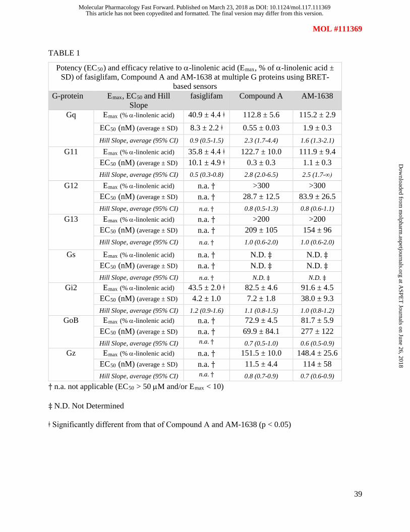
TABLE 1
Potency (EC50) and efficacy relative to α-linolenic acid (Emax, % of α-linolenic acid ± SD) of fasiglifam, Compound A and AM-1638 at multiple G proteins using BRET-
based sensors G-protein Emax, EC50 and Hill
Slope fasiglifam Compound A AM-1638
Gq
Emax (% α-linolenic acid) 40.9 ± 4.4 ǂ 112.8 ± 5.6 115.2 ± 2.9 EC50 (nM) (average ± SD) 8.3 ± 2.2 ǂ 0.55 ± 0.03 1.9 ± 0.3 Hill Slope, average (95% CI) 0.9 (0.5-1.5) 2.3 (1.7-4.4) 1.6 (1.3-2.1)
G11
Emax (% α-linolenic acid) 35.8 ± 4.4 ǂ 122.7 ± 10.0 111.9 ± 9.4 EC50 (nM) (average ± SD) 10.1 ± 4.9 ǂ 0.3 ± 0.3 1.1 ± 0.3 Hill Slope, average (95% CI) 0.5 (0.3-0.8) 2.8 (2.0-6.5) 2.5 (1.7-∞)
G12
Emax (% α-linolenic acid) n.a. † >300 >300 EC50 (nM) (average ± SD) n.a. † 28.7 ± 12.5 83.9 ± 26.5 Hill Slope, average (95% CI) n.a. † 0.8 (0.5-1.3) 0.8 (0.6-1.1)
G13
Emax (% α-linolenic acid) n.a. † >200 >200 EC50 (nM) (average ± SD) n.a. † 209 ± 105 154 ± 96 Hill Slope, average (95% CI) n.a. † 1.0 (0.6-2.0) 1.0 (0.6-2.0)
Gs
Emax (% α-linolenic acid) n.a. † N.D. ‡ N.D. ‡ EC50 (nM) (average ± SD) n.a. † N.D. ‡ N.D. ‡ Hill Slope, average (95% CI) n.a. † N.D. ‡ N.D. ‡
Gi2
Emax (% α-linolenic acid) 43.5 ± 2.0 ǂ 82.5 ± 4.6 91.6 ± 4.5 EC50 (nM) (average ± SD) 4.2 ± 1.0 7.2 ± 1.8 38.0 ± 9.3 Hill Slope, average (95% CI) 1.2 (0.9-1.6) 1.1 (0.8-1.5) 1.0 (0.8-1.2)
GoB
Emax (% α-linolenic acid) n.a. † 72.9 ± 4.5 81.7 ± 5.9 EC50 (nM) (average ± SD) n.a. † 69.9 ± 84.1 277 ± 122 Hill Slope, average (95% CI) n.a. † 0.7 (0.5-1.0) 0.6 (0.5-0.9)
Gz
Emax (% α-linolenic acid) n.a. † 151.5 ± 10.0 148.4 ± 25.6 EC50 (nM) (average ± SD) n.a. † 11.5 ± 4.4 114 ± 58 Hill Slope, average (95% CI) n.a. † 0.8 (0.7-0.9) 0.7 (0.6-0.9)
† n.a. not applicable (EC50 > 50 µM and/or Emax < 10)
‡ N.D. Not Determined
ǂ Significantly different from that of Compound A and AM-1638 (p < 0.05)
This article has not been copyedited and formatted. The final version may differ from this version.Molecular Pharmacology Fast Forward. Published on March 23, 2018 as DOI: 10.1124/mol.117.111369
at ASPE
T Journals on June 26, 2018
molpharm
.aspetjournals.orgD
ownloaded from

This article has not been copyedited and formatted. The final version may differ from this version.Molecular Pharmacology Fast Forward. Published on March 23, 2018 as DOI: 10.1124/mol.117.111369
at ASPE
T Journals on June 26, 2018
molpharm
.aspetjournals.orgD
ownloaded from

This article has not been copyedited and formatted. The final version may differ from this version.Molecular Pharmacology Fast Forward. Published on March 23, 2018 as DOI: 10.1124/mol.117.111369
at ASPE
T Journals on June 26, 2018
molpharm
.aspetjournals.orgD
ownloaded from
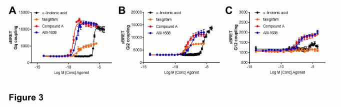
This article has not been copyedited and formatted. The final version may differ from this version.Molecular Pharmacology Fast Forward. Published on March 23, 2018 as DOI: 10.1124/mol.117.111369
at ASPE
T Journals on June 26, 2018
molpharm
.aspetjournals.orgD
ownloaded from
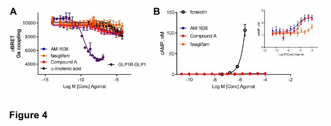
This article has not been copyedited and formatted. The final version may differ from this version.Molecular Pharmacology Fast Forward. Published on March 23, 2018 as DOI: 10.1124/mol.117.111369
at ASPE
T Journals on June 26, 2018
molpharm
.aspetjournals.orgD
ownloaded from
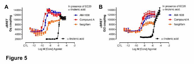
This article has not been copyedited and formatted. The final version may differ from this version.Molecular Pharmacology Fast Forward. Published on March 23, 2018 as DOI: 10.1124/mol.117.111369
at ASPE
T Journals on June 26, 2018
molpharm
.aspetjournals.orgD
ownloaded from
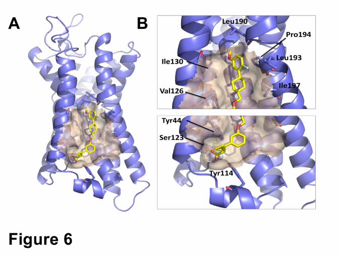
This article has not been copyedited and formatted. The final version may differ from this version.Molecular Pharmacology Fast Forward. Published on March 23, 2018 as DOI: 10.1124/mol.117.111369
at ASPE
T Journals on June 26, 2018
molpharm
.aspetjournals.orgD
ownloaded from
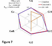
This article has not been copyedited and formatted. The final version may differ from this version.Molecular Pharmacology Fast Forward. Published on March 23, 2018 as DOI: 10.1124/mol.117.111369
at ASPE
T Journals on June 26, 2018
molpharm
.aspetjournals.orgD
ownloaded from