at last! how to attract money online easily and effortlessly by
TRANSCRIPT

Drosophila Myc regulates ribosome biogenesis and cell growth via LTV1
1
Drosophila LTV1 (Low Temperature Viability Protein 1) is Required for Ribosome Biogenesis and Cell Growth Downstream of dMyc
Wonho Kim1,2, Hag Dong Kim3, Youjin Jung3, Joon Kim3, Jongkyeong Chung2
1Department of Biological Sciences, Korea Advanced Institute of Science and Technology, 291 Daehak-ro,
Yuseong-gu, Daejeon 305-701, Republic of Korea 2National Creative Research Initiatives Center for Energy Homeostasis Regulation, Institute of Molecular
Biology and Genetics and School of Biological Sciences, Seoul National University, 599 Gwanak-ro, Gwanak-gu, Seoul 151-742, Republic of Korea
3Laboratory of Biochemistry, Division of Life Sciences, Korea University, Seoul 136-701, Republic of Korea
*Running title: Drosophila Myc regulates ribosome biogenesis and cell growth via LTV1
To whom correspondence should be addressed: Dr. Jongkyeong Chung, School of Biological Sciences, Seoul National University, 599 Gwanak-ro, Gwanak-gu, Seoul 151-742, Republic of Korea, Phone: +82-2-880-4399, Fax: +82-2-876-4401, E-mail: [email protected]; Dr. Joon Kim, Division of Life Sciences, Korea University, Seoul 136-701, Republic of Korea, Phone: +82-2-3290-3442, Fax: +82-2-927-9028, E-mail: [email protected] Keywords: cell growth; ribosome biogenesis; ribosomal RNA processing; LTV1; dMyc Background: To understand the molecular mechanism of cell growth, novel regulators of cell growth should be identified. Results: LTV1 mutation impairs 40S ribosome subunit synthesis, and it blocks cell growth and ribosome biogenesis induced by dMyc. Conclusion: LTV1 is essential for cell growth and ribosome biogenesis downstream of dMyc. Significance: Characterization of LTV1 will be helpful for understanding Myc-induced tumorigenesis. ABSTRACT
During animal development, various signaling pathways converge to regulate cell growth. In this study, we identify LTV1 as a novel cell growth regulator in Drosophila. LTV1 mutant larvae exhibit developmental delays and lethality at the second larval stage. Using biochemical studies, we discover that LTV1 interacts with ribosomal protein S3 (RpS3) and co-purifies with free 40S ribosome subunits. We further demonstrate that LTV1 is crucial for ribosome biogenesis through 40S ribosome subunit synthesis and pre-ribosomal RNA processing, suggesting that LTV1 is required
for cell growth by regulating protein synthesis. We also demonstrate that Drosophila Myc (dMyc) directly regulates LTV1 transcription and requires LTV1 to stimulate ribosome biogenesis. Importantly, the loss of LTV1 blocks the cell growth and endoreplication induced by dMyc. Combined, these results suggest that LTV1 is a key downstream factor of dMyc-induced cell growth by properly maintaining ribosome biogenesis. INTRODUCTION
Cell growth is coordinated with cell differentiation and apoptosis in animal development. To control cell growth precisely, proto-oncogenes and tumor suppressors are tightly regulated by developmental cues and environmental changes. To understand the molecular mechanism of cell growth, novel cell growth regulators should be identified and their in vivo functions should be demonstrated in animal models. Although multiple cell growth regulators have been discovered and investigated (1,2), our understanding of cell growth regulation still remains elusive.
During cell growth, de novo synthesis of
http://www.jbc.org/cgi/doi/10.1074/jbc.M114.607036The latest version is at JBC Papers in Press. Published on April 9, 2015 as Manuscript M114.607036
Copyright 2015 by The American Society for Biochemistry and Molecular Biology, Inc.
by guest on Decem
ber 6, 2018http://w
ww
.jbc.org/D
ownloaded from

Drosophila Myc regulates ribosome biogenesis and cell growth via LTV1
2
the ribosome, the machinery required for mRNA translation, is highly induced (3). The ribosome is generated through coordinated multiple processes occurred in the nucleolus, nucleoplasm, and cytosol (4-6). In yeast, the precursor of ribosomal RNA (pre-rRNA) is transcribed, and simultaneously assembled with the ribosomal proteins imported from the cytoplasm to form the 90S precursor (pre-90S) ribosome particles in the nucleolus (4). The 35S pre-rRNA, the longest precursor, contains 18S, 5.8S and 25S mature rRNAs that are separated by internal transcribed spacers (ITS) and are flanked by external transcribed spacers (ETS). These extra spacer sequences are sequentially removed by endo- and exo-nucleases to make mature rRNAs (7). In the nucleolus, an internal cleavage of ITS in pre-rRNA separates pre-90S ribosomes into pre-40S and -60S ribosome subunits. Both of these precursor ribosome subunits in the nucleus are exported to the cytosol in a Crm1-Ran GTPase-dependent manner (8,9). After the export from the nucleus, the precursor ribosomal subunits are further processed to fully mature in the cytosol.
There are approximately 200 non-ribosomal proteins that associate and dissociate dynamically with pre-ribosomes during ribosome biogenesis (10). These proteins have indispensable roles in ribosome biogenesis by assisting pre-rRNA processing and modifications, ribosomal protein folding and association, etc. For the synthesis of fully matured 40S ribosome subunits, multiple non-ribosomal proteins, such as Rio2p, Tsr1p, Ltv1p, Enp1p, Nob1p, Hrr25p, Dim1p and Dim2p, interact with pre-40S ribosome subunits (11). They have various protein domains like methyl transferase, protein kinase, endoribonuclease and GTPase, implicating that they are involved in 40S ribosome biogenesis in diverse ways. These non-ribosomal proteins are structurally highly conserved, suggesting that they have similar functions in ribosome biogenesis from yeast to multicellular animals.
Diverse signaling molecules regulate ribosome biogenesis to control cell growth (3,12). Among these signals, Myc proto-oncogene plays the most important roles at several stages including rRNA transcription (13-15), rRNA processing (16) and the export of ribosome
subunits from the nucleus to the cytosol (17,18). Consistently, Myc transcriptionally induces multiple genes critical for ribosome biogenesis, including the genes for ribosomal proteins (19), upstream binding factors (the transcription factors for RNA polymerase I-mediated transcription) (14) and nucleophosmin (a nuclear export chaperone for ribosome) (18,20).
In this study, we attempted to discover a novel cell growth regulator using a fruit fly system and successfully identified LTV1. LTV1 specifically interacted with RpS3 and co-purified with free 40S ribosome subunits. We found that LTV1 is crucial for the biogenesis of 40S ribosome subunits by affecting pre-rRNA processing and the nuclear export of pre-40S ribosome subunits. Furthermore, we showed that LTV1 is transcriptionally regulated by dMyc and is required for dMyc-dependent ribosome biogenesis, cell growth and endoreplication. Together, our results strongly suggested that dMyc controls ribosome biogenesis and cell growth by directly regulating the gene expression of LTV1 in Drosophila.
EXPERIMENTAL PROCEDURES Fly stocks The following fly lines were used in this study: Cg-GAL4 (Bloomington #7011), Ptc-GAL4 (Bloomington #2017), UAS-dMyc (Bloomington #9674), Act>CD2>GAL4 (Bloomington #4780), UAS-LTV1 RNAi [Vienna Drosophila Resource Center (VDRC) #33650], UAS-Crm1 RNAi (VDRC #3347), UAS-RpI135 RNAi (VDRC #37581) and UAS-RpS2-3xHA (FlyORF #F000781), dm4 and the revertant for dm4 (a gift from Dr. Robert Eisenman). Generation of LTV1E1 mutant LTV1E1 was generated in this study through imprecise excision of the P-element from CG7686CB-6202-3 (Kyoto Drosophila Genetic Resource Center, #123972). Generation of LTV1 transgenic flies LTV1 cDNA was amplified by PCR from EST [Drosophila Genomics Resources Center (DGRC), #LD21529] and subcloned into the EcoRI and XbaI sites followed by HA sequences in pUAST
by guest on Decem
ber 6, 2018http://w
ww
.jbc.org/D
ownloaded from
Drosophila Myc regulates ribosome biogenesis and cell growth via LTV1
3
vector. pUAST LTV1-HA plasmid was microinjected into w1118 embryo. Clone generation Homozygous LTV1E1 fat body cells were generated by the FRT/FLP-mediated mitotic recombination (21). Embryos with proper genotypes were collected for 6 hours and subsequently incubated at 37°C for 2 hours. To generate transgene-expressing clones, the FRT/FLP-mediated flipout technique was used (22). Generation of the flipout clones in the fat body cells did not require heat shock. To induce the flipout recombination in wing discs, larvae were incubated at 37°C for 20 minutes at 2nd instar stage. RNA extraction and quantitative reverse transcription PCR analysis To induce gene expression of dMyc in the whole body, the 3rd instar larvae with genotypes of Hs-GAL4, Hs>dMyc (on second chromosome) and Hs>dMyc (on third chromosome) were incubated for 30 minutes at 37°C. After recovery for 6 hours at 25°C, the larvae were collected to isolate RNA. For extracting RNA from control and dMyc mutant, first instar larvae of the revertant and dm4 were collected. Total RNA was extracted by Trizol (Invitrogen) according to the manufacturer’s manual. RNA (3 μg) was transcribed reversely by MMLV reverse transcriptase (Promega). For quantitative real time PCR (qPCR), the synthesized cDNA was mixed with SYBR Green (Enzynomics, South Korea, #RT500) and appropriate primers (300 nM), and then applied to Bio-Rad iQ5 Real-time PCR detection system. The following primers were used to amplify LTV1 cDNA: 5’- CAGCAGGCACATGTGTTTCTCTGTATTTAC-3’, 5’- CACGTTTCTTCAGATGCTGCATGTAATCATAG-3’ and Actin cDNA: 5’-CACACCAAATCTTACAAAATGTGT-3’, 5’-AATCCGGCCTTGCACATG-3’. Statistical analysis Student unpaired t-test and one-way ANOVA Dunnett test (Graphpad Prism 5) were used for statistical comparison.
Immunostaining and cell size measurement Wandering larvae (for the salivary gland cells) or 3rd instar larvae (for the fat body cells) were cut and turned inside out in phosphate-buffered saline (PBS) and then directly fixed with 4% paraformaldehyde for 20 minutes at room temperature. After washing for 10 minutes twice with PBS containing 0.1% Triton X-100 (PBST), the samples were permeabilized in PBS with 0.5% Triton X-100 for 5 minutes at room temperature and washed once with PBST. The samples were blocked with PBST containing 3% BSA. Primary antibodies and TRITC-labeled phalloidin (Sigma) were applied, and the samples were incubated at 4°C for overnight on rotator. Following three washes for 10 minutes in PBST, the samples were incubated at room temperature for 2 hours with fluorescent-labeled secondary antibodies and Hoechst 33258 (Invitrogen). Larvae were washed for 10 minutes three times with PBST. The salivary glands or the fat body cells were isolated and mounted in 80% glycerol-PBS solution on slide glass. The samples were observed with the confocal microscope LSM710 (Zeiss). Primary antibodies used in this study: anti-fibrillarin mouse monoclonal antibody (1:200, Abcam, #4566), anti-HA rabbit monoclonal antibody [1:100, Cell Signaling Technology (CST), #3724] and anti-cleaved caspase-3 rabbit polyclonal antibody (1:100, CST, #9661). To measure cell size, the fat body cells of 3rd instar larvae were dissected, fixed and stained by phalloidin and Hoechst to visualize cell boundary and the nucleus, respectively. The samples were mounted with 80% glycerol-PBS solution, and then were observed by LSM 710 (Zeiss). The cell area was measured by ImageJ. BrdU incorporation Wandering larvae (for the salivary glands) or mid-3rd instar larvae (for the fat body cells) were dissected on Shields and Sang M3 media (Sigma, #S8398) and then incubated in M3 media containing 0.1 mg/ml BrdU (Roche, #10 280879 001) for 30 minutes on rotator at room temperature. After three times brief washing with PBS, larvae were fixed in 4% paraformaldehyde for 20 minutes. The fat body samples were
by guest on Decem
ber 6, 2018http://w
ww
.jbc.org/D
ownloaded from
Drosophila Myc regulates ribosome biogenesis and cell growth via LTV1
4
washed three times in PBST and then incubated with 5 units of DNase I (Promega, #M6101) in 200 μl PBS containing DNase I buffer for 1.5 hours at 37°C. The salivary gland samples were washed three times in PBST and then incubated in 2 N hydrochloric acid for 30 minutes. Larvae were washed three times in PBST followed by incubation with anti-BrdU mouse monoclonal antibody (1:100, BD Biosciences, #555627) in blocking solution. TUNEL assay Wing discs were dissected on PBS and subsequently fixed with 4% paraformaldehyde for 20 minutes on room temperature. After washing with PBST three times, discs were incubated in 0.1% sodium citrate in PBST for 30 minutes at 65°C. The samples were washed three times in PBST followed by incubation in 3% BSA PBST blocking solution, and incubated with labeling and enzyme solution (In Situ Cell Death Detection Kit, Fluorescein, Roche Applied Science, #11 684 795 910) for 2 hours at 37°C. After staining with Hoechst and washing with PBST, wing discs were mounted in 80% glycerol-PBS. Drosophila S2 cell culture Drosophila S2-DRSC cells were obtained from DGRC and maintained in Shields and Sang M3 media with 10% fatal bovine serum (GIBCO) and 10% insect medium supplement (Sigma, #17267) at 25°C. Transfection was performed using Effectene (QIAGEN, #301425) according to manufacturer’s instruction on 6-well plates. Generation of RpS3 antibody The RpS3-specific antibody was generated by immunizing rabbits with the peptide, MNANLPISKKRKFVS. Cloning of LTV1 and ribosomal protein S3 for transfection in S2 cells To generate the C-terminal V5-tagged LTV1 plasmid, LTV1 cDNA was subcloned into the EcoRI and XbaI sites of pAc5.1-V5/His vector (Invitrogen). For the C-terminal HA-tagged LTV1 plasmid, LTV1-HA containing stop codon was amplified by PCR from the template, pUAST LTV1-HA (see above), and cloned into the EcoRI
and NotI sites of pAc5.1-V5/His vector. The ribosomal protein S3 (RpS3) ORF was amplified by PCR from Expressed Sequence Tag (DGRC, #LD21061). To generate the C-terminal mCherry-tagged RpS3 plasmid, the RpS3 ORF without stop codon was subcloned into the EcoRI and XhoI sites of pAc5.1-V5/His vector. Subsequently, the mCherry ORF containing stop codon was subcloned into the XhoI and XbaI sites of pAc5.1-RpS3-V5/His plasmid. The C-terminal V5-tagged RpS3 plasmid was generated by subcloning RpS3 ORF into the EcoRI and XhoI sites of pAc5.1-V5/His vector. Double-stranded RNA synthesis and bathing For synthesis of double-stranded RNA (dsRNA), double-stranded DNA was amplified by PCR from genomic DNA which was extracted from w1118 with the primers containing T7 polymerase binding site (5’-TAATACGACTCACTATAGGGAGA-3’) at the 5’ end. dsRNA was transcribed using PCR product as a template with MEGAscript T7 kit (Ambion, #AM1334). Synthesized dsRNA was purified by MEGAclearTM kit (Ambion, #AM1908). The following primers were used for dsRNA synthesis: Luciferase dsRNA: 5’-TAATACGACTCACTATAGGGAGAGGCCCGGCGCCATTCTATC-3’ and 5’-TAATACGACTCACTATAGGGAGAGATTGGGAGCTTTTTTTGCACG-3’; LTV1 dsRNA: 5’-TAATACGACTCACTATAGGGAGACGGAACTGGGCGACTTGGCTCT-3’ and 5’- TAATACGACTCACTATAGGGAGAACCCCACCACGCAGGACATT-3’; dMyc dsRNA: 5’-TAATACGACTCACTATAGGGAGACAAAGTGACGCATAGCTCCA-3’ and 5’-TAATACGACTCACTATAGGGAGAGGTTATCCTAGCCCTACGCC-3’. For bathing dsRNA, culture media were replaced by serum-free M3 media, and S2 cells were diluted to a proper concentration. 1 × 10⁶ cells were applied into 6-well plate, and 16 μg of dsRNA was added. After incubation for 45 minutes at 25°C, serum-containing M3 media were supplied. S2 cells were lysed after 4 days incubation at 25°C. Generation of LTV1 expressing stable cell line To generate stably expressing S2 cell lines, each
by guest on Decem
ber 6, 2018http://w
ww
.jbc.org/D
ownloaded from
Drosophila Myc regulates ribosome biogenesis and cell growth via LTV1
5
pAc5.1-V5/His or pAc5.1 LTV1-V5/His vector was co-transfected with pCoHygro plasmids (Invitrogen) by Effectene in 6-well plates. Fresh M3 media containing serum and 300 μg/ml hygromycin (Sigma, #H0654) were replaced at every four days. After one month, survived hygromycin-resistant cells began to proliferate and they were used for further experiments. Identification of LTV1 binding protein S2 cells stably expressing each empty vector and pAc5.1 LTV1-V5/His were cultured at T-75 flask, and 1 × 10⁸ cells were collected. After washing twice with 20 ml of cold PBS, S2 cells were lysed with 4 ml of lysis buffer (25 mM Tris pH7.9, 150 mM sodium chloride, 10% glycerol, 0.1% NP-40, 1 mM ethylenediaminetetraacetic acid (EDTA), 1 mM dithiothreitol (DTT), 1 μg/ml leupeptin and 1 mM phenylmethylsulfonyl fluoride). The lysates were incubated for 15 minutes at 4°C on rotator. After centrifugation at 13,200 rpm for 15 minutes at 4°C, the clarified lysates were transferred. Protein concentration in lysates was measured by Bradford assay. 80 μl of protein G Sepharose (GE healthcare, #17-0618-02) was applied to the same concentration of lysates and then the samples were incubated for 30 minutes at 4°C to remove non-specifically bound proteins to Sepharose. After brief centrifugation, the supernatant was collected. 80 μl of V5-agarose (Sigma, #A7345) in 50% slurry was applied and incubated for 4 hours at 4°C on rotator. The beads were washed sequentially once with 1 ml of BC150, once with 1 ml of BC300, twice with 1 ml of BC500, and once with 1 ml of BC150. The following washing buffers were used: BC150: 20 mM Tris-HCl pH7.9, 15% glycerol, 1 mM EDTA, 0.05% NP-40 and 150 mM potassium chloride (KCl), BC300: 20 mM Tris-HCl pH7.9, 15% glycerol, 1 mM EDTA, 0.05% NP-40 and 300 mM KCl, BC500: 20 mM Tris-HCl pH7.9, 15% glycerol, 1 mM EDTA, 0.05% NP-40 and 500 mM KCl. The sample buffer (70 mM Tris-HCl pH6.8, 2% glycerol, 0.002% bromophenolblue, 3% sodium dodecyl sulfate and β-mercaptoethanol) was directly applied to the washed beads. The samples were boiled and subjected to SDS-PAGE and coomassie blue staining. Excised gels were sequenced by LC/MS/MS in Taplin Mass
Spectrometry Facility (Harvard Medical School, USA). Immunoblotting and immunoprecipitation S2 cells were grown to a density 6 × 10⁶ cells/ml, and 1 × 10⁷ cells were collected by pipetting and washed twice with 1 ml of cold PBS. S2 cells were lysed in lysis buffer (described above). The lysates were clarified by centrifugation at 13,200 rpm for 15 minutes at 4°C. The whole cell lysates for western blotting was mixed with the sample buffer and boiled at 95°C. Anti-HA (MBL International) or -V5 (Invitrogen) or -RFP (Abcam) antibodies were applied into the remaining lysates for immunoprecipitation. The mixture was incubated for 3 hours on rotator at 4°C. 35 μl of protein G Sepharose (GE healthecare, #17-0618-02) in 50% slurry was added, followed by incubating for 1 hour on rotator at 4°C. The Sepharose beads were washed five times with 500 μl of washing buffer, BC150 (described above). After washing, the immunoprecipitate was boiled with the sample buffer. For immunoblotting of the lysates from flies, fifteen adult flies were collected in a tube, and homogenized in 150 μl of lysis buffer. After centrifugation at 13,200 rpm for 15 minutes at 4°C, the clear lysates were carefully transferred and boiled with the sample buffer. For the treatment of RNase A, the lysates were incubated with 0.1 μg/ml RNase A for 2 hours at room temperature, and immunoprecipitation was further conducted. To confirm 18S rRNA degradation, total RNA was extracted by Trizol-LS (Invitrogen) and reversely transcribed by MMLV reverse transcriptase (Promega). Synthesized cDNA was amplified by PCR with the following primers: 5’-CATTCATGTTGGCAGTAAAATGCTTATTGTGTTTG-3’ and 5’-GATCCTTCCGCAGGTTCACCTACG-3’. For immunoblotting, primary antibodies were applied as follow: anti-V5 mouse monoclonal antibody (1:5,000, Invitrogen, #46-0705), anti-HA 3F10 rat monoclonal antibody (1:1,000, Roche, #11867423001), anti-β-tubulin mouse monoclonal antibody (E7, 1:500, Developmental Studies Hybridoma Bank) and anti-RFP rabbit polyclonal antibody (1:1,000, Abcam, #Ab62341).
by guest on Decem
ber 6, 2018http://w
ww
.jbc.org/D
ownloaded from
Drosophila Myc regulates ribosome biogenesis and cell growth via LTV1
6
Sucrose density gradient analysis After 200 μg/ml cycloheximide was treated for 10 minutes at 25°C, 1 × 10⁷ of S2 cells were washed twice with 1 ml of cold PBS and lysed by incubation for 10 minutes at 4°C with 900 μl of lysis buffer [10 mM Tris-HCl pH 7.3, 5 mM MgCl2, 100 mM KCl, 0.5% Triton X-100, 2 mM DTT, 100 μg/ml cycloheximide, protease inhibitor cocktail (Calbiochem, #535140) and 200 units Superase-In RNase inhibitor (Ambion, #AM2696)]. For analysis of ribosome content in LTV1-HA overexpressed larvae, thirty 3rd instar larvae were collected and washed twice by 1 ml of cold PBS. They were homogenized in 900 μl of lysis buffer. After centrifugation at 10,000 rcf for 15 minutes at 4°C, 850 μl of the cleared lysates were transferred. Protein concentration in the lysates was measured by Bradford assay, and the samples were adjusted to the same protein concentration and volume. 10 ml of 10%-50% sucrose gradient in 20 mM HEPES-KOH, 5 mM MgCl2, 100 mM KCl, 2 mM DTT and 100 μg/ml cycloheximide was prepared one day before cell harvest. 800 μg of the lysates was carefully applied on the top of sucrose gradient and then separated by centrifugation at 32,000 rpm for 3.5 hours at 4°C with SW41Ti rotor (Beckman). The fractions were collected by a fraction collector (Biorad) and UV absorbance at 260 nm was measured from each fraction. Chromatin-immunoprecipiation assay The dsRNA for luciferase and dMyc were bathed in 1 × 10⁶ cells in 6 well plates. After four days, S2 cells were incubated in 1% formaldehyde for 10 minutes at 25°C for cross-linking. The reaction was terminated by incubating in 0.24 M glycine for 5 minutes at room temperature. S2 cells were washed twice by 1 ml of cold PBS, collected in 1.5 ml tube, and lysed by 500 μl of freshly prepared SDS lysis buffer (1% SDS, 10 mM EDTA, 50 mM Tris-HCl pH8.1 and protease inhibitor cocktail). The samples were incubated for 5 minutes at 4°C, and sonicated 10 times with 5 seconds/30 seconds pulses in cold ice-water slurry to be sheared to 200 bp ~ 1,000 bp of genomic fragments. The samples were clarified by centrifugation of 13,200 rpm for 10 minutes at 4°C. 10 μl of the lysates was kept for input, and
400 μl of the lysates was mixed with 3.6 ml of Chip dilution buffer (0.01% SDS, 1.1% Triton X-100, 1.2 mM EDTA, 16.7 mM Tris-HCl pH8.1, 167 mM NaCl and protease inhibitor cocktail). 50 μl of protein A Sepharose (GE healthcare, #17-0974-04) was applied into the diluted lysates, followed by incubating on rotator for 30 minutes at 4°C to remove non-specifically bound proteins to Sepharose in the lysates. After brief centrifugation, the supernatant was transferred, and 1.2 μg of dMyc antibody (Santa Cruz Biotechnology, #sc-28208) was added. The same amount of rabbit immunoglobin G (Santa Cruz Biotechnology, #sc-2027) was added for control. The samples were incubated on rotator for overnight at 4°C. 50 μl of protein A Sepharose (GE healthcare, #17-0974-04) was applied into the samples, and incubated on rotator for 1 hour at 4°C. The Sepharose beads were sequentially washed by following washing buffers: once with low salt washing buffer (0.1% SDS, 1% Triton X-100, 2 mM EDTA, 20 mM Tris-HCl pH8.1, 150 mM NaCl and protease inhibitor cocktail), once with high salt washing buffer (0.1% SDS, 1% Triton X-100, 2 mM EDTA, 20 mM Tris-HCl pH8.1, 500 mM NaCl and protease inhibitor cocktail), once with lithium chloride (LiCl) washing buffer (0.25 M LiCl, 1% NP-40, 1% deoxycholate, 1 mM EDTA, 10 mM Tris-HCl pH8.1 and protease inhibitor cocktail) and twice with TE buffer (10 mM Tris-HCl pH8.0, 1 mM EDTA and protease inhibitor cocktail). The immunoprecipitates were eluted twice by incubating in elution buffer (0.1% SDS and 0.1 M sodium bicarbonate) on rotator for 15 minutes at room temperature. The DNA fragments of input and immunoprecipitate were reversely cross-linked in 200 mM NaCl for 4 hours at 65°C. The samples were further incubated in 10 mM EDTA, 40 mM Tris-HCl pH 6.5, 40 μg/ml proteinase K for 1 hour at 65°C. The DNA fragments were purified by phenol/chloroform extraction. The contents of DNA fragment were amplified by qPCR. The results were quantified by the ∆Ct method and normalized to the input control. The following primers were used for qPCR: LTV1 region A: 5’-CAAATTTGTCTTTCCATACTTATTTCAGACTTG-3’ and 5’-
by guest on Decem
ber 6, 2018http://w
ww
.jbc.org/D
ownloaded from

Drosophila Myc regulates ribosome biogenesis and cell growth via LTV1
7
CATGAAATTAATAGTTTTTGAGATTTTGTTCTG-3’; LTV1 region B: 5’-GGCGCCTTTTTTCGAATTTGAGTGTGC-3’ and 5’-ATTGGATTGTTTTCGTAAAATCTTTTGTAAATAC-3’; RpI135: 5’-CTTTTATACAACTTGGATACAATTCAAAATTGG-3’ and 5’-CAGATTAATAGAACACACTAAAATAACCTTAAAG-3’ Northern blotting Total RNA was isolated from 2nd instar larvae by Trizol. Equal amount of denatured RNA (4 μg) was loaded on 1.5% agarose gel. The separated RNA was transferred by capillary method to Hybond N membrane (GE Healthcare, #RPN303). The cross-linked membranes were blocked by hybridization buffer (5× SSC and 0.5% SDS) and then incubated with hybridization buffer containing probes at 65°C for overnight. The probe for 5ITS1 was synthesized by random prime labeling system (GE Healthcare, #RPN1633) with manufacturer’s instruction. To synthesize the template for random prime labeling, the following primers were used: 5ITS1: 5’-TTGTATAATATCCTTACCGTTAATAAATATTTGTAATTATAC-3’ and 5’-GAAACGCCGTTGTTGTAAGTACTCGCCAC-3’. Synthesized probes were purified by Sephadex G-50 column (GE Healthcare, #27-5330-01) and denatured at 95°C. The probes for 18S and 28S rRNA were prepared by oligonucleotide 5’ end labeling. The 10 pmole of oligonucleotides were mixed with T4 polynucleotide kinase (TAKARA, #2021A), reaction buffer and [γ-32P] ATP (3,000 Ci/mmol, 10.0 mCi/ml, PerkinElmer, #NEG502A). The mixture was incubated for 1 hour at 37°C and the probes were purified by Sephadex G-50 column. The following primers were used for oligonucleotide 5’ end labeling: 18S rRNA: 5’-CTTCCTCTAAATAATCAAGTTCGGTCAACTTTTGCGAAACAACCGTAACACGC-3’; 28S rRNA: 5’-GTAACTAGCGCGGCATCAGGTGATCGAAGATCCTCCC-3’. Electron microscopy The salivary glands of wandering larvae were
fixed with 3% glutaraldehyde in PBS for 3 hours. They were washed five times with 0.1 M cacodylate buffer (pH 7.2) containing 0.1% calcium chloride (CaCl2) at 4°C. Then, they were postfixed with 1% osmium tetroxide in 0.1 M cacodylate buffer (pH 7.2) containing 0.1% CaCl2 for 2 hours at 4°C. After rinsing with cold distilled water, the tissue samples were dehydrated slowly with ethanol series and propylene oxide at 4°C. The samples were embedded in Embed-812 (EMS, USA). After polymerization of the resin at 60°C for 36 hours, serial sections were cut with a diamond knife on an ULTRACUT UC7 ultramicrotome (Leica, Austria) and mounted on formvar-coated slot grids. The sections were stained with 4% uranyl acetate for 10 minutes and lead citrate for another 10 minutes. They were observed using Tecnai G2 Spirit Twin transmission electron microscope (FEI Company, USA). RESULTS LTV1 is a novel cell growth regulator
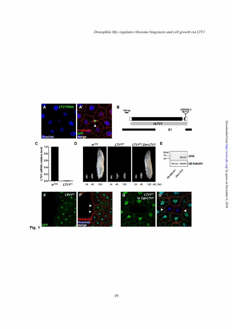
We conducted an RNAi knockdown-based screen of novel cell growth regulators in Drosophila. Because many growth regulating genes were critical for development, the majority of their mutants exhibited lethality at early larval stages (data not shown). Therefore, we randomly generated RNAi transgene-expressing clone cells by flip-out recombination (22). We examined the effects of RNAi transgenes on the size of the fat body cells because they could be easily observed. Fortunately, compared with the neighboring cells expressing no transgenes, CG7686 RNAi-expressing fat body cells (the GFP-positive cell in Fig. 1A and A’) displayed decreased cell and nucleus size phenotypes when the cell boundary and DNA were stained with phalloidin and Hoechst, respectively (Fig. 1A and A’). A BLAST search indicated that CG7686 is the Drosophila orthologue of the mammalian and yeast low temperature viability protein 1 (LTV1). To characterize the function of LTV1 in vivo, we generated deletion mutants of LTV1 in Drosophila. For this purpose, we performed imprecise excisions using the CG7686CB-6202-3 line that contains a P-element in the 5’ untranslated region of LTV1. As a result, we isolated one deletion
by guest on Decem
ber 6, 2018http://w
ww
.jbc.org/D
ownloaded from
Drosophila Myc regulates ribosome biogenesis and cell growth via LTV1
8
mutant, LTV1E1, in which the first half of the coding region of LTV1 (including the start codon) was removed (Fig. 1B). The mutant did not express LTV1 transcripts as shown by quantitative RT-PCR in the mutant (Fig. 1C), and was arrested at the 2nd larval stage and died approximately 6 days after egg hatching (Fig. 1D and data not shown). To confirm that all these phenotypes were caused by the loss of LTV1, we generated LTV1 transgenic flies. We first expressed LTV1 transgene using a ubiquitously expressed GAL4 driver, Daughterless (Da)-GAL4, and detected the LTV1 protein at approximately 65-kDa by immunoblotting (Fig. 1E). When LTV1 was ectopically expressed in LTV1E1 mutant by Da-GAL4, the developmental arrest and lethality of LTV1E1 mutant were completely rescued, confirming that these defective phenotypes are due to the LTV1 mutation (Fig. 1D).
We also generated mosaic clones of the LTV1 mutant in the fat body using FLP/FRT-mediated mitotic recombination (21). Similar to the cell growth defects of LTV1 RNAi-expressing cells (Fig. 1A and A’), homozygous LTV1 deletion mutant cells (GFP-negative cells) were much smaller than neighboring control cells (GFP-positive cells) (Fig. 1F and F’). Moreover, the decreased cell size of LTV1-deleted cells was completely rescued when we overexpressed exogenous LTV1 in the fat body using Cg-GAL4, a fat body specific driver (Fig. 1G and G’). These results indicated that LTV1 is required for normal cell growth. LTV1 interacts with RpS3 and co-purifies with free 40S ribosome subunits
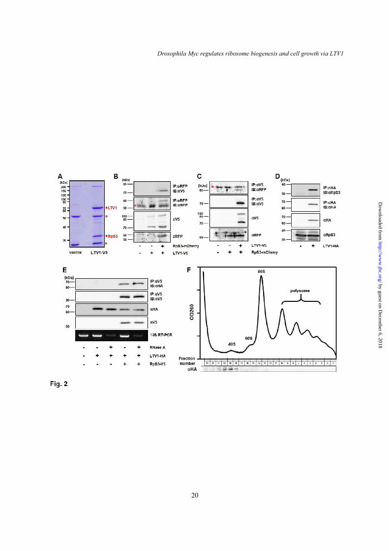
To investigate the mechanisms by which LTV1 regulates cell growth, we decided to identify novel proteins that interact with LTV1. We generated an S2 cell line stably expressing the C-terminal V5-tagged LTV1 and immunoprecipitated LTV1 using anti-V5 antibody. Notably, we detected a 32-kDa protein specifically bound to the LTV1 protein (Fig. 2A) and identified the protein as ribosomal protein S3 (RpS3) in the subsequent mass spectrometry analysis (data not shown). To validate the result, LTV1-V5 and the C-terminal mCherry-tagged RpS3 were transfected in S2 cells. We observed
that LTV1 was co-precipitated with RpS3 (Fig. 2B). Additionally, when we immunoprecipitated LTV1, exogenous RpS3 was also co-precipitated (Fig. 2C). Moreover, we generated an antibody against RpS3 to determine whether endogenous RpS3 interacts with LTV1. As expected, endogenous RpS3 was co-precipitated with overexpressed LTV1, further confirming the specific interaction between these two proteins (Fig. 2D). Because ribosomal proteins are assembled with rRNA to form ribosome subunits, we examined whether rRNA is required for the interaction between LTV1 and RpS3. Notably, when we degraded rRNA by treating RNase A, LTV1 still interacted with RpS3 (Fig. 2E). This result indicated that the interaction between LTV1 and RpS3 is not dependent on the integrity of 40S ribosome subunits.
Moreover, previous studies in yeast demonstrated that LTV1 associates with pre-40S ribosome subunits, and LTV1-RpS3 subcomplex could be released from pre-40S ribosome subunits in a high salt buffer (11,23). Therefore, we questioned whether Drosophila LTV1 also associates with 40S ribosome subunits. We expressed LTV1-HA in S2 cells and analyzed its co-localization with 40S ribosome subunits using sucrose gradient experiments. Notably, our immunoblot analyses revealed that LTV1 was co-purified with the fractions containing 40S ribosome subunits but not with 60S ribosome subunits or 80S monosomes (Fig. 2F), suggesting that LTV1 can specifically associate with free 40S ribosome subunits. LTV1 is required for the biogenesis of 40S ribosome subunit
Next, we determined whether LTV1 is required for ribosome biogenesis. We examined the ribosomes isolated from LTV1 knockdown cells on sucrose density gradient. The LTV1 transcripts were efficiently depleted by double stranded RNA (dsRNA)-based knockdown in S2 cells (data not shown). In LTV1 knockdown cells, the amount of free 40S ribosome subunits and 80S monosomes were severely reduced whereas free 60S ribosome subunits accumulated compared to the luciferase knockdown controls (Fig. 3A). This imbalanced ratio of 40S/60S ribosome subunits
by guest on Decem
ber 6, 2018http://w
ww
.jbc.org/D
ownloaded from

Drosophila Myc regulates ribosome biogenesis and cell growth via LTV1
9
indicated that 40S ribosome biogenesis was defective without LTV1. In addition, we analyzed the amount of 40S and 60S ribosome subunits in LTV1 mutant larvae by hybridizing 18S and 28S mature rRNA, respectively. Consistent with the results from the sucrose gradient analysis, the ratio of 18S to 28S rRNA was significantly reduced by approximately 25% in the LTV1 mutant (Fig. 3B and C). This defect was also rescued by the ectopic expression of LTV1 in the entire body, indicating that it was caused by LTV1 mutation (Fig. 3B and C).
Next, we examined whether LTV1 overexpression increases the biogenesis of 40S ribosome subunits and 80S monosomes. Exogenous LTV1 was expressed in the entire body using Da-GAL4 driver, and the ribosome content of the LTV1-expressing larvae was analyzed using sucrose density gradient experiments. As a result, we did not observe significant differences in the amount of 40S ribosome subunits and 80S monosomes between LTV1-expressing larvae and controls (Fig. 3D). In addition, LTV1 overexpression did not increase the size of the fat body cells (data not shown). These results indicated that LTV1 overexpression is not sufficient for increasing ribosome contents and cell size.
Previous studies in yeast demonstrate that LTV1 is required for the nuclear export of pre-40S ribosome subunits to the cytosol (24). Therefore, we examined the role of LTV1 in the nuclear export of pre-40S ribosome subunits in Drosophila. In yeast and human cells, the localization of pre-40S ribosome subunits can be monitored by immunostaining of ribosomal protein S2 (RpS2) (25-28). Because no available antibody detects endogenous RpS2, we overexpressed HA-tagged RpS2 and immunostained with an HA antibody. When RpS2 was expressed alone in the fat body cells, it dominantly localized in the cytosol (Fig. 3E-E’’). To verify whether RpS2 could serve as a reporter for the nuclear export of pre-40S ribosome subunits in Drosophila cells, we co-overexpressed an RNAi transgene for Crm1, the receptor for the nuclear export of pre-40S and pre-60S ribosome subunits (29-31). Interestingly, Crm1 knockdown highly induced the accumulation of RpS2 in the
nucleus, consistent with yeast and human cells (Fig. 3F-F’’). Notably, similar to the Crm1 knockdown result, LTV1 RNAi expression also increased the concentration of the nuclear RpS2 (Fig. 3G-G’’), suggesting that LTV1 is important for the nuclear export of pre-40S ribosome subunits. LTV1 is required for pre-rRNA processing
During ribosome synthesis, pre-rRNA is processed over multiple steps by several nucleases to yield mature rRNA (4,6). Because LTV1 is required for 40S ribosome biogenesis, we examined whether LTV1 also functions in pre-rRNA processing. In Drosophila, pre-rRNA is processed by two different pathways (Fig. 4A) (32). To observe the pre-rRNA processing for 18S rRNA synthesis, we examined pre-rRNA intermediates using P32-radiolabeled 5’ end of the internally transcribed spacer 1 (5ITS1) as a hybridization probe. In the Northern blotting using the 5ITS1 probe, we detected pre-rRNA and the intermediate d in w1118 larvae (Fig. 4B, first lane). We did not observe the intermediate a in w1118, likely because of its rapid turnover rate (Fig. 4B, first lane). However, the LTV1 mutant larvae accumulated the intermediate a and the unreported 2.5 kb intermediate * (Fig. 4B). The size of the intermediate * suggested that this intermediate corresponds to mammalian 21S rRNA, which contains 18S rRNA and 5ITS1 (32). This altered pre-rRNA processing pattern suggested that the cleavage of pre-rRNA intermediates is defective in LTV1 mutant larvae. In addition, ectopic expression of LTV1 fully rescued the pre-rRNA processing defects in LTV1 mutant larvae, confirming that the pre-rRNA processing defects were caused by the loss of LTV1. Loss of LTV1 induces cell death
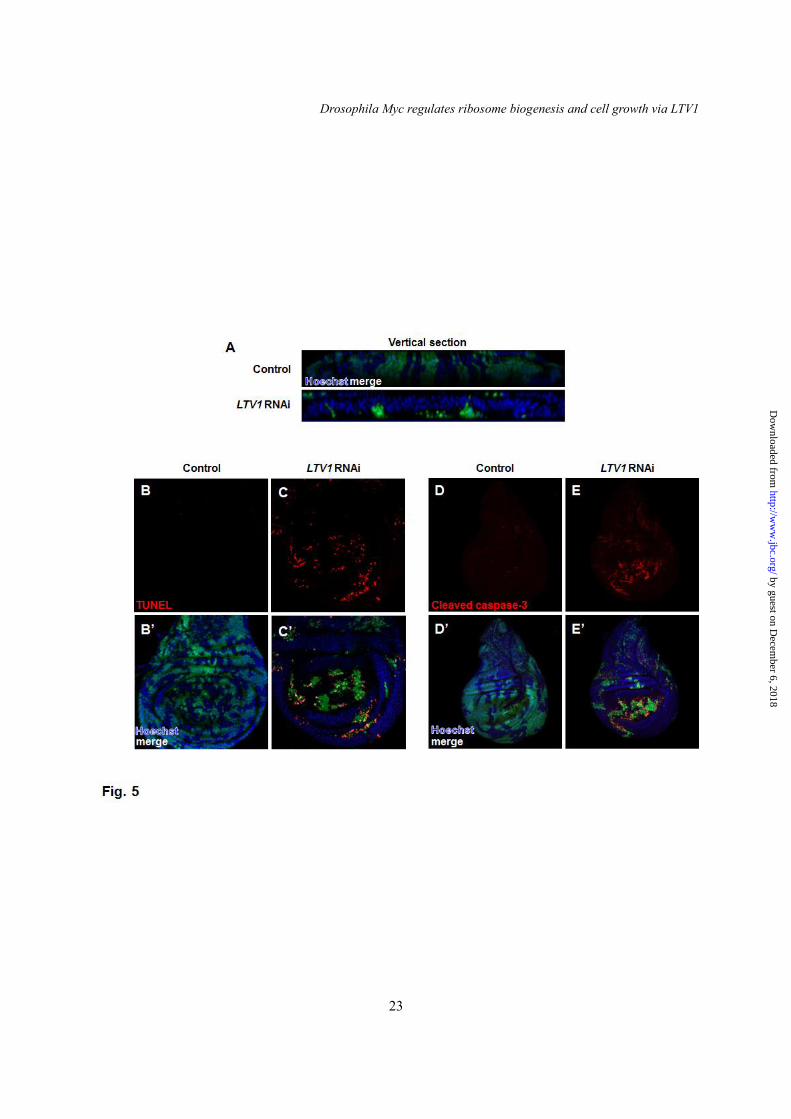
To find the in vivo functions of LTV1 other than cell growth regulation, we expressed LTV1 RNAi in developing tissues. When LTV1 RNAi-expressing GFP-positive clones were generated in wing imaginal discs using flip-out recombination (22), their clone size (Fig. 5A, lower panel) was much smaller than the control clones-expressing GFP alone (Fig. 5A, upper panel). Moreover, LTV1 RNAi-expressing clones
by guest on Decem
ber 6, 2018http://w
ww
.jbc.org/D
ownloaded from

Drosophila Myc regulates ribosome biogenesis and cell growth via LTV1
10
were accumulated in the basal side of the wing epithelia layers (Fig. 5A, lower panel). Because dead cells were shown to be extruded basally (33,34), we examined whether LTV1 knockdown induces cell death. Surprisingly, LTV1 RNAi expressing wing disc cells showed increased TUNEL signals (Fig. 5C and C’) whereas control cells did not (Fig. 5B and B’). In addition, we immunostained control and LTV1 knockdown wing imaginal disc cells with the antibody against cleaved caspase-3. In control cells, we observed weak caspase-3 signals (Fig. 5D and D’). However, we detected increased cleaved and active caspase-3 signals in LTV1 RNAi-expressing cells (Fig. 5E and E’), suggesting that LTV1 loss-of-function induces a caspase-dependent cell death. Combined, these data demonstrated that LTV1 is critical for cell survival. LTV1 is a direct transcriptional target of dMyc
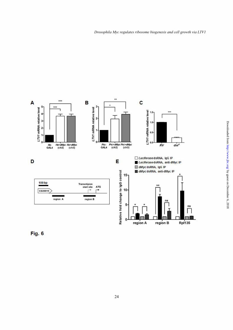
To understand the upstream regulator of LTV1, we examined dMyc [also known as diminutive (dm)], the oncogenic transcription factor that regulates multiple genes involved in ribosome biogenesis. We determined whether dMyc overexpression increases the level of LTV1 mRNA. For this experiment, we used two transgenic flies of dMyc in which dMyc transgene was inserted in two different chromosome locations. Notably, the ectopic expression of dMyc in the entire body of two different transgenic flies using Heat shock (Hs)-GAL4 increased the level of LTV1 mRNA by ~3.5-fold (Fig. 6A). Because dMyc regulates cell growth and ribosome biogenesis in the salivary glands (35,36), we also examined LTV1 transcripts in the salivary gland tissues expressing ectopic dMyc. Similar to the results expressing in the entire body, the amount of LTV1 mRNA increased by ~2-fold when dMyc transgene was expressed in the salivary glands using Patched (Ptc)-GAL4 (Fig. 6B). By contrast, the transcript level of LTV1 decreased by ~75% in dm4 (dMyc null mutant) compared to that of control flies (a revertant of dm4) (Fig. 6C). These results indicated that the level of LTV1 mRNA is positively regulated by dMyc.
To further determine whether dMyc directly regulates LTV1 transcription, we conducted a chromatin immunoprecipitation (Chip)
assay on S2 cells. We used the identical dMyc antibody used in a previous genome-wide Chip-seq analysis of Drosophila cells (37). As a positive control, we observed that dMyc associates with the promoter region of RpI135, a known target gene of dMyc (Fig. 6E) (35). To confirm the specificity of the association, we depleted dMyc by treating with dsRNA, which blocked the association of dMyc with the RpI135 promoter region (Fig. 6E). We then examined two different regions (region A and B) in the putative LTV1 promoter region. Region A is located away from the LTV1 transcription start site, and region B contains the LTV1 transcription start site (Fig. 6D). Notably, dMyc binded to both region A and B. However, when dMyc was down-regulated, region A was still enriched in dMyc immunoprecipitates, indicating that region A was not specifically associated with dMyc. By contrast, region B was barely detected in dMyc immunoprecipitates from dMyc knockdown cells (Fig. 6E). These results suggested that dMyc directly associates with the promoter region nearby the transcription start site of LTV1 and regulates LTV1 transcription. LTV1 is critical for the ribosome biogenesis induced by dMyc
Because dMyc is a well-established regulator of ribosome biogenesis, we investigated whether LTV1 is required for ribosome biogenesis downstream of dMyc. To assess this possibility, we examined the nucleolus size, an index of ribosome biogenesis (35). When the nucleolus was stained in dMyc-overexpressed salivary glands using an antibody for fibrillarin (a nucleolar marker), the size of the nucleolus dramatically increased, indicating that ribosome biogenesis was stimulated (Fig. 7A and B). By contrast, LTV1 knockdown suppressed the enlargement of the nucleolus of the dMyc-expressing salivary glands (Fig. 7B and C). A transmission electron microscope analysis also showed that LTV1 depletion reduced the enlarged nucleolus of the dMyc-overexpressed salivary glands (Fig. 7D-F). Moreover, the amount of ribosomes and rough endoplasmic reticulum (RER) network increased after dMyc overexpression as previously described (Fig. 7D’ and E’) (35). These
by guest on Decem
ber 6, 2018http://w
ww
.jbc.org/D
ownloaded from
Drosophila Myc regulates ribosome biogenesis and cell growth via LTV1
11
increases were also suppressed by LTV1 RNAi expression, even lower than control (Fig. 7D’-F’). Collectively, these results showed that LTV1 is critical for the ribosome biogenesis stimulated by dMyc. dMyc requires LTV1 to promote cell growth and endoreplication
Because ribosome biogenesis is critical for cell growth, we hypothesized that LTV1 is also important for dMyc-induced cell growth. To test this possibility, we determined whether dMyc overexpression can increase the size of LTV1 RNAi-expressing cells. To examine cell size, we expressed transgenes clonally in the fat body cells and measured the cell area of the transgene-expressing cells relative to that of the neighboring cells expressing no transgenes. Control cells expressing only GFP exhibited a similar cell size with neighboring cells (Fig. 8A and G). However, as shown in the previous results (Fig. 1A and A’), LTV1 RNAi expression dramatically decreased cell size up to ~90% (Fig. 8B and G). We also observed highly increased cell sizes of ~50% (Fig. 8D and G) when we overexpressed dMyc, as shown by others (36). However, when dMyc was simultaneously overexpressed in LTV1 RNAi-expressing cells, dMyc failed to increase cell size (Fig. 8E and G), indicating that LTV1 is highly important for dMyc-induced cell growth. We speculated that other target genes of dMyc involved in ribosome biogenesis are also important for dMyc-induced cell growth. To investigate this possibility, we expressed RNAi for RpI135 (a subunit of the RNA polymerase I complex) with or without dMyc transgene. RpI135 RNAi expression also decreased cell size by up to ~90% (Fig. 8C and G). Moreover, dMyc overexpression did not increase the cell size of RpI135-knockdown cells, similar to the results obtained from LTV1-knockdown cells (Fig. 8F and G). These results suggested that ribosome biogenesis is critical for dMyc-induced cell growth, and LTV1 is one of the critical dMyc target genes involved in ribosome biogenesis. In Drosophila polyploidy cells, such as the salivary glands and the fat body, dMyc promotes endoreplication and cell growth (36). We decided to examine whether LTV1 is required
for dMyc-promoted endoreplication in the fat body cells. Drosophila polyploid cells replicate DNA by repeating cycles of G and S phases (38). Therefore, we thought that dMyc-overexpressed fat body cells enter S phase more frequently than controls. To observe S phase cells, we incorporated 5-bromo-2'-deoxyuridine (BrdU) into replicating DNA. We observed that ~9% of control clone cells replicated DNA in the mid-3rd instar larvae (96 hours after egg laying) (Fig. 8H). However, LTV1 RNAi-expressing clone cells did not replicate during the identical time period (Fig. 8H). Consistent with these results, the nuclear size of LTV1 mutant cells was smaller than that of control cells (Fig. 1A, A’, E and E’). When ectopic dMyc was clonally expressed, the number of cells replicating DNA was increased by ~6-fold more than control clone cells (Fig. 8H). However, when both dMyc and LTV1 RNAi were clonally expressed, the number of replicating cells drastically decreased to the level of control cells (Fig. 8H). In addition, a previous study reported that dMyc expression also extended the endoreplicating period (36). Similar to the claims of this previous report, when we expressed dMyc in the salivary glands of wandering larvae, we observed the cells replicating DNA (Fig. 8J and J’) whereas control cells halted replication (Fig. 8I and I’). Notably, LTV1 knockdown almost completely blocked the endoreplication induced by dMyc expression (Fig. 8K and K’). Based on these results, we concluded that LTV1 is critical for the cell growth and endoreplication driven by dMyc. DISCUSSION
In this study, we identified LTV1 as a novel cell growth regulator using a fly system. We generated an LTV1 null animal to investigate the in vivo functions of LTV1 and discovered that LTV1 is required for normal cell growth (Fig. 1F and G). Subsequently, we found that LTV1 interacts with RpS3 (Fig. 2A-D) and co-sedimented with 40S ribosome subunits in sucrose density gradient (Fig. 2F). We also observed that the amount of 40S ribosome subunits was diminished in LTV1 mutants (Fig. 3A-C), and further demonstrated that the nuclear export of pre-40S ribosome subunits (Fig. 3E-G) and the
by guest on Decem
ber 6, 2018http://w
ww
.jbc.org/D
ownloaded from
Drosophila Myc regulates ribosome biogenesis and cell growth via LTV1
12
processing of pre-rRNA (Fig. 4) were impaired in the mutants. These results suggested that LTV1 is required for cell growth through regulating ribosome biogenesis. We further characterized LTV1 mutant phenotypes and found that LTV1 functions in cell survival and endoreplication (Fig. 5 and 8H). In addition, we discovered that dMyc transcription factor positively regulates the level of LTV1 mRNA and directly binds to the LTV1 promoter (Fig. 6). Notably, LTV1 RNAi expression suppressed dMyc-induced ribosome biogenesis, endoreplication and cell growth (Fig. 7 and 8), suggesting that LTV1 is an important downstream factor of dMyc in cell growth regulation.
In yeast, LTV1p was reported to bind with RpS3 (11,39), which is highly consistent with this study (Fig. 2A-D). Furthermore, LTV1p associates with pre-40S ribosome subunits (11,23). However, in our anti-LTV1 immunoprecipitation assay (Fig. 2A), we could not identify other proteins associated in pre-40S ribosomal subunits than RpS3. We postulated that they were washed out from LTV1 immunoprecipitates during the washing steps as our washing buffers contained higher salt concentrations than the buffers used in a previous study (Fig. 2A) (11). Consistent with this postulation, we observed that LTV1 was co-purified with 40S ribosome subunits in sucrose gradient experiments (Fig. 2F). Moreover, we examined whether rRNA is required for the interaction between LTV1 and RpS3. Notably, when we degraded rRNA by treating RNase A, the interaction between two proteins was not affected (Fig. 2E). These results indicated that the interaction between LTV1 and RpS3 is highly specific and not dependent on the integrity of 40S ribosome subunits in Drosophila.
Mature 40S ribosome subunits are generated through sequential processing steps from the nucleolus to the cytosol. Here, we observed increased nuclear-localized RpS2, a marker for pre-40S ribosome subunits, in LTV1 mutant cells, similar to a previous study in yeast (Fig. 3E-G) (24). In yeast, 20S rRNA assembled in pre-40S ribosome subunits is exported from the nucleus and subsequently processed in the cytosol. Notably, we found that 21S rRNA that corresponds to yeast 20S rRNA accumulated in
Drosophila LTV1 mutant larvae, supporting that pre-40S ribosome subunits are stalled in the nucleus of LTV1 mutant cells (Fig. 4B). These data consistently supported that LTV1 is required for the nuclear export of pre-40S ribosome subunits both in yeast and Drosophila. In addition, LTV1 may also be involved in other steps of ribosome biogenesis, although we could not further address this issue in this study. Interestingly, the yeast LTV1p mutant showed a defective maturation of pre-40S ribosome subunits in the cytosol (40).
In this study, we showed that LTV1 transcription is directly regulated by dMyc transcription factor (Fig. 6). However, LTV1 could also be regulated at the translation level. The target of rapamycin complex 1 (TORC1) regulates translation of the mRNAs displaying a structural characteristic of the 5’ terminal oligopyrimidine tract (41). Notably, Drosophila LTV1 mRNA has ‘CCCTTTCT’ at the 5’ terminal end (NCBI Accession # NM_136776), suggesting that mRNA translation of LTV1 can be a target of TORC1. Consistent with this suggestion, we observed that LTV1 knockdown suppressed the cell growth induced by a constitutive active insulin receptor (an upstream activator of TORC1) in the developing Drosophila eye (data not shown).
Myc regulates transcription of the diverse genes involved in ribosome biogenesis, cell cycle progression, metabolism, etc. to promote cell growth (42). In this study, we identified LTV1 as another direct transcription target of dMyc (Fig. 6) and found that LTV1 is crucial for dMyc-induced ribosome biogenesis (Fig. 7). Notably, dMyc overexpression could not induce cell growth in LTV1-depleted cells (Fig. 8A, B, D, E and G), indicating that LTV1 is critical target of dMyc in cell growth regulation. Moreover, dMyc also regulates transcription of other ribosome biogenesis-related genes including RpI135, a subunit of the RNA polymerase complex I that synthesizes pre-rRNA (35). Interestingly, the knockdown of RpI135 blocked the cell growth stimulated by dMyc overexpression, similar to LTV1. Furthermore, the heterozygotic loss-of-function mutation of the ribosomal protein L24 suppressed the cell size increase and lymphoma formation induced by
by guest on Decem
ber 6, 2018http://w
ww
.jbc.org/D
ownloaded from
Drosophila Myc regulates ribosome biogenesis and cell growth via LTV1
13
Myc-overexpression in B cells (43). All of these results consistently supported that ribosome biogenesis is crucial for Myc-dependent cell size regulation and tumorigenesis.
In summary, we demonstrated that LTV1 is required for Myc-induced ribosome biogenesis
and cell growth in Drosophila. We expect that our data may assist in understanding the molecular mechanism of the tumorigenesis induced by Myc in cancers.
REFERENCES 1. Hariharan, I. K., and Bilder, D. (2006) Regulation of imaginal disc growth by tumor-suppressor
genes in Drosophila. Annual review of genetics 40, 335-361 2. Neto-Silva, R. M., Wells, B. S., and Johnston, L. A. (2009) Mechanisms of growth and
homeostasis in the Drosophila wing. Annual review of cell and developmental biology 25, 197-220
3. Ruggero, D., and Pandolfi, P. P. (2003) Does the ribosome translate cancer? Nature reviews. Cancer 3, 179-192
4. Tschochner, H., and Hurt, E. (2003) Pre-ribosomes on the road from the nucleolus to the cytoplasm. Trends in cell biology 13, 255-263
5. Venema, J., and Tollervey, D. (1999) Ribosome synthesis in Saccharomyces cerevisiae. Annual review of genetics 33, 261-311
6. Fromont-Racine, M., Senger, B., Saveanu, C., and Fasiolo, F. (2003) Ribosome assembly in eukaryotes. Gene 313, 17-42
7. Henras, A. K., Soudet, J., Gerus, M., Lebaron, S., Caizergues-Ferrer, M., Mougin, A., and Henry, Y. (2008) The post-transcriptional steps of eukaryotic ribosome biogenesis. Cellular and molecular life sciences : CMLS 65, 2334-2359
8. Zemp, I., and Kutay, U. (2007) Nuclear export and cytoplasmic maturation of ribosomal subunits. FEBS letters 581, 2783-2793
9. Johnson, A. W., Lund, E., and Dahlberg, J. (2002) Nuclear export of ribosomal subunits. Trends in biochemical sciences 27, 580-585
10. Karbstein, K. (2011) Inside the 40S ribosome assembly machinery. Current opinion in chemical biology 15, 657-663
11. Schafer, T., Maco, B., Petfalski, E., Tollervey, D., Bottcher, B., Aebi, U., and Hurt, E. (2006) Hrr25-dependent phosphorylation state regulates organization of the pre-40S subunit. Nature 441, 651-655
12. Lempiainen, H., and Shore, D. (2009) Growth control and ribosome biogenesis. Current opinion in cell biology 21, 855-863
13. Arabi, A., Wu, S., Ridderstrale, K., Bierhoff, H., Shiue, C., Fatyol, K., Fahlen, S., Hydbring, P., Soderberg, O., Grummt, I., Larsson, L. G., and Wright, A. P. (2005) c-Myc associates with ribosomal DNA and activates RNA polymerase I transcription. Nature cell biology 7, 303-310
14. Poortinga, G., Hannan, K. M., Snelling, H., Walkley, C. R., Jenkins, A., Sharkey, K., Wall, M., Brandenburger, Y., Palatsides, M., Pearson, R. B., McArthur, G. A., and Hannan, R. D. (2004) MAD1 and c-MYC regulate UBF and rDNA transcription during granulocyte differentiation. The EMBO journal 23, 3325-3335
15. Shiue, C. N., Berkson, R. G., and Wright, A. P. (2009) c-Myc induces changes in higher order rDNA structure on stimulation of quiescent cells. Oncogene 28, 1833-1842
16. Schlosser, I., Holzel, M., Murnseer, M., Burtscher, H., Weidle, U. H., and Eick, D. (2003) A role for c-Myc in the regulation of ribosomal RNA processing. Nucleic acids research 31, 6148-6156
17. Wu, C. H., Sahoo, D., Arvanitis, C., Bradon, N., Dill, D. L., and Felsher, D. W. (2008) Combined analysis of murine and human microarrays and ChIP analysis reveals genes associated with the ability of MYC to maintain tumorigenesis. PLoS genetics 4, e1000090
by guest on Decem
ber 6, 2018http://w
ww
.jbc.org/D
ownloaded from
Drosophila Myc regulates ribosome biogenesis and cell growth via LTV1
14
18. Maggi, L. B., Jr., Kuchenruether, M., Dadey, D. Y., Schwope, R. M., Grisendi, S., Townsend, R. R., Pandolfi, P. P., and Weber, J. D. (2008) Nucleophosmin serves as a rate-limiting nuclear export chaperone for the Mammalian ribosome. Molecular and cellular biology 28, 7050-7065
19. Boon, K., Caron, H. N., van Asperen, R., Valentijn, L., Hermus, M. C., van Sluis, P., Roobeek, I., Weis, I., Voute, P. A., Schwab, M., and Versteeg, R. (2001) N-myc enhances the expression of a large set of genes functioning in ribosome biogenesis and protein synthesis. The EMBO journal 20, 1383-1393
20. Zeller, K. I., Haggerty, T. J., Barrett, J. F., Guo, Q., Wonsey, D. R., and Dang, C. V. (2001) Characterization of nucleophosmin (B23) as a Myc target by scanning chromatin immunoprecipitation. The Journal of biological chemistry 276, 48285-48291
21. Xu, T., and Rubin, G. M. (1993) Analysis of genetic mosaics in developing and adult Drosophila tissues. Development 117, 1223-1237
22. Pignoni, F., and Zipursky, S. L. (1997) Induction of Drosophila eye development by decapentaplegic. Development 124, 271-278
23. Schafer, T., Strauss, D., Petfalski, E., Tollervey, D., and Hurt, E. (2003) The path from nucleolar 90S to cytoplasmic 40S pre-ribosomes. The EMBO journal 22, 1370-1380
24. Seiser, R. M., Sundberg, A. E., Wollam, B. J., Zobel-Thropp, P., Baldwin, K., Spector, M. D., and Lycan, D. E. (2006) Ltv1 is required for efficient nuclear export of the ribosomal small subunit in Saccharomyces cerevisiae. Genetics 174, 679-691
25. Merwin, J. R., Bogar, L. B., Poggi, S. B., Fitch, R. M., Johnson, A. W., and Lycan, D. E. (2014) Genetic analysis of the ribosome biogenesis factor Ltv1 of Saccharomyces cerevisiae. Genetics 198, 1071-1085
26. Zemp, I., Wild, T., O'Donohue, M. F., Wandrey, F., Widmann, B., Gleizes, P. E., and Kutay, U. (2009) Distinct cytoplasmic maturation steps of 40S ribosomal subunit precursors require hRio2. The Journal of cell biology 185, 1167-1180
27. Grandi, P., Rybin, V., Bassler, J., Petfalski, E., Strauss, D., Marzioch, M., Schafer, T., Kuster, B., Tschochner, H., Tollervey, D., Gavin, A. C., and Hurt, E. (2002) 90S pre-ribosomes include the 35S pre-rRNA, the U3 snoRNP, and 40S subunit processing factors but predominantly lack 60S synthesis factors. Molecular cell 10, 105-115
28. Milkereit, P., Strauss, D., Bassler, J., Gadal, O., Kuhn, H., Schutz, S., Gas, N., Lechner, J., Hurt, E., and Tschochner, H. (2003) A Noc complex specifically involved in the formation and nuclear export of ribosomal 40 S subunits. The Journal of biological chemistry 278, 4072-4081
29. Moy, T. I., and Silver, P. A. (2002) Requirements for the nuclear export of the small ribosomal subunit. Journal of cell science 115, 2985-2995
30. Moy, T. I., and Silver, P. A. (1999) Nuclear export of the small ribosomal subunit requires the ran-GTPase cycle and certain nucleoporins. Genes & development 13, 2118-2133
31. Ho, J. H., Kallstrom, G., and Johnson, A. W. (2000) Nmd3p is a Crm1p-dependent adapter protein for nuclear export of the large ribosomal subunit. The Journal of cell biology 151, 1057-1066
32. Long, E. O., and Dawid, I. B. (1980) Alternative pathways in the processing of ribosomal RNA precursor in Drosophila melanogaster. Journal of molecular biology 138, 873-878
33. Sedlak, B. J., Manzo, R., and Stevens, M. (1984) Localized cell death in Drosophila imaginal wing disc epithelium caused by the mutation apterous-blot. Developmental biology 104, 489-496
34. Mitchell, N. C., Johanson, T. M., Cranna, N. J., Er, A. L., Richardson, H. E., Hannan, R. D., and Quinn, L. M. (2010) Hfp inhibits Drosophila myc transcription and cell growth in a TFIIH/Hay-dependent manner. Development 137, 2875-2884
35. Grewal, S. S., Li, L., Orian, A., Eisenman, R. N., and Edgar, B. A. (2005) Myc-dependent regulation of ribosomal RNA synthesis during Drosophila development. Nature cell biology 7, 295-302
by guest on Decem
ber 6, 2018http://w
ww
.jbc.org/D
ownloaded from

Drosophila Myc regulates ribosome biogenesis and cell growth via LTV1
15
36. Pierce, S. B., Yost, C., Britton, J. S., Loo, L. W., Flynn, E. M., Edgar, B. A., and Eisenman, R. N. (2004) dMyc is required for larval growth and endoreplication in Drosophila. Development 131, 2317-2327
37. Yang, J., Sung, E., Donlin-Asp, P. G., and Corces, V. G. (2013) A subset of Drosophila Myc sites remain associated with mitotic chromosomes colocalized with insulator proteins. Nature communications 4, 1464
38. Budirahardja, Y., and Gonczy, P. (2009) Coupling the cell cycle to development. Development 136, 2861-2872
39. Loar, J. W., Seiser, R. M., Sundberg, A. E., Sagerson, H. J., Ilias, N., Zobel-Thropp, P., Craig, E. A., and Lycan, D. E. (2004) Genetic and biochemical interactions among Yar1, Ltv1 and Rps3 define novel links between environmental stress and ribosome biogenesis in Saccharomyces cerevisiae. Genetics 168, 1877-1889
40. Fassio, C. A., Schofield, B. J., Seiser, R. M., Johnson, A. W., and Lycan, D. E. (2010) Dominant mutations in the late 40S biogenesis factor Ltv1 affect cytoplasmic maturation of the small ribosomal subunit in Saccharomyces cerevisiae. Genetics 185, 199-209
41. Meyuhas, O. (2000) Synthesis of the translational apparatus is regulated at the translational level. European journal of biochemistry / FEBS 267, 6321-6330
42. Dang, C. V. (2012) MYC on the path to cancer. Cell 149, 22-35 43. Barna, M., Pusic, A., Zollo, O., Costa, M., Kondrashov, N., Rego, E., Rao, P. H., and Ruggero, D.
(2008) Suppression of Myc oncogenic activity by ribosomal protein haploinsufficiency. Nature 456, 971-975
ACKNOWLEDGEMENT This work was supported by National Creative Research Initiatives Program Grant 2010-0018291 and 2012R1A2A1A01009027 from the Korean Ministry of Science, ICT and Future Planning. We thank Dr. Robert Eisenman for providing Drosophila stocks. We are grateful to Bloomington Stock Center, Kyoto Drosophila Genetic Resource Center, FlyORF and Vienna Drosophila Resource Center. We thank Drs. Narry Kim and Jun Cho for kind support for sucrose density gradient analysis. We appreciate Dr. Sang-Hee Lee for electron microscopic analysis. We thank Taplin Mass Spectrometry Facility for mass spectrometry analysis. We thank Drs. Eunjoo Cho, Minjeong Ryu, Jinwook Choi, Shin Jeon and Ji Min Lee for helpful discussion. FOOTNOTES The abbreviations used are: LTV1, low temperature viability protein 1; dMyc, Drosophila Myc; RpS3, ribosomal protein S3; RpS2, ribosomal protein S2; rRNA, ribosomal RNA; pre-rRNA, pre-ribosomal RNA; dsRNA, double-stranded RNA; ETS, external transcribed spacer; ITS1, internal transcribed spacer 1; 5ITS1, 5’ end of internal transcribed spacer 1; ITS2, internal transcribed spacer 2; RER, rough endoplasmic reticulum; TORC1, target of rapamycin complex 1; AEL, after egg laying; Da, Daughterless; Ptc, Patched; Hs, Heat shock. FIGURE LEGENDS Fig. 1. LTV1 is required for cell growth and normal development. (A) LTV1 RNAi expressing clones (GFP-positive) indicated by arrowhead were generated, and the nuclei and cell size were examined by staining with Hoechst (blue) and phalloidin (red), respectively. (B) A schematic representation of LTV1 genomic locus and the genomic deletion region of LTV1E1. The exons of LTV1 are marked by boxes and the coding regions of LTV1 are by colored black. The P-element, LTV1CB6202-3, is inserted in the 5’ untranslated region of LTV1. (C) qPCR analysis of LTV1 transcripts in w1118 and LTV1E1 flies. Actin expression was used as a loading control. (D) Larvae of w1118, LTV1 mutants, and LTV1 mutants expressing exogenous LTV1 in the whole body were photographed at 24, 48 and 120
by guest on Decem
ber 6, 2018http://w
ww
.jbc.org/D
ownloaded from
Drosophila Myc regulates ribosome biogenesis and cell growth via LTV1
16
hours after egg laying (AEL). (E) LTV1 was overexpressed by Da-GAL4. The whole cell lysates were immunoblotted by anti-HA and -β-tubulin antibodies. (F) FRT/FLP-mediated recombination was used to generate homozygous LTV1E1 mutant clones (arrowhead) marked by GFP-negative. (G) In the fat body expressing exogenous LTV1, LTV1E1 mutant clones (arrowhead) indicated by GFP-negative were generated by FRT/FLP-mediated recombination. Ectopic expression of LTV1 rescued the decreased cell size of the mutant clones back to that of control cells. (F and G) Hoechst (blue) and phalloidin (red) were stained to observe the nuclei and cell boundary, respectively. Genotypes: (A) y w hsflp; UAS-LTV1 RNAi; Act>CD2>GAL4 UAS-GFP. (F) y w hsflp; Cg-GAL4 FRT42D UAS-GFP/FRT42D LTV1E1. (G) y w hsflp; Cg-GAL4 FRT42D UAS-GFP/FRT42D LTV1E1; UAS-LTV1-HA. Fig. 2. LTV1 interacts with RpS3 and co-purifies with 40S ribosome subunits. (A) LTV1-V5 and RpS3-mCherry were co-transfected in S2 cells. The immunoprecipitates of LTV1-V5 were separated by SDS-PAGE and subsequently stained with coomassie blue. RpS3 was identified by mass spectrometry analysis. Immunoglobin heavy and light chains were indicated by asterisks. (B) LTV1-V5 and RpS3-mCherry were expressed in S2 cells by transfection. The whole cell lysates were immunoblotted by anti-RFP and -V5 antibodies, and RpS3-mCherry was immunoprecipitated by anti-RFP antibody followed by immunoblotting with anti-RFP and -V5 antibodies. The asterisk indicates inmmunoglobin heavy chain. (C) LTV1-V5 and RpS3-mCherry were transfected in S2 cells. The whole cell lysates were immunoblotted by anti-V5 and -RFP antibodies. LTV1-V5 was immunoprecipitated by anti-V5 antibody and the immunoprecipitates were blotted by anti-V5 and -RFP antibodies. The asterisk indicates non-specifically bound proteins. (D) Transfected LTV1-HA was immunoprecipitated by anti-HA antibody, and LTV1-HA and endogenous RpS3 were detected by anti-HA and -RpS3 antibodies, respectively. The whole cell lysates were blotted by anti-HA and -RpS3 antibodies. (E) LTV1-HA and RpS3-V5 were transfected in S2 cells. The lysates were incubated without or with 0.1 μg/ml RNase A for two hours at room temperature, and RpS3-V5 was immunoprecipitated. The whole cell lysates and immunoprecipitates were immunoblotted by anti-HA and -V5 antibodies. Degradation of 18S rRNA was confirmed by RT-PCR. (F) LTV1-HA was transfected in S2 cells. The clarified lysates were applied to 10%-50% sucrose gradient. After centrifugation, the absorbance at 260 nm was measured, and each fraction was collected and analyzed by immunoblotting with anti-HA antibody. The peaks of 40S and 60S ribosome subunits, 80S monosomes and polysomes are indicated. Fig. 3. LTV1 is crucial for 40S ribosome biogenesis. (A) The lysates were extracted from S2 cells treated with dsRNA for luciferase (red) and LTV1 (blue). Ribosomes were separated on 10%-50% sucrose gradient. The absorbance at 260 nm was measured. Luciferase dsRNA-treated cell lysates were used for control. (B) Total RNA was extracted from the 2nd instar larvae of w1118, LTV1 mutant, and LTV1 mutant expressing exogenous LTV1. RNA was subjected to formaldehyde agarose gel electrophoresis and assessed by Northern blotting. The amount of 18S and 28S rRNA was analyzed by Northern blotting. (C) The relative ratio of 18S and 28S rRNA amounts was quantified. The intensity of each band was measured by Multi gauge. The means of three independent experiments were presented. Error bars show SEM. **, P<0.005, n = 3. (D) LTV1 was ectopically expressed by Da-GAL4. Control (red) and LTV1-HA-expressed (green) 3rd instar larvae were collected and lysed. Ribosomes were separated by 10%-50% sucrose gradient from the lysates, and the absorbance at 260 nm was measured. (E-G) RpS2-HA was immunostained by anti-HA antibody (red) in the fat body cells expressing RpS2-HA (E), RpS2-HA and Crm1 RNAi (F) and RpS2-HA and LTV1 RNAi (G). Transgene-expressing cells were marked by the presence of GFP. Hoechst (blue) was used to stain the nucleus. Genotypes: (E) y w hsflp; Act>CD2>GAL4 UAS-GFP/UAS-RpS2-3xHA. (F) y w hsflp; UAS-Crm1 RNAi; Act>CD2>GAL4 UAS-GFP/UAS-RpS2-3xHA. (G) y w hsflp; UAS-LTV1 RNAi; Act>CD2>GAL4 UAS-GFP/UAS-RpS2-3xHA.
by guest on Decem
ber 6, 2018http://w
ww
.jbc.org/D
ownloaded from
Drosophila Myc regulates ribosome biogenesis and cell growth via LTV1
17
Fig. 4. LTV1 is required for pre-rRNA processing. (A) A schematic representation of Drosophila pre-rRNA processing. Pre-rRNA and its intermediates are designated pre, a and d according to the previous report (32). ETS (external transcribed spacer), ITS1 (internal transcribed spacer 1) and ITS2 (internal transcribed spacer 2) were indicated. The 5ITS1 probe, which was used in hybridizing 5’ region of ITS1, was indicated by black bar. (B) Total RNA was extracted from the 2nd instar larvae of w1118, LTV1 mutant, and LTV1 mutant-expressing exogenous LTV1. Total RNA was separated in formaldehyde agarose gel. Pre-rRNA and its intermediates were examined by Northern blotting with the probe for 5ITS1. Each intermediate is indicated. Asterisk marks the intermediate of 2.5 kb. Fig. 5. LTV1 is critical for cell survival in wing imaginal disc. (A) Confocal z-stack analyses of control clones (Top) and LTV1 RNAi-expressing clones (Bottom) in wing discs. The clonal cells were marked by GFP. DNA (blue) was stained by Hoechst. (B-E) Control clones (B and D) and LTV1 RNAi expressing clones (C and E) (GFP-positive) were immunostained for TUNEL (B and C) and cleaved caspase-3 (D and E) in wing discs. Hoechst (blue) was stained to mark the nuclei. Genotypes: (A) y w hsflp; Act>CD2>GAL4 UAS-GFP (Top), y w hsflp; UAS-LTV1 RNAi; Act>CD2>GAL4 UAS-GFP (Bottom). (B and D) y w hsflp; Act>CD2>GAL4 UAS-GFP. (C and E) y w hsflp; UAS-LTV1 RNAi; Act>CD2>GAL4 UAS-GFP. Fig. 6. dMyc directly regulates LTV1 transcription. (A) LTV1 transcripts were measured upon induction of dMyc by heat shock with Hs-GAL4 in the 3rd instar larvae. Two transgenic flies for dMyc were used. One has dMyc transgene in second chromosome (ch2) and the other in third chromosome (ch3). dMyc was induced by heat shock at 37°C for 30 minutes and the larvae were subsequently incubated at 25°C for 6 hours. (B) The transcript level of LTV1 in the dMyc-expressed salivary glands was determined. (C) The mRNA levels for LTV1 were examined in the 1st instar larvae of the revertant (RV) and dMyc null mutant, dm4. (A-C) Following total RNA extraction and reverse transcription, qPCR was used to quantify LTV1 mRNA. Actin was used as a loading control. Error bars show SEM. (A) ***, P<0.0005, n = 3; (B) *, P<0.05 and **, P<0.01, n = 3; (C) ***, P<0.0001, n = 4. (D) A schematic representation of putative LTV1 promoter region. The transcription start site and the start codon of LTV1 were indicated by arrows. The genomic region A and B used for Chip-qPCR were indicated by black bars. (E) S2 cells were treated with the dsRNA for luciferase and dMyc. After 4 days, protein and DNA were cross-linked by formaldehyde. S2 cells were lysed and immunoprecipitated by anti-dMyc antibody. Immunoglobin G was used in controls. Genomic sequence (region A and B) of the LTV1 promoter region was amplified by qPCR. The promoter sequence of RpI135 was used as a positive control. Relative fold changes over IgG controls were measured. Error bars show SEM. Region A: *, P<0.05, n = 3; Region B: **, P<0.01, n = 3; RpI135: *, P<0.05, n = 3; ns, non-significant. Genotypes: (A) Hs-GAL4, UAS-dMyc (ch2); Hs-GAL4, UAS-dMyc (ch3)/Hs-GAL4. (B) Ptc-GAL4, Ptc-GAL4/UAS-dMyc (ch2), Ptc-GAL4; UAS-dMyc (ch3). Fig. 7. LTV1 knockdown suppresses dMyc-induced ribosome biogenesis. (A-C) The nucleus (blue), nucleolus (green) and cell boundary (red) were stained by Hoechst, anti-fibrillarin antibody and phalloidin, respectively, in the salivary glands of the wandering larvae with indicated genotypes. (D-F) The nucleus, nucleolus and rough endoplasmic reticulum (RER) of the salivary glands of the wandering larvae with indicated genotypes were examined by transmission electron microscope. The nuclei were marked by asterisks. The nucleoli were indicated by arrows. The RER and ribosomes were marked by arrowheads. Scale bars: (A-C) 500 μm, (D-F) 5 μm and (D’-F’) 500 nm. Genotypes: (A and D) Ptc-GAL4. (B and E) Ptc-GAL4/UAS-dMyc (ch2). (C and F) Ptc-GAL4/UAS-dMyc (ch2), UAS-LTV1 RNAi.
by guest on Decem
ber 6, 2018http://w
ww
.jbc.org/D
ownloaded from

Drosophila Myc regulates ribosome biogenesis and cell growth via LTV1
18
Fig. 8. dMyc requires LTV1 to promote cell growth and endoreplication. (A-F) The fat body clone cells expressing only GFP (control) (A), LTV1 RNAi (B), RpI135 RNAi (C), dMyc (D), both dMyc and LTV1 RNAi (E) and both dMyc and RpI135 RNAi (F) were generated by flipout technique. The clone cells were marked by the presence of GFP. The cell boundary and nuclei were stained by phalloidin and Hoechst, respectively, at the mid-3rd instar larvae (96 hours AEL). (G) The area of the transgene-expressing cells relative to that of the neighboring cells was measured. Error bars show SEM. ***, P<0.0001; ns, non-significant. n = 23 (control), n = 21 (dMyc), n = 22 (LTV1 RNAi), n = 41 (dMyc+LTV1 RNAi), n = 21 (RpI135 RNAi) and n = 22 (dMyc+RpI135 RNAi). (H) The fat body clone cells expressing only GFP (control), dMyc, LTV1 RNAi and both dMyc and LTV1 RNAi were induced by flipout recombination. DNA replication of the fat body cells in the mid-3rd instar larvae (96 hours AEL) was examined by immunostaining with incorporated BrdU, and the BrdU-positive clone cells were counted. BrdU-positive clone cells/total clone cells: 10/109 (control), 0/71 (LTV1 RNAi), 35/66 (dMyc), 6/83 (dMyc+LTV1 RNAi). (I-K) BrdU (green) was incorporated in the salivary glands of the wandering larvae with indicated genotypes. The nuclei were stained by Hoechst (blue). FB and dotted line indicate the fat body cells which were contaminated in the preparation of the salivary glands. Genotypes: (A) y w hsflp; Act>CD2>GAL4 UAS-GFP. (B) y w hsflp; UAS-LTV1 RNAi, Act>CD2>GAL4 UAS-GFP. (C) y w hsflp; Act>CD2>GAL4 UAS-GFP/UAS-RpI135 RNAi. (D) y w hsflp; UAS-dMyc (ch2); Act>CD2>GAL4 UAS-GFP. (E) y w hsflp; UAS-dMyc (ch2)/UAS-LTV1 RNAi; Act>CD2>GAL4 UAS-GFP. (F) y w hsflp; UAS-dMyc (ch2); Act>CD2>GAL4 UAS-GFP/UAS-RpI135 RNAi. (H) y w hsflp; Act>CD2>GAL4 UAS-GFP (control); y w hsflp; UAS-LTV1 RNAi, Act>CD2>GAL4 UAS-GFP (LTV1 RNAi); y w hsflp; UAS-dMyc (ch2); Act>CD2>GAL4 UAS-GFP (dMyc); y w hsflp; UAS-dMyc (ch2)/UAS-LTV1 RNAi; Act>CD2>GAL4 UAS-GFP (dMyc+LTV1 RNAi). (I) Ptc-GAL4. (J) Ptc-GAL4/UAS-dMyc (ch2). (K) Ptc-GAL4/UAS-dMyc (ch2), UAS-LTV1 RNAi.
by guest on Decem
ber 6, 2018http://w
ww
.jbc.org/D
ownloaded from
Drosophila Myc regulates ribosome biogenesis and cell growth via LTV1
19
by guest on Decem
ber 6, 2018http://w
ww
.jbc.org/D
ownloaded from
Drosophila Myc regulates ribosome biogenesis and cell growth via LTV1
20
by guest on Decem
ber 6, 2018http://w
ww
.jbc.org/D
ownloaded from

Drosophila Myc regulates ribosome biogenesis and cell growth via LTV1
21
by guest on Decem
ber 6, 2018http://w
ww
.jbc.org/D
ownloaded from

Drosophila Myc regulates ribosome biogenesis and cell growth via LTV1
22
by guest on Decem
ber 6, 2018http://w
ww
.jbc.org/D
ownloaded from
Drosophila Myc regulates ribosome biogenesis and cell growth via LTV1
23
by guest on Decem
ber 6, 2018http://w
ww
.jbc.org/D
ownloaded from
Drosophila Myc regulates ribosome biogenesis and cell growth via LTV1
24
by guest on Decem
ber 6, 2018http://w
ww
.jbc.org/D
ownloaded from

Drosophila Myc regulates ribosome biogenesis and cell growth via LTV1
25
by guest on Decem
ber 6, 2018http://w
ww
.jbc.org/D
ownloaded from

Drosophila Myc regulates ribosome biogenesis and cell growth via LTV1
26
by guest on Decem
ber 6, 2018http://w
ww
.jbc.org/D
ownloaded from
Wonho Kim, Hag Dong Kim, Youjin Jung, Joon Kim and Jongkyeong ChungBiogenesis and Cell Growth Downstream of dMyc
Drosophila LTV1 (Low Temperature Viability Protein 1) is Required for Ribosome
published online April 9, 2015J. Biol. Chem.
10.1074/jbc.M114.607036Access the most updated version of this article at doi:
Alerts:
When a correction for this article is posted•
When this article is cited•
to choose from all of JBC's e-mail alertsClick here
by guest on Decem
ber 6, 2018http://w
ww
.jbc.org/D
ownloaded from