aus dem institut für physiologie - uni-due.de
TRANSCRIPT
Mediziniche Facultät
der
Universität Duisburg-Essen
Aus dem Institut für Physiologie
Arsenic trioxide (As2O3) interacts with [Ca2+
]i of
human SY-5Y neuroblastoma and human embryonic kidney 293 (HEK) cells and induces
cytotoxicity
Inaugural – Dissertation
zur
Erlangung des Doktorgrades der Naturwissenschaften in der Medizin
durch die Mediziniche Fakultät
der Universität Duisburg-Essen
Vorgelegt von
Dr.rer.nat. Ana-Maria Florea
aus Iasi, Rumänien
2007

2
Dekan: Herr Univ. Prof. Dr. rer. nat. K.-H.Jöckel
1. Gutachter: Herr Prof. Dr. rer. nat. D. Büsselberg
2. Gutachter: Herr Univ. Prof. Dr. med. E. Gulbins
Tag der mündlichen Prüfung: 16 Januar 2008

3
Publications:
Florea A.-M, Büsselberg D. Toxic effects of metals: use, benefits and toxic cellular effects Material
Wissenschaft 2005, 36: 757-760.
Florea A.-M, Büsselberg D.: Metals and metal compounds: occurrence, use, benefits and toxic
cellular effects. Biometals 2006, 19: 419-427.
Florea, A.-M., Splettstoesser, F. and D. Büsselberg. Mechanisms of arsenic trioxide (As2O3) induced
calcium signals and cellular death in two human SY-5Y Neuroblastoma and 293 embryonic kidney
(HEK) cells. TAAP. 2007, 220:292-301.
Splettstoesser, F., Florea, A.-M. and D. Büsselberg. IP3-receptor-antagonist 2-APB attenuates
cisplatin induced Ca2+
-influx in HeLa-S3 cells and prevents activation of calpain. Brit. J.
Pharmacology. 2007 Accepted with Revision.
In preparation
Florea, A.-M., Splettstoesser, F. and D. Büsselberg. Picomolar concentrations of arsenic trioxide
(As2O3) induce calcium signals and interfere with cell survival of human SY-5Y Neuroblastoma and
293 embryonic kidney (HEK) cells. (ready for submission)
Florea A.-M, Kunz, I., Büsselberg D.: Cisplatin, a short review
Florea, A.-M., Splettstoesser, F. and D. Büsselberg. Calcium stores involvement in arsenic trioxide
(As2O3) induced calcium signals.
The experiments regarding present work have been completed at the Institute of
Physiology, University-Hospital Essen, Germany

4
Content
1 Introduction 1 1.1 Human exposure to arsenic and health effects 2
1.2 Medical use of arsenic 2
1.3 Manifestation of arsenic intoxications 2 1.3.1 Acute arsenic poisoning 3
1.3.2 Chronic arsenic toxicity 3
1.3.3 Arsenic neurotoxicity 4
1.4 Molecular mechanisms of arsenic interaction with living cells 4
1.5 Calcium signalling and cell death induced by arsenic trioxide 5
1.5.1 Calcium as a second messenger in living cells 5
1.5.2 Calcium – cell death link 6 1.5.3 The process of apoptosis 6
1.5.4 Arsenic induced cell death 7
1.6 Aims of the study 10
2 Material and Methods 11
2.1 Material 11
2.1.1 Cell lines 11
2.1.2 Cell culture 12
2.1.3 Calcium sensitive dyes for calcium imaging 12
2.1.4 Other chemicals and reagents 13
2.2 Methods 14
2.2.1 Confocal laser scanning microscopy 14 2.2.2 Fluorescent activated cell sorting (FACS) 14
2.2.3 Trypan Blue Cytotoxicity Test 15
2.2.4 MTT-cytotoxicity test 15
2.2.5 Hoechst staining to score micronucleated cells, condensed nuclei, and cells in
mitosis 16
3 Results 17
3.1 As2O3 induced changes in [Ca2+
]i in neuroblastoma and HEK cells 17
3.2 As2O3 induced different types of [Ca2+
]i changes in neuroblastoma and HEK cells 19
3.3 The changes in [Ca2+
]i induced by As2O3 are concentration dependent in
neuroblastoma and HEK cells 22
3.4 Ca2+
stores are involved in the As2O3 mediated [Ca2+
]i changes 25
3.5 Ca2+
receptors are involved in the As2O3 mediated [Ca2+
]i changes 25
3.6 Other indications about Ca2+
pools that are involved in the As2O3 mediated [Ca2+
]i
changes 27
3.7 No Ca2+
rise in the absence of As2O3 in neuroblastoma and HEK cells 29
3.8 As2O3 determines cell death and damages DNA of neuroblastoma and HEK cells 29
3.9 Differential effects of nanomolar and picomolar concentrations of As2O3 on cell death and damages DNA of neuroblastoma and HEK cells 34
4 Discussion 36
4.1 As2O3 influences [Ca2+
]i homeostasis 36
4.2 [Ca2+
]i changes induced by As2O3 induces cell toxicity 40
4.3 Anticancer drugs could have different effects on calcium signalling e.g. As2O3 vs.
cisplatin 43
4.4 Synergistic effects of drug combinations 43
4.5 Outlook 44
5 Summary 46
6 References 47
Curriculum Vitae
Acknowledgements

5
1 Introduction
1.1 Human exposure to arsenic and health effects
Arsenic toxicity is a health problem affecting millions of people all over the world, especially
in India and Bangladesh. Human exposure to arsenic is mainly represented by intake of food
and drinking water contaminated with arsenic. Epidemiological studies show that a long time
arsenic intake correlates with the occurrence of several illnesses: abnormal development,
neurological and neurobehavioral disorders, cardiovascular and haematological diseases,
diabetes, hearing loss, fibrosis of the liver and lung, blackfoot disease, and several types of
cancers (Abernathy et al., 1999; Tchounwou et al., 1999; Sordo et al., 2001). The permissible
level of arsenic in drinking water was, before 2001, as high as 50 ppb but was further reduced
by the Environment Protection Agency (USA) down to 10 ppb (National Research Council,
2001; Ratnaike, 2003). Nevertheless, 10 ppb arsenic concentration in drinking water might
not be sufficiently low to avoid the toxicity and carcinogenity induced by arsenic (Florea and
Büsselberg, 2006; Ratnaike, 2003).
The contamination of drinking water with arsenic could be the result of (1) natural geological
sources and, may also occur from (2) human activity: mining, industry or agriculture. In
industry, arsenic is used for producing paints, fungicides, insecticides, pesticides, herbicides,
wood preservatives, cotton desiccants, semiconductors, light emitting diodes, lasers, and a
variety of transistors. Additionally, arsenic has been used over the time in agriculture as
pesticide (Florea and Büsselberg, 2006; Ratnaike, 2003; Florea, 2005).
In previous times, humans were more often exposed to arsenic toxicity as compared to today.
Arsenic was constituent in cosmetics as well as in paints (e.g. pigment in “Paris green”)
(Ratnaike, 2003; Florea and Büsselberg, 2006). Arsenic was used in formal times as a poison
weapon since arsenic compounds are tasteless and odourless. A famous example is the death
of Napoleon Bonaparte, which is still a matter of discussion today, whether he was
intentionally or accidentally poisoned with arsenic. In addition, several cases of death caused
by paints containing arsenic have been documented. One reason was a fungus (Scopulariopsis
breviculis), which metabolises arsenic from the wallpaper; process that results in very
poisonous vapours of arsenic (mixture of arsine, dimethyl and trimethyl arsine). A second
source of arsenic induced intoxications was the use of coal fires that emitted hydrogen which,
combined with the gas for lighting and with arsenic found in “Paris green” formed the toxic
gas arsine. Fortunately, those sources of human exposure are no longer of concern, while

6
other sources (especially drinking water) remain (Ratnaike et al., 2003; Florea and
Büsselberg. 2006).
1.2 Medical use of arsenic
While arsenic compounds are regarded as potent toxic and carcinogens, they also have been
medically used for over 2000 years, and are still used in diverse treatments (e.g. leukaemia,
leishmaniosis, trypanosomiasis) (Bergstrom et al., 1998; Shim et al., 2002; Florea, 2005;
Griffin et al., 2005). Arsenic was a “healing agent” used by Greek physicians (e.g.
Hippocrates). Later on, Fowler’s solution, a 1% arsenic trioxide preparation, was widely used
during the 19th century. The indications were: leukaemia, skin conditions (psoriasis,
dermatitis herpetiformis, and eczema), stomatitis and gingivitis in infants,
and Vincent’s
anginas, as well as a health tonic. Thus, long-term use of Fowler’s solution caused
haemangiosarcoma, angiosarcoma of the liver and nasopharyngeal carcinoma. Arsenic was
the primary treatment for syphilis until World War II; (arsphenamine, neoarsphenamine- 30%)
and some protozoan infections (Florea and Büsselberg, 2006; Ratnaike, 2003).
Also in traditional Chinese medicine, arsenous acid or arsenic trioxide (As2O3) was often used
to treat tooth marrow disease (devitalizing agent), but also against psoriasis, syphilis, and
rheumatosis with the saying: "using a toxic against another toxic" (Chen et al., 1995;
Ratnaike, 2003). In the 1970s, As2O3 was introduced into the treatment of acute
promyelocytic leukemia (APL) and showed immense success in China. The clinical complete
remission rate with As2O3 treatment (10 mg/d, intravenous infusion for 28 to 60 days) was in
the range from 65.6% to 84% (Sun et al., 1992; Zhang et al., 1999; Zhang, 1999; Wang et al.,
1996). As2O3 is now widely used to induce remission in patients with APL based on its
mechanism of induction of apoptosis specifically in tumour cells (Shen et al., 1997;
Bergstrom et al., 1998; Soignet et al., 2001; Soignet et al., 1998; Fenaux et al, 2001; Zhu et
al., 2002).
1.3 Manifestation of arsenic intoxications
In humans, after ingestion, the absorption of arsenic occurs mainly in the small intestine.
Minimal arsenic absorption is due to skin contact and inhalation. Arsenic exerts its general
toxicity by inactivating up to 200 enzymes that are involved in cellular energy pathways as
well as in DNA synthesis and repair. There is no established treatment to handle chronic
arsenic poisoning. The uses of antioxidants have been discussed as helpful but their benefit is

7
not fully proven. The health management today is to reduce arsenic ingestion from drinking
water and food by using alternative supplies of water (Kitchin, 2001; Florea and Büsselberg,
2006; Ratnaike et al., 2003).
1.3.1 Acute arsenic poisoning
Acute arsenic intoxications shows following clinical manifestation: nausea, vomiting, colicky
abdominal pain, profuse watery diarrhoea, and excessive salivation, up to acute psychosis,
toxic cardiomyopathy, haematological abnormalities, renal
failure, respiratory failure,
pulmonary oedema as well as neurological manifestations such as peripheral neuropathy or
encephalopathy (Goddard et al., 1992; Ratnaike, 2003). The indicator of a recent poisoning is
the arsenic present in the urine, 1–2 days after arsenic intake. Analyses of blood, urine, and
hair samples are used to quantify and monitor arsenic exposure: levels between 0.1 and 0.5
mg/kg on a hair sample indicate chronic poisoning while 1.0 to 3.0 mg/kg
indicates acute
poisoning (Ratnaike, 2003).
1.3.2 Chronic arsenic poisoning
Chronic arsenic toxicity manifests in all body systems. Arsenic accumulates in the liver,
kidneys, heart and lungs, as well as in the muscles, nervous system, gastrointestinal tract,
spleen, and lungs but, in smaller amounts. Arsenic is deposited in keratin-rich
tissues (nails,
hair, and skin) and an arsenic intoxication indication could be given by “Mee’s lines” that
appear after to arsenic exposure in the fingernails and toenails. Dermatological
changes are
common expressed as hyper-pigmentation and keratoses. Upon chronic exposure to arsenic
the risk of cardiovascular
disease, peripheral vascular disease, diabetes
mellitus, and
neutropenia are significantly increased. The most important consequence of long time arsenic
exposure is a malignant transformation of the cells. In Bangladesh and India arsenic is
associated with skin, lung-, liver-, kidney-, bladder-, nasal cavity-, bone-, liver-, larynx-,
colon- and stomach-cancer as well as lymphoma (Ratnaike, 2003). Guo and co-workers
(1997) analysed cancer registry data (1980–1987) of tumours of the bladder
and kidney in
Taiwan and reported that high arsenic levels in drinking water were associated with cell
carcinomas of the bladder, kidney, ureter, urethral cancers (in males and females), and
adenocarcinomas of the bladder in males (Guo et al., 1997). Effective treatment of chronic
arsenic toxicity is not yet established (Ratnaike, 2003).

8
1.3.3 Arsenic neurotoxicity
The nervous system is an important target of arsenic (Piao et al., 2005; Florea et al., 2005;
Florea and Büsselberg, 2006). Arsenic induces peripheral neuropathy as well as changes in
behaviour, confusion, and memory loss. Cognitive impairment was reported in two
workers
after 14–18 months of exposure, but mental function returned to normal after withdrawal of
the arsenic exposure. An increased prevalence of cerebro-vascular disease was observed in a
large study of 8102 men and women who experienced long-term arsenic exposure in drinking
water (Chiou et al., 1997; Morton and Caron, 1989; Schenk and Stolk, 1967; Florea et al.,
2005; Florea and Büsselberg, 2006; Ratnaike, 2003).
1.4 Molecular mechanisms of arsenic interaction with living cells
The mechanisms of arsenic interaction with living cells are not fully understood. After intake,
arsenic undergoes a biomethylation, process that results in formation of organic trivalent and
pentavalent arsenic compounds. For a long time, the biotransformation of arsenic was
believed to be a detoxification process because the pentavalent organic forms of arsenic
showed little or no toxic effects in “in vitro” systems. But today, the arsenic methylation is
regarded as a toxification process. The reasons are the trivalent organic forms of arsenic that
are produced intracellular upon biotrasformation, products that are by far more toxic than the
arsenic inorganic forms (for review see Florea, 2005; Florea et al., 2005, Florea and
Büsselberg, 2005, 2006).
Arsenic compounds have the ability to replace physiological metals (e.g. zinc, selenium) from
their binding sites in molecules and therefore, interferes with many physiological processes
(Qian et al., 2003). Arsenic contributes to reactive oxygen species (ROS) production that
could further damages DNA, lipids, or proteins. It was also shown that arsenic activates the
proto-oncogene c-myc. Interference of arsenic with signalling transduction pathways related to
cell growth, cell proliferation, and apoptosis was also proven. Not at last, arsenic may act as a
co-carcinogen, tumour promoter, or tumour progressor, under certain circumstances inducing
cancer development (Abernathy et al., 1999; Qian et al., 2003; Yang and Frenkel, 2002;
Ratnaike, 2003; Florea and Büsselberg, 2006).
Different possible modes of action of arsenic induced carcinogenesis have been proposed:
chromosomal damage, oxidative stress, modification of gene expression, modulation of DNA
repair or DNA methylation and interactions with growth factors or cell proliferation. It could

9
also determine promotion/progression, gene amplification, suppression of p53, as well as
global DNA hypomethylation or malignant transformation (Kitchin, 2001, Zhao et al., 1997).
In the last years, there was a high research interest regarding the genotoxic effects of arsenic
species. Cytogenetic studies showed that arsenic organic derivatives (especially trivalent
forms) are particularly damaging the DNA (micronuclei, chromosome aberrations and sister
chromatid exchanges) by ROS formation. Only little arsenic is up taken in the in vitro cell
models but after forced uptake pentavalent forms of organic arsenic have shown clastogenic
effects (Florea et al., 2005; Florea, 2005).
1.5 Calcium signalling and cell death induced by arsenic trioxide
1.5.1 Calcium as a second messenger in living cells
Calcium is a universal second messenger. Intracellular calcium ([Ca2+
]i) signals regulate
cellular events, including muscle contraction, neurotransmitter release, fertilization, gene
expression, motility, secretion of hormones and neurotransmitters, changes in energy
metabolism cell growth and cell death by apoptosis or necrosis. Therefore, calcium signalling
could be an important mechanism of arsenic interaction with physiological cellular events. To
survive, living cells need to maintain a tight control over [Ca2+
]i that ranges from basal levels
of 100 nM to signalling levels up to millimolar concentrations (Mattson et al., 2000; Berridge
et al., 2003; Orrenius et al., 2003).
The classical calcium signalling theory affirms that the increase in [Ca2+
]i can be due to:
(1) calcium entry from the extracellular space, through channels in the plasma membrane or
from (2) intracellular calcium stores. In living cells (e.g. epithelial cells, blood cells,
fibroblasts), agonists (e.g. G-protein linked receptors, drugs) could activate phospholipase C
(PLC) causing hydrolysis of phosphatidylinositol (4,5) biphosphate (PIP2) to release the
signalling molecule inositol-1,4,5-trisphosphate (IP3). The receptor for IP3 (IP3R) located on
the cell membrane and at the internal stores functions as a ligand-gated calcium channel. Its
activation by IP3 leads to an opening of calcium conducting channels and therefore to an
increase in [Ca2+
]i (Mattson et al., 2000; Berridge et al., 2003; Orrenius et al., 2003).
The return of [Ca2+
]i to resting level is done by: (1) plasma membrane pumps or exchangers
or, through (2) re-entry to the calcium stores (mitochondria, endoplasmic reticulum) via
Ca2+
-ATPases (most likely active transport mechanisms driven by a co-transport or by the use
of ATP). Intracellular calcium could also be bound by calcium-buffering proteins that can
further modulate [Ca2+
]i levels. One of the most important events resulting from the calcium
10
signalling is the activation of biological events that are modulated by binding of calcium to
calcium-sensor proteins (Mattson et al., 2000; Berridge et al., 2003; Orrenius et al., 2003).
Therefore calcium signalling is essential for function of living cells.
1.5.2 Calcium – cell death link
Calcium signalling is involved in regulation of accidental or programmed cell death
(apoptosis). Apoptosis is a highly regulated process of cell death and plays an important role
in tissue homeostasis. This suicide programme is expressed in most of the cells and can be
triggered by a variety of extrinsic and intrinsic signals.
Many human diseases can be attributed directly or indirectly to malfunction of apoptosis. This
could result in:
(1) cell accumulation when the cell death is impaired or,
(2) cell loss where the apoptotic programme is abnormally triggered.
Inefficient apoptosis results in uncontrolled cell growth and tumour formation. Therefore,
proper function of the apoptotic process is critical for cancer treatment. Understanding the
signalling pathways involved in apoptosis and removal of dying cells may be novel
therapeutic strategies in human cancers and other illnesses (Zhivotovsky and Orrenius, 2006;
Fadeel and Orrenius, 2005; Green and Reed, 1998; Robertson and Orrenius, 2002; Orrenius
and Zhivotovsky; 2006; Zimmermann and Pinkoski, 2001; Zimmermann and Green, 2001).
1.5.3 The process of apoptosis
Apoptosis is characterized as programmed cell death with certain cellular and molecular
events. Apoptotic cells can be recognised after their morphological feature, such as: (1)
membrane blebbling, (2) cell shrinkage, (3) cytosolic and nuclear condensation, (4)
breakdown of chromosomal DNA, (5) formation of vesicles containing intact organelles. Two
well-characterized apoptotic pathways determine caspase (aspartate-specific cysteine
protease) activation and execution of apoptosis:
(1) the death receptor pathway involves the interaction of a death receptor with its ligand and
(2) the intrinsic or mitochondrial pathway that depends upon the participation of
mitochondria (receptor-independent) (Zimmermann et al., 2001, Green and Reed, 1998;
Orrenius et al., 2003, Robertson and Orrenius, 2002; Orrenius and Zhivotovsky; 2006;
Zimmermann and Pinkoski, 2001; Zimmermann and Green, 2001).

11
The death receptor pathway involves the ligation of death receptors, like the tumour necrosis
factor receptor-1 and the Fas receptor, which causes the activation of procaspase-8. The
activated caspase may directly activate procaspase-3 or cleave the pro-apoptotic Bcl-2
homology 3-only protein Bid that induces cytochrome c release. Nevertheless, in the end
caspases are activated and the cleavage of specific cellular substrates, results in the
morphological and biochemical changes associated with the apoptotic phenotype
(Zimmermann et al., 2001; Orrenius and Zhivotovsky; 2006; Zimmermann and Pinkoski,
2001; Zimmermann and Green, 2001).
The intrinsic apoptotic pathway is triggered by various extracellular and intracellular
“stresses” such as: withdrawal of the growth-factor, hypoxia, ROS, DNA damage or
oncogenes. Its induction is triggered by the activation of the caspase cascade. Besides the
mitochondrial role in the energy metabolism another mitochondrial function is the regulation
of cell death mechanisms. Pro- and anti-apoptotic members of the Bcl-2 family regulate the
mitochondrial pathway. Death signals determine the induction of members of the pro-
apoptotic Bcl-2 family to translocation from the cytosol to the mitochondria, followed by
cytochrome c release. The anti-apoptotic Bcl-2 proteins prevent cytochrome c release from
mitochondria, and therefore preserve cell survival. In the cytoplasm, cytochrome c catalyzes
the oligomerization of apoptotic protease activating factor-1, thereby promoting the activation
of procaspase-9, which in turn activates procaspase-3 ending with the execution of apoptosis
after reaching “the point of no return” (Zimmermann et al., 2001; Green and Reed, 1998,
Orrenius et al., 2003; Robertson and Orrenius, 2002, Orrenius et al., 2007, Bossy-Wetzel and
Green, 1999; Orrenius and Zhivotovsky; 2006; Zimmermann and Pinkoski, 2001;
Zimmermann and Green, 2001).
Thus, mitochondria play a decisive role in the regulation of apoptotic and necrotic cell death.
The outer mitochondrial membrane permeabilization induces a release of intermembrane
space proteins are important features of necrosis and apoptosis. Thus, mitochondrial
permeability transition appears to be associated mainly with necrosis, whereas primarily the
Bcl-2 family of proteins regulates the release of caspase activating proteins during early
apoptosis (Gogvadze et al., 2006; Gogvadze and Orrenius, 2006).
1.5.4 Arsenic induced cell death
Although toxicologists have traditionally associated cell death with necrosis a body of
evidence suggests that different types of environmental and clinical relevant chemicals could

12
exert their toxicity by triggering apoptosis. The mechanism responsible for pro-apoptotic
effects of a given chemical is often unknown (Robertson and Orrenius, 2002) but many
toxicants target the mitochondria to promote the cytochrome c release and other pro-apoptotic
proteins, followed by caspase activation and apoptosis. The Bcl-2 family (Bax and Bak) of
proteins could control the release of cytochrome c, or by Ca2+
-triggered mitochondrial
permeability transition; whereas other proteins can modulate the caspase activation, including
inhibitor of apoptosis proteins and heat shock proteins (Orrenius, 2004).
As2O3 is actually used to induce remission in patients with APL based on its
mechanism of
induction of apoptosis specifically in tumour cells (Shen et al., 1997; Zhang et al., 1999;
Zhang et al., 2000; Bergstrom et al., 1998; Soignet et al., 1998; Soignet et al., 2001; Fenaux et
al, 2001; Zhu et al., 2002). Previous studies have demonstrated that arsenic compounds cause
direct damage to mitochondria (Nutt et al., 2005; Bustamate et al., 2005). At low
concentrations, arsenic stimulated cytochrome c release and apoptosis via a Bax/Bak-
dependent mechanism whereas at higher concentrations (125 µM-1 mM), cells died via a
Bax/Bak-independent mechanism mediated by oxidative stress resulting in necrosis (Nutt et
al., 2005; Bustamate et al., 2005). It has been also shown that arsenic directly inhibits the
complex I of the mitochondrial electron transport chain that further results in mitochondrial
permeability transition (MPT), generation of ROS and thiol oxidation. Thus, these effects
occurred at concentrations of As2O3 of 50 µM and higher where the oxidative stress
associated with these effects blocked the caspase activation. At high concentrations of arsenic
the cytochrome c release occurs indirectly via the activation of Bax/Bak rather than via direct
mitochondrial damage. Furthermore, the results implicate ROS in a concentration-dependent
mechanistic switch between apoptosis and necrosis (Bustamate et al., 2005; Nutt et al., 2005).
It was affirmed that arsenic compounds are effective in the treatment of APL by down
regulating the Bcl-2 expression. It induces apoptosis by releasing an apoptosis-inducing factor
(AIF) from the mitochondrial intermembrane space from where it translocates to the cell
nucleus. AIF then continues the apoptosis process, resulting in altered nuclear biochemistry,
chromatin condensation, DNA fragmentation, and cell death (Lorenzo et al., 1999). These
effects result in complete remission with minimal toxicity in patients with refractory APL
(Ora et al., 2000; Wei et al., 2001; Chen et al., 2001a,b).
In vitro, As2O3 exerts a dose-dependent dual effect: (1) it triggers apoptosis at relatively high
concentrations (0.5 to 2.0 µM) associated with the collapse of mitochondrial transmembrane
potentials and (2) induces partial differentiation at low concentrations (0.1 to 0.5 µM) where
the retinoic acid signalling is required for APL cell differentiation (Chen et al., 2001a). As2O3

13
induced about 40 % - 60 % of apoptosis in leukaemia NB4, K562, and HL-60 cells at the
concentration of 0.6, 2.7, and 8.1 µM respectively, as well as down-regulated telomerase
activities (Wei et al., 2001).
Furthermore, As2O3 could be effectively used for treatment of other forms of cancer. Positive
effects have been documented for neuroblastoma as well as prostate, ovary and cervix
carcinoma (Ora et al., 2000; Chun et al., 2002; Lindskog et al., 2006; Cheung et al., 2007).
Like APL cells, neuroblastoma (NB) cells are arrested at an early stage of differentiation, and
cells of highly malignant tumours fail to undergo spontaneous maturation if treated with
As2O3. In vitro, 1 µM As2O3 concentration can reduce the number of viable NB cells, after 72
h of exposure. The IC50 in six different neuroblastoma cell lines treated for 3 days was
between 1.5 to 5 µM, the most sensitive being SK-N-BE(2) cells derived from a
chemotherapy resistant tumour. As2O3 induced apoptotic death of NB cells and involved
decreased expression of Bcl-2 and stimulation of caspase-3 activity (Ora et al., 2000). The
effect of As2O3 was also investigated in vivo, in nude mice bearing tumours of NB cells.
As2O3 treatment reduced tumour growth but, complete remission was not achieved.
Therefore, it was suggested that As2O3 in combination with existing treatment modalities,
might be a treatment approach for high-risk euroblastoma patients (Ora et al., 2000).
Arsenic compounds can inhibit growth and induce apoptosis in human ovarian and cervical
cancer cells (CI80-13S, OVCAR and HeLa cells) at clinically achievable concentrations
indicating that these compounds could be effectively used for treating gynecological cancer
(Du and Ho, 2001; Chun et al., 2002). In addition, As2O3 could sensitise human cervical
cancer cells to ionizing radiation in vitro and in vivo. This has a synergistic effect in
decreasing clonogenic survival and in the regression of established human cervical tumours
(Chun et al., 2002). Apoptosis of the cells by combined treatment of As2O3 and radiation was
associated with ROS generation and loss of mitochondrial membrane potential, resulting in
the activation of caspase-9 and caspase-3; increased G2/M cell cycle distribution at the
concentration of As2O3 which did not alter cell cycle when applied alone (Chun et al., 2002).
In vivo, As2O3 induces a high complete remission rate in patients with both primary and
relapsed APL (85% - 90%) (Chen et al., 2001b). After complete remission obtained in
relapsed patients, chemotherapy in combination with As2O3 as post remission therapy has
yielded better survival than treatment with As2O3 alone (Chen et al., 2001a, b).
The mechanisms of interaction of arsenic with living cells look to be more complicated as by
now believed. Into a study using cDNA microarray, Zheng and co-workers (2005) showed
that in APL, NB4 cell lines the effects of As2O3 combinations involve several molecular

14
networks including transcription factors and cofactors, activation of calcium signalling
(especially endoplasmic reticulum related calcium events), stimulation of the interferon
pathway, activation of the proteasome system, restoration of the nuclear body, cell-cycle
arrest, and gain of apoptotic potential (Zheng et al., 2005). Having such a large body of
evidence in the background this study focuses on determining the role of As2O3 induced
[Ca2+
]i signals in induction of cytotoxicity using two target cell models: human
neuroblastoma SY-5Y and human embryonic kidney (HEK) cells.
1.6 Aims of the study
The paradox of arsenic compounds is that, on the one hand, they are considered extremely
dangerous for the human`s health, the most dangerous outcome of the long term exposure to
arsenic is the progression to cancer. On the other hand, arsenic compounds are regarded as
potential drugs against cancer. Although arsenic compounds are known and used for
centuries, their mechanisms of interaction with living cells are not fully elucidated. Since
calcium signalling is important for regulation of physiological and pathological processes
arsenic could decisively modify the calcium homeostasis leading to deregulation of normal
function of the cells. Therefore, this work was focussed on investigation of
clinical/environment relevant concentrations of As2O3 interaction with calcium homeostasis
and calcium rise and the effects were related with induction of cell death.
The specific aims of this work were:
1. To test if As2O3 interferes with calcium homeostasis of neuroblastoma and human
embryonic kidney cells;
2. To identify if As2O3 triggers different types of calcium signals in the cell models tested;
3. To examine if the calcium rise depends on the As2O3 concentration;
4. To investigate the sources from where the calcium originates, after exposure of cells to
As2O3;
5. To determine the types of calcium receptors involved in As2O3 induced calcium signalling;
6. To study if As2O3 induced calcium rise is related to cytotoxicity/apoptosis.

11
2 Material and Methods
2.1 Material
2.1.1 Cell lines
Fig. 1: Microscopic view of neuroblastoma and HEK cells.
The Neuroblastoma SH-SY5Y cell line (Fig. 1) was ordered from American Tissue Culture
Collection (ATTC). The cell line stems from SH-SY5, SH-SY, and from SK-N-SH. The
original cell line was isolated from a woman's metastatic bone tumour, in 1970. The SH-
SY5Y cells are genetically female since they posses two X chromosomes, and have blood
type A with a positive Rh group (A+). The cells have very different growth phases: propagate
via mitosis and differentiate by extending neurites to the surrounding area. While dividing, the
aggregated cells can look very different from the differentiated cells. The dividing cells can
form clusters of cells that are reminders of their cancerous nature. SY-5Y neuroblastoma cells
were maintained in RPMI 1640 (Gibco) supplemented with 10% heat-inactivated foetal calf
serum (Cambrex Biowhiteker), 100IU/ml penicillin and streptomycin (Gibco).
Human Embryonic Kidney 293 cells (HEK 293) (Fig. 1) were also purchased from ATCC.
Human Embryonic Kidney cells, also known as HEK cells, HEK 293 or 293 cells, are
epithelial cell line originally derived from embryonic human kidney. HEK cells are widely
used cell line in cell biology research. HEK 293 cells were generated by transformation of
cultures of normal human embryonic kidney cells with sheared adenovirus 5 DNA. HEK 293
cells were maintained in DMEM (Gibco) supplemented with 10% heat-inactivated foetal calf
serum (FCS, Gibco), 100IU/ml penicillin and streptomycin. Cells were kept incubated at
37°C, under an atmosphere of 5% CO2.
Neuroblastoma cells (SY5Y) Human Embryonic Kidney cells (HEK 293)

12
2.1.2 Cell culture
Cells were grown in cell culture flasks and split for confocal microscopy in “petri dishes”, and
in “chamber slides” for microscopic counting of cells. Splitting is the division of a cell rich
culture into less dense cultures to preventing overcrowding, senescence, or for expanding the
number of cultured flasks. The general procedure was as follows: the old cell media was
aspirated using vacuum, the cell monolayer was rinsed with sterile phosphate saline buffer
(D-PBS), and about 2 ml of Trypsin was add (20 sec), in order to break apart all of the cellular
proteins that make the cells adhere to the flask. After removal of the used Trypsin, the cells
were further incubated at 37°C for another 3-5 minutes. The detachment of the cells was
controlled under the microscope. Fresh, previously warmed feeding media was added to the
cells and then split to fresh culture dishes as needed.
2.1.3 Calcium sensitive dyes for calcium imaging
Calcium sensitive dyes used for calcium imaging were Fluo4/AM (fluo-4) for intracellular
calcium concentration and rhod2/AM (rhod-2) used for imaging calcium in the internal
calcium stores. Calcium dyes were ordered from Molecular Probes (OR, USA). The
acetoxymethyl (AM) ester derivatives of fluorescent indicators are very useful compounds to
study living cells. AM ester groups results in permeability through cell membranes. Inside the
cell, the lipophilic blocking groups are cleaved by non-specific esterases, resulting in a
charged form that cannot leave the cells easily. Thus, the hydrolysis of the esterified groups is
essential for binding of the target ion. In some cases (e.g., calcein AM), the AM ester is
colourless and non-fluorescent until hydrolyzed.
Fluo-4 is a fluorescent indicator that provides brighter images of intracellular calcium
dynamics compared to its predecessor fluo-3. Fluo-4 has applications for imaging calcium
fluxes that support cellular signal transduction and the transmission and propagation of
impulses in excitable cells. It has also been used for flow cytometry, for experiments
involving photoactivation of caged chelators, second messengers and neurotransmitters, and
for cell-based drug discovery screening. Fluo-4 has an absorption spectrum which is
compatible with an excitation at 488 nm by argon-ion laser sources, a high calcium binding
affinity and selectivity, and a very large (>100-fold) fluorescence intensity increase in
response to calcium binding (Kd = 345 nM). Fluo-4 is available as a cell-permeant
acetoxymethyl (AM) ester and it was stored and protected from light at ≤–20°C (Molecular
Probes, OR, USA).
13
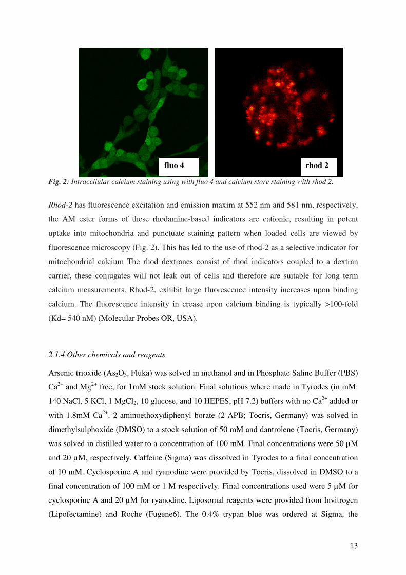
Fig. 2: Intracellular calcium staining using with fluo 4 and calcium store staining with rhod 2.
Rhod-2 has fluorescence excitation and emission maxim at 552 nm and 581 nm, respectively,
the AM ester forms of these rhodamine-based indicators are cationic, resulting in potent
uptake into mitochondria and punctuate staining pattern when loaded cells are viewed by
fluorescence microscopy (Fig. 2). This has led to the use of rhod-2 as a selective indicator for
mitochondrial calcium The rhod dextranes consist of rhod indicators coupled to a dextran
carrier, these conjugates will not leak out of cells and therefore are suitable for long term
calcium measurements. Rhod-2, exhibit large fluorescence intensity increases upon binding
calcium. The fluorescence intensity in crease upon calcium binding is typically >100-fold
(Kd= 540 nM) (Molecular Probes OR, USA).
2.1.4 Other chemicals and reagents
Arsenic trioxide (As2O3, Fluka) was solved in methanol and in Phosphate Saline Buffer (PBS)
Ca2+
and Mg2+
free, for 1mM stock solution. Final solutions where made in Tyrodes (in mM:
140 NaCl, 5 KCl, 1 MgCl2, 10 glucose, and 10 HEPES, pH 7.2) buffers with no Ca2+
added or
with 1.8mM Ca2+
. 2-aminoethoxydiphenyl borate (2-APB; Tocris, Germany) was solved in
dimethylsulphoxide (DMSO) to a stock solution of 50 mM and dantrolene (Tocris, Germany)
was solved in distilled water to a concentration of 100 mM. Final concentrations were 50 µM
and 20 µM, respectively. Caffeine (Sigma) was dissolved in Tyrodes to a final concentration
of 10 mM. Cyclosporine A and ryanodine were provided by Tocris, dissolved in DMSO to a
final concentration of 100 mM or 1 M respectively. Final concentrations used were 5 µM for
cyclosporine A and 20 µM for ryanodine. Liposomal reagents were provided from Invitrogen
(Lipofectamine) and Roche (Fugene6). The 0.4% trypan blue was ordered at Sigma, the
fluo 4 rhod 2

14
“Vybrant®
MTT Cell Proliferation Assay Kit” provided by Molecular Probes (Invitrogen,
Germany) and Hoechst 33342 at Molecular Probes, Germany.
2.2 Methods
2.2.1 Confocal laser scanning microscopy
To study [Ca2+
]i modulations by As2O3 two Ca2+
sensitive dyes were used: fluo-4/AM to
observe changes of [Ca2+
]i and rhod-2/AM to determine the changes of Ca2+
within the
calcium stores (Florea et al., 2005 a, b, 2007). Fluorescence images were collected at room
temperature every 30s. To observe faster events the time interval was reduced to 1s. The
resolution was 512x512 for the fluo-4 and 1024x1024 for rhod-2 experiments. Full screen
images were taken which allows analysing offline selected regions of interest (ROI’s). As2O3
(1 µM, 100 nM, 10 nM, 1 nM, 100 pM) was applied using an flow system at a rate of 1
ml/min. Reversibility on [Ca2+
]i–homeostasis was tested by applying Tyrodes buffer.
For determination of [Ca2+
]i concentration the “ion concentration” option of the META
software (Zeiss) was used. Images were background subtracted and the [Ca2+
]i–concentration
was calculated using the following equation:
[Ca2+
]i=Kd*(F-Fmin/Fmax-F),
where “Kd” is the dissociation constant (2.1.3 and see Molecular Probes) and “F” is the fluo-4
intensity. The basal [Ca2+
]i concentration was considered 100 nM (Orrenius et al., 2003). To
illustrate [Ca2+
]i changes over time, the “subtraction” option from the META software was
used. In the calculated images with “rainbow scale”, blue illustrates the minimum and red the
maximum change of [Ca2+
]i.
2.2.2 Fluorescent activated cell sorting (FACS)
Cells were grown in 125 cm2 flasks and collected by trypsin treatment in culture media (with
FCS), centrifuged and washed with phosphate saline buffer (PBS). The cell suspension was
aliquoted in 1.5 ml eppendorf tubes, and re-suspended in 1 ml Tyrodes buffer. Fluo-4 (50 µg)
was dissolved in DMSO (20 µl) and 1 µl was added to each sample of 1 ml Tyrodes buffer.
Unstained and stained controls were used. After 30 min they were washed once with buffer
solution and As2O3 was applied to the cell suspension for 30 min. After incubation with
As2O3 the cells were washed twice with Tyrodes buffer. Cells were fixed in 2.5%

15
formaldehyde in PBS and measured by FACS. 10,000 events (cells) were counted for each
sample. The parametric analysis of FITC/ fluo-4 green fluorescence (x-axis) was performed
with the “WinMDI” software. The modification of fluo-4 intensity was observed by a shift of
the cell population to the right on the x-axis.
2.2.3 Trypan Blue Cytotoxicity Test
For determination of cell viability, treated as well as untreated cells were used. Non-confluent
cell monolayers were exposed to As2O3, in cell culture flasks, for 24, 48 and 72 h. All
experiments were repeated twice. After treatment, the culture media was collected in a 50 ml
centrifugation tube, since it might contain dead cells necessary for cell counts. The cell
monolayer was washed with PBS and then collected in the same tube. Cells were treated with
Trypsin and, the Trypsin with the cells was collected in the same 50 ml tube. The suspension
was centrifuged (2min, 1200rpm) and the supernatant removed. The pellet was washed with
PBS, centrifuged (2min, 1200rpm) and re–suspended again in complete culture media. Then,
a small aliquot of the cell suspension (50 µl) was mixed with the same volume of 0.4% trypan
blue (Sigma) solution and the sample was counted after 3 min of staining using a
haemocytometer. The number of bright (viable) cells and blue cells (non-viable) were
evaluated using a light microscope with a 20-fold magnification. After counting, the cell
viability (CV) was expressed as the percentage of surviving cells compared to the total
number of cells:
CV = (viable cells/total number of cells)x100.
As2O3 was considered to be cytotoxic, when it induced a decrease of cell viability of more
than 50%. Controls and exposed samples were compared using the two-tailed Student’s t-test
with equal variance.
2.2.4 MTT-cytotoxicity test
For the MTT test we used a “Vybrant®
MTT Cell Proliferation Assay Kit” provided by
Molecular Probes (Invitrogen, Germany) as suggested by the manufacturer recommendation.
Briefly, 5,000 cells were seeded in each well of a 96 well plate, using 100 µl culture media.
They were allowed to attach over night. As2O3 (1 µM) was applied to non-confluent cultures,
to avoid grow inhibition, for different time of exposure (2 h to 72 h). Stock solution of 12 mM

16
MTT was prepared by adding 1 ml of sterile PBS to one 5 mg vial of MTT 3-(4,5-
domethylthiazol-2-yl)-2,5-diphenyltetrazolim (component A). Stock solution of 1 gm SDS
(sodium dodecyl sulphate) was prepared by adding 10 ml of DMSO to one tube of component
B included in the kit. Before exposure to As2O3, the culture medium was exchanged. For
controls and blanks, complete culture medium was used (100 µl/well). The test cells were
exposed to 1 µM As2O3 in a final volume of 100 µl culture medium. After exposure 10 µl of
the solubilized MTT (component A) was add to the controls and exposure wells, but not to the
blanks. The blanks were used to correct the microplate readings. The plate was further
incubated 4h at 37°C. After labelling with MTT, 25 µl of the supernatant was removed and 50
µl DMSO-component B was added and mixed and further incubated at 37°C (10 min). Again
the plate was carefully mixed/shacked and red at a wavelength of 540 nm. For analysis the
blanks were subtracted and calculated using the formula:
CV = (absorbance treated wells/absorbance control wells) X 100.
Controls and exposed samples were compared using the two-tailed Student’s t-test with equal
variance.
2.2.5 Hoechst staining to score micronucleated cells, condensed nuclei, and cells in mitosis
Four well chamber slides (LabTek) were seeded with 100,000 cells, in 1 ml culture media and
incubated overnight. 1 µM, 10 nM, 100 pM of As2O3 concentrations was applied for 24, 48
and 72 h. After the incubation period, the cells were washed twice with PBS, and for fixation,
treated with cold methanol (-20°C) and left overnight at -20°C. After fixation, slides were air-
dried, stained with 10 µM Hoechst 33342 (Molecular Probes) for 30 s and mounted with
cover slips. Cells were analysed using a 63x magnification with an Axiovert fluorescent
microscope. The two-tailed Student’s t-test was used to compare the difference between
controls and exposed samples. For the treatment with liposomes 1 µl of 1 mM arsenic trioxide
was mixed with 1.5 µl Lipofectamine or Fugene6 and further incubated with 50 µl culture
media for 15 min. This mixture was given to cells cultured on chamber-slides to final volume
of 1 ml per well. Cells were fixed and stained as previously described. Controls and exposed
samples counts were compared using the two-tailed Student’s t-test with equal variance.

17
3 Results
3.1 As2O3 induced changes in [Ca2+
]i in neuroblastoma and HEK cells
As2O3 is an anticancer drug used for APL treatment that could be potentially used for other
types of cancer. Unfortunately, secondary effects may occur upon treatment such as
neurotoxicity, hepatotoxicity or nephrotoxicity. To understand the cellular mechanisms of
As2O3 interactions with living cells, the study was focused on [Ca2+
]i signals that could be a
trigger for cytotoxicity. The aims of the first part of this study were to:
(1) investigate whether clinical relevant concentrations of As2O3 influence [Ca2+
]i,
(2) define the types of [Ca2+
]i-signals and,
(3) investigate from which sources the Ca2+
originates.
Experiments using LSM and FACS (Fig. 3) demonstrated that 1 µM As2O3 induced an
increase of [Ca2+
]i in neuroblastoma and in HEK cells. Confocal images show the increase of
the intracellular calcium concentration over time with traces of 3 different cells (Fig. 3A for
neuroblastoma, Fig. 3B for HEK). Before application of As2O3 a stable base line of [Ca2+
]i
was established. The application of As2O3 elevated [Ca2+
]i, with variations in time course
(Fig. 3). Overall, the pattern of [Ca2+
]i increase was similar in the two cell lines until a steady
state was reached. The [Ca2+
]i rise was not reversible after removal of As2O3 from the
application system, and during the time of the experiment. The averaged [Ca2+
]i rise was
170+4.84% in neuroblastoma (7 experiments, 58 cells) and 166.88+1.68% in HEK cells (3
experiments, 13 cells), a difference that was not significant in t-test (p>0.05).
As2O3 induced [Ca2+
]i-rise in both cell lines was also confirmed by using a different
experimental approach: measuring large cellular populations (10,000 cells) in FACS
experiments (Fig. 4). Examples of FACS plot after 30 min of incubation with arsenic are
shown in Fig. 4. The auto-fluorescence level of cells is presented with red trace (a) and the
fluorescence level of the cells after staining with fluo4 is presented in trace (b). Observe that
the intensity of cell population shifted to the right after incubation with As2O3 as shown in
trace c (compare trace (b) with trace (c)), an indication that [Ca2+
]i rose in neuroblastoma as
well as in HEK cells.

18
Fig. 3: Arsenic trioxide (As2O3) triggers an increase of [Ca2+
]i in neuroblastoma SY-5Y (A) und in
HEK cells (B) if cell cultures were exposed to 1µM As2O3. The black line marks the application As2O3
and the arrow marks the removal of the drug (washout). The traces illustrate the increase of [Ca2+
]i
during the application of As2O3 using selected regions of interest (ROI). The image series show the
overtime modification in fluo 4 intensity that is in direct relationship with the concentration of [Ca2+
]i.
A
B

19
Fig. 4: As2O3 induced increase of [Ca
2+]i in FACS experiments. FACS plots of neuroblastoma (A) and
HEK cells (B) are shown. Cells were stained with fluo 4 and incubated with As2O3 (1µM). The traces
show the auto fluoresce level of unstained cells (a), the green fluorescence after staining with fluo 4 for
30min (b) and (c) is fluo 4 fluorescence after incubation with 1µM As2O3 (30min). Notice the shift of the
intensity to the right that is in concordance with increased [Ca2+
]i.
3.2 As2O3 induced different types of [Ca2+
]i changes in neuroblastoma and HEK cells
In addition to the slow [Ca2+
]i increase to a steady state, two other types of [Ca2+
]i elevations
were found:
(1) transient [Ca2+
]i-elevations and
(2) [Ca2+
]i spikes.
To analyse fast [Ca2+
]i rises LSM images were taken at an time interval as short as 1s. NO
bleaching of the calcium sensitive dye was observed. Transient [Ca2+
]i–elevations measured
with the fast scanning are illustrated in Fig. 5. Apparently, transient calcium elevations and
calcium spikes were generated independently of each other and varied in time and intensity.
[Ca2+
]i traces of independent cells show that the cells could express synchronized calcium rise
as it can be observed at the time point of ~1500s; and also unsynchronised transient [Ca2+
]i
increases that occurred simultaneously with a sustained increase.
1 0 0 1 01 1 02 1 03 104
F L 1
a b c
10 0 1 01 1 02 1 03 104 F L 1
0
3 2
a
b c
B A
20
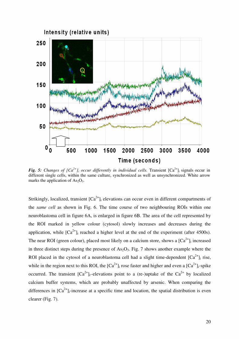
Fig. 5: Changes of [Ca
2+]i occur differently in individual cells. Transient [Ca
2+]i signals occur in
different single cells, within the same culture, synchronized as well as unsynchronized. White arrow
marks the application of As2O3.
Strikingly, localized, transient [Ca2+
]i elevations can occur even in different compartments of
the same cell as shown in Fig. 6. The time course of two neighbouring ROIs within one
neuroblastoma cell in figure 6A, is enlarged in figure 6B. The area of the cell represented by
the ROI marked in yellow colour (cytosol) slowly increases and decreases during the
application, while [Ca2+
]i reached a higher level at the end of the experiment (after 4500s).
The near ROI (green colour), placed most likely on a calcium store, shows a [Ca2+
]i increased
in three distinct steps during the presence of As2O3. Fig. 7 shows another example where the
ROI placed in the cytosol of a neuroblastoma cell had a slight time-dependent [Ca2+
]i rise,
while in the region next to this ROI, the [Ca2+
]i rose faster and higher and even a [Ca2+
]i-spike
occurred. The transient [Ca2+
]i–elevations point to a (re-)uptake of the Ca2+
by localized
calcium buffer systems, which are probably unaffected by arsenic. When comparing the
differences in [Ca2+
]i-increase at a specific time and location, the spatial distribution is even
clearer (Fig. 7).

21
Fig. 6: Localized calcium signals. Images of neuroblastoma cells were taken under control conditions
and, after the application of As2O3 (white arrow). False colours highlight the [Ca2+
]i. changes. The
arrows (A) mark specific points of interest that show intracellular localised spikes. A magnification of
the area marked in figure 4A is presented in part 4B, with time course of two neighbouring ROIs of
the same cell; white arrow marks the application of As2O3. While one trace (yellow ROI) has a small
but transient increase, a neighbour area (green ROI) showed a stepwise elevation of [Ca2+
]i.
control 10 min 20 min
30 min 40 min 50 min
A
B

22
Fig. 7: Localized calcium signals are presented as fast [Ca2+
]i increases, which returned to base level
within 1 min (red line) could be neighboured by regions which show a slow sustained increase of
[Ca2+
]i (green line).
3.3 The changes in [Ca2+
]i induced by As2O3 are concentration dependent in neuroblastoma
and HEK cells
In this study it was further tested whether the As2O3 induced calcium increase is concentration
dependent. As2O3 was applied to cells in also in submicromolar concentrations of 100 nM, 10
nM, 1 nM and 100 pM (Fig. 8). At these low environmental and clinical relevant
concentrations As2O3 interacted with calcium homeostasis in both cell lines with the same
patterns as observed a concentration of 1µM of As2O3.
The calcium rise in neuroblastoma and HEK cells was concentration dependent as presented
in Fig. 9. Only the 1µM concentration showed a lower increase probably due to a biphasic
dose-response shown by As2O3. The averaged calcium rise for neuroblastoma cells was: 100
nM: 232%+3.2 (3 experiments; 19 cells), 10 nM: 189%+4 (3 experiments; 45 cells), 1 nM:
177%+1.7 (3 experiments; 40 cells), 100 pM: 122%+0.46 (4 experiments; 35 cells). For HEK
cells the calculated calcium increase was: 100 nM: 263%+2 (3 experiments; 30 cells), 10 nM:
232%+1.4 (3 experiments; 31 cells), 1nM: 197%+2.2 (3 experiments; 31 cells), 100pM:
153%+4.3 (4 experiments; 30 cells) (Fig. 9). The lowest concentration tested was still able to
trigger calcium rise to steady state as well as fast calcium signals (spikes) as shown in Fig. 10.
6 0 0
4 0 0
2 0 0
1 0 0 0 2 0 0 0
T im e (s )
A s2 O
3
w a s h o u t

23
Fig. 8: Calcium rise induced by application of low concentrations of As2O3. Application of As2O3
concentration down to 100 pM still induced elevations of [Ca2+
]i in (A) neuroblastoma and (B) HEK
cells.
A B

24
100
150
200
250
300
neuroblastoma HEK
1 µM 100 nM 10 nM 1 nM 100 pM
Fig. 9: [Ca2+
]i rise induced by As2O3 in neuroblastoma (A) and HEK cells (B) was concentration
depended in the range between 100 nM and 100 pM. Surprisingly a concentration of 1 µM gave a
smaller increase in intracellular calcium than concentrations between 100 nM and 1 nM probably due
of biphasic As2O3 dose-response.
Fig. 10: Example of fast [Ca2+
]i rise induced by 100 pM As2O3 : while some cells had a over time
gradual calcium rise to a steady state, other cells showed fast calcium spikes in (A) neuroblastoma, (B)
HEK cells, white arrows mark the application of the drug.
A
B

25
3.4 Ca2+
stores are involved in the As2O3 mediated [Ca2+
]i changes
To analyse from which sources the increase of [Ca2+
]i originates, we performed experiments
where:
(1) Ca2+
in the stores of neuroblastoma and HEK cells were labelled with rhod-2 before As2O3
was applied, and
(2) [Ca2+
]i was stained with fluo-4 under two experimental conditions; (a) no calcium added
to the external solution (to exclude a Ca2+
entry from the extracellular space) and (b) these
results were compared with the data obtained in extracellular solution which contained 1.8
mM Ca2+
.
Exposure of neuroblastoma and HEK cells to As2O3 showed a Ca2+
release from the internal
calcium stores since an application of 1 µM As2O3 decreased over time the intensity of rhod-2
(Fig. 11A - neuroblastoma and Fig. 11B - HEK cells). Experiments with fluo-4 staining and
no extracellular Ca2+
underline that the [Ca2+
]i-rise depend on calcium stores (Fig. 12). While
in neuroblastoma cells the increase of [Ca2+
]i was not influenced by the presence of Ca2+
in
the external solution (164.8+2.93% increase; p>0.05, Student`s t-test, not significant, 3
experiments, 13 cells), surprisingly the [Ca2+
]i in HEK cells rose nearly twice as high
(241+0.59%, p<0.001 in t-test, highly significant) (Fig. 10). This is an other indication that
the rise of [Ca2+
]i did not result from Ca2+
entry from the extracellular space but from Ca2+
release. Possible explanations, why the absence of extracellular Ca2+
triggered a larger release
from the stores in HEK cells will be given in the discussion.
3.5 Ca2+
receptors are involved in the As2O3 mediated [Ca2+
]i changes
To determine whether inositol-1,4,5-triphosphate (IP3) and/or ryanodine (Ry) receptors,
which are found at the endoplasmatic reticulum (ER) and/or the Golgi apparatus (Pinton et al.,
1998), are involved in the regulation of [Ca2+
]i we used specific blockers for these receptors
(2-APB and dantrolene). A pre-incubation (20 min) with 2-APB (50 µM) and/or dantrolene
(20 µM) reduced significantly the As2O3 induced [Ca2+
]i–rise (in 1.8 mM calcium buffer, Fig.
12). After preincubation with 2-APB, As2O3 determined a [Ca2+
]i-increase of: 121±0.17%, (3
experiments, 23 cells) in neuroblastoma and 113±0.91% in HEK cells (3 experiments, 28
cells). Similar effects were observed with dantrolene: 137±1%, (3 experiments, 28 cells)
increase of [Ca2+
]i in neuroblastoma and 125±2.45% in HEK cells (3 experiments, 18 cells).
The rise was highly significantly reduced (p<0.001) compared with the [Ca2+
]i-rise induced

26
by As2O3 (1 µM) in normal containing calcium buffer. This results drive the conclusion that
[Ca2+
]i-rise induced by As2O3 is basically modulated by IP3- and Ry-receptors.
A B
Fig. 11: As2O3 releases Ca
2+ from the calcium stores. During the application of As2O3 the internal
calcium stores of neuroblastoma (A) and HEK cells (B) got depleted over the time of application as
presented by the confocal images of the stores (blue = no calcium, yellow = medium calcium and red
= high calcium) and with 2.5 D false colour images (colour coding as in figure 1). The first row (from
top to bottom of the figure) illustrates the calcium stores under control conditions, while following
rows show decreased intensity after 15 min (2nd
row) and 30 min (3rd
row) of incubation with As2O3 (1
µM).
27
260
220
180
140
100
SY-5Y (neuroblastoma) HEK 293
2mM calcium
no calcium added
2-APB
danthrolene
C
no calcium added control HEK no calcium added 1µM As2O3 HEKno calcium added control HEK no calcium added 1µM As2O3 HEK
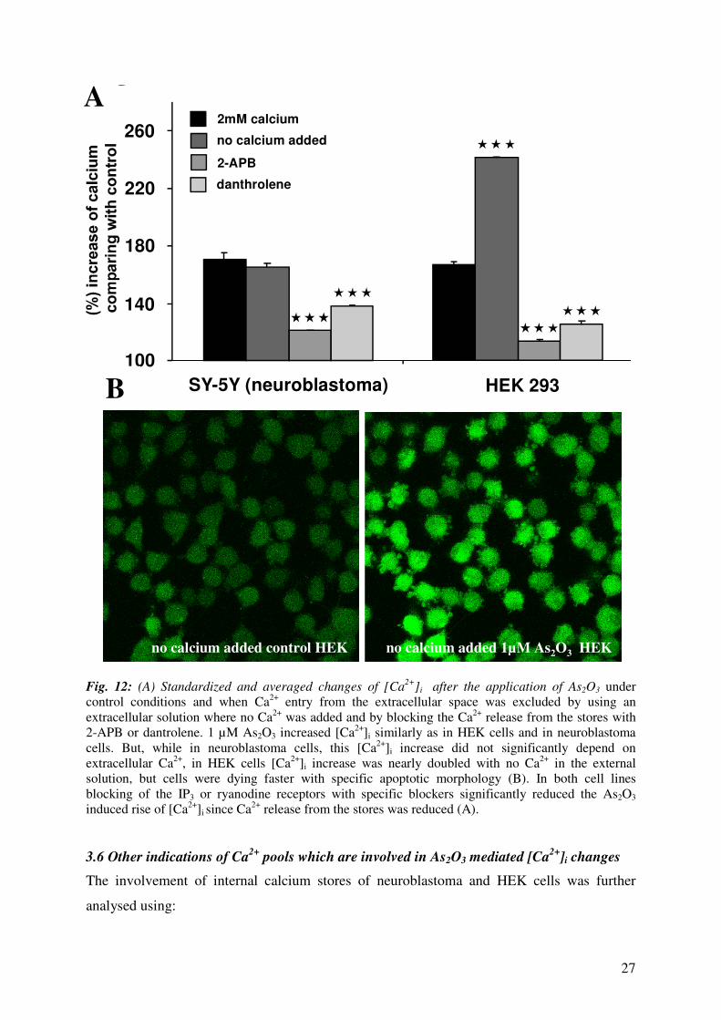
Fig. 12: (A) Standardized and averaged changes of [Ca2+
]i after the application of As2O3 under
control conditions and when Ca2+
entry from the extracellular space was excluded by using an
extracellular solution where no Ca2+
was added and by blocking the Ca2+
release from the stores with
2-APB or dantrolene. 1 µM As2O3 increased [Ca2+
]i similarly as in HEK cells and in neuroblastoma
cells. But, while in neuroblastoma cells, this [Ca2+
]i increase did not significantly depend on
extracellular Ca2+
, in HEK cells [Ca2+
]i increase was nearly doubled with no Ca2+
in the external
solution, but cells were dying faster with specific apoptotic morphology (B). In both cell lines
blocking of the IP3 or ryanodine receptors with specific blockers significantly reduced the As2O3
induced rise of [Ca2+
]i since Ca2+
release from the stores was reduced (A).
3.6 Other indications of Ca2+
pools which are involved in As2O3 mediated [Ca2+
]i changes
The involvement of internal calcium stores of neuroblastoma and HEK cells was further
analysed using:
A
B
28
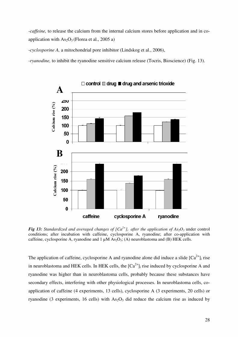
-caffeine, to release the calcium from the internal calcium stores before application and in co-
application with As2O3 (Florea et al., 2005 a)
-cyclosporine A, a mitochondrial pore inhibitor (Lindskog et al., 2006),
-ryanodine, to inhibit the ryanodine sensitive calcium release (Tocris, Bioscience) (Fig. 13).
Fig 13: Standardized and averaged changes of [Ca2+
]i after the application of As2O3 under control
conditions; after incubation with caffeine, cyclosporine A, ryanodine; after co-application with
caffeine, cyclosporine A, ryanodine and 1 µM As2O3; (A) neuroblastoma and (B) HEK cells.
The application of caffeine, cyclosporine A and ryanodine alone did induce a slide [Ca2+
]i rise
in neuroblastoma and HEK cells. In HEK cells, the [Ca2+
]i rise induced by cyclosporine A and
ryanodine was higher than in neuroblastoma cells, probably because these substances have
secondary effects, interfering with other physiological processes. In neuroblastoma cells, co-
application of caffeine (4 experiments, 13 cells), cyclosporine A (3 experiments, 20 cells) or
ryanodine (3 experiments, 16 cells) with As2O3 did reduce the calcium rise as induced by
A
B
29
As2O3 alone. This could mean that As2O3 induced signals are positively modulated by RyR,
ER and mitochondria. Interestingly, in HEK cells different effects were observed compared to
neuroblastoma cells. The co-application of As2O3 with caffeine (4 experiments, 15 cells) and
respectively ryanodine (3 experiments, 13 cells) slightly increased calcium rise comparing
with [Ca2+
]i elevation induced by As2O3 alone, a fact that could underline that RyR and ER
regulates in the negative way the As2O3 induced [Ca2+
]i elevation. Thus, cyclosporine A (3
experiments, 16 cells) did reduce the As2O3 induced [Ca2+
]i rise probably because
mitochondria positively modulated As2O3 induced [Ca2+
]i elevation.
3.7 No Ca2+
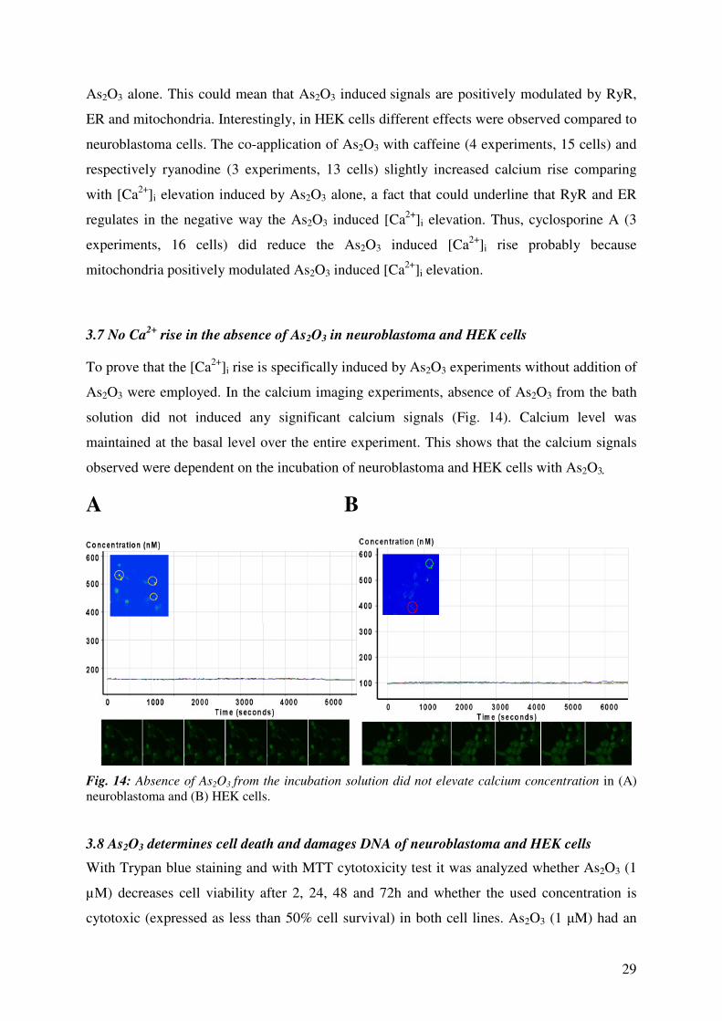
rise in the absence of As2O3 in neuroblastoma and HEK cells
To prove that the [Ca2+
]i rise is specifically induced by As2O3 experiments without addition of
As2O3 were employed. In the calcium imaging experiments, absence of As2O3 from the bath
solution did not induced any significant calcium signals (Fig. 14). Calcium level was
maintained at the basal level over the entire experiment. This shows that the calcium signals
observed were dependent on the incubation of neuroblastoma and HEK cells with As2O3.
A B
Fig. 14: Absence of As2O3 from the incubation solution did not elevate calcium concentration in (A)
neuroblastoma and (B) HEK cells.
3.8 As2O3 determines cell death and damages DNA of neuroblastoma and HEK cells
With Trypan blue staining and with MTT cytotoxicity test it was analyzed whether As2O3 (1
µM) decreases cell viability after 2, 24, 48 and 72h and whether the used concentration is
cytotoxic (expressed as less than 50% cell survival) in both cell lines. As2O3 (1 µM) had an

30
increased toxic effect on both cell lines. In Trypan blue exclusion assay, after exposure to
1µM As2O3, the cell viability was significantly decreased (t-test, p<0.05), with an
approximate 80% survival after 72h of exposure. However, it did not drop under 50% cell
survival. The effect was similar for both cell types (Fig. 15). When applying MTT test, the
neuroblastoma cells had higher sensitivity to 1µM As2O3 than HEK cells (70% vs. 85%), but
the cell survival did also not drop under 50% (Fig. 15).
0 2 24 48 72Time of exporure (h)
A B
0
40
80
120 SY-5Y MTT-Test
HEK293 MTT-Test
SY-5Y Trypanblue
HEK293 Typanblue
Fig. 15: Cytotoxic effects induced by As2O3. A trypan blue staining of cells after incubation with As2O3
(1 µM) shows slight cytotoxic effects since cell viability was significantly reduced over time in
neuroblastoma (black squares) and HEK cells (grey triangles). These results were confirmed with the
MTT toxicity test as indicated by the black bars for neuroblastoma and the white bars for HEK cells.
After exposure to As2O3, neuroblastoma and HEK cells were scored for DNA damage
(micronucleated cells) apoptotic cells (condensed nuclei) and cells in mitosis (Fig. 16). The
number of cells with damaged DNA as well as apoptotic cells was significantly increased
(p<0.05, t-test) in neuroblastoma (Fig. 17A and C) and HEK cells (Fig. 17B and D). The
number of mitotic cells was not significantly affected in any of the cell lines (compare Tab. I).
Time of exposure (h)

31
neuroblastomaneuroblastoma HEKHEK
Fig. 16: Specific morphology of cells with DNA damage (micronuclei-loss of DNA) after application
of As2O3; apoptotic nuclei, smaller nuclei and condensed chromatin are shown in for neuroblastoma
and for HEK cells as well as cells in mitosis.
Tab. I: The level of DNA damage (MN) and apoptosis (AP) compared with control levels, induced
by1µM As2O3 in neuroblastoma and HEK cells.
Liposomal therapy is an option in anticancer drug delivery; therefore an increased uptake of
As2O3 was tried using liposomes with As2O3, in order to see consolidated effects. Fugene6
and Lipofectamine did not modify the background level of DNA damage, apoptosis or mitosis
(Fig. 17). In Figs. 17 C (neuroblastoma) and D (HEK) it is illustrated that Fugene6 and
Lipofectamine had similar effects. After application of As2O3 with Lipofectamine/Fugene6,
Neuroblastoma MN AP
24 h 370% 266%
48 h 514% 152%
72 h 400% 214%
HEK MN AP
24 h 335% 156%
48 h 481% 192%
72 h 316% 200%

32
neuroblastoma and HEK cells showed significant increase in the number of cells with
damaged DNA. In the same experimental conditions the number of apoptotic cells was
significantly increased (p<0.05) while the number of cells in mitosis was significantly
decreased (p<0.05). However, the DNA damaged and apoptotic level of cells was not
significantly different compared to the experiments without usage of liposomes, an indication
that intracellular concentration of As2O3 is possibly regulated (Tab. II).
Tab. II: The level of DNA damage (MN) and apoptosis (AP) compared with control levels, induced by
liposomic treatment with As2O3 in neuroblastoma and HEK cells.
Neuroblastoma MN AP
As2O3 and Fugene 6 (24h) 231% 246%
As2O3 and Lipofectamine (24h) 264% 242%
HEK MN AP
As2O3 and Fugene 6 (24h) 348% 252%
As2O3 and Lipofectamine (24h) 356% 240%

33
****
**
0
2 0
4 0
6 0
c t r 2 4 h c t r 4 8 h c t r 7 2 h
**
*
***
c t r 2 4 h c t r 4 8 h c tr 7 2 h
m ic ro n u c le a te d c e l ls
a p o p to tic n u c le i
m it o t ic c e l ls
m ic r o n u c le a t e d c e lls
a p o p to t ic n u c le i
m i t o tic c e lls
***
***
***
***
***
*
***
**
*
***
***
******
**
***** *
0
2 0
4 0
6 0
m ic ro n u c le a te dc e l ls
a p o p to t icn u c le i
c e l lsin m it o s is
c o n t ro l F u g e n e 6
F u g e n e 6 a n d A s 2 O 3
c tr L ip o fe c ta m in e
L ip o fe c ta m in e a n d A s 2 O 3
c o n t ro l F u g e n e 6
F u g e n e 6 a n d A s 2 O 3
c tr L ip o fe c ta m in e
L ip o fe c ta m in e a n d A s 2 O 3
m ic r o n u c le a t e d
c e l ls
a p o p t o t ic
n u c le i
c e lls
in m i to s is
C D
E F
Fig. 17: As2O3 significantly induced DNA damage; rise of apoptotic rate but no significant influence on the mitosis rate was observed in neuroblastoma (A)
and HEK (B) cells. Cells were exposed to As2O3 for 24h, 48h as well as 72h of exposure and then Hoechst stained. In panel (C) and (D) the levels of DNA
damage, apoptosis and the number of cell in mitosis after liposomic treatment are presented for neuroblastoma (C) and HEK (D). The level of DNA damage,
apoptosis and mitosis is significantly increased compared to control.
C D
A B

34
3.9 Differential effects of nanomolar and picomolar concentrations of As2O3 on cell death
and damages DNA of neuroblastoma and HEK cells.
Exposure of neuroblastoma and HEK cell to lower concentrations than 1µM of As2O3 showed
a concentration dependent effect (Fig. 18 and Tab.III). While nanomolar concentration of
As2O3 did significantly induced the level of DNA damage expressed as micronuclei, As2O3 in
the same concentration range was able to induce apoptosis in neuroblastoma cells but not in
HEK cells. This underlines again the specificity of As2O3 for tumour cells. The number of
mitotic cells was not significantly affected at any of the concentrations tested.
Tab. III: Significance analysis applied on lower concentration range experiments with As2O3.
Student’s t-test was applied where (*) is p<0.05, (**) is p<0.01, (***) is p<0.001.
neuroblastom a HEK
24 h M N AP M I 24 h M N A P M I
100pM - - - 100pM ** - -
10nM * - - 10nM *** - -
1µM *** *** - 1µM *** ** -48 h M N AP M I 48 h M N A P M I
100pM * * - 100pM ** - -
10nM ** *** - 10nM *** - -
1µM *** *** - 1µM *** * -72 h M N AP M I 72 h M N A P M I
100pM - - - 100pM - - -
10nM ** *** - 10nM *** - -
1µM *** *** - 1µM ** - -

35
Fig. 18: Dose and time dependence of As2O3 induced DNA damage, apoptosis rate and the mitosis rate in neuroblastoma and HEK cells. Cells were exposed to 100 pM, 10
nM and 1µM As2O3 concentration for 24h, 48h as well as 72h of exposure and then Hoechst stained. The effect of As2O3 on DNA damage, apoptosis and was concentration
dependent (see text)

36
4 Discussion
Ca2+
is an important signal transducer in excitable and none excitable cells. [Ca2+
]i signalling
is involved in physio- as well as pathological processes and therefore the level of [Ca2+
]i is
tightly regulated. [Ca2+
]i dynamics are modulated by calcium channels and calcium stores.
[Ca2+
]i could be increased by Ca2+
-entry from the extracellular space such as opening of Ca2+
selective pores; Ca2+
release from the stores; impairment of Ca2+
selective transport proteins
which pump Ca2+
in the extracellular space and/or in the calcium stores (Ferguson et al.,
2000). [Ca2+
]i could be decreased by a reduction of Ca2+
entry by blocking calcium selective
pores (Büsselberg et al., 1994, Büsselberg, 1995) or by an enhancement of the efficiency of
calcium transport proteins.
Besides its physiological function, [Ca2+
]i-rises as well as deregulation of in local intracellular
Ca2+
distribution could lead to accidental (necrosis) or programmed cell death (apoptosis)
(Orrenius et al., 2003). The calcium stores represented mainly by mitochondria and
endoplasmic reticulum (ER) play important roles in cellular Ca2+
homeostasis and signalling
like (a) regulating crucial processes (e.g. motility, secretion, gene expression); (b) signalling
cascades that drive proliferation, differentiation, and various metabolic reactions as well as
(c) cell death in physiological settings or during injury or diseases (Mattson et al., 2000;
Berridge et al., 2003). Therefore, calcium signalling could play a major role in As2O3 induced
toxicity.
4.1 As2O3 influences [Ca2+
]i homeostasis
Metallic compounds could interact with [Ca2+
]i homeostasis of living cells (Florea and
Büsselberg, 2005, 2006) although [Ca2+
]i is highly controlled the deregulation of [Ca2+
]i
induced by metallic species could affects the plasma membrane, mitochondria, or ER
(Orrenius et al., 2003; Florea and Büsselberg, 2005). One of the many metallic compounds,
As2O3, is used effectively to treat APL (Shen et al., 1997; Bergstrom et al., 1998; Soignet et
al., 1998; Fenaux et al, 2001; Zhu et al., 2002; Diaz et al., 2005) and it could be useful to treat
other types of cancer. In to a recent study using cDNA microarray technology and applying
As2O3 on APL, NB4 cells, several molecular signalling pathways modulated the cell response
to As2O3 including the activation of calcium signalling (with ER stress and involvement of
calcium receptors) with end point leading to cell death (Zheng et al., 2005). It was also shown
that an As2O3 triggered increase of [Ca2+
]i inhibited cell growth and induces apoptosis in

37
human malignant cell lines by an increase of cellular H2O2, a decreased of mitochondrial
membrane potential and activation of caspase-3 (Zhang et al., 1999; Kajiguki et al., 2003;
Miller et al., 2002; Diaz et al., 2005). While it is documented that [Ca2+
]i overloads could
trigger apoptosis (Orrenius et al., 2003), there is no detailed work describing how As2O3
induced [Ca2+
]i modulations are involved in programmed cell death. Therefore in this work it
was investigated the mechanisms of [Ca2+
]i elevation triggered by As2O3, using two different
target models: neuroblastoma and embryonic kidney cells.
In this study it was demonstrated that As2O3 triggers: an (1) increase of [Ca2+
]i that was
irreversible and reached a steady state level or/and induces (2) faster and slower calcium
transients. The effects on calcium homeostasis induced by 1µM As2O3 were similar in the
two cell lines; suggesting that As2O3 could target these cell models in a similar manner. Ca2+
-
release from intracellular calcium stores was most the important effect induced by As2O3
since extracellular calcium did not significantly influenced [Ca2+
]i elevation. This shows
minimal effect of As2O3 on Ca2+
entry from the extracellular space; however, a Ca2+
intake
from the extracellular space is not totally excluded by the experiments presented here since
we were unable to clamp the Ca2+
-concentration in the extracellular solution (e.g. using
BAPTA) because the cells did not survive the course of the experiments (up to 4h). Therefore
we compared data were no calcium was added to the extracellular solution with results were
this solution contained 2 mM Ca2+
.
Other authors affirmed that As2O3 induces an uptake of calcium from the extracellular matrix.
Ma and colleagues (2006) showed that in low extracellular Ca2+
concentration As2O3 (10 µM)
did not induce the opening of the mitochondrial transition pore (PTP) and cytochrome c
release from mitochondria, while the same concentration of As2O3 with high extracellular
Ca2+
concentration induced PTP opening and cytochrome c release. A possible explanation
could be the different cell systems, as well as the at least 10-fold lower concentration of
As2O3 used in our study.
In this study we also demonstrated that As2O3 triggered different kinds of Ca2+
signals: slow
(sustained), transient elevations and calcium spikes. Calcium signals were often not
synchronized between the different cells and they could occur highly localized within the
different compartments of the cell. During the slow increase of [Ca2+
]i the over time reduction
of Ca2+
concentration in the stores was observed. This effect could be explained by an
enhanced calcium release (e.g. IP3 and/or ryanodine mediated opening of calcium pores) or

38
defective re-uptake of calcium into the stores (reduced transport rate of calcium pumps).
Therefore, calcium stores play a major role in the As2O3 induced changes of [Ca2+
]i.
Additionally, the calcium stores could take up Ca2+
from the cytosol by calcium transport
proteins. Calcium overload of mitochondria and ER unless regulated could result in oxidative
stress, caspases activation and cell death by apoptosis, hypothesis supported also by Zheng et
al., 2005. Since an impaired re-uptake of calcium in the stores for the slower transient of
[Ca2+
]i cannot be excluded, the fast calcium spikes are not in the support of this hypothesis.
Calcium spikes do depend on functional extrusion mechanisms for the cytosolic Ca2+
that are
still functional.
Transient increases of [Ca2+
]i occur fast, are localized and appeared independently, in
different regions of same cell. Also, cells have individual changes of [Ca2+
]i after the
application of As2O3. These effects could be explained by interaction of As2O3 with
hetrogenous cells. The heterogenicity of the cells could be given by:
(1) different protein expression at the cell membrane (e.g. membrane receptors),
(2) different activation level of signalling pathways or
(3) variable efficiency of the extrusion mechanisms as well as
(4) different stages of the cell cycle.
More difficult to explain are the fast calcium spikes. An extensive analysis of fast calcium
signalling effects was impossible due to the limitations of our recording parameters such as:
(1) taking one scanning for each 30s to avoid bleaching and data overload of the computer;
(2) the ROIs set-up of a specific seize where very small and localized events could hardly be
analyzed; (3) or searching for events which occurred in the 3D-structure of the cell when
analyzing a 2D confocal image. Therefore, fast calcium signalling events such as spikes are
difficult to judge because they could occur any time in the interval when no images were
taken. Thus, when faster scanning was applied (1 image/sec.) spikes that lasted 3-5 seconds or
longer were recorded.
An important question that arise from this study is whether As2O3 needs to enter the cytosol to
accomplish the rise of [Ca2+
]i. This study was not designed to check to which level As2O3
permeate the membrane of the cells, but it was previously documented that arsenite, an
inorganic trivalent arsenic form has a very low uptake in vitro (below 5% of the substrate
(Florea, 2005)). Since As2O3 is structurally very similar with arsenite this is an indication that
39
As2O3 does not need to enter the cells to complete its effect but it could directly interact with
receptors at the cell membrane and therefore activating signalling pathways through the rise
of [Ca2+
]i. This conclusion is underlined by the ability of under-nanomolar concentrations of
As2O3 to induce a calcium rise in neuroblastoma and HEK cells, which were concentration
and time dependent. In addition, the experiments using liposomes, to increase the amount of
As2O3 in the cell, did not result in a higher cytotoxicity, probably due to a fast export out of
the cells. Therefore it could be speculated whether As2O3 interaction with the cell models
tested in two different ways. Either (1) As2O3 binds to receptors at the extracellular
membrane, which trigger intracellular pathways resulting in an elevated [Ca2+
]i-level, or/and
(2) relatively small amount of As2O3 which enters the cytosol is sufficient to release Ca2+
from the stores.
In other work regarding the interaction of As2O3 with tumours cells the involvement of
mitochondria on the rise of [Ca2+
]i was demonstrated (Ma et al. 2006; Shen et al., 2002).
Thus, little or no investigations have been done in analysing the involvement of ER in As2O3
induced [Ca2+
]i rise and cell death. Only the study of Zheng et al., 2005 using microarray
technology have underlined the importance of calcium signalling and ER in the As2O3
induced effects in tumour cells. The control of Ca2+
dynamics are complex and are regulated
not only by ion channels and pumps at the plasma membrane but the ER could play a major
role in regulation of rapid and long-term changes in [Ca2+
]i e.g. Ca2+
is released from the ER
in response to signals that activate receptors coupled to IP3 production (Mattson et al., 2000;
Berridge et al., 2003).
In this work it is shown that IP3- and Ry-receptors are involved in regulation of [Ca2+
]i rise
induced by As2O3. This could be an important finding for improving anti-cancer treatments
since the involvement of ER, IP3 and RyR was also proven by Zheng et al., 2005 using cDNA
micoarray study. These authors show that calcium signalling play an important role in As2O3
induced apoptosis of APL cells that could be also in connection with other cellular events.
Additionally, Zheng and coworkers (2005) have shown that calcium binding proteins (S100
family of proteins) could be also involved in calcium signalling by As2O3 as well as PKC,
PKA, aquaporin 9 (Zheng et al., 2005).
An other issue, specifically in regard to HEK cells, is the higher increase of [Ca2+
]i in the
absence of extracellular Ca2+
. We also observed that those cells were dying faster in no
calcium conditions, with signs of apoptosis (membrane blebs, apoptotic bodies - Fig. 12). We
have no conclusive explanation for this phenomenon. A hypothesis is that feedback

40
mechanisms, which are involved in the Ca2+
induced Ca2+
release, are not functional when
As2O3 is present.
In addition to experiments with low/high calcium in the external solution and the use of
calcium sensitive dyes for the calcium stores, the analysis of involvement of other calcium
pools that could be involved in As2O3 induced calcium signals revealed that there is a
difference in mechanism of calcium release between neuroblastoma and HEK cells. In
experiments using caffeine to release the calcium stores before application of As2O3
neuroblastoma cells showed a clear reduction of the calcium increase dependent of As2O3
while HEK cells reacted in the opposite way. An explanation could be that feedback
mechanisms, which are involved in the Ca2+
induced Ca2+
release, are not functional when
As2O3 is present, as discussed for the experiments in low calcium. Treatment with
cyclosporine A, a mitochondrial pore inhibitor, showed that the mechanism of calcium
increase involving mitochondria is similar in booth cell lines, while blocking RyR again
differential effects were observed. These results could give the indication that probably the
calcium modulation by ER makes the difference between As2O3 in tumour cells and non
tumour cells fact that is supported by the work of Zheng et al., 2005.
4.2 [Ca2+
]i changes induced by As2O3 induces cell toxicity
Several authors describe the cytotoxicity of As2O3; e. g. As2O3 induced cytotoxicity in vitro
(human chronic leukaemia cell line) with an IC50 of 10 µM after 24h of exposure (Shim et al.,
2002). The same authors described DNA fragments, morphological changes, and chromatin
condensation of the cells undergoing apoptosis after incubation with a two times higher As2O3
concentration with caspase 3 and p38 activation that was confirmed by other authors using
different cell lines (Verma et al, 2002; Shim et al., 2002). Zhang et al. (1999) showed that 1
µM As2O3 had cytotoxic effects in malignant cells but not in none malignant human
embryonic cells which is in contradiction with our results. This could be explained by the use
of different in vitro models that could have different sensibility, since kidneys represent a
target for As2O3 toxicity. In our study it is shown that As2O3 (1 µM) exhibits cytotoxic effects
by significantly decreasing cell viability, enhanced cell death by apoptosis, and induces DNA
damage in neuroblastoma as well as in HEK cells; while no As2O3 application did not have an
effect on calcium homeostasis and cytotoxicity. The increase in micronucleus and apoptotic
rate could be a result of mitochondrial and ER stresses with formation of ROS that can further
damage the DNA and can signal to induce programmed cell death.

41
Other authors have established an involvement on ROS production in As2O3 induced
cytotoxicity. Haga et al., 2005 have shown in human glioblastoma cell lines (A172) that
As2O3 causes mitochondrial damage mediated by the production ROS and the dissipation of
mitochondrial transmembrane potential leading to apoptosis. These authors have hypothesised
that mitochondria might be the key target of antitumour activity by As2O3 that we also show
in our study. To support this finding, into a study of Shen et al., 2002 using SHEEC1 cells
exposed to As2O3 the authors observed characteristic morphological and functional changes of
mitochondria such as hyperplasia, swelling and disruption, accompanied by a decrease of the
transmembrane potential. The Ca2+
level increased immediately after adding As2O3 and the
nitric oxide (NO) concentration increased continuously over 24 h, finally apoptotic
morphology of cells occurred. The authors suggested that As2O3 induces a rise of Ca2+
and
NO, the apoptotic signal messengers, which will trigger a mitochondria-dependent apoptotic
pathway (Shen et al., 2002).
Additionally, As2O3 concentrations of 0.01-1 µM applied to human malignant cell lines,
MGC-803, HIC, MCF-7, HeLa, BEL-7402 and A549 cells showed growth inhibition and
apoptosis in a time dose-dependent manner. Changes in [Ca2+
]i, correlated with the sensitivity
of these cells to As2O3 indicating that a critical intracellular Ca2+
signal transduction pathway
could be involved in As2O3 -mediated cell-death (Zhang, et al., 1999). In other investigations,
arsenite applied to Chinese hamster ovary cells (CHO-K1) perturbed intracellular calcium
homeostasis, activated PKC activity and induced genotoxicity represented a micronuclei
production. Also, it was shown that the protein kinase C activator 12-O-tetradecanoylphorbol-
13-acetate increased the apoptotic action of As2O3, an effect mediated by ERK activation and
GSH depletion (Fernandez et al., 2004). Therefore activation of PKC activity may play an
important role in arsenite-induced DNA damage and toxicity (Jarvis et al., 1994; Liu and
Huang, 1997; Fernandez et al., 2004).
Strikingly, lower concentration than 1 µM As2O3 were able to damage the DNA in
neuroblastoma and HEK cells, however apoptosis was triggered in neuroblastoma cells but
not in HEK cells, showing As2O3 specificity for tumour cells. As shown with low As2O3
concentrations, the induction of DNA damage could be a problem when the apoptosis is not
longer triggered because cells undergo division and proliferate with a damaged DNA. This
effect could be a supplementary risk in the case of non-tumour cells, since clastogenesis
increases the possibility of secondary malignancies. Cell line specific effects exhibited by
As2O3 have been shown also by other authors. In human glioblastoma cell lines, As2O3

42
induced apoptosis in A172 cells but not in T98G cells even when As2O3-induced ROS
production was observed in both cell lines but the authors affirmed that mitochondrial
aggregation played an important role in regulating the sensitivity to As2O3-induced apoptosis
(Haga et al., 2005).
Thus, micromolar concentrations of As2O3 rose [Ca2+
]i, triggered apoptosis, damaged DNA,
and stopped the cells to enter mitosis. [Ca2+
]i rise from mitochondria and/or ER could induce
mitochondrial and ER stress, formation of ROS that could further damage DNA. Therefore,
localised Ca2+
accumulation could be a result of a Ca2+
re-uptake, Ca2+
overload, PTP opening
and apoptosis. The non-reversibility of the calcium elevation indicates that [Ca2+
]i disturbance
could also lead to ”a point of no return” and cell death. The different types of [Ca2+
]i-signals
induced by As2O3 could also be interpreted as modulators for specific processes such as
resistance, transport and gene expression. In addition, the number of cells in mitosis was
significantly decreased only in the co-treatment of As2O3 with liposomes, a fact that
underlines that the cells do not divide anymore. Unfortunately, this effect was not strictly
related to tumour cells but also affected non-tumour cells, finding that emphasize a
therapeutic strategy that protects non-tumour cells.
Trying to determine the involvement of IP3 and Ry receptors in DNA damage, apoptosis and
mitosis induced by As2O3 we faced the challenge that the blockers themselves induced a high
rate of micronuclei and apoptotic cells after 24 h of incubation, and therefore it was
impossible to obtain dependable results (data not shown). Another non-cytotoxic
experimental set up (e.g. siRNA, over-expression or IP3 and RyR) will be necessary to be
established in order to investigate these effects.
To conclude, arsenic induces apoptosis in several tumour and non-tumour cell lines. Several
[Ca2+
]i dependent apoptotic pathways were emphasised: (1) an intrinsic mitochondrial
pathway, (2) an activation/repression MAPK, and/or of (3) other protein kinases (Chen et al.,
2001a; Fernandez et al., 2004; Davidson et al., 2004; Verma et al., 2002; Maeda et al., 2001;
Zhou et al. 2004; Zhu et al. 2002; Jarvis et al. 1994; Lorenzo et al., 1999; Cheung et al., 2007;
Zheng et al., 2005). The same concentration of As2O3 (1 µM) as used in our work induced
apoptosis in a NKM-1 cell line. It increased oxidative stress, decreased mitochondrial
membrane potential and activation of caspase-3 and C-Jun-terminal kinase activation (JNK)
(Kajiguki et al., 2003). This finding was confirmed by other authors in different cell lines.
As2O3 activated p38, JNK, and caspase-3 concentration dependently with ROS formation in
human androgen-independent prostate cancer cell lines, PC-3, DU-145, and TSU-PR1 (Maeda

43
et al, 2001). Experiments completed with APL and NB4 support an essential role for JNK
signalling in the induction of growth inhibition and apoptosis by As2O3 and suggest that
activating JNK may provide a therapeutic advantage in the treatment of cancers that do not
respond to arsenic alone (Davison et al., 2002).
4.3 Anticancer drugs could have different effects on calcium signalling e.g. As2O3 vs.
cisplatin
In this thesis it was shown that As2O3 interferes with calcium signalling leading to
cytotoxicity. Another question that arises after discussing all these effects induced by As2O3 is
if other anticancer drugs have similar effects as showed by As2O3 on intracellular calcium
homeostasis. Therefore we also investigated the effect exhibited by cisplatin on human cervix
carcinoma cells (HeLa-S3) (Splettstoesser et al., 2007), showing that cisplatin also interferes
with [Ca2+
]i-signalling of Hela cells. But we found a different mechanism: uptake of Ca2+
from the external space was the main source if the [Ca2+
]i rise. This rise was dependent on IP3
receptors which - most likely – were expressed at the cell membrane. Interestingly, the
increase of [Ca2+
]i was related to the activation of calpain but not caspase-8 to trigger
apoptosis. These finding support the possibility of using combinations for successful
anticancer therapy especially if the tumour cells are resistant or become resistant to a certain
chemotherapeutic. This fact is supported by other authors (see 4.4) and in addition it was
specifically shown that arsenic compounds can inhibit growth and induce apoptosis in human
ovarian and cervical cancer cells resistant to cisplatin at clinically achievable concentrations
(Du et al., 2001).
4.4 Synergistic effects of drug combinations
Even if As2O3 is generally efficient to treat different forms of cancer, combinations of As2O3
with other anticancer drugs could enforce the effect. Therefore, I will discuss some of the
therapeutic strategies for use of combinations with As2O3. The classical example for the
clinical treatment of APL is the combination of retinoic acid (RA) and As2O3. Treatment of
patients with this combination results in a high-quality disease-free survival in most patients
after consolidation with conventional chemotherapy (Zheng et al., 2005). Using cDNA
microarray, 2D gel electrophoresis with mass spectrometry, and methods of computational
biology Zheng and co-workers (2005) investigated in vitro (APL, NB4 cell lines) the effects

44
of RA and As2O3 combinations. They found that several molecular networks are involved in
RA and As2O3 effects including transcription factors and cofactors, activation of calcium
signalling, stimulation of the interferon pathway, activation of the proteasome system,
degradation of the putative transcription factor (PML) and the retinoic acid receptor (RAR
alpha) known as PML-RAR-alpha oncoprotein, restoration of the nuclear body, cell-cycle
arrest, and gain of apoptotic potential (Zheng et al., 2005).
Another therapeutic strategy is the induction of tumour cells apoptosis by combined treatment
of As2O3 and ionized radiation (Chun et al., 2002; Griffin et al., 2005). Chun and his group
(2002) found that this combination applied to human cervix carcinoma cells results in the
generation of ROS and loss of mitochondrial membrane potential and the activation of
caspase-9 and caspase-3. The combined treatment also resulted in an increased G2/M cell
cycle distribution at a concentration of As2O3 that did not alter cell cycle when applied alone
(Chun et al., 2002). Diaz and co-workers (2005) have tested the combinations of As2O3 and
trolox on APL, myeloma, and breast cancer cells. Combined therapy of As2O3 and trolox
increased intracellular oxidative stress, JNK activation, and protein and lipid oxidation.
Interestingly, trolox protected non-malignant cells from As2O3-mediated cytotoxicity (Diaz et
al., 2005). A treatment strategy that combines As2O3 with the tyrosine kinase inhibitor
imatinib-mesylate- induces markedly more cell apoptosis than imatinib-mesylate alone in
chronic myeloid leukemia (CML) (Du et al., 2006). Du and colleagues (2006) showed that
matinib-mesylate appears to induce mainly the intrinsic pathway of cell apoptosis, whereas
As2O3 induces the ER stress-mediated pathway of cell apoptosis, a fact that was shown also in
this thesis. The combination of the two agents is more effective to induce the intrinsic,
extrinsic, and ER stress-mediated pathways of cell apoptosis (Du et al., 2006).
4.5 Outlook
Despite its many therapeutic qualities, As2O3 has been mentioned mainly as a poison and
public health problem than as an effective anticancer drug. The ability of As2O3 to treat APL
has changed the point of view in regard with its positive evaluation (Zhu et al., 2002). Arsenic
affects many cellular and physiological pathways, and therefore malignancies such as
hematologic cancer as well as solid tumours might be treated with As2O3 (Miller et al., 2002).
In the last years research was focussed on arsenic compounds bringing new insights into the
pathogenesis of tumours and a rising hope that arsenic coumpounds might be useful in
treating other types of cancer (Zhu et al., 2002). Thus, these multiple actions of As2O3 also

45
show the need for additional mechanistic studies to determine which actions mediate the
diverse biological effects of this agent. These informations will be critical to realize the
potential for synergy between As2O3 and other chemotherapeutic agents, thus providing
enhanced benefit in cancer therapy (Miller et al., 2002).
46
5 Summary
Arsenic trioxide (As2O3) is an anticancer drug used in humans to treat some forms of cancer.
However its clinical application could also result in secondary effects such as neuro-, hepato-
or nephro-toxicity. As2O3 interactions with cells are not fully understood, but effects on
calcium signalling could be a major factor of As2O3 interactions with living cells. In this work
it was investigated whether As2O3 influences the intracellular calcium ([Ca2+
]i) homeostasis in
two human derived cells lines: human neuroblastoma SY-5Y and embryonic kidney cells
(HEK 293) and whether As2O3 induced [Ca2+
]i modifications are related to cytotoxicity.
To measure Ca2+
changes during the application of As2O3 calcium sensitive dyes (fluo-4 and
rhod-2) were used combined with laser scanning microscopy or fluorescence activated cell
sorting. In addition cytotoxicity tests were employed (Trypan blue extrusion and MTT
assays).
As2O3 (1 µM) increased [Ca2+
]i in SY-5Y and HEK 293 cells. Three forms of [Ca2+
]i
elevations were found: (1) steady-state increases, (2) transient [Ca2+
]i–elevations and (3) Ca2+
-
spikes. [Ca2+
]i modifications were independent from extracellular Ca2+
but dependent on
internal calcium stores. The effect was not reversible. As2O3 is able to modulate calcium
signalling even with a low concentration of 100 pM. Steady state increase of [Ca2+
]i and
calcium-spikes were observed. Calcium rise depended on time and drug concentration. The
calcium rise in neuroblastoma cells was: 1 µM: 171+5%, 100 nM: 232+3.2%, 10 nM:
189+4%, 1 nM. 177+1.7%, 100 pM: 122+0.46% and in HEK 1 µM: 167+1.68%, 100 nM:
263+2% 10 nM: 232+1.4%, 1 nM: 197+2.2%, 100 pM: 153+4.3%. Inositol triphosphate (IP3)
and ryanodine (Ry) receptors are involved in regulation of calcium signals induced by As2O3.
In addition, cyclosporine A sensitive calcium pools are similarly modulated in neuroblastoma
and HEK cells, while the caffeine and ryanodine sensitive calcium pools are differentially
regulated in the two cell line. In cytotoxicity tests As2O3 (1µM) significantly reduced cell
viability in both cell types. Staining with Hoechst 33342 showed occurrence of apoptosis and
DNA damage with 1µM As2O3. Lower concentrations have a specific apoptotic effect for
neuroblastoma cells but not for HEK cells. The data suggest that [Ca2+
]i is an important
messenger in As2O3 induced cell death and that low concentrations of As2O3 are able to
interfere with physiological processes in diverse cell models.

47
6 References
1. Arsenic in Drinking Water: 2001 Update. National Research Council. National Academy
Press, Washington DC. 2001.
2. Abernathy CO, Liu YP, Longfellow D, Aposhian HV, Beck B, Fowler B, et al. Arsenic: health
effects, mechanisms of actions, and research issues. Environ Health Perspect
1999;107(7):593-7.
3. Bergstrom SK, Gillan E, Quinn JJ, Altman AJ. Arsenic trioxide in the treatment of a patient
with multiply recurrent, ATRA-resistant promyelocytic leukemia: a case report. J Pediatr
Hematol Oncol 1998;20(6):545-7.
4. Berridge MJ, Bootman MD, Roderick HL. Calcium signalling: dynamics, homeostasis and
remodelling. Nat Rev Mol Cell Biol 2003;4(7):517-29.
5. Bossy-Wetzel E, Green DR. Apoptosis: checkpoint at the mitochondrial frontier. Mutat Res
1999;434(3):243-51.
6. Büsselberg D. Calcium channels as target sites of heavy metals. Toxicol Lett 1995;82-83:255-
61.
7. Büsselberg D, Platt B, Michael D, Carpenter DO, Haas HL. Mammalian voltage-activated
calcium channel currents are blocked by Pb2+
, Zn2+
, and Al3+
. J Neurophysiol
1994;71(4):1491-7.
8. Bustamante J, Nutt L, Orrenius S, Gogvadze V. Arsenic stimulates release of cytochrome c
from isolated mitochondria via induction of mitochondrial permeability transition. Toxicol
Appl Pharmacol 2005;207(2 Suppl):110-6.
9. Chen F, Lu Y, Zhang Z, Vallyathan V, Ding M, Castranova V, et al. Opposite effect of NF-
kappa B and c-Jun N-terminal kinase on p53-independent GADD45 induction by arsenite. J
Biol Chem 2001a;276(14):11414-9.
10. Chen Z, Chen GQ, Shen ZX, Chen SJ, Wang ZY. Treatment of acute promyelocytic leukemia
with arsenic compounds: in vitro and in vivo studies. Semin Hematol 2001b; 38(1):26-36.
11. Chen ZY, Liu, T.P., Yang, Y. Manual of Clinical Drugs. Shanghai, China: Shanghai Science
and Technology; 1995.
12. Cheung WM, Chu, P.W., Kwong, Y.L. Effects of arsenic trioxide on the cellular proliferation,
apoptosis and differentiation of human neuroblastoma cells. Cancer Letters 2007;246(1-
2):122-8.
13. Chiou HY, Huang WI, Su CL, Chang SF, Hsu YH, Chen CJ. Dose-response relationship
between prevalence of cerebrovascular disease and ingested inorganic arsenic. Stroke
1997;28(9):1717-23.
14. Chun YJ, Park IC, Park MJ, Woo SH, Hong SI, Chung HY, et al. Enhancement of radiation
response in human cervical cancer cells in vitro and in vivo by arsenic trioxide (As2O3).
FEBS Lett 2002;519(1-3):195-200.
15. Davison K, Mann KK, Waxman S, Miller WH, Jr. JNK activation is a mediator of arsenic
trioxide-induced apoptosis in acute promyelocytic leukemia cells. Blood 2004;103(9):3496-
502.
16. Diaz Z, Colombo M, Mann KK, Su H, Smith KN, Bohle DS, et al. Trolox selectively
enhances arsenic-mediated oxidative stress and apoptosis in APL and other malignant cell
lines. Blood 2005;105(3):1237-45.
17. Du Y, Wang K, Fang H, Li J, Xiao D, Zheng P, Chen Y, Fan H, Pan X, Zhao C, Zhang Q,
Imbeaud S, Graudens E, Eveno E, Auffray C, Chen S, Chen Z, Zhang J.Coordination of

48
intrinsic, extrinsic, and endoplasmic reticulum-mediated apoptosis by imatinib mesylate
combined with arsenic trioxide in chronic myeloid leukemia. Blood 2006; 107(4):1582-90.
18. Du YH, Ho PC. Arsenic compounds induce cytotoxicity and apoptosis in cisplatin-sensitive
and -resistant gynecological cancer cell lines. Cancer Chemother Pharmacol 2001; 47(6):481-
90.
19. Fadeel B, Orrenius S. Apoptosis: a basic biological phenomenon with wide-ranging
implications in human disease. J Intern Med 2005;258(6):479-517.
20. Fenaux P, Chomienne C, Degos L. Treatment of acute promyelocytic leukaemia. Best Pract
Res Clin Haematol 2001;14(1):153-74.
21. Ferguson C, Kern M, Audesirk G. Nanomolar concentrations of inorganic lead increase Ca2+
efflux and decrease intracellular free Ca2+
ion concentrations in cultured rat hippocampal
neurons by a calmodulin-dependent mechanism. Neurotoxicology 2000; 21(3):365-78.
22. Fernandez C, Ramos AM, Sancho P, Amran D, de Blas E, Aller P. 12-O-
tetradecanoylphorbol-13-acetate may both potentiate and decrease the generation of apoptosis
by the antileukemic agent arsenic trioxide in human promonocytic cells. Regulation by
extracellular signal-regulated protein kinases and glutathione. J Biol Chem 2004;279(5):3877-
84.
23. Florea A. Toxicity of Alkylated Derivatives of Arsenic, Antimony and Tin: Cellular Uptake,
Cytotoxicity, Genotoxic Effects, Perturbation of Ca2+
Homeostasis and Cell Death. Aachen:
Shaker Verlag; 2005.
24. Florea AM, Büsselberg, D. Toxic effects of metals: use, benefits and toxic cellular effects.
Materialwiss. u. Werkstofft. 2005(36):757-760.
25. Florea AM, Büsselberg D. Occurrence, use and potential toxic effects of metals and metal
compounds. Biometals 2006;19(4):419-27.
26. Florea AM, Dopp E, Büsselberg D. Elevated Ca2+
(i) transients induced by trimethyltin chloride
in HeLa cells: types and levels of response. Cell Calcium 2005;37(3):251-8.
27. Florea AM, Splettstoesser F, Dopp E, Rettenmeier AW, Büsselberg D. Modulation of
intracellular calcium homeostasis by trimethyltin chloride in human tumour cells:
neuroblastoma SY5Y and cervix adenocarcinoma HeLa S3. Toxicology 2005;216(1):1-8.
28. Florea AM, Yamoah EN, Dopp E. Intracellular calcium disturbances induced by arsenic and
its methylated derivatives in relation to genomic damage and apoptosis induction. Environ
Health Perspect 2005;113(6):659-64.
29. Florea AM, Splettstoesser F, Büsselberg D. Mechanisms of arsenic trioxide (As2O3) induced
calcium signals and cellular death in two human SY-5Y Neuroblastoma and 293 embryonic
kidney (HEK) cells. TAAP. 2007; May 220: 292-301.
30. Goddard MJ, Tanhehco JL, Dau PC. Chronic arsenic poisoning masquerading as Landry-
Guillain-Barre syndrome. Electromyogr Clin Neurophysiol 1992;32(9):419-23.
31. Gogvadze V, Orrenius S. Mitochondrial regulation of apoptotic cell death. Chem Biol Interact
2006;163(1-2):4-14.
32. Gogvadze V, Orrenius S, Zhivotovsky B. Multiple pathways of cytochrome c release from
mitochondria in apoptosis. Biochim Biophys Acta 2006;1757(5-6):639-47.
33. Green DR, Reed JC. Mitochondria and apoptosis. Science 1998;281(5381):1309-12.
34. Griffin RJ, Williams BW, Park HJ, Song CW. Preferential action of arsenic trioxide in solid-
tumor microenvironment enhances radiation therapy. Int J Radiat Oncol Biol Phys
2005;61(5):1516-22.
49
35. Guo HR, Chiang HS, Hu H, Lipsitz SR, Monson RR. Arsenic in drinking water and incidence
of urinary cancers. Epidemiology 1997;8(5):545-50.
36. Haga N, Fujita N, Tsuruo T. Involvement of mitochondrial aggregation in arsenic trioxide
(As2O3)-induced apoptosis in human glioblastoma cells. Cancer Sci 2005;96(11):825-33.
37. Jarvis WD, Turner AJ, Povirk LF, Traylor RS, Grant S. Induction of apoptotic DNA
fragmentation and cell death in HL-60 human promyelocytic leukemia cells by
pharmacological inhibitors of protein kinase C. Cancer Res 1994;54(7):1707-14.
38. Kajiguchi T, Yamamoto K, Hossain K, Akhand AA, Nakashima I, Naoe T, et al. Sustained
activation of c-jun-terminal kinase (JNK) is closely related to arsenic trioxide-induced
apoptosis in an acute myeloid leukemia (M2)-derived cell line, NKM-1. Leukemia
2003;17(11):2189-95.
39. Kitchin KT. Recent advances in arsenic carcinogenesis: modes of action, animal model
systems, and methylated arsenic metabolites. Toxicol Appl Pharmacol 2001;172(3):249-61.
40. Lindskog M, Gleissman H, Ponthan F, Castro J, Kogner P, Johnsen JI. Neuroblastoma cell
death in response to docosahexaenoic acid: sensitization to chemotherapy and arsenic-induced
oxidative stress. Int J Cancer 2006;118(10):2584-93.
41. Liu YC, Huang H. Involvement of calcium-dependent protein kinase C in arsenite-induced
genotoxicity in Chinese hamster ovary cells. J Cell Biochem 1997;64(3):423-33.
42. Lorenzo HK, Susin SA, Penninger J, Kroemer G. Apoptosis inducing factor (AIF): a
phylogenetically old, caspase-independent effector of cell death. Cell Death Differ 1999;
6(6):516-24.
43. Ma XD, Qiao DF, Tian XM, Yan F, Ma AD. [Mechanism of opening of mitochondrial
permeability transition pore induced by arsenic trioxide]. Ai Zheng 2006;25(1):17-21.
44. Maeda H, Hori S, Nishitoh H, Ichijo H, Ogawa O, Kakehi Y, et al. Tumor growth inhibition
by arsenic trioxide (As2O3) in the orthotopic metastasis model of androgen-independent
prostate cancer. Cancer Res 2001;61(14):5432-40.
45. Mattson MP, LaFerla FM, Chan SL, Leissring MA, Shepel PN, Geiger JD. Calcium signaling
in the ER: its role in neuronal plasticity and neurodegenerative disorders. Trends Neurosci
2000;23(5):222-9.
46. Miller WH, Jr., Schipper HM, Lee JS, Singer J, Waxman S. Mechanisms of action of arsenic
trioxide. Cancer Res 2002;62(14):3893-903.
47. Morton WE, Caron GA. Encephalopathy: an uncommon manifestation of workplace arsenic
poisoning? Am J Ind Med 1989;15(1):1-5.
48. Nutt LK, Gogvadze V, Uthaisang W, Mirnikjoo B, McConkey DJ, Orrenius S. Indirect effects
of Bax and Bak initiate the mitochondrial alterations that lead to cytochrome c release during
arsenic trioxide-induced apoptosis. Cancer Biol Ther 2005;4(4):459-67.
49. Ora I, Bondesson L, Jonsson C, Ljungberg J, Porn-Ares I, Garwicz S, et al. Arsenic trioxide
inhibits neuroblastoma growth in vivo and promotes apoptotic cell death in vitro.
Biochem.Biophys.Res.Commun. 2000;277:179-185.
50. Orrenius S. Mitochondrial regulation of apoptotic cell death. Toxicol Lett 2004; 149(1-3):19-
23.
51. Orrenius S, Gogvadze V, Zhivotovsky B. Mitochondrial oxidative stress: implications for cell
death. Annu Rev Pharmacol Toxicol 2007;47:143-83.
52. Orrenius S, Zhivotovsky B. The future of toxicology--does it matter how cells die? Chem Res
Toxicol 2006;19(6):729-33.
50
53. Orrenius S, Zhivotovsky B, Nicotera P. Regulation of cell death: the calcium-apoptosis link.
Nat Rev Mol Cell Biol 2003;4(7):552-65.
54. Piao F, Ma, N., Hiraku, Y., Murata, M., Oikawa, S., Cheng, F., Zhong, L., Yamauchi, T.,
Kawanishi S, Yokoyama, K. Oxidative DNA damage in relation to neurotoxicity in the brain
of mice exposed to arsenic at environmentally relevant levels. J Occup Health.
2005;47(5):445-9.
55. Pinton P, Pozzan T, Rizzuto R. The Golgi apparatus is an inositol 1,4,5-trisphosphate-
sensitive Ca2+
store, with functional properties distinct from those of the endoplasmic
reticulum. Embo J 1998;17(18):5298-308.
56. Qian Y, Castranova V, Shi X. New perspectives in arsenic-induced cell signal transduction. J
Inorg Biochem 2003;96(2-3):271-8.
57. Ratnaike RN. Acute and chronic arsenic toxicity. Postgrad Med J 2003; 79(933):391-6.
58. Robertson JD, Orrenius S. Role of mitochondria in toxic cell death.Toxicology 2002; 181-
182:491-6.
59. Schenk VW, Stolk PJ. Psychosis following arsenic (possibly thallium) poisoning. A clinical-
neuropathological report. Psychiatr Neurol Neurochir 1967;70(1):31-7.
60. Shen ZX, Chen GQ, Ni JH, Li XS, Xiong SM, Qiu QY, et al. Use of arsenic trioxide (As2O3)
in the treatment of acute promyelocytic leukemia (APL): II. Clinical efficacy and
pharmacokinetics in relapsed patients. Blood 1997;89(9):3354-60.
61. Shen ZY, Shen WY, Chen MH, Shen J, Cai WJ, Zeng Y. Mitochondria, calcium and nitric
oxide in the apoptotic pathway of esophageal carcinoma cells induced by As2O3. Int J Mol
Med 2002;9(4):385-90.
62. Shim MJ, Kim HJ, Yang SJ, Lee IS, Choi HI, Kim T. Arsenic trioxide induces apoptosis in
chronic myelogenous leukemia K562 cells: possible involvement of p38 MAP kinase. J
Biochem Mol Biol 2002;35(4):377-83.
63. Soignet SL, Frankel SR, Douer D, Tallman MS, Kantarjian H, Calleja E, et al. United States
multicenter study of arsenic trioxide in relapsed acute promyelocytic leukemia. J Clin Oncol
2001;19(18):3852-60.
64 Soignet SL, Maslak P, Wang ZG, Jhanwar S, Calleja E, Dardashti LJ, et al. Complete
remission after treatment of acute promyelocytic leukemia with arsenic trioxide. N Engl J Med
1998;339(19):1341-8.
65. Sordo M, Herrera LA, Ostrosky-Wegman P, Rojas E. Cytotoxic and genotoxic effects of As,
MMA, and DMA on leukocytes and stimulated human lymphocytes. Teratog Carcinog
Mutagen 2001;21(4):249-60.
66. Splettstoesser, F., Florea, A.-M. and D. Büsselberg. IP3-receptor-antagonist 2-APB attenuates
cisplatin induced Ca2+
-influx in HeLa-S3 cells and prevents activation of calpain. Brit. J.
Pharmacology. 2007 Accepted with minor revision.
67. Tchounwou PB, Wilson B, Ishaque A. Important considerations in the development of public
health advisories for arsenic and arsenic-containing compounds in drinking water. Rev
Environ Health 1999;14(4):211-29.
68. Verma A, Mohindru M, Deb DK, Sassano A, Kambhampati S, Ravandi F, et al. Activation of
Rac1 and the p38 mitogen-activated protein kinase pathway in response to arsenic trioxide. J
Biol Chem 2002;277(47):44988-95.
69. Wei YM, Ou YX, Bai H, Lu JH, Zheng RL. Down-regulation of four arsenic antagonists on
apoptosis and telomerase activity induced by arsenic trioxide in three myelocytic leukemia
cell lines. Acta Pharmacol Sin 2001;22(8):725-30.

51
70. Yang C, Frenkel K. Arsenic-mediated cellular signal transduction, transcription factor
activation, and aberrant gene expression: implications in carcinogenesis. J Environ Pathol
Toxicol Oncol 2002;21(4):331-42.
71. Zhang P. The use of arsenic trioxide (As2O3) in the treatment of acute promyelocytic
leukemia. J Biol Regul Homeost Agents 1999;13(4):195-200.
72. Zhang P, Wang S, Hu L, Qiu F, Yang H, Xiao Y, Li X, Han X, Zhou J, Liu P. [Seven years'
summary report on the treatment of acute promyelocytic leukemia with arsenic trioxide--an
analysis of 242 cases]. Zhonghua Xue Ye Xue Za Zhi 2000;21(2):67-70.
73. Zhang TC, Cao EH, Li JF, Ma W, Qin JF. Induction of apoptosis and inhibition of human
gastric cancer MGC-803 cell growth by arsenic trioxide. Eur J Cancer 1999;35(8):1258-63.
74. Zhao CQ, Young MR, Diwan BA, Coogan TP, Waalkes MP. Association of arsenic-induced
malignant transformation with DNA hypomethylation and aberrant gene expression. Proc Natl
Acad Sci U S A 1997;94(20):10907-12.
75. Zheng PZ, Wang KK, Zhang QY, Huang QH, Du YZ, Zhang QH, Xiao DK, Shen SH,
Imbeaud S, Eveno E, Zhao CJ, Chen YL, Fan HY, Waxman S, Auffray C, Jin G, Chen SJ,
Chen Z, Zhang J.Systems analysis of transcriptome and proteome in retinoic acid/arsenic
trioxide-induced cell differentiation/apoptosis of promyelocytic leukemia. Proc Natl Acad Sci
U S A 2005; 102(21): 7653-8.
76. Zhivotovsky B, Orrenius S. Carcinogenesis and apoptosis: paradigms and paradoxes.
Carcinogenesis 2006;27(10):1939-45.
77. Zhou J, Meng R, Sui XH, Lu M, Wang DS, Yang BF. [Effects of protein tyrosine kinase,
protein tyrosine phosphatase and protein kinase C on the apoptosis of arsenic trioxide treated
NB4 cells and human cortex neurons]. Zhonghua Xue Ye Xue Za Zhi 2004;25(10):600-4.
78. Zhu J, Chen Z, Lallemand-Breitenbach V, de The H. How acute promyelocytic leukaemia
revived arsenic. Nat Rev Cancer 2002;2(9):705-13.
79. Zimmermann KC, Bonzon C, Green DR. The machinery of programmed cell death.
Pharmacol Ther 2001;92(1):57-70.
80. Zimmermann KC, Green DR. How cells die: apoptosis pathways. J Allergy Clin Immunol
2001;108(4 Suppl):S99-103.
81. Zimmermann KC, Pinkoski MJ. In the company of killers. Trends Mol Med 2001;7(5):195-7.

52
Abbreviations AIF apoptosis-inducing
factor
APL acute promyelocytic leukemia
ATTC American Tissue Culture Collection
As2O3 arsenic trioxide
[Ca2+
]i intracellular calcium
CML chronic myeloid leukemia
CV cell viability
DMSO dimethylsulphoxide
ER endoplasmatic reticulum
FACS fluorescent activated cell sorting
fluo-4 fluo4/AM
FCS heat-inactivated foetal calf serum
HEK 293 embryonic kidney cells
IP3R inositol triphosphate receptor
JNK c-Jun-terminal kinase
MTT 3-(4,5-domethylthiazol-2-yl)-2,5-diphenyltetrazolim
NB neuroblastoma
NO nitric oxide
PBS phosphate saline buffer
PTP mitochondrial transition pore
RA retinoic acid
rhod 2/AM rhod-2
ROI regions of interest
ROS reactive oxygen species
RyR ryanodine receptor
SDS sodium dodecyl sulphate
SY-5Y neuroblastoma cells
2-APB 2-aminoethoxydiphenyl borate

53
Acknowledgements
This study was carried out at the Institute of Physiology, University-Hospital Essen, Germany
beginning with January 2005, after I have acquired the title of “Doctor in science”
(Dr.rer.nat.) in the field of Environmental Toxicology, under the supervision of Prof. Dr.
A.W. Rettenmeier Institute of Hygiene and Occupation Medicine, University-Hospital Essen.
Here I would like to express my acknowledgments to the supervisor of this work, Prof.
Dietrich Büsselberg (Institute of Physiology, University-Hospital Essen), for making this
project possible, for guidance, and constant support in completing this work. I would like to
thank also to Frank Splettstoesser and Kirstin Göpelt for all support. In addition I would like
to thank to Prof. Dieter Bingmann (Institute of Physiology, University-Hospital Essen) for
support.
At the end, but not at last I would like to tank to everybody that believed in me and supported
me in my way to scientific carrier; I really appreciate it.

54
Curriculum Vitae
Dr. rer. nat. Ana-Maria Florea
Current as of May, 2007
University Hospital Tübingen,
Institute of Medical Virology,
Section of Experimental Virology,
Tübingen,
Germany
e-mail: [email protected]
Date of birth: 26.06.1975
Memberships
Society for Neuroscience
Education:
2001-2005: PhD degree (Dr. rer. nat) in Environmental Toxicology, at the Faculty of Geo- and
Biology, University of Duisburg-Essen, Germany
1995–1999: University Diploma in Marine Ecology, Faculty of Biology, Department of Ecology and
Environment Protection, University ‘Al. I. Cuza’ of Iasi, Romania
1994–1997: Vocational Diploma, Medical-Theological Vocational School "Sfanta Parascheva" Iasi,
Romania.
Prizes, grants and awards:
Travelling grant sponsored by “Physiologische Gesellschaft”, to attend the workshop held on 8-10
December, 2005, Xanten, Germany.
Travelling award, sponsored by German Physiological Society to attend “German Physiology
Meeting”, held on 18 to 21 March 2006, in Munich, Germany.
Travelling award, sponsored by International Brain Research Organisation (IBRO) to attend “World
Neuroscience Meeting-IBRO 2007”, held on 12 to 17 July 2007, in Melbourne, Australia.
Poster Prize sponsored by German Physiological Society for the poster Presented at German
Physiology Meeting, held on 18 to 21 March 2006, in Munich, Germany
55
Current appointment:
Postdoctoral fellow at the University Hospital Tübingen, Institute of Medical Virology, Section of
Experimental Virology
Positions and appointments:
2006 Jan-present: postdoctoral fellow at the University Hospital Tübingen, Institute of Medical
Virology Section of Experimental Virology
2005 Jan-Dec: scientist (postdoctoral level) at the University Hospital Bergmannsheil, Department of
Internal Medicine, Endocrinology, University of Ruhr-Bochum, Germany
2005 Jan-present: Dr. rer. medic candidate in Cell Physiology, at the University Hospital Essen,
Germany, Institute of Physiology
2004 Nov-Dec: visitor scientist at the University Hospital Essen, Germany, Institute of Physiology
2004 May-Oct: visitor scientist at the University of California, Davis, USA; Center for Neuroscience;
Department of Otolaryngology
2001–2005: PhD student and scientist at the University Hospital Essen, Germany, Institute of Hygiene
and Occupational Medicine
1999–2001: stuff biologist at the Institute for Public Health and Medical Research ‘M.Ciuca’ of Iasi,
Romania, Occupational Medicine Section, Industrial Toxicology Lab, Cytogenetics Department
1997–1999: technical assistant at the Institute for Public Health and Medical Research ‘M.Ciuca’ of
Iasi, Romania, Occupational Medicine Section
Specific achievements: Foreign languages: Romanian-native speaker
English–fluent, German–beginner, French–beginner,
Italian–beginner, Spanish–beginner.
Computer skills include: MS Office
Diverse lab analysis programs for LSM, FACS, etc.
Scientific interests (in alphabetical order):
Cancer Research, Cell signalling and mechanisms of cell death, Genetics Toxicology, Molecular
Biology, Neuroscience, Physiology.

56
Publications
Publications related to this work are marked with *)
Articles
1. Dopp, E., Hartmann, L.M., Florea, A.-M., Rettenmeier, A.W., Hirner, A.V.: (2004) Environmental
distribution, analysis and toxicity of organometal(loid) compounds. Critical Reviews in
Toxicology May 15 (34) 3.
2. Dopp, E.; Hartmann, L.M.; Florea, A.-M.; von Recklinghausen, U.; Piper, R.; Shokouhi, B.;
Rettenmeier, A.W.; Hirner, A.V.; Obe, G.: (2004) Uptake of trivalent and pentavalent inorganic
and methylated arsenicals and association with cytotoxic and genotoxic effects in Chinese hamster
ovary (CHO) cells. Toxicol. Appl. Pharmacol. 201, 156-165.
3. Florea A.-M, Dopp E., Büsselberg D.: Elevated calcium transients in HeLa cells: types and levels
of response. Cell calcium 37, 252-258
4. Seck T., Pellegrini M., Florea A. M., Grignoux V., Baron R., Mierke D.F., Horne W.C. The
{Delta}e13 isoform of the calcitonin receptor forms a six transmembrane domain receptor with
dominant negative effects on receptor surface expression and signaling. Mol Endocrinol. 2005 Apr
28.
5. Dopp E., Hartmann L.M., von Recklinghausen U., Florea A. M., Rabieh S., Zimmermann U.,
Shokouhi B., Yadav S., Hirner A.V., Rettenmeier A.W. Forced uptake of trivalent and pentavalent
methylated and inorganic arsenic and its cyto-/genotoxicity in fibroblasts and hepatoma cells.
Toxicol Sci. 2005 Jun 9.
6. Florea, A.-M., Yamoah, E.N., Dopp, E. Intracellular calcium disturbances induced by arsenic and
its methylated derivatives in relation to genomic damage and apoptosis induction: a mini-review.
Environ Health Perspect. 2005 Jun;113(6):659-64.
7. Florea A.-M., Splettstoesser F., Dopp E., Rettenmeier A.W., Büsselberg D. Modulation of
intracellular calcium by trimethyltin chloride in human tumour cells: neuroblastoma SY5Y and
cervix adenocarcinoma HeLa S3. Toxicology. 2005 Dec 1;216(1):1-8.
8. *) Florea A.-M, Büsselberg D. Toxic effects of metals: use, benefits and toxic cellular effects
Matterial Wissenschaft 2005, 36: 757-760.
9. Dopp E, Hartmann LM, Florea AM, von Recklinghausen U, Rabieh S, Shokouhi B, Hirner AV,
Rettenmeier AW. Trimethylantimony dichloride causes genotoxic effects in Chinese hamster
ovary cells after forced uptake. Toxicol In Vitro. 2006 Sep;20(6):1060-5. Epub 2006 Mar 9.
10. *) Florea A.-M, Büsselberg D.: Metals and metal compounds: occurrence, use, benefits and toxic
cellular effects. Biometals 2006, 19: 419-427.
11. Dopp E, Hartmann LM, von Recklinghausen U, Florea AM, Rabieh S, Shokouhi B, Hirner AV,
Obe G, Rettenmeier AW. The cyto- and genotoxicity of organotin compounds is dependent on the
cellular uptake capability. Toxicology. 2007 Apr 11;232(3):226-34. Epub 2007 Jan 24.
12. *) Florea, A.-M., Splettstoesser, F. and D. Büsselberg. Mechanisms of arsenic trioxide (As2O3)
induced calcium signals and cellular death in two human SY-5Y Neuroblastoma and 293
embryonic kidney (HEK) cells. TAAP. 2007; May 220: 292-301.
13. *) Splettstoesser, F., Florea, A.-M. and D. Büsselberg. IP3-receptor-antagonist 2-APB attenuates
cisplatin induced Ca2+
-influx in HeLa-S3 cells and prevents activation of calpain. Brit. J.
Pharmacology. 2007 Accepted with Revision.
In preparation
1. *) Florea, A.-M., Splettstoesser, F. and D. Büsselberg. Picomolar concentrations of arsenic trioxide
(As2O3) induce calcium signals and interfere with cell survival of human SY-5Y Neuroblastoma
and 293 embryonic kidney (HEK) cells. (ready for submission)

57
2. *) Florea A.-M, Kunz, I., Büsselberg D.: Cisplatin, a short review
3. *) Florea, A.-M., Splettstoesser, F. and D. Büsselberg. Calcium stores involvement in arsenic
trioxide (As2O3) induced calcium signals.
Book publications
Florea A.-M.: Toxicity of Alkylated Derivatives of Arsenic, Antimony and Tin: Cellular Uptake,
Cytotoxicity, Genotoxic Effects, Perturbation of Ca2+
Homeostasis and Cell Death. Shaker Verlag,
Aachen, 2005.
Book articles
1. Florea A.-M, Dopp E, Obe G, Rettenmeier AW (2004) Genotoxicity of organometallic species.
In: Hirner AV and Emons H (Eds). Organic metal and metalloid species in the Environment:
Analysis, Distribution, Processes and Toxicological Evaluation. Springer-Verlag, Heidelberg. pp.
205-219.
2. Florea, A.-M., Splettstoesser, F., Dopp, E., Rettenmeier, A. W., and D. Büsselberg. The
environmentally relevant trimethylltin (TMT) influences calcium homeostasis in excitable and
non-excitable cell systems. In: Neuro-Visionen 2; K.-A. Hossmann (Ed.), Verlag Ferdinand
Schöningh, Paderborn, München, Wien, Zürich, 2005, 23-25.
3. *) Florea, A.-M., Splettstoesser, F., Tomaszewski, A. and D. Büsselberg. Anti cancer drugs
(As2O3 and cis-platin) interfere wit calcium signals in in tumour cells. In: Neuro-Visionen 3; K.-A.
Hossmann (Ed.), Verlag Ferdinand Schöningh, Paderborn, München, Wien, Zürich, 2006, 34-36.

58
Poster abstracts
• Micronucleus And Chromosomal Aberrations In The Peripheric Blood Lymphocytes Of
Workers Professionally Exposed To A Chemical Complex Mixture; Roman, S. Simirad, A.-
M. Florea; Zilele Academice Ieşene, 4-6 oct. 2001 Iasi; Romania.
• Cytogenetic Changes in Workers Occupationally Exposed to Complex Mixtures in a Metal-
plating Factory. I. Roman, S. Simirad, A.-M. Florea, Free communications Section III,
XXXVI Conference at The Institute for Public Health Bucharest, Romania, 2001.
• Cytogenetic Effects Of Organic Arsenic And Organic Tin Compounds; A.-M. Florea, E.
Dopp, L.M. Hartmann, U. von Recklinghausen, R. Piper, B. Shokouhi, A.V. Hirner, A.W.
Rettenmeier, G. Obe; University Hospital Essen, 18th of July, 2003, “40 Jahre Medizinische
Fakültat Essen-Tag der Forschung”, Essen, Germany.
• Elevated Ca2+
Transients By Trimethyltinchloride In HeLa Cells: Types And Levels Of
Response; A.-M. Florea, E. Dopp, A.W. Rettenmeier, D. Büsselberg; Deutsche
Physiologische Gesellschaft, Congres 2004, Leipzig, Germany, March, 13-17, 2004.
• Uptake Of Trivalent Inorganic And Methylated Arsenicals And Asociation With Cytotoxic
And Genotoxic Effects In Mammalian Cells In Vitro; A.-M. Florea, L.M. Hartmann, U. von
Recklinghausen, A.W. Rettenmeier, A.V. Hirner, G. Obe, E. Dopp; 1st International
Conference “Molecular Research in Environmental Medicine”, Düsseldorf, Germany, March
18-20, 2004.
• Genotoxische Eigenschaften organischer Arsen- und Zinnverbindungen. E. Dopp, A.-M.
Florea, G. Obe, A.V. Hirner, A.W. Rettenmeier; Deutsche Gesellschaft für Arbeitsmedizin
und Umweltmedizin e.V. 21-24 April 2004, Innsbruck, Austria.
• Trimethyltin Chloride Induces Intracellular Calcium Elevation And Apoptosis In HeLa S3
cells. A.-M. Florea, D. Büsselberg, E. Dopp; EEMS 2004, September 4-8 in Maastricht, The
Netherlands.
• Uptake Of Trivalent Inorganic And Methylated Arsenicals Is Asociated With Cytotoxic And
Genotoxic Effects In Mammalian Cells In Vitro; E. Dopp, A.-M. Florea, L.M. Hartmann, U.
von Recklinghausen, A.V. Hirner, G. Obe, A.W. Rettenmeier, ; EEMS 2004, September 4-8
in Maastricht, The Netherlands.
• Trimethyltin Chloride (TMT) Modulates Calcium Signalling In Neuroblastoma And HeLa
Cells; A.-M. Florea, E. Dopp, D. Büsselberg 7th International and Interdisciplinary Essen
Symposium Biomaterials and Biomechanics: Fundamentals and Clinical Applications. Essen,
October 6-8, 2004.
• The Environmentally Relevant Trimethyltin (TMT) Influences Calcium Homeostasis in
Excitable and Non Excitable Cell Systems. A.-M. Florea, F. Splettstoesser, E. Dopp, A.W.
Rettenmeier, D. Büsselberg. Neuro- Visionen Dusseldorf 16.11.2004 Nordrhein-Westfälische
Akademie der Wissenschaften
• Trimethyltin (TMT) Influences Calcium Homeostasis in Excitable and Non-Excitable Cell
Systems. A.-M. Florea, F. Splettstoesser, E. Dopp, A.W. Rettenmeier, D. Büsselberg
University Hospital Essen, 19th Nov, 2004, “41 Jahre Medizinische Fakültat Essen-Tag der
Forschung”, Essen, Germany.
• Arsenic Cytotoxic And Genotoxic Effects are Associated With Their Uptake In Mammalian
Cells In Vitro A.-M. Florea, L.M. Hartmann, U. von Recklinghausen, A.V. Hirner, G. Obe,
A.W. Rettenmeier, E. Dopp, University Hospital Essen, 19th Nov, 2004, “41 Jahre
Medizinische Fakültat Essen-Tag der Forschung”, Essen, Germany.
59
• *) Metal containing compounds: differential interaction with calcium homeostasis. A.-M.
Florea, F. Splettstoesser, A. Tomaszewski, and D. Büsselberg. September 2005, Proc of the
Symposium on Biomaterials and Biomechanics in Essen, Germany.
• *) Picomolar concentrations of arsenic trioxide modulate calcium signals in human
neuroblastoma SYSY cells. A.-M. Florea and D. Büsselberg. 11 November, 2005, Proc. of
the Neuroscience meeting in Washington D.C.
• F. Splettstoesser, A.-M. Florea, and D. Büsselberg. Modulation of calcium homeostasis in
different cell lines by cisplatin and trimethyltin. 11 November, 2005, Proc. of the
Neuroscience meeting in Washington D.C.
• *) Anti-cancer drugs (As2O3 and cis-Platin) interfere with calcium signals in tumour cells. A.-
M. Florea, F. Splettstoesser, A. Tomaszewski, and D. Büsselberg. 29. Nov. 2005Proc.of
NeuroVision in Düsseldorf.
• *) [Ca2+
]i-homeostasis in tumour cells is modified by anti-cancer drugs (As2O3 and cis-Platin).
A.-M. Florea, F. Splettstoesser, and D. Büsselberg. Proc.of Forschungstag der Medizinischen
Fakultät Essen am 18. November 2005.
• *) Splettstoesser, F., Florea, A.-M. and D. Büsselberg. Cisplatin modulates intracellular
calcium ([Ca2+
]i). Proc. Deutsche Physiologentagung in München, 2006.
• *) Florea, A.-M., Sptettstoesser, F. and D. Büsselberg. Low arsenic trioxide concentrations
releases calcium from internal calcium stores and triggers cell death. Proc. Deutsche
Physiologentagung in München, 2006.
• *) Splettstoesser, F., Florea, A.-M. and D. Büsselberg. Cisplatin and As2O3 increase
intracellular calcium by two different mechanisms in maligne cells. Proc. Deutsche
Physiologentagung in München, 2006.
• *) Günes, D., Florea, A.-M., Splettstoesser, F. and D. Büsselberg. Additive effects on
intracellular calcium concentration [Ca2+
]i after co-application of cisplatin and arsenic
trioxide. Proc. Deutsche Physiologentagung in München, 2006.
• *) Splettstoesser, F., Tomaszewski, A., Florea, A.-M., Günes, D. and D. Büsselberg.
Intracellular calcium ([Ca2+
]i) is elevated after the application of anticancer drug cisplatin.
Proc. DGPT in Mainz 2006.
• *) Florea, A.-M., Splettstoesser, F. and D. Büsselberg. Arsenic trioxide triggers calcium signals
and apoptosis in human cells: SY-5Y neuroblastoma and 293 embryonic kidney (HEK). Proc.
DGPT in Mainz 2006.
• *) Florea, A.-M., Splettstoesser, F., and D. Büsselberg. Calciumsignals triggered by arsenic
trioxide (As2O3) are able to induce cytotoxicity in two human cell lines: SY-5Y
neuroblastoma and 293 embryonic kidney (HEK). Proc of the Neuroscience meeting in
Atlanta, 2006.
• *) Günes, D. A., Florea, A.-M., Splettstoesser F., King, W. M. and D. Büsselberg. Synergistic
effects of cisplatin and arsenic trioxid on intracellular calcium concentration in human
Nneuroblastoma (5Y-SY) cells. Proc of the Neuroscience meeting in Atlanta, 2006.
• *) Günes, D. A., Florea, A.-M.; Splettstoesser, F. and D. Büsselberg Intracellular calcium
homeostasis is modified by metal compounds. Proc of the Symposium on Biomaterials and
Biomechanics in Essen, 2006.
• *) Günes, D. A., Florea, A.-M.; Splettstoesser, F., Bingmann, D. and D. Büsselberg.
Synergistic effects of cisplatin and arsenic trioxide on intracellular calcium homeostasis in
neuroblastoma and hippocampal neurones. 8th young scientist meeting of the DGZ,
Heidelberg 21st-23
rd of September 2006.

60
• *) Günes, D.A., Florea, A-M., Splettstoesser, F. and D. Büsselberg. Gesteigerte Effektivität
von Chemotherapeutika durch Co-applikation? Ein Vergleich der induzierten
Calciumanstiege in malignen und nicht-malignen Zellen. Proc. Of the 181. Tagung der
Rheinisch-Westfälischen Gesellschaft für Innere Medizin e.V. 2.-4. Nov. 2006 in Essen.
• *) Kunz, I., Günes, D.A., Florea, A-M., Splettstoesser, F. and D. Büsselberg. Die
intrazelluläre Calcium-Konzentration wird durch Anti-Krebs-Mittel in malignen Zellen
unterschiedlich verändert. Proc. Of the 181. Tagung der Rheinisch-Westfälischen Gesellschaft
für Innere Medizin e.V. 2.-4. Nov. 2006 in Essen.
• *) Günes, D.A., Florea, A-M., Splettstoesser, F., Kunz, I. and D. Büsselberg. Anti-cancer drugs
elevate intracellular calcium by different mechanisms. Proc. Of “Neuro-Visionen 4” held in
Düsseldorf on 28th Nov. 2006.
• *) Kunz, I., Günes, D.A., Florea, A-M., Splettstoesser, F. and D. Büsselberg. [Ca2+
]i is
differentially modulated by the chemotherapeutings oxali- and cis-platin in maligne and non-
maligne neuronal cells. Presentation at the “Science Day” of the medical faculty in Essen, 2006.
• *) Günes, D.A., Florea, A-M., Splettstoesser, F., and D. Büsselberg. Increased efficiency of
chemotherapie by co-application? A comparison of induced calcium-elevations in maligne
and non-maligne cell-lines. Presentation at the “Science Day” of the medical faculty in Essen,
2006.
• *) Florea, A-.M., Splettstoesser, F. and D. Büsselberg. Arsenic trioxide (As2O3) in picomolar
concentrations interacts with calcium homeostasis and induces apoptosis in different cell
lines. Proc. Deutsche Physiologentagung in Hannover, 2007.
• *) Günes, D., Florea, A.-M., Splettstoesser, F. and D. Büsselberg. Boosted increase of the
intracellular calcium concentration by co-application of anti-cancer drugs. Proc. Deutsche
Physiologentagung in Hannover, 2007.
• *) Florea, A.-M., Splettstoesser, F. and D. Büsselberg. Interaction of arsenic trioxide on
tumor and non-tumor cell models. Proc. DGPT in Mainz, 2007.
• *) Günes, D. A., Florea, A.-M., Splettstoesser, F. and D. Büsselberg. The cisplatin and
arsenic trioxide induced increase of [Ca2+
]i is independent of each other and a co-application
results in a synergistic increase of [Ca2+
]i. Proc. DGPT in Mainz, 2007.
• *) Florea, A.-M., Splettstoesser, F. and D. Büsselberg. Trimethyltin chloride modulates
intracellular calcium homeostasis and triggers cell death in „in vitro“ cell models. Proc. of
IBRO in Melbourne 2007.
61
Presentations
o Genotoxicity of Metalloids; A.-M. Florea, Seminar at the Institute for Hygiene and
Occupational Medicine, University Hospital Essen, March, 2002.
o Genotoxicity of Organometallic Species. A.-M. Florea, E. Dopp, G. Obe, A. W. Rettenmeier;
at the Workshop in Organometallics, October 2003, University Duisburg Essen, Germany.
o Trimethyltinchloride induces elevated Ca2+
transients in HeLa Cells: types and levels of
response; A.-M. Florea, E. Dopp, A.W. Rettenmeier, D. Büsselberg; at Physiologische
Gesellschaft, December, 2003, Xanten, Germany.
o Uptake Of Trivalent Inorganic And Methylated Arsenicals And Asociation With Cytotoxic
And Genotoxic Effects In Mammalian Cells In Vitro; A.-M. Florea, L.M. Hartmann, U. von
Recklinghausen, A.W. Rettenmeier, A.V. Hirner, G. Obe, E. Dopp; 1st International
Conference “Molecular Research in Environmental Medicine”, Düsseldorf, Germany, March
18-20, 2004.
o Trimethyltin chloride (TMT) modulates calcium signalling in neuroblastoma and HeLa cells,
A.-M. Florea, E. Dopp, D. Büsselberg, Society for Neuroscience Annual Meeting, October
23-27, San Diego, CA.
o Toxicity of Alkylated Derivatives of Arsenic, Antimony and Tin: Cellular Uptake,
Cytotoxicity, Genotoxic Effects, Perturbation of Ca2+
Homeostasis and Cell Death. A.-M.
Florea, seminar held on the 27th of January 2005, at the Institute of Physiology, University
Hospital Essen, Germany.
o Trimethyltinchloride perturbes Ca2+
homeostasis in HeLa Cells; A.-M. Florea, E. Dopp, A.W.
Rettenmeier, D. Büsselberg; Deutsche Physiologische Gesellschaft, Congres 2005, Gottingen,
Germany, March, 6-9, 2005.
o Toxicity of Alkylated Derivatives of Arsenic, Antimony and Tin: Cellular Uptake,
Cytotoxicity, Genotoxic Effects, Perturbation of Ca2+
Homeostasis and Cell Death. A.-M.
Florea, seminar held on the 15th of April 2005, at the Institute of Radiation Biology,
University Hospital Essen, Germany.
o Calcium as a second messenger in tumor and nontumor cell models: in vitro. A.-M. Florea,
seminar held on the 26th of September 2005, at the Institute of Virology, University Hospital
Tubingen, Germany.
o Cytotoxic effects induced by As2O3 in Human Embryonic Kidney and neuroblastoma cells;
A.-M. Florea, Frank Splettstoesser and Dietrich Büsselberg at Physiologische Gesellschaft, 8-
10 December, 2005, Xanten, Germany.
o Human Papilloma Viruse and cervical carcinoma, A.-M. Florea, seminar held on September
2006 at MPI Berlin.
o Are PDZ proteins involved in Human Papilloma Viruses induced carcinogenesis? A.-M.
Florea, seminar held on the 21st of January, 2007, at the Institute of Virology, University
Hospital Tubingen, Germany.
o Are PDZ proteins involved in Human Papilloma Viruses induced carcinogenesis? A.-M.
Florea, seminar held on the 21st of January, 2007, at the Institute of Virology, University
Hospital Tubingen, Germany.
o Identification of changes in cellular gene expression involved in cervical cancer that are linked
to viral infection. A.-M. Florea, S. Krüger-Kjaer, C. Munk, M. Bonin, F. Stubenrauch, T.
Iftner, at Role of Chronic Infections in the Development of Cancer (INCA) Annual Meeting,
March 2007, Sesimbra, Portugal.
o A rapid assay to determine E6 interaction partners and their E6-dependent
modifications. Peter Münch, Sonja Probst, A.-M. Florea, F. Stubenrauch, T. Iftner, at
Role of Chronic Infections in the Development of Cancer (INCA) Annual Meeting, March
2007, Sesimbra, Portugal.
o Looking for predictive biological markers for development of cervical cancer, a
microarray approach. A.-M. Florea, seminar held on the 14th of May, 2007, at the Institute
of Virology, University Hospital Tubingen, Germany.

62
Teaching experience Institute of Virology, University Hospital Tübingen, Germany
• WS 2005-2006: Practicum-Molecular Imaging
• SS 2006: Practicum-Molecular Imaging
• WS 2006-2007: Practicum-Molecular Imaging