australian and new zealand audit of surgical mortality ...€¦ · national case note review...
TRANSCRIPT

NATIONAL CASE NOTE REVIEW BOOKLETVOLUME 9 / MAY 2016
AUSTRALIAN AND NEW ZEALAND AUDIT OF SURGICAL MORTALITY

2 National Case Note Review Booklet / Volume 9 / May 2016
The information contained in this case note review booklet has been prepared on behalf of the Royal Australasian College of Surgeons. The Australian and New Zealand Audit of Surgical Mortality, including the Western Australian, Tasmanian, South Australian, Australian Capital Territory, Northern Territory, New South Wales, Victorian and Queensland Audits of Surgical Mortality, has protection under the Commonwealth Qualified Privilege Scheme under Part VC of the Health Insurance Act 1973 (gazetted 23 August 2011).
Royal Australasian College of Surgeons Australian and New Zealand Audit of Surgical Mortality 199 Ward Street North Adelaide SA 5006 Australia
Telephone: +61 8 8219 0900 Facsimile: +61 8 8219 0999 Email: [email protected]
Website: surgeons.org/for-health-professionals/audits-and-surgical-research/anzasm.aspx
3National Case Note Review Booklet / Volume 9 / May 2016
ContentsChairman’s Report ......................................................................................4
ANZASM Clinical Editor’s Report ...............................................................5
Recommendations .....................................................................................6
Case Studies ..............................................................................................7
Case study 1: Collaboration and communication – a core competency for all surgeons ... 7
Case study 2: Consultant communication - simple yet serious .......................................... 8
Case study 3: Non-operative management and communication ...................................... 11
Case study 4: Poor communication in a patient with a hip wound infection .................... 13
Case study 5: Bleed after angiogram with inability to contact consultant ........................ 14
Case study 6: Communication failure results in death from postoperative bleeding. ....... 17
Case study 7: Team decisions needed in complex cholecystitis case .............................. 19
Case study 8: Poor immediate postoperative communication in a bleeding patient ........ 20
Case study 9: Communication failures and inaction in missed small bowel obstruction . 21
Case study 10: Communication problems around transfer ............................................... 24
Case study 11: Poor communication between surgical and renal teams ......................... 25
Case study 12: Early cholecystectomy in a high risk patient ............................................ 26
Case Study 13: Acute severe necrotising pancreatitis in a morbidly obese patient ......... 28
Case Study 14: Fournier’s gangrene – delayed treatment due to poor communication. .. 30
Shortened Forms ......................................................................................33
4 National Case Note Review Booklet / Volume 9 / May 2016
Chairman’s ReportOne of the greatest challenges facing health care facilities and practitioners is the difficulty in maintaining and delivering high quality communication. Issues of handover, communication with patients, communication between professionals and with general practice are constantly being brought to the attention through media and coroners’ findings. This report, which is the 9th Case Note Review Booklet, highlights many of the communication breakdowns that can lead to adverse outcomes for our patients. While the examples highlighted here led to death, many serious non-fatal complications can occur due to poor communication. Systems need to be developed to minimise these types of errors and ensure high quality care is provided, not only at the level of technical training, which most surgeons achieve, but also in areas such as communication leading to clear instructions to other health professionals involved in the complex care environment in which our patients find themselves.
Electronic solutions have been suggested as a way to ensure high quality communication is consistently delivered, however this is not always practical or available and clinical staff need to ensure that vital communication is delivered and understood by those receiving it. Even when our healthcare facilities have almost fool proof electronic systems in place, information needs also to be transmitted reliably to patients who are often elderly, not always able to take on instructions and sometimes simply misunderstand what they are told. It is likely that improved communication will lead to a dramatic improvement in our healthcare system. The way, however, this is to be achieved remains elusive and should be the focus of our ongoing attention and research.
I trust this booklet provides some insights into communication challenges and constructive feedback would be most welcome.
Professor Guy Maddern
Chair, Australian and New Zealand Audit of Surgical Mortality (ANZASM)

5National Case Note Review Booklet / Volume 9 / May 2016
ANZASM Clinical Editor’s ReportThe ninth booklet includes cases from all states and territories and forms part of the feedback process that is seen as essential in the quality improvement processes of the audits of surgical mortality. A national booklet is produced to provide a wider readership for cases from various states. It also assists smaller states who do not have enough cases to produce their own booklet and may have difficulty in adequately de-identifying cases. The larger states will continue to publish their own case note review booklets as well as contribute to the national booklet.
The cases in this booklet are focussed on events arising as a consequence of communication failures – between medical staff, between medical and nursing staff and even with patients and their families. No specialty is exempt from this issue. Some of the cases have been edited to focus on a few points in a complex story or to reduce the length of the report. There is variability in the writing style as the text is, in general, written by assessors and treating surgeons and not by the editor.
There may be cases where readers may not entirely agree with the assessment and comments but if we have stimulated you to think about the case we have succeeded in our aim. Correspondence and questions about specific cases are welcome, and while ANZASM cannot provide identifying information, we may be able to explain the case in more detail than we have in this booklet.
As the ANZASM office is in the same building as the South Australian Audit of Perioperative Mortality (SAAPM) office, it seemed logical that the final clinical editing process would be done by the Clinical Director of SAAPM on behalf of ANZASM. I must emphasise that I did not write this booklet. The real authors are the treating surgeons, the clinical directors, and the first- and second-line assessors of the various states and territories. To the assessors and the treating surgeons, we all owe a debt of gratitude as this publication would not be possible without them. Please learn from these cases.
Glenn McCulloch
Clinical Director, SAAPM Clinical Editor, National Case Note Review Booklet, ANZASM

6 National Case Note Review Booklet / Volume 9 / May 2016
Recommendations• In complex cases there needs to be clear, demonstrable leadership in
patient management. There should be regular team meetings involving all disciplines to ensure the treatment plan is understood by all.
• Communication is one of the most essential factors in good patient care. This includes communication between surgeons and their junior staff, between disciplines, and between hospitals, particularly in relation to the transfer of critically ill patients.
• All clinicians should provide clear and relevant records. Some of the cases in this report had record keeping deficiencies.
• The surgical case form must contain good and accurate information. It should be completed by a team member who was involved in the care of the patient and has sufficient experience to contribute in a useful fashion to the audit process. In instances where the surgical case form is completed by a junior staff member, a consultant should check the completed form or provide advice in advance on salient points that need to be recorded. Even unpalatable truths should be stated on the form.
• All clinicians should keep in mind that the clinical deterioration of a patient, in the absence of a clear cause, may be related to something outside their particular specialty.
• Elderly, frail, confused or very sick patients are at greater risk of falls. Caregivers must be vigilant in this group of patients.
• Proper deep vein thrombosis prophylaxis is critical in the care of acute surgical patients. Proper care includes the correct dosage, the correct drug and timely commencement of treatment.
• Consultants should be actively involved in the care of their patients, including in the decision-making process. They have an obligation to make personal entries in the case record of the reasoning that led to the decision. They should also be willing to obtain other opinions if something is not right.

7National Case Note Review Booklet / Volume 9 / May 2016
Case StudiesCase study 1: Collaboration and communication – a core competency for all surgeonsCASE SUMMARY
A patient was presented to the emergency department (ED) by ambulance, awake and alert, but with no limb movement or sensation below the C6 level. The patient had fallen whilst intoxicated and was discovered unconscious on the day prior to admission. X-rays and a computed tomography (CT) scan of the cervical spine showed an unstable fracture/dislocation at C6/7. A CT head scan showed an area of lowered density within the cerebellum, consistent with infarction. A neurosurgical opinion was obtained: there were no specific advice or treatment suggestions other than the comment that the cerebellar infarction may have been the cause of the fall.
The unstable spinal fracture was treated with corpectomy and fusion was performed. The operation was uneventful and the patient was taken to the intensive care unit (ICU) intubated and ventilated. Evidence of cardiovascular instability occurred throughout the following day with episodes of bradycardia. Later the
following day both pupils became fixed and dilated.
Emergency CT head scan and CT angiogram confirmed the previous cerebellar infarction with more mass effect, and obstructive hydrocephalus. The CT angiogram showed evidence of a vertebral artery dissection. Neurosurgical input was sought once more and the patient was returned to theatre where a ventricular drain was placed and a posterior fossa decompression was performed.
The patient recovered some papillary function but progress was poor and a subsequent CT scan some days later showed extensive posterior fossa and brainstem infarction. The situation was thought to be irretrievable. The patient was extubated and soon died.
CLINICAL LESSONS
1. The first aid (or lack of it) administered by this patient’s peers was clearly inadequate, and may have contributed to the overall outcome.
2. Failure to call for an ambulance and the decision to lift and carry the patient after the fall may well have worsened the injuries.

8 National Case Note Review Booklet / Volume 9 / May 2016
3. At hospital, a CT head scan identified a probable cerebellar infarction. At that point management should have changed, in that the planned cervical surgery should have been delayed
4. At presentation, a CT angiogram of the neck would have been appropriate. If the cerebellar infarction and vertebral artery dissection had been identified, the decision to proceed with the cervical fusion surgery may have been postponed. The risk with the cerebellar infarction was the cerebellar swelling and posterior fossa mass effect which caused brainstem compression and hydrocephalus.
5. Knowing that a surgical procedure (for the fracture dislocation) would occur and that surgery and postoperative care in ICU may last for some hours, during which time clinical assessment could not be undertaken, it may have been prudent to insert a ventricular drain prior to surgery. Intracranial pressure could then have been monitored. In this case, because the neurological condition and the intracranial pressure were not monitored for many hours, when it became obvious that the cerebellar infarction was causing
significant problems it was really too late. Despite subsequent neurosurgical intervention the damage had already been done.
Probably the main issue to be learned from this case is that collaboration and communication is an essential part of the management of the multiply injured patient. Collaboration remains a core competency for all surgeons.
Case study 2: Consultant communication - simple yet seriousCASE SUMMARY
A morbidly obese middle-aged patient underwent a total cystectomy with ileal conduit for bladder malignancy. At three months post-surgery a CT scan was performed to ascertain the cause of increasing abdominal pain. The scan showed a mass in the left pelvis adjacent to the proximal sigmoid colon. A decision not to offer radiation therapy was made as bowel involvement was considered highly likely.
At five months post-surgery the patient presented once more with a history of fevers and abdominal pain, and was admitted as a urology patient. The provisional diagnosis was urosepsis. The patient had experienced some diarrhoea during the previous week, although no

9National Case Note Review Booklet / Volume 9 / May 2016
major bowel symptoms were noted. There was a satisfactory response to treatment with antibiotics.
Daily review was undertaken by the urology team and on day seven the consultant was present. Whilst surgical consultation was requested to clarify continuing management of the residual/recurrent disease, a further CT scan showed a marked increase in the size of the mass.
The colorectal registrar reviewed the patient within an hour of being asked. It was determined that colonoscopy/biopsy/stent was the most appropriate course and the gastroenterology team was requested to undertake the procedure. The registrar did not indicate which consultant they were representing, or on whose behalf they were seeing the patient. Additionally, documentation does not clarify whether the consultant was aware of the plan.
The patient was seen, the same day, by the gastroenterology registrar and colonoscopy/biopsy/stent was deemed appropriate, with the gastroenterology registrar indicating their intent to discuss this case with their consultant. The colonoscopy was undertaken a week later. There was no documentation to explain the delay, or any suggestion that it had been scheduled earlier than this and then cancelled. The
medical record indicated that the colonoscopy preparation was not well tolerated, and after plain x-ray revealed colonic dilatation, the preparation was aborted. However, flexible sigmoidoscopy was a possible alternative and revealed an obstructing tumour in the sigmoid colon. A stent was placed with minimal difficulty and the medical record indicates several bowel actions over the next day suggesting some decompression of the obstruction.
The patient continued with clinical and radiological signs of obstruction and was reviewed by another colorectal registrar. This registrar indicated that they were having difficulty in establishing colorectal consultant ownership of the patient. The rostered consultant was on leave. Another colorectal consultant who was in the hospital was consulted and a plan was made for a further attempt at stenting.
The patient’s urology consultant contacted the second colorectal consultant directly due to their frustration with the inappropriate delays and poor communication surrounding the case. The procedure was done the same day. The original stent was patent and had not migrated. Further malignant obstruction was noted but could not be stented. The following day,

10 National Case Note Review Booklet / Volume 9 / May 2016
abdominal pain increased and a CT showed free gas and fluid indicating perforation. Careful but open discussion with the patient, their partner, two colorectal consultants, an ICU consultant and the consultant urologist promptly followed. It was decided that palliative surgery to remedy the situation was not feasible. Palliative care was initiated and the patient died one week later.
CLINICAL LESSONS
From the outset this patient’s management appears to have been fraught with difficulties due to a lack of consultant input. Lack of early management planning by the consultant was partly due to the registrar’s ignorance or lack of desire to identify the consultant to whom they were responsible. It was also partly due to the failure of the hospital service to clarify and ensure that an on-call consultant was available and willing to advise. There were deficiencies in all of the teams involved in this patient’s care, suggesting a systemic problem in the hospital.
Lack of clear leadership and consultant ownership caused an inappropriate delay in undertaking the first stent procedure. Poor communication and/or confusion amongst staff regarding the surgical response to the perforation were obvious. In the chart, ICU
staff indicated that further team discussions were urgently required. They had obviously perceived that surgery was a poor option in this case.
In summary the lessons that should be learned from this case are:
1. Difficult cases like this require early and consultant-led decision-making by all of the involved specialties. Clear documentation of the agreed management plan is essential, especially when the decision may be to withhold major interventions.
2. When this process does not appear to be progressing as it should, only consultant-to-consultant communication is likely to address the deficiency.
3. The decision to stent a large bowel obstruction is an emergent one and should not be unduly delayed. Progression to complete obstruction increases the difficulty of the procedure and the complication rate.
4. Within any specialty service there must be clear designation of the responsible consultant, clear delegation of handover and all of this must be fully documented.
11National Case Note Review Booklet / Volume 9 / May 2016
Case study 3: Non-operative management and communicationCASE SUMMARY
A previously well elderly patient had a colonoscopy which confirmed a lesion at the hepatic flexure. This lesion was biopsied but no attempt was made at endoscopic resection. The gastroenterologist also excised several smaller polyps, including one in the caecum, and organised a staging CT. The patient was appropriately consented for “laparoscopic right hemicolectomy, ascending colon cancer” due to a right iliac fossa mass.
A colorectal surgeon performed a routine lateral-to-medial laparoscopic right hemicolectomy with stapled functional end-to-end anastomosis. At pre-anaesthetic clinic, the patient was rated American Society of Anesthesiologists (ASA) 3. At the preoperative check the patient was rated ASA 2. The patient spent about two hours in recovery with blood pressure (BP) 120/65 mmHg and with acceptable pain scores. Given that the preoperative electrocardiogram (ECG) revealed ‘normal left ventricle size and function’, the patient was admitted to the ward rather than the high dependency unit (HDU) or ICU.
The patient was reviewed on the ward on the afternoon following
surgery. Observations at that time were normal. The following day on the morning ward round, observations had not changed and pain scores at rest were acceptable. At 15:00 a code blue was called because the patient had vomited twice.
Despite O2 15 L/min, arterial saturations did not exceed 80%. The patient was in atrial fibrillation (AF) with ventricular response rate of 100-120 per minute. ECG showed depression caused by ischaemia (BP was around 80 mmHg systolic).
Chest x-ray was ordered and demonstrated pneumoperitoneum. The haemoglobin and white cell count (WCC) were normal. Significant lactic acidosis (lactate 8.5) was evident on the blood gas estimation. The patient was transferred to ICU. There was no record in the chart that this dramatic deterioration had been communicated to the consultant surgeon.
In ICU the patient remained febrile (up to 39°C) and was started on Timentin for presumed aspiration pneumonia. The patient was commenced on noradrenaline and Bilevel Positive Airway Pressure (BiPAP) for acute pulmonary oedema (APO) (chest x-ray showed bilateral pulmonary infiltrates). The patient was oliguric when reviewed by the
12 National Case Note Review Booklet / Volume 9 / May 2016
on-call surgical registrar who felt “nil input useful at this time”.
The following morning this unwell patient was reviewed by the on-call surgeon but not the operating surgeon. The progress notes indicated that because the patient’s troponin was >90 ug/L, and creatine kinase 4800 U/L, the decision was made “to review again for possible theatre this afternoon if stable enough”. Within an hour the patient was intubated due to worsening fatigue and lactic acidosis. The patient’s abdomen was tender with tinkling bowel sounds. The possibility of anastomotic leak was raised by the ICU registrar but the consultant intensivist later stated “unlikely to be a primary surgical component”.
The patient was then seen by the cardiology registrar who discussed the situation with the consultant. An ECG was recommended during the “next two days”. The patient continued to deteriorate and further management was felt to be futile. The patient died later that day.
Note: histology showed no evidence of malignancy in a 25 mm sessile polyp but the death certificate lists “myocardial infarction in the setting of colon cancer”.
CLINICAL LESSONS
This patient died on day two postoperation (right hemicolectomy
for a benign polyp). The cause of death was said to be a myocardial infarct without an attempt at excluding an intra-abdominal cause, pulmonary thromboembolism or acute pancreatitis. The fact that this occurred in a tertiary referral hospital is of concern.
The consent form states the surgery was for ‘cancer’ even though the colonoscopic findings were not conclusive and the biopsies showed high grade dysplasia only. The surgeon did not reconcile the discrepancy between their clinical finding of a right iliac fossa mass, small polyp seen at colonoscopy and a CT which did not show advanced malignancy.
The cardiology team felt the patient was not unwell enough to warrant urgent investigation – at a time when acutely deteriorating triple-organ failure was clearly present. It was felt an ECG was not necessary as it would not change treatment. However, ECG would have distinguished regional from global cardiac dysfunction and confirmed whether the cause of shock was cardiac or noncardiac. No mention is made of whether more aggressive cardiac investigations or treatment were even considered or appropriate.
The surgical team failed to provide an explanation for why this patient was febrile. The cause of ‘vomiting with

13National Case Note Review Booklet / Volume 9 / May 2016
tinkling bowel sounds’ and ‘lactic acidosis’ was never established. The registrar involved failed to keep the consultant informed. At no stage was a serious attempt made to search for an intra-abdominal catastrophe.
The death certificate (and presumably the information provided to the coroner) was simply not correct. Not only did the patient not have a confirmed malignancy, but aspiration pneumonitis and APO were not listed, despite the patient receiving specific treatment for these conditions. It remains unclear why the ICU team did not intubate earlier and at least perform an abdominal CT scan to look for evidence of intra-abdominal catastrophe. Cardiac output monitoring (either non-invasive or invasive) was not considered - this may have confirmed the clinical suspicion that the only postoperative complication was an isolated myocardial infarct.
In this case, an alternative explanation for the patient’s deterioration needed to be explored; communication with the operating surgeon needed to take place and the death certificate needed to be correct.
Case study 4: Poor communication in a patient with a hip wound infectionCASE SUMMARY
An elderly patient with significant medical comorbidities, including Parkinson’s disease, diabetes and inflammatory bowel disease, underwent a Moore’s arthroplasty. Postoperatively there was a myocardial infarct with APO; however, the patient improved and went to rehabilitation where the wound was described as indurated and “leaking”. The patient was transferred back to the orthopaedic team. The Moore’s prosthesis was removed in a subsequent operation, the wound washed out and the patient transferred to the critical care unit (CCU) because of persistent hypotension. Shortly after admission to the CCU, the patient developed APO and died.
CLINICAL LESSONS
It seems the decision to reoperate was taken, and the operation performed, by an advanced trainee assisted by a basic trainee.
The postoperative management was well described by a senior physician. It seems that the medical team had reservations against the surgery and was not involved in the actual decision, but that the anaesthetic

14 National Case Note Review Booklet / Volume 9 / May 2016
team did accept the patient for surgery. It was suggested that the anaesthetic team then refused to provide postoperative orders and transferred the patient to CCU without a central venous or arterial line. This high-risk patient was then simply handed over to the medical team to manage.
There was no anaesthetic note or comment regarding the high risk of a fatal outcome. The notes recorded the anaesthetic department’s refusal to be involved in the patient’s fluid management. Additionally it seems that the orthopaedic team were not contactable. Quite clearly there was a major problem in communication between the three teams involved in this patient’s care.
Another issue was the notes made by the three teams involved in the patient’s care. The medical team clearly and concisely wrote a diagnosis and treatment plan. The orthopaedic surgeons and anaesthetists did not provide a similar level of detail.
The patient did not necessarily need to return to the operating room for drainage of the discharging wound. This may be optimal in a well patient, but this patient was febrile, not in pain, had a falling C-reactive protein (CRP) and was in a parlous state with cardiopulmonary problems.
The decision to operate was made by an inexperienced trainee who was perhaps influenced by rigid treatment protocols.
Conclusions:
1. Poor interdisciplinary communication
2. There appears to have been poor supervision of the orthopaedic trainee in terms of the decision taken, the actual surgery and the postoperative management.
Case study 5: Bleed after angiogram with inability to contact consultantCLINICAL SUMMARY
This elderly patient was initially seen by the vascular unit. The patient had experienced three weeks of ischaemic change in the right leg and had an ultrasound (US) confirming a popliteal aneurysm with possible embolic complications. The patient was a non-insulin-dependent diabetic and hypertensive with no history of ischaemic heart disease or cardiac issues. Furthermore, there was a history of idiopathic thrombocytopenia and the patient was on prednisolone and Persantin.
On examination there were pulses present bilaterally apart from a dorsalis pedis on the right, and a palpable aneurysm was present in
15National Case Note Review Booklet / Volume 9 / May 2016
the right popliteal fossa. According to the nursing record of medications administered, 70 mg of Clexane had been given subcutaneously twice-daily on the day of the angiogram as well as another dose of 60 mg at 2:25 p.m. on the same day. Other records of Clexane administration were contradictory and it was impossible to ascertain how much Clexane had been given in the peri-operative period and when the doses had been administered
Just before midday the patient underwent a right femoral angiogram. The puncture was made in the right common femoral artery under US guidance. This revealed the presence of a popliteal aneurysm with probable embolic occlusion of the posterior tibial artery from its origin. The pre-angiogram intention was to treat this by endoluminal stent grafting, but the plan changed during the angiogram with the preferred treatment being a bypass procedure. Groin pressure was applied to control the puncture site. Nursing observations showed that there was no change in the patient’s haemodynamic condition, but despite a normal pulse and BP it was noted that the leg appeared cool and pale. At around 4:00 a.m. the next day a medical emergency team (MET) call was made because the patient became hypotensive with a drop in BP to 70 mm systolic and
there was a mass palpable in the patient’s right lower quadrant.
It was felt that the patient had a bleed into the retroperitoneal region and a CT of the abdomen confirmed this. The haemoglobin was 9.5 and the surgical registrar had been contacted. The patient remained in the ward until transfer to ICU. Further Clexane was withheld. The patient required intensive fluid replacement including blood transfusions, but became unresponsive and acidotic with a haemoglobin now of 7.7. The patient was seen by the vascular surgeons at noon and was taken directly to the operating theatre, where the cause of bleeding was found to be a high puncture from the angiogram together with a large retroperitoneal haematoma. The haematoma was evacuated and the puncture site in the external iliac artery was repaired with a suture. The patient received 5 units of packed cells together with other blood products intraoperatively.
On return to ICU the patient was still grossly acidotic with a pH of 7.15. The patient had not passed urine since returning to ICU and required inotropic support to keep their BP up. Just before midnight it was noted that the abdomen was distended and that wounds were oozing and a probable coagulopathy was present. Despite blood transfusions the
16 National Case Note Review Booklet / Volume 9 / May 2016
haemoglobin continued to fall. An attempt was made to contact the vascular surgeon shortly thereafter with no response, so a message was left to contact the ICU.
Just after midnight the surgical registrar was contacted and another CT scan of the abdomen was obtained. This revealed further right retroperitoneal haemorrhage. Again the original vascular surgeon was uncontactable so another consultant was informed. The patient’s haemoglobin had continued to drop despite repeated blood transfusions, so the patient underwent a repeat laparotomy.
At this time there were numerous bleeding vessels in the retroperitoneal region, which were clipped, and were thought to be the cause of the ongoing bleeding. Two gauze packs were placed and the abdomen closed and the patient returned to the ICU. Over 20 units of cells had been transfused together with other blood products. Although the bleeding was now controlled, the patient progressively developed multi-organ failure with ongoing worsening acidosis over the next 24 hours. It was felt that no further active management should be undertaken and the patient died shortly thereafter.
There were a few major problems in the management of this patient.
Initially, there appears to have been a possible over-anticoagulation following the angiogram. This would probably not have been an issue if there had not been a high puncture of the femoral artery (despite using US guidance for the puncture). The high puncture was the cause of the initial bleed. Also, there appears to have been a significant inability to contact the vascular surgeon at multiple points during the crisis.
After the MET call, when the haemorrhage was first diagnosed, it was decided to leave this patient in the ward in the early hours of the morning. This patient would have been much better managed in a HDU or ICU setting.
CLINICAL LESSONS
The primary reason that this patient had a complication was the initial incorrect puncture of the artery. The main error in this patient’s care was the delay in communication with the treating consultant because staff could not contact the responsible surgeon. The delays in treatment of the different phases of this patient’s complicated course are quite extreme and should be addressed as a matter of utmost urgency.
Postoperative haemorrhage occurred requiring surgery, which was delayed resulting in a coagulopathic state. This event was

17National Case Note Review Booklet / Volume 9 / May 2016
avoidable and the adverse outcome was the result of multifactorial issues. High punctures of the femoral artery are not uncommon and are well known to cause retroperitoneal bleeding. The recommended treatment for this complication is early surgical repair when diagnosed. The chief issue was the inability to contact the vascular surgeon involved in the patient’s treatment
Case study 6: Communication failure results in death from postoperative bleeding.CASE SUMMARY
A middle-aged patient with type 2 diabetes, chronic obstructive pulmonary disease and morbid obesity presented to a regional ED with abdominal pain. The pain had been present for several hours prior to presentation and was associated with vomiting and diarrhoea.
Examination findings were of generalised tenderness but the patient was afebrile and observations were unremarkable. A plain abdominal film showed some dilated small bowel, a full blood count showed a high WCC but CRP of only 12. The patient was held in the ED from late evening to the early hours of the morning prior to being transferred to the
ward. Several hours later the patient was febrile, had tachycardia and was tachypnoeic. Medical review occurred again and noted increased pain in the right iliac fossa but pneumonia was felt to be the most likely diagnosis due to widespread wheeze. However, a surgical review was requested.
Surgical review took place a couple of hours later and a CT was ordered. Based on this a diagnosis of appendicitis was made and the patient was prepared for surgery noting the underlying comorbidities. Antibiotics and salbutamol were commenced. All bloods were repeated, the only change being an increase in the CRP.
Due to scheduling the patient was taken to theatre by a second consultant around 4 hours later. Findings were of a gangrenous appendix without perforation and minimal suppuration, although the operation note simply states “appendicectomy”. Intraoperatively, an arterial line was inserted and all observations were within normal limits. The operation took approximately 1 hour and the patient was transferred to recovery, where observations were stable for a further few hours when the BP dropped significantly. No medical documentation of fluid resuscitation was apparent but about an hour

18 National Case Note Review Booklet / Volume 9 / May 2016
later a metaraminol infusion was commenced with some effect.
The patient was transferred to the HDU and again became hypotensive. Fluid resuscitation was initiated by a member of the emergency staff and appears to have consisted of 500 mL gelofusin stat and 1000 mL of Hartman’s solution over one hour. The metaraminol infusion was increased with little effect and the patient died a couple of hours later.
At no stage was the anaesthetist, admitting surgeon or operating surgeon contacted regarding the postoperative hypotension, and the reviewing doctor listed septic shock as the diagnosis. A postmortem revealed a large tear in the mesentery between the appendix and terminal ileum associated with well over a litre of blood. There was one stitch in this area. The patient also had significant coronary artery disease but there was no evidence of acute infarction and no evidence of septic shock.
CLINICAL LESSONS
The diagnosis of appendicitis can be extremely difficult in any group of patients but particularly the obese, where even obvious peritoneal signs can be difficult to detect. Admitting this patient overnight with continual reassessment can hardly be criticised. Even if a surgical review was arranged earlier
it would have been inadvisable to take such a patient to theatre in the middle of the night. The approach of fluid resuscitation, antibiotics and appropriate investigations with an operation by a consultant surgeon and anaesthetist in daylight hours was entirely appropriate.
The operative note simply states “appendicectomy”, and with no indication of difficulty or bleeding the presumption is that the procedure was uneventful. However, in view of the postmortem finding and the stitch in the area there may have been some intraoperative difficulties. This is impossible to determine from the documentation provided.
The postoperative treatment and documentation was poor, and the reasons for the treatment that was administered are unclear. The operating surgeon should have been contacted, and this may have led to the sepsis diagnosis being challenged and altered the course of events. Septic shock from a gangrenous non-perforated appendix is a difficult pathophysiological diagnosis to sustain. No thought seems to have been given to the possibility of haemorrhage, and the fluid resuscitation was inadequate for the management of either condition.
That the surgeon involved was not contacted about this patient as they deteriorated represents a complete

19National Case Note Review Booklet / Volume 9 / May 2016
system and communication failure. Whether the outcome would have been altered is uncertain. HDU policies must highlight the importance of communication with the admitting doctor in the management of their patients.
Case study 7: Team decisions needed in complex cholecystitis caseCASE SUMMARY
This woman was admitted as an emergency with sepsis (possibly cholangitis) secondary to CBD stones. She had a long history of symptoms from suspected gallstones which had been diagnosed by ultra-sound. She was admitted on a Friday evening under the care of another surgeon and the initial plan was to treat with antibiotics and perform an endoscopic retrograde cannulation of pancreatic duct (ERCP) on the following Monday morning.
On the Saturday morning a third surgeon decided that the patient should proceed to theatre over the weekend and an attempted laparoscopic cholecystectomy was performed on the Sunday afternoon. It soon became apparent that this would be impossible and the procedure was converted to open. This initial procedure was commenced by a fellow who soon called in the admitting surgeon.
The gallbladder was encased in dense adhesions and it proved impossible to adequately dissect out the gallbladder and remove it. The gallbladder was opened and stones extracted and a cholangiogram was performed via a tube inserted into the gallbladder. This confirmed drainage into the CBD and the presence of stones in the CBD without obstruction of bile flow into the duodenum.
The operating surgeon was inexperienced in this situation and sought telephone advice from two other colleagues. Consequently the surgeon abandoned attempts to remove the gallbladder and inserted a tube into this to act as a cholecystostomy, and also placed a drain adjacent to the gall bladder and closed the patient.
Postoperative course was difficult due to inadequate analgesia with a deterioration in respiratory function due to atelectasis and hospital acquired pneumonia. There was persistent leak of bile via the adjacent drain so plans were made to proceed with ERCP and clearance of the CBD. This was performed 8 days after the initial procedure.
During the ERCP the patient developed severe cardiovascular instability due to runs of supra-ventricular tachycardia. It proved impossible to clear the CBD but it
20 National Case Note Review Booklet / Volume 9 / May 2016
was possible to insert a stent beyond the stones to facilitate bile drainage into the duodenum.
The patient was transferred to ICU, ventilated and subsequently developed marked elevation of liver function tests (thought to be ischaemic in origin and reflecting multi-organ failure).She also developed renal failure requiring large doses of inotropes and deteriorating gas exchange.
Initial improvement in haemodynamic status was short-lived and she was returned to the operating theatre with suspected bile peritonitis due to an uncontrolled bile leak. This was confirmed at laparotomy where it was also confirmed that the biliary stent was on view in the base of the gallbladder confirming the presence of a large cholecyst-choledochal fistula. By this time the patient was requiring massive doses of inotropes, had a very labile blood pressure and, despite surgical control of the bile leak and return to ICU, she died of septic complications related to bile peritonitis.
CLINICAL LESSONS
One must question the decision made to perform a laparoscopic cholecystectomy on Sunday in a case of cholangitis with sepsis
due to CBD stones in which there was a clear plan for an ERCP on the following day. The consultant making this decision should have also considered the complexity of the case and the fact that there was no specialist hepato-biliary cover available and should have communicated with the admitting surgeon rather than making a major decision regarding the care of a complex case in isolation.
Case study 8: Poor immediate postoperative communication in a bleeding patientCASE SUMMARY
An elderly patient was admitted for a rigid cystoscopy and resection of a bladder tumour. There was a medical history of hypertension and an abdominal aortic aneurysm repair. A transurethral resection of the bladder tumour was performed.
Postoperatively on the ward the patient had active bleeding. Continuous bladder irrigation was performed and traction applied to the indwelling catheter. The urologist was not informed of the active bleeding, but when the patient was reviewed he did notice the active haematuria and that the BP and haemoglobin levels had been low.
The patient was taken to theatre

21National Case Note Review Booklet / Volume 9 / May 2016
for evacuation of blood clots and control of the bleeding. A blood transfusion was required. A cardiac arrest occurred shortly post-surgery and the patient was intubated, resuscitated and transferred to ICU. An emergency echo-cardiogram confirmed the presence of anterior wall and apical left ventricular hypokinesia. The patient received several units of blood and other blood products. Inotropic drugs were required to maintain the BP. Over the next several hours the patient deteriorated and required increasing inotropes. A second echo-cardiogram showed akinesia of the anterior wall and a left ventricular function of less than 20%. The patient progressed to palliative care and died soon thereafter.
CLINICAL LESSONS
The case adhered to reasonable and routine well-established clinical pathways for an elective endoscopic resection of a bladder tumour that was complicated by active haemorrhage. No communication occurred between the ward staff and the surgeon regarding the active postoperative bleeding nor regarding the clinical deterioration of the patient.
This lack of communication between the surgical ward staff and the surgeon is an area of concern. With continuous active bleeding and low
systolic blood pressure it is probable that the surgeon should have been called. The active bleeding led to hypovolemia, which contributed to the onset of acute myocardial infarction, resulting in multisystem failure and the death of the patient.
Case study 9: Communication failures and inaction in missed small bowel obstructionCASE SUMMARY
An elderly independent patient was admitted with increasing agitation and confusion, offensive smelling urine and lower abdominal pain following a laminectomy complicated by a urinary tract infection. The patient had undergone an abdominoperineal resection 10 years previously.
On admission the patient was afebrile, tachycardic and normotensive. Abdominal examination revealed lower abdominal tenderness with no rigidity or guarding. Urine analysis showed leucocytes and blood. The full blood count was essentially normal. The patient was admitted under the care of the ED physician with a diagnosis of urosepsis. Urine micro culture and sensitivity, blood cultures and other investigations were requested, and intravenous gentamicin and amoxycillin commenced. The next
22 National Case Note Review Booklet / Volume 9 / May 2016
day the patient was still confused and was now febrile.
A nursing entry noted ‘stoma is not active’. The patient was reviewed by the on-call medical team who noted lower abdominal tenderness, concurred with the diagnosis of urosepsis and accepted the patient to the medical unit. The following day the patient was still complaining of abdominal pain and had tenderness to light and deep palpation. The colostomy bag was still empty. An urgent abdominal CT scan was requested.
The CT scan was performed the following day and showed a distal small bowel obstruction. A nasogastric tube was inserted and a surgical review requested. That evening the patient was reviewed by the on-call surgical registrar (A), who noted ‘nausea, vomiting’, the ‘stoma stopped working’, the patient ‘looks fine’, ‘afebrile’ and that the nasogastric tube had drained 2L of fluid. The registrar also detailed ‘lower abdominal tenderness’, the presence of ‘bowel sounds’ and a CRP of 370 but a normal white blood cell count. The registrar documented discussion of the abdominal CT scan with the radiologist and noted ‘bowel obstruction with a huge stomach and duodenum’, ‘gas in the lower small bowel wall’ and ‘gas in the left iliac fossa’.
The registrar documented the discussion with the on-call general surgical consultant (A), who felt that there was possible bowel perforation and infection, and that the patient would benefit from conservative therapy overnight.
A generally tender abdomen was noted the next day and at laparotomy there were extensive small bowel adhesions in the pelvis from previous radiotherapy and two feet of intact gangrenous mid-small bowel. The ‘distal half of the small bowel was matted and fixed in the true pelvis’ and ‘freed with blunt finger dissection’. The gangrenous small bowel was resected and a side-to-side stapled anastomosis performed, some serosal tears repaired and an appendicectomy performed. This was performed by surgical registrar (B) and took 3.5 hours.
Postoperatively the patient was managed in ICU but failed to progress. The patient had a second emergency laparotomy by the same surgical registrar (B), assisted by general surgical consultant (B). There was a small bowel anastomotic leak. The anastomosis was taken down, a proximal jejunostomy formed with an end mucus fistula, as well as a gastrostomy and feeding jejunostomy. Postoperatively the patient experienced considerable problems with malabsorption. A

23National Case Note Review Booklet / Volume 9 / May 2016
variety of feeding methods were employed including jejunostomy feeds, total parenteral nutrition, gastrostomy feeds, and re-feeding jejunal effluent through ileostomy. The patient eventually died two months after admission.
CLINICAL LESSONS
There are a number of matters that are of concern in this case. There was a clear delay in diagnosis. While in hospital, the patient complained of abdominal pain for four days prior to the first operation, and the stoma bag was not active for this period, and yet no plain abdominal x-ray or surgical review was sought by the medical team until day four of admission. There was a delay in getting a CT scan by the radiology department (>24 hours) in a patient with peritonitis.
The first surgical review of the patient was by general surgical registrar (A). The subsequent discussions between registrar (A) and on-call general surgical consultant (A) are of concern. There was a failure to appreciate that the patient had a high-grade bowel obstruction with focal peritonism, this in turn being suggestive of ischaemic gut. Clinically, the stoma had not worked and the nasogastric tube had drained 2L of fluid in under 6 hours. There is no mention of whether the fluid
was bile-stained or faeculent. The documented ‘lower abdominal tenderness’ and presence of ‘bowel sounds’ suggests inexperience, with no mention of percussion or rebound tenderness and guarding. Moreover, the knowledge of a raised serum CRP of 370 and a radiologist’s verbal report of the abdominal CT scan showing a ‘bowel obstruction with a huge stomach and duodenum’, ‘gas in the lower small bowel wall’ and ‘gas in the left iliac fossa’, should have raised alarm bells. The decision to manage this patient conservatively overnight was an error of clinical judgment and reflected the poor communication between the clinical teams.
Supervision was an issue, as was seniority of the operating surgeon. There are doubts as to whether it was appropriate for surgical registrar (B) to perform surgery of this magnitude without a consultant. The length of the procedure (3.5 hours), the numerous serosal tears, the use of ‘blunt finger dissection’ to take down ‘matted and fixed’ post-radiotherapy small bowel pelvic adhesions, and the performance of an appendicectomy when the pathology was in the pelvis and the left iliac fossa, all suggest inexperience. The subsequent small bowel anastomotic leak also supports this as small bowel anastomoses are usually very forgiving.

24 National Case Note Review Booklet / Volume 9 / May 2016
This case highlights a major systemic issue in the relationship between registrars and consultants in the acute surgical setting. It is now frequent practice for consultants to be on call with registrars whom they do not know well. In particular they may have minimal knowledge of their clinical and operative skills. In this case there were two registrars and two consultants involved in the management of a patient with an adhesive proximal small bowel obstruction with compromised small bowel. There was a delay in diagnosis (4 days), a failure to act surgically when the evidence was clear that the patient had ischaemic/gangrenous small bowel, and the first operation was carried out by an inexperienced registrar without a consultant present. While it is easy to blame poor outcomes on inexperienced registrars, the responsibility must ultimately lie with the supervising consultant surgeon. The onus is on consultants to make sure they know the competencies and limitations of the registrars they are on call with.
Case study 10: Communication problems around transferCASE SUMMARY
An elderly patient presented to a peripheral hospital with a one week
history of abdominal pain. A CT scan the next day reported acute diverticulitis. The CRP was 97 and WCC 13,700. The patient self-discharged the next day but it is not clear whether discharge antibiotics were given.
The patient re-presented in the evening, three days later, with an acute abdomen. The WCC was now 23,000 and the CRP was 282. The ED doctor wrote “worsening to generalised peritonitis”. The patient was admitted overnight. The morning bloods revealed a CRP of 334. A repeat CT scan at 14:00 showed perforated diverticular disease. The patient was transferred to a tertiary hospital, arriving in the ED at 15:40, assessed at 16:00, but not seen by the surgical team until 22:00. Surgery started at 23:00. There was faeculent peritonitis secondary to perforated sigmoid colonic diverticular disease and a Hartmann’s procedure was performed.
In recovery at 05:30, the patient was hypotensive and the pH at that time was 7.25. The patient was then admitted to a general ward, but at 14:00 was admitted to the ICU with severe acidosis and initially managed with supportive therapy and no ventilation.
On the morning of the second day in the ICU (some 30 hours after surgery), a MET call was placed due to the patient’s reduced
25National Case Note Review Booklet / Volume 9 / May 2016
consciousness. The patient was reviewed by the on-call consultant surgeon 90 minutes later. A CT head (normal) was undertaken.
Eight days after the first laparotomy the patient underwent a second laparotomy by a different surgeon. There was extensive faecal contamination with a retracted stoma that was leaking faeces. The colostomy was refashioned, the abdomen washed out and the patient returned to the ICU. The patient died from sepsis four days later.
CLINICAL LESSONS
Issues associated with this case include communication, delay in transfer between hospitals and delay to surgery. Admission to the ICU should have been considered immediately after the first operation. Closer consultant involvement would have been appropriate in this elderly patient.
Case study 11: Poor communication between surgical and renal teamsCASE SUMMARY
A 94-year-old patient was admitted for a left neck of femur fracture. The patient had multiple comorbidities including renal impairment, hypertension, gout, gastroesophageal reflux disease and AF. His initial bloods showed mild
renal impairment (Creatinine 163, estimated glomerular filtration rate 31 and Urea 16.6) and he underwent an uncomplicated fracture fixation the next day. He was making a good recovery from this in the early postoperative period. Bloods on postoperative day four showed a slight deterioration in his renal function (Cr 196, estimated GFR 31, Ur 24.2) but no further bloods were noted. He was reviewed regularly by the surgical and medical team.
The patient was then started on Celebrex for his knee pain (as requested by the patient) and continued to make progress. Two weeks postoperatively he was reviewed for rib/chest pain which seemed mechanical in nature, but was noted to have pitting oedema up to the sacrum/buttocks. He was noted by the physiotherapists to have deteriorated in his mobility and was short of breath on exertion. Two days later he was seen by a medical team who repeated troponins and an ECG. He had several MET calls the next day and later that day was found unresponsive with acute kidney failure. He passed away despite attempts at correcting the metabolic disturbances.
CLINICAL LESSONS
There appears to have been a lack of urgency regarding his deteriorating condition. He started deteriorating

26 National Case Note Review Booklet / Volume 9 / May 2016
three days before his demise and this was noted by the ward call, but there are no notes to indicate that his increasing oedema was being investigated. By the time he was found unresponsive on the morning of his passing he had probably deteriorated too far.
In summary, this patient’s deteriorating condition two weeks after surgery should have been investigated with greater urgency, and there appears to have been a lack of communication between the surgical and medical teams. It is also worth reflecting on whether the use of Celebrex in an elderly patient with impaired renal function may have contributed to his kidney failure.
Case study 12: Early cholecystectomy in a high risk patientCASE SUMMARY
A patient in her early 60’s presented to a remote hospital (A) with weakness, fatigue, malaise, and poor urine output. Bloods showed hyperkalaemia of 5.8 mM, hyponatraemia 122, and acute-on-chronic renal failure. She was found to be in urinary retention but remained oliguric after catheter insertion. Urine micro showed leucocytes >400 and bacteria 3+ but only mixed skin flora were grown. Her comorbidities included type 2
diabetes, ischaemic heart disease with coronary artery bypass graft, and chronic renal failure with a recent creatinine of 150-200. She had also previously been treated conservatively for cholecystitis.
She made little improvement over the next two days and was transferred to a tertiary hospital (B). She deteriorated acutely during transfer and was diverted to a closer hospital (C) with hyperkalaemia, acidosis and bradycardia. She required intraosseous access and was managed with glucose/insulin, adrenaline and external pacing, later developing AF with a rapid ventricular response. She was transferred to hospital B as soon as her condition permitted, arriving in the ICU on day three after her initial presentation. There was a dramatic improvement in several parameters, with a remarkably normal creatinine only 18 hours later on continuous veno-venous haemodialysis. Inotropic support was able to be weaned quite quickly. Meropenem and vancomycin commenced. A surgical registrar review noted pancreatitis, and dilated biliary system on ultra-sound. He suggested a gastroenterology review to consider (ERCP).
She was considered stable enough to proceed to ERCP on day five. The findings were suggestive of
27National Case Note Review Booklet / Volume 9 / May 2016
a recently passed stone with no material found in the common bile duct which was found to be 14 mm in diameter. The pancreatic duct was slightly irregular in the head region. Sphincterotomy was performed and a balloon trawl performed. No stent was left in place.
On day six she was transferred to the ward. An advanced care directive form was completed with “Not for resuscitation” effect. Over days six to eight her bilirubin continued to rise. A surgical registrar note concludes “Not ready for lap chole on [day 10]. Suggest repeat US +/- magnetic resonance cholangiopancreatography and further gastro consult”.
On day nine the surgical registrar discussed timing and utility of planned surgery with the consultant surgeon. Liver function tests that day were improving but it is unclear whether those results were available at the time of review. An anaesthetic review that day does not mention the raised troponin or an echo-cardiogram result. A plan was made for operation after dialysis the following day.
On day 10 after dialysis a laparoscopic cholecystectomy was carried out by the accredited trainee with a consultant in attendance. The duration of surgery was 45 minutes with stable haemodynamics. Postoperatively she
remained in the post-anaesthetic care unit for 2.5 hours and was discharged with a BP of 105/45. She was increasingly hypotensive on the ward but despite a BP of 70/41 there was no MET call. She was managed initially by phone by the on-call resident who reviewed her and ordered a 200 mL fluid bolus, encouragement of oral fluids, and asked the night resident medical officer to monitor. There was no communication between the residents and registrar or consultant. A MET call was eventually made after midnight when she was peri-arrest, but the situation was not retrievable and she was declared dead shortly after.
An autopsy was performed which showed no intra-abdominal complications relating to the surgery, and a critically stenosed left coronary artery graft with corresponding fibrosis. Changes of gallstone pancreatitis were confirmed with severe autolysis of the distal pancreas.
CLINICAL LESSONS
This patient had severe comorbidities for her age. Critically unwell in the first few days of her illness, she responded well to appropriate ICU management. The decision for ERCP was correct. The rising bilirubin post-ERCP then posed a management problem.
In retrospect there were issues
28 National Case Note Review Booklet / Volume 9 / May 2016
arising from the decision to proceed to cholecystectomy at day 10:
1. The liver function tests were falling significantly over the 48 hours before surgery. This should have prompted a review of that plan.
2. The advantage of cholecystectomy over repeat ERCP was not entirely clear, as there was no picture of worsening sepsis or worsening cholecystitis.
There were no deficits in the frequency or quality of the ongoing review of the patient (particularly by the registrar involved), and the patient’s best interests appear to have been the primary consideration at all stages. There were no apparent issues with the operation.
The surgical team naturally relies on the anaesthetic team for risk assessment. Unless there was an ECG result missing it appears that the troponin was overlooked. If this fact was known it might have prompted re-consideration of the surgery. The anaesthetic assessment was otherwise complete. Clear communication about the relative necessity of surgery is needed to guide the anaesthetic team in their decisions regarding acceptable risk.
The postoperative course on the ward was clearly deficient and should
prompt further analysis. MET call system is designed to avoid patients with developing problems sliding into irreversible decline. That a junior resident did not appreciate the seriousness of the patient’s persisting hypotension is not uncommon. The escalation to registrar level should have occurred automatically.
Case Study 13: Acute severe necrotising pancreatitis in a morbidly obese patientCASE SUMMARY
This female patient in her early 40s was admitted with acute necrotising pancreatitis. She was morbidly obese with a past history of gastroesophageal reflux disease, hypertension, asthma and previous upper gastrointestinal bleeding. She was admitted via the ED in the evening with abdominal pain and was diagnosed with pancreatitis with lipase levels of 12,200. She was admitted to the ward for fluid resuscitation and analgesia. Due to her body habitus her imaging was limited to a bedside US. This limited examination demonstrated what appeared to be a single gallstone and no gallbladder wall thickening.
On day two of her admission she was noted to be hypotensive and tachycardic. She was given empirical antibiotics and fluid resuscitation

29National Case Note Review Booklet / Volume 9 / May 2016
with a plan to move to the ICU. On day three of her admission she required intubation for her worsening respiratory function, as a trial of BiPAP had not stabilised her condition. She had multi-organ dysfunction at this stage requiring inotropic support and dialysis. It was noted that she would require further imaging once she was stable.
On day seven of her admission it was thought that she had developed necrotising pancreatitis. Her WCC was 33 and her abdomen was noted to be markedly distended. Due to her size, it was thought that her width would prohibit entry through the CT scanner. Her case was discussed with a tertiary level hospital for opinion regarding imaging options. On day eight she had ongoing multi-organ dysfunction and she was again discussed with a tertiary level hospital for opinion regarding intervention at this stage. The advice was that she should only undergo laparotomy if there was severe deterioration of her condition. On day 21 it was noted that she had developed sacral pressure wounds, again due to her size and immobility.
On day 22 of her admission she was rediscussed with the radiology department to further obtain information on scanning options. At this point they were advised that she was able to have a CT scan and
that the initial information had been incorrect. She went on to have a CT of her abdomen that demonstrated an apparent perforation of her sigmoid colon with a large intra-abdominal collection. She was taken to theatre for an exploratory laparotomy and was found to have a perforated left sided colon, a large abscess cavity with faeces in the collection. She had a resection of the necrotic bowel and a laparostomy.
She had a repeated laparotomy two days following this for a pancreatic necrosectomy and at this point sustained a superior mesenteric vein injury. Cultures from the necrotic tissue were sent and grew Pseudomonas that was resistant to meropenem. The following evening she had an asystolic episode overnight and was resuscitated. She continued to display cardiac instability through her admission.
She had a further laparotomy and necrosectomy that also required further partial colectomy. This was followed by a bleeding that required a re-laparotomy that night. The bleeding was thought to be from the spleen and this was packed. The next morning she returned to theatre with ongoing bleeding requiring splenectomy. Following this, she had multiple further relook laparotomies with closure of her laparostomy and a colostomy formed. Several days
30 National Case Note Review Booklet / Volume 9 / May 2016
following this she developed an enterocutaneous fistula associated with her right sided drain. She had ongoing episodes of bradycardia and asystole and required temporary pacing wires.
On day 62 of her admission she had a repeat CT of her abdomen for investigation of ongoing sepsis. The CT suggested a likely perforated ileum with an undrained intraperitoneal collection. This appeared not amenable to percutaneous drainage. On day 67 of her admission she was noted to be drowsy and this appeared to be related to her ongoing sepsis. On day 68 of her admission she had drainage of the intra-abdominal collection in theatre via a small, localised incision and a washout.
On day 79 of her admission she was noted to have type 2 respiratory failure thought to be associated with a left sided multilobar consolidation. She was commenced on BiPAP and intravenous antibiotics. At this stage there were multiple discussions with the involved teams and her family regarding the futility of ongoing treatment. Active treatment was ceased and she passed away on day 80 of her admission.
CLINICAL LESSONS
This case highlights the importance of communication between the
departments and a multi-team approach to managing such complicated patients. Earlier discussions with the radiology department and the head radiographer, particularly around the dimensions of the CT scanner, may have led to earlier scanning of this patient and earlier identification of intra-abdominal complications.
However, it is difficult to know, given the severe nature of her disease, the protracted length of stay and ongoing issues such as cardiac instability and obesity, whether the outcome would have been any different. It is possible that earlier identification and treatment may have shortened her illness, and that this may have increased her chances of survival, but this is difficult to predict.
Case Study 14: Fournier’s gangrene – delayed treatment due to poor communication.CASE SUMMARY
This woman in her early sixties had a long history of clinical depression, significant morbid obesity and diabetes with a healed below knee amputation. She was transferred from Nursing Home care following increasing lower abdominal pain, in the context of chronic urinary catheter dependence, and a recent

31National Case Note Review Booklet / Volume 9 / May 2016
complex bacterial UTI, treated with oral antibiotics.
Emergency Room assessment showed a very high WCC, mild fever, tenderness in the mons area. She was admitted under a medical unit. Despite increasing perineal swelling and redness, a diagnosis of urinary tract cause was pursued. Forty eight hours after admission, the mons rash had worsened and some purulent discharge occurred. On the third day of admission the patient was transferred to a General Medical Unit who then suspected the deteriorating mons skin infection represented necrotising fasciitis. A consultant opinion from General Surgery was obtained who agreed with the diagnosis but surgery was not suggested, instead better antibiotics. An US assessment from the previous day failed to show an abscess resulting in a diagnosis of a superficial wound infection.
That evening she was seen by a junior plastic surgery registrar, after a request from general surgery. The registrar came to another diagnosis, was reluctant to advise surgery, and made no mention of discussion with a supervising consultant. The urology team saw her early the subsequent day, and urged surgical debridement that subsequently was undertaken by the plastic surgery team, within three hours of request.
That first surgery occurred 110 hours after her initial institutional admission. Thereafter three more operations ensued to ensure complete tissue resection, and left an open wound of the lower abdomen, both upper thighs and perineum. She was having full ICU care and support. She expressed a clear personal wish to avoid extra-ordinary medical measures to preserve her life.
Despite control of the tissue necrosis, this injury added to her considerable physical impairments and she refused further active treatment. At a family and patient meeting, she requested withdrawal of active surgical management and succumbed three days after that documented decision, after 20 days in hospital.
CLINICAL LESSONS
I do feel the time delay to diagnosis represents an adverse event in the management of this case and contributed to the ultimate outcome.
A delay to diagnosis is a significant factor in the management of this illness. Necrotising Fasciitis is a difficult diagnosis, made harder in the context of morbid obesity and antibiotic modification. In retrospect, the diagnosis was missed on presentation to the hospital, and her care progressed in the medical stream rather than surgical, until the

32 National Case Note Review Booklet / Volume 9 / May 2016
skin issues became most obvious. Even then, despite the medical team conviction of the diagnosis, a delay in surgical debridement ensued.
The surgical care was delayed, with confusion over the appropriate surgical team, problems with junior staff opinion and delay in senior opinion and action. At the time of eventual surgery, by experienced surgeons, the tissue necrosis was extensive, and the resulting defect after very suitable management too great for the patient to allow subsequent reconstruction.
Expeditious surgical debridement of necrotising fasciitis is a keystone of successful management of the condition.
33National Case Note Review Booklet / Volume 9 / May 2016
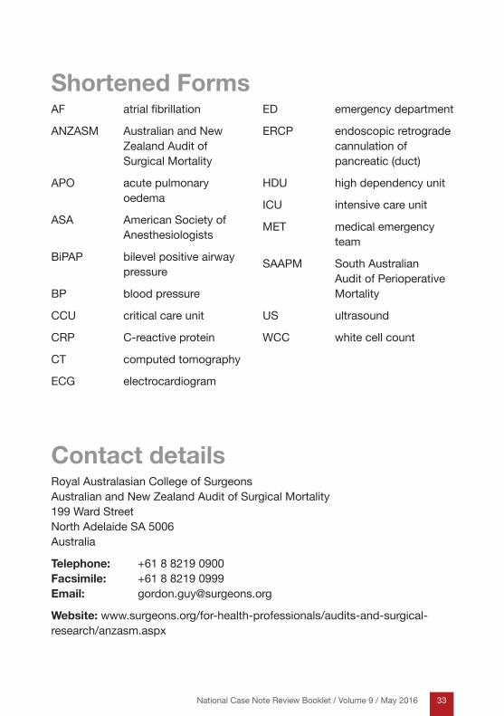
Shortened FormsAF atrial fibrillation
ANZASM Australian and New Zealand Audit of Surgical Mortality
APO acute pulmonary oedema
ASA American Society of Anesthesiologists
BiPAP bilevel positive airway pressure
BP blood pressure
CCU critical care unit
CRP C-reactive protein
CT computed tomography
ECG electrocardiogram
ED emergency department
ERCP endoscopic retrograde cannulation of pancreatic (duct)
HDU high dependency unit
ICU intensive care unit
MET medical emergency team
SAAPM South Australian Audit of Perioperative Mortality
US ultrasound
WCC white cell count
Contact detailsRoyal Australasian College of Surgeons Australian and New Zealand Audit of Surgical Mortality 199 Ward Street North Adelaide SA 5006 Australia
Telephone: +61 8 8219 0900 Facsimile: +61 8 8219 0999 Email: [email protected]
Website: www.surgeons.org/for-health-professionals/audits-and-surgical-research/anzasm.aspx

34 National Case Note Review Booklet / Volume 9 / May 2016
Notes..............................................................................................................................................................
..............................................................................................................................................................
..............................................................................................................................................................
..............................................................................................................................................................
..............................................................................................................................................................
..............................................................................................................................................................
..............................................................................................................................................................
..............................................................................................................................................................
..............................................................................................................................................................
..............................................................................................................................................................
..............................................................................................................................................................
..............................................................................................................................................................
..............................................................................................................................................................
..............................................................................................................................................................
..............................................................................................................................................................
..............................................................................................................................................................
..............................................................................................................................................................

35National Case Note Review Booklet / Volume 9 / May 2016
Notes..............................................................................................................................................................
..............................................................................................................................................................
..............................................................................................................................................................
..............................................................................................................................................................
..............................................................................................................................................................
..............................................................................................................................................................
..............................................................................................................................................................
..............................................................................................................................................................
..............................................................................................................................................................
..............................................................................................................................................................
..............................................................................................................................................................
..............................................................................................................................................................
..............................................................................................................................................................
..............................................................................................................................................................
..............................................................................................................................................................
..............................................................................................................................................................
..............................................................................................................................................................

Royal Australasian College of Surgeons Australian and New Zealand Audit of Surgical Mortality 199 Ward Street North Adelaide SA 5006 Australia
Telephone: +61 8 8219 0900 Facsimile: +61 8 8219 0999
Email: [email protected]