author's personal copy - all catfish species...
TRANSCRIPT

This article appeared in a journal published by Elsevier. The attachedcopy is furnished to the author for internal non-commercial researchand education use, including for instruction at the authors institution
and sharing with colleagues.
Other uses, including reproduction and distribution, or selling orlicensing copies, or posting to personal, institutional or third party
websites are prohibited.
In most cases authors are permitted to post their version of thearticle (e.g. in Word or Tex form) to their personal website orinstitutional repository. Authors requiring further information
regarding Elsevier’s archiving and manuscript policies areencouraged to visit:
http://www.elsevier.com/copyright

Author's personal copy
Tissue and Cell 43 (2011) 8–23
Contents lists available at ScienceDirect
Tissue and Cell
journa l homepage: www.e lsev ier .com/ locate / t i ce
Sperm of Doradidae (Teleostei: Siluriformes)
I. Quagio-Grassiottoa,∗, R.J. Ortiza,b, M.H. Sabaj Pérezc, C. Oliveiraa
a Departamento de Morfologia, Instituto de Biociências, Universidade Estadual Paulista – UNESP, 18618-970 Botucatu, São Paulo, Brazilb Graduate Program on Zoology, Instituto de Biociências de Botucatu, Unesp, Botucatu-SP, Brazilc Department of Ichthyology, Academy of Natural Sciences, 1900 Benjamin Franklin Parkway, Philadelphia 19103, PA, USA
a r t i c l e i n f o
Article history:Received 4 April 2009Received in revised form 28 October 2010Accepted 31 October 2010Available online 16 December 2010
Keywords:Thorny catfishesSpermatogenesisSpermiogenesisSperm ultrastructure
a b s t r a c t
Spermatic characteristics were studied in 10 species representing several distinct groups within the cat-fish family Doradidae. Interestingly, different types of spermatogenesis, spermiogenesis and spermatozoaare correlated with intrafamilial groups previously proposed for Doradidae. Semi-cystic spermatogene-sis, modified Type III spermiogenesis, and biflagellate sperm appear to be unique within Doradidae to thesubfamily Astrodoradinae. Other doradid species have sperm with a single flagellum, cystic spermatogen-esis, and spermiogenesis of Type I (Pterodoras granulosus, Rhinodoras dorbignyi), Type I modified (Oxydoraskneri), or Type III (Trachydoras paraguayensis). Doradids have an external mode of fertilization, and sharea few spermatic characteristics, such as cystic spermatogenesis, Type I spermiogenesis and uniflagel-late sperm, with its sister group Auchenipteridae, a family exhibiting sperm modifications associatedwith insemination and internal fertilization. Semi-cystic spermatogenesis and biflagellate spermato-zoa are also found in Aspredinidae, and corroborate recent proposals that Aspredinidae and Doradoidea(Doradidae + Auchenipteridae) are sister groups and that Astrodoradinae occupies a basal position withinDoradidae. The co-occurrence in various catfish families of semi-cystic spermatogenesis and eitherbiflagellate spermatozoa (Aspredinidae, Cetopsidae, Doradidae, Malapturidae, Nematogenyidae) or uni-flagellate sperm with two axonemes (Ariidae) reinforces the suggestion that such characteristics arecorrelated. Semi-cystic spermatogenesis and biflagellate sperm may represent ancestral conditions forLoricarioidei and Siluroidei of Siluriformes as they occur in putatively basal members of each suborder,Nematogenyidae and Cetopsidae, respectively. However, if semi-cystic spermatogenesis and biflagel-late sperm are ancestral for Siluriformes, cystic spermatogenesis and uniflagellate sperm have arisenindependently in multiple lineages including Diplomystidae, sister group to Siluroidei.
© 2010 Elsevier Ltd. All rights reserved.
1. Introduction
Doradidae is a family of freshwater catfishes endemic to SouthAmerica that comprises about 90 valid extant species and one fossilspecies arranged in 31 genera. Doradids commonly exhibit a row ofthorny scutes along the lateral line, and are promptly diagnosed bythe presence of the infranuchal scute, a laminar bone co-formed byfusion of an expanded ossicle of the lateral line canal and an ossifiedligament extending from beneath the posterior nuchal plate to thetip of the first rib (Birindelli et al., 2008; Birindelli, 2010).
Doradidae often is separated into two major groups, one withsimple barbels and more or less depressed head, and the otherwith fimbriate barbels and relatively deep head (Kner, 1853; Sabajand Ferraris, 2003; Birindelli and Sousa, 2010). Doradids with sim-
∗ Corresponding author at: Depto. de Morfologia, Instituto de Biociências, Univer-sidade Estadual Paulista, Campus de Botucatu, 18618-970, CP 510, Distrito de RubiãoJr, Botucatu, São Paulo, Brazil. Tel.: +55 14 3811 6264; fax: +55 14 3811 6264.
E-mail address: [email protected] (I. Quagio-Grassiotto).
ple barbels are non-monophyletic and include the most basal taxaaccording to both morphological and molecular cladistic analysessummarized below.
In the first cladistic analysis of intrafamilial relationshipsHiguchi (1992, unpublished Ph.D. Dissertation; cladogram andsynapomorphies published in Pinna de, 1998) used morphologi-cal characteristics to support the monophyly of the family, andrecovered Wertheimeria and Franciscodoras, respectively, as suc-cessive sister groups to all other doradids. For the remainingtaxa Higuchi (1992) recognized three monophyletic subfamiliesin an unresolved trichotomy: “Doradinae”, “Platydoradinae”, andAstrodoradinae, the lattermost formally named and diagnosed inHiguchi et al. (2007).
Moyer et al. (2004) subsequently used mitochondrial andnuclear DNA sequence data to examine phylogenetic relationshipsamong doradids. Their topology conflicted with the supra-genericclassification proposed by Higuchi (1992), however, their molec-ular analysis did not include several key genera (e.g., Centrochir,Franciscodoras, Kalyptodoras and Wertheimeria). Only one of theintra-familial groups proposed by Higuchi (1992), Astrodorad-
0040-8166/$ – see front matter © 2010 Elsevier Ltd. All rights reserved.doi:10.1016/j.tice.2010.10.006
Author's personal copy
I. Quagio-Grassiotto et al. / Tissue and Cell 43 (2011) 8–23 9
inae, was supported as monophyletic, and Astrodoradinae andAcanthodoras were recovered as deep lineages forming a basal tri-chotomy with a third group comprising all other doradids in theiranalysis.
In a separate cladistic study based on morphology Birindelli(2006 unpublished Ph.D. Dissertation) recovered a new topol-ogy wherein Kalyptodoras and Wertheimeria formed a basaltrichotomy with a clade containing all other doradid genera.Birindelli’s (2006) study supported Higuchi’s (1992) subfamilialgroup “Platydoradinae” as sister to Astrodoradinae + Doradinae.Later, Birindelli (2010, unpublished Ph.D. Dissertation) expandedhis original study to include all genera of Auchenipteridaeplus several additional catfish families as outgroups. His newstudy recovered Kalyptodoras + Wertheimeria as basal, sister toFranciscodoras + a clade containing the remaining doradid taxaanalyzed. Within the remaining taxa, a clade composed ofAcanthodoras, Agamyxis and two genera of Astrodoradinae wassister to a trichotomy formed by Centrochir, Platydoras, and aclade subdivided into three informally named tribes: “Pterodor-adini” sister to “Rhinodoradini” + “Doradini”. Finally, Sousa (2010,Unpublished Ph.D. Dissertation) used morphology to investigatephylogenetic relationships of Astrodoradinae. Sousa’s analysisrecovered Anadoras as the most basal member of Astrodoradi-nae, and expanded the subfamily to include Acanthodoras andAgamyxis.
Although the phylogenies differed, those studies have hypoth-esized several lineages within Doradidae. Morphological analysesconsistently recover Franciscodoras, Kalyptodoras and Wertheime-ria as the most basal doradids with the latter two as sister taxain Birindelli (2010). Morphological and molecular analyses iden-tify Acanthodoras and Astrodoradinae as deep lineages, and the twoappear to be closely related based on morphology (Birindelli, 2010;Sousa, 2010). Most of the more derived taxa group into three lin-eages, two with simple barbels (“Pterodoradini”, “Rhinodoradini”),and one inclusive of all fimbriate barbel genera. The monophylyof doradids sharing fimbriate barbels is well supported by mor-phological (Higuchi, 1992; Birindelli, 2006; 2010; Sousa, 2010) andmolecular (Moyer et al., 2004) data. However, the sister group rela-tionship between the fimbriate-barbel clade and Oxydoras, a genuswith simple barbels, is only supported by the morphological stud-ies.
A particularity of the Doradidae is the presence of anelastic-spring apparatus formed by a special arrangement of theparapophyses of the fourth vertebra (i.e., Müllerian rami), gas(swim) bladder, and associated muscles and ligaments (see Sabajand Ferraris, 2003 and Birindelli et al., 2009 for review). Thisparticularity is shared with the South American Auchenipteri-dae and with the African Mochokidae. According to Pinna de(1998) and Birindelli (2010), the South America families Doradidaeand Auchenipteridae constitute a monophyletic group assem-bled in the superfamily Doradoidea, and Doradoidea with theAfrican Mochokidae form the suborder Doradoidei. The occur-rence of a similar elastic-spring apparatus in Ariidae has beenused to suggest a sister group relationship with Doradoidei (Mo,1991; Lundberg, 1993; Royero, 1999). Friel (1994) alternativelyproposed Aspredinidae as the sister group to the Doradoidea(Doradidae + Auchenipteridae) based on phylogenetic analysis ofmorphological data; his hypothesis was later supported by phylo-genetic analyses of molecular data (Hardman, 2005; Sullivan et al.,2006). Aspredinidae is alternatively considered a member of theotherwise Asian Sisoroidea (Chen, 1994; Pinna de, 1993, 1996,1998; Diogo et al., 2002, 2003; Birindelli, 2010). A molecular phy-logeny by Sullivan et al. (2008), however, recovered Sisoroidea asa monophyletic group restricted to the Asian Akysidae, Amblycip-itidae and Sisoridae, and again placed Aspredinidae sister to SouthAmerican Doradoidea.
Studies of phylogenetic relationships within and between fam-ilies of Siluriformes have been based on bony and/or soft anatomyand molecular sequence data. It is known that sexual characteris-tics pertaining to spermatogenesis and spermiogenesis, as well assperm morphology, may yield phylogenetically informative char-acters useful for cladistic analyses (Jamieson, 2009).
In an attempt to evaluate their phylogenetic significance,characteristics of spermatogenesis, spermiogenesis, and/or spermultrastructure of 10 representative species of Doradidae aredescribed herein and discussed with respect to previously hypoth-esized relationships: within Doradidae, between Doradidae andpurported sister group Auchenipteridae, between Doradoidea(Doradidae + Auchenipteridae) and purported sister group Aspre-dinidae, and between Doradoidea and purported related groupAriidae.
2. Materials and methods
2.1. Examined material
Museum specimens were examined from ichthyological collec-tions at the Academy of Natural Sciences of Philadelphia (ANSP);Laboratório de Biologia de Peixes, Departamento de Morfologia,Universidade Estadual Paulista, Campus de Botucatu (LBP); andMuseu de Zoologia da Universidade de São Paulo (MZUSP).
Descriptions of spermatic characteristics are based on anal-yses at the ultrastructural level of testis from adult males ofAnadoras weddellii (LBP 672), Amblydoras sp. (ANSP 167626),Wertheimeria maculata (MZUSP 93658), Franciscodoras marmoratus(MZUSP 84224), Kalyptodoras bahiensis (MZUSP 100737), Acan-thodoras cataphractus (MZUSP 6831), Pterodoras granulosus (LBP4322), Oxydoras kneri (LBP 4323), Rhinodoras dorbignyi (LBP 4326)and Trachydoras paraguayensis (LBP 5627).
2.2. Preparation of specimens for observation of spermaticcharacteristics
Live specimens were anesthetized with 0.1% benzocaine andeuthanized (according to institutional protocols and approval) forremoval of the testis. Gonad fragments from freshly sacrificed fishwere fixed overnight in 2% glutaraldehyde and 4% paraformalde-hyde in 0.1 M Sorensen phosphate buffer, pH 7.4. The materialwas post-fixed in the dark for 2 h in 1% osmium tetroxide in thesame buffer, stained in block with aqueous solution of 5% uranylacetate for 2 h, dehydrated in acetone, embedded in araldite, andsectioned and stained with a saturated solution of uranyl acetate in50% ethanol and lead citrate. Electron micrographs were obtainedusing a Phillips-CM 100 transmission electron microscope.
“Dead” specimens from ichthyological collections (i.e., previ-ously fixed in 10% formalin and conserved in 70% ethanol) weredissected and the removed testis gradually rehydrated in a decreas-ing ethanol concentration (60%, 50%, 40% . . . distilled water). Oncerehydrated the material was re-fixed and prepared for observationas described for the live specimens.
Instances when the condition of the testis did not permitcomplete or accurate observations (e.g., previously fixed museumspecimens) are noted as “not available” (NA).
3. Results
Various features of spermatogenesis, spermiogenesis and sper-matozoa are summarized for the doradids analyzed herein andcompared to other catfishes in Table 1.

Author's personal copy
10 I. Quagio-Grassiotto et al. / Tissue and Cell 43 (2011) 8–23
Tab
le1
Com
par
ison
ofsp
erm
atic
char
acte
rist
ics
inD
orad
idae
toot
her
catfi
shes
(Sil
uri
form
es).
Dat
aco
mp
iled
from
this
stu
dy
and
Bu
rns
etal
.(20
02:A
uch
enip
teri
dae
),B
urn
set
al.(
2009
:Ari
idae
,Au
chen
ipte
rid
aean
dN
emat
ogen
yid
ae),
Parr
eira
etal
.(20
09:A
uch
enip
teri
dae
),Q
uag
io-G
rass
iott
oet
al.(
2001
:Dip
lom
ysti
dae
),Sh
ahin
(200
6:M
alap
teru
rid
ae),
Spad
ella
etal
.(20
06:A
spre
din
idae
and
Cet
opsi
dae
),Sp
adel
laet
al.(
2007
:Cal
lich
thyi
dae
).D
orad
idsu
bgro
up
sad
opte
dfr
omB
irin
del
liet
al.(
2009
).
Fam
ily
Subg
rou
pG
enu
ssp
ecie
sM
axil
lary
barb
elFe
rtil
izat
ion
Sper
mat
ogen
esis
Sper
mio
gen
esis
Typ
eC
hro
mat
inN
ucl
ear
rota
tion
Cen
trio
lem
igra
tion
Dor
adid
ae“B
asal
”A
cant
hodo
ras
cata
phra
ctus
Sim
ple
Exte
rnal
NA
NA
NA
NA
NA
Dor
adid
ae“B
asal
”Fr
anci
scod
orda
sm
arm
orat
usSi
mp
leEx
tern
alN
AN
AN
AN
AN
AD
orad
idae
“Bas
al”
Kal
ypto
dora
sba
hien
sis
Sim
ple
Exte
rnal
NA
NA
NA
NA
NA
Dor
adid
ae“B
asal
”W
erth
eim
eria
mac
ulat
aSi
mp
leEx
tern
alN
AN
AN
AN
AN
AD
orad
idae
Ast
rod
orad
inae
Ana
dora
sw
edde
llii
Sim
ple
Exte
rnal
Sem
icys
tic
Typ
eII
Imod
ified
Dif
fuse
,hom
ogen
ous,
irre
gula
rou
tlin
eA
bsen
tPr
esen
ta
Dor
adid
aeA
stro
dor
adin
aeA
mbl
ydor
assp
.Si
mp
leEx
tern
alN
AN
AN
AN
AN
AD
orad
idae
Cla
de
3Pt
erod
oras
gran
ulos
usSi
mp
leEx
tern
alC
ysti
cTy
pe
ID
iffu
se,h
omog
enou
s,ci
rcu
lar
outl
ine
Com
ple
tePr
esen
tD
orad
idae
Cla
de
4R
hino
dora
sdo
rbig
nyi
Sim
ple
Exte
rnal
Cys
tic
Typ
eI
Dif
fuse
,hom
ogen
ous,
circ
ula
rou
tlin
eC
omp
lete
Pres
ent
Dor
adid
aeC
lad
e5
Trac
hydo
ras
para
guay
ensi
sFi
mbr
iate
Exte
rnal
Cys
tic
Typ
eII
ID
iffu
se,h
omog
enou
s,ci
rcu
lar
outl
ine
Abs
ent
Abs
ent
Dor
adid
aeO
xydo
ras
kner
iSi
mp
leEx
tern
alC
ysti
cTy
pe
Imod
ified
Dif
fuse
,hom
ogen
ous,
circ
ula
rou
tlin
eC
omp
lete
Abs
enta
Au
chen
ipte
rid
aeTr
ache
lyop
teru
ssp
p.Si
mp
leIn
tern
al(i
nse
min
atin
g)C
ysti
cTy
pe
ID
iffu
se,h
omog
enou
s,ci
rcu
lar
outl
ine
Com
ple
tePr
esen
t
Asp
red
inid
aeBu
noce
phal
usam
azon
icus
Sim
ple
Exte
rnal
Sem
icys
tic
Typ
eII
Imod
ified
Dif
fuse
,hom
ogen
ous,
irre
gula
rou
tlin
eab
sen
tPr
esen
ta
Cal
lich
thyi
dae
Cor
ydor
adin
aeCo
rydo
ras
flave
olus
Sim
ple
Exte
rnal
Sem
icys
tic
Typ
eII
Imod
ified
Dif
fuse
,hom
ogen
ous,
irre
gula
rou
tlin
eA
bsen
tA
bsen
tA
riid
aeG
enid
ens
geni
dens
Sim
ple
Exte
rnal
Sem
icys
tic
Typ
eIm
odifi
edD
iffu
se,h
omog
enou
s,ci
rcu
lar
outl
ine
Com
ple
teA
bsen
ta
Nem
atog
enyi
dae
Nem
atog
enys
iner
mis
Sim
ple
Exte
rnal
Sem
icys
tic
Typ
eII
Imod
ified
Dif
fuse
,hom
ogen
ous,
circ
ula
rou
tlin
eab
sen
tPr
esen
ta
Mal
apte
ruri
dae
Mal
apte
ruru
sel
ectr
icus
Sim
ple
Exte
rnal
Sem
icys
tic
NA
Dif
fuse
,hom
ogen
ous,
circ
ula
rou
tlin
eN
AN
AC
etop
sid
aeCe
tops
isco
ecut
iens
Sim
ple
Exte
rnal
Sem
icys
tic
Typ
eII
ID
iffu
se,h
omog
enou
s,ci
rcu
lar
outl
ine
Abs
ent
Abs
ent
Dip
lom
ysti
dae
Dip
lom
yste
sm
esem
brin
usSi
mp
leEx
tern
alC
ysti
cTy
pe
ID
iffu
se,h
omog
enou
s,ci
rcu
lar
outl
ine
Pres
ent
Pres
ent
Gen
us
spec
ies
Sper
mat
ozoa
Nu
cleu
sC
hro
mat
inN
ucl
ear
foss
aC
entr
iole
sM
idp
iece
Mit
och
ond
ria
Cyt
opla
smic
can
alFl
agel
la
Aca
ntho
dora
sca
taph
ract
usSu
bsp
her
ical
,tip
slig
htl
yfl
atte
ned
Hig
hly
con
den
sed
,h
omog
enou
sPr
esen
t(m
oder
atel
yd
eep
,in
clu
des
pro
xim
alce
ntr
iole
and
mos
tof
dis
talc
entr
iole
)
Alm
ost
per
pen
dic
ula
rSl
igh
tly
asym
met
ric,
con
tain
sm
itoc
hon
dri
a,ve
sicl
esan
dcy
top
lasm
icca
nal
NA
NA
1(1
axon
eme)
,fi
ns
lack
ing
Fran
cisc
odor
das
mar
mor
atus
Ovo
id,t
ipfl
atte
ned
Hig
hly
con
den
sed
,h
omog
enou
sPr
esen
t(m
oder
atel
yd
eep
,in
clu
des
pro
xim
alce
ntr
iole
and
mos
tof
dis
talc
entr
iole
)
Alm
ost
per
pen
dic
ula
rSh
orte
r,as
ymm
etri
c,co
nta
ins
mit
och
ond
ria,
few
vesi
cles
and
cyto
pla
smic
can
al
NA
NA
1(1
axon
eme)
,fi
ns
lack
ing
Kal
ypto
dora
sba
hien
sis
Ovo
id,t
ipw
eakl
yfl
atte
ned
Hig
hly
con
den
sed
,h
omog
enou
sPr
esen
t(m
oder
atel
yd
eep
,in
clu
des
pro
xim
alce
ntr
iole
and
mos
tof
dis
talc
entr
iole
)
Alm
ost
per
pen
dic
ula
rLo
nge
r,as
ymm
etri
c,co
nta
ins
mit
och
ond
ria
and
cyto
pla
smic
can
al
NA
NA
1(1
axon
eme)
,fi
ns
lack
ing
Wer
thei
mer
iam
acul
ata
Ovo
id,t
ipex
trem
ely
flat
ten
edH
igh
lyco
nd
ense
d,
hom
ogen
ous
Pres
ent
(mod
erat
ely
dee
p,i
ncl
ud
esp
roxi
mal
cen
trio
lean
dm
ost
ofd
ista
lcen
trio
le)
Alm
ost
per
pen
dic
ula
rSh
orte
r,as
ymm
etri
c,co
nta
ins
mit
och
ond
ria,
few
vesi
cles
and
cyto
pla
smic
can
al
Ver
yel
onga
teN
A1
(1ax
onem
e),
fin
sla
ckin
g
Ana
dora
sw
edde
llii
Bel
l-sh
aped
Hig
hly
con
den
sed
,h
omog
enou
sPr
esen
ta(v
ery
dee
p,
incl
ud
esce
ntr
iole
s,m
itoc
hon
dri
aan
din
itia
lse
gmen
tof
flag
ella
)
Para
llel
Lon
g,co
nta
ins
mit
och
ond
ria,
vesi
cles
and
two
cyto
pla
smic
can
als
Ovo
id(s
ligh
tly
elon
gate
)R
esu
lts
from
sim
ult
aneo
us
pro
ject
ion
ofn
ucl
eus
and
cyto
pla
smto
war
dfl
agel
laan
dm
igra
tion
ofce
ntr
iole
s
2(2
axon
emes
),fi
ns
lack
ing
Am
blyd
oras
sp.
Bel
l-sh
aped
Hig
hly
con
den
sed
,h
omog
enou
sPr
esen
t(v
ery
dee
p,
incl
ud
esce
ntr
iole
san
dm
itoc
hon
dri
a)
Para
llel
Lon
g,fi
lled
wit
hve
sicl
esO
void
(sli
ghtl
yel
onga
te)
NA
2(2
axon
emes
),fi
ns
lack
ing
Pter
odor
asgr
anul
osus
Ovo
id,t
ipfl
atte
ned
Hig
hly
con
den
sed
,h
omog
enou
sPr
esen
t(m
oder
atel
yd
eep
,in
clu
des
pro
xim
alce
ntr
iole
and
mos
tof
dis
talc
entr
iole
)
Alm
ost
per
pen
dic
ula
rSl
igh
tly
asym
met
ric,
con
tain
sm
itoc
hon
dri
a,ve
sicl
esan
dcy
top
lasm
icca
nal
Obl
ong
Res
ult
sfr
omp
roje
ctio
nof
cyto
pla
smto
war
ds
flag
ellu
m
1(1
axon
eme)
,fi
ns
lack
ing

Author's personal copy
I. Quagio-Grassiotto et al. / Tissue and Cell 43 (2011) 8–23 11
Tabl
e1
(Con
tinu
ed)
Gen
us
spec
ies
Sper
mat
ozoa
Nu
cleu
sC
hro
mat
inN
ucl
ear
foss
aC
entr
iole
sM
idp
iece
Mit
och
ond
ria
Cyt
opla
smic
can
alFl
agel
la
Rhi
nodo
ras
dorb
igny
iSu
bsp
her
ical
,tip
slig
htl
yfl
atte
ned
Hig
hly
con
den
sed
,h
omog
enou
sPr
esen
t(m
oder
atel
yd
eep
,in
clu
des
pro
xim
alce
ntr
iole
and
mos
tof
dis
talc
entr
iole
)
Alm
ost
per
pen
dic
ula
rA
sym
met
ric,
con
tain
sm
itoc
hon
dri
a,ve
sicl
esan
dcy
top
lasm
icca
nal
Elon
gate
Res
ult
sfr
omp
roje
ctio
nof
cyto
pla
smto
war
ds
flag
ellu
m
1(1
axon
eme)
,fi
ns
lack
ing
Trac
hydo
ras
para
guay
ensi
sSp
her
ical
Hig
hly
con
den
sed
,h
omog
enou
sA
bsen
tO
bliq
ue
Asy
mm
etri
c,co
nta
ins
mit
och
ond
ria,
larg
eve
sicl
esan
dcy
top
lasm
icca
nal
Elon
gate
Res
ult
sfr
omp
roje
ctio
nof
mid
pie
ceve
sicl
esto
war
ds
flag
ellu
m
1(1
axon
eme)
,fi
ns
lack
ing
Oxy
dora
skn
eri
Sph
eric
alH
igh
lyco
nd
ense
d,
hom
ogen
ous
Pres
ent
(mod
erat
ely
dee
p,i
ncl
ud
esp
roxi
mal
cen
trio
lean
dm
ost
ofd
ista
lcen
trio
le)
Perp
end
icu
lar
Slig
htl
yas
ymm
etri
c,co
nta
ins
mit
och
ond
ria,
abu
nd
ant
vesi
cles
and
cyto
pla
smic
can
al
Elon
gate
Res
ult
sfr
omp
roje
ctio
nof
cyto
pla
smto
war
ds
flag
ellu
m
1,fi
ns
lack
ing
Trac
hely
opte
rus
spp
.El
onga
teH
igh
lyco
nd
ense
d,
hom
ogen
ous
Pres
ent
Obl
iqu
ePe
culi
arV
ery
elon
gate
Res
ult
sfr
omp
roje
ctio
nof
cyto
pla
smto
war
ds
flag
ellu
m
1(1
axon
eme)
Buno
ceph
alus
amaz
onic
usB
ell-
shap
edFl
occu
len
tPr
esen
ta(d
eep
,in
clu
des
cen
trio
les)
Para
llel
Lon
g,co
nta
ins
mit
och
ond
ria,
vesi
cles
and
two
cyto
pla
smic
can
als
Elon
gate
Res
ult
sfr
omsi
mu
ltan
eou
sp
roje
ctio
nof
nu
cleu
san
dcy
top
lasm
tow
ard
flag
ella
and
mig
rati
onof
cen
trio
les
2(2
axon
emes
)
Cory
dora
sfla
veol
usN
AN
APr
esen
ta(s
hal
low
,not
occu
pie
dby
cen
trio
les)
Para
llel
NA
NA
NA
1(1
axon
eme)
,fi
ns
lack
ing
Gen
iden
sge
nide
nsH
igh
lyco
nd
ense
d,
hom
ogen
ous
Pres
ent
(dee
p,i
ncl
ud
esce
ntr
iole
san
dm
itoc
hon
dri
a)
Para
llel
Shor
t,sy
mm
etri
c,co
nta
ins
mit
och
ond
ria
Elon
gate
Abs
ent
1(2
axon
emes
),fi
ns
lack
ing
Nem
atog
enys
iner
mis
Subs
ph
eric
alH
igh
lyco
nd
ense
d,
hom
ogen
ous
Abs
ent
Para
llel
Sym
met
ric,
con
tain
sm
itoc
hon
dri
aan
dve
sicl
es
Elon
gate
Abs
ent
2(2
axon
emes
)
Mal
apte
ruru
sel
ectr
icus
Subs
ph
eric
alH
igh
lyco
nd
ense
dh
omog
enou
sPr
esen
tPa
rall
elLo
ng,
con
tain
sm
itoc
hon
dri
a,ve
sicl
esan
dtw
ocy
top
lasm
icca
nal
s
Elon
gate
Pres
ent
2(2
axon
emes
)
Ceto
psis
coec
utie
nsSu
bsp
her
ical
Hig
hly
con
den
sed
hom
ogen
ous
Pres
enta
(dee
p,i
ncl
ud
esce
ntr
iole
s)Pa
rall
elSh
ort,
sym
met
ric,
con
tain
sm
itoc
hon
dri
aan
dve
sicl
es
Rou
nd
edPr
esen
t2
(2ax
onem
es)
Dip
lom
yste
sm
esem
brin
usSu
bsp
her
ical
Floc
cule
nt
Pres
ent
(dee
p,i
ncl
ud
esce
ntr
iole
san
dst
art
ofax
onem
e)
Perp
end
icu
lar
Shor
tSi
ngl
e,la
rge,
C-s
hap
edA
bsen
t1
(1ax
onem
e)
NA
ind
icat
esco
mp
arat
ive
dat
an
otav
aila
ble.
aIn
dic
ates
mod
ifica
tion
from
clas
sic
Typ
eIo
rII
Isp
erm
ioge
nes
is.

Author's personal copy
12 I. Quagio-Grassiotto et al. / Tissue and Cell 43 (2011) 8–23
3.1. A. weddellii and Amblydoras sp. (Figs. 1–4)
3.1.1. SpermatogenesisIn A. weddellii spermatogenesis is semi-cystic. In this kind of
spermatogenesis, although spermatogonia proliferation and mei-otic divisions of the spermatocytes occur inside the spermatocysts(Fig. 1A), spermatid differentiation is extra-cystic and occurs out-side the cysts in the luminal compartment of the testis (Fig. 1B). Inthe luminal compartment, clusters of spermatids recently releasedfrom the cysts remain connected to one another by cytoplasmicbridges. Spermatids gradually lose those connections and differ-entiate. Spermatid differentiation is not synchronous and cells indistinct phases of development can be seen together in the lumi-nal compartment (Fig. 1C). Spermatozoa are also present in theluminal compartment (Fig. 1A). Information on spermatogenesisin Amblydoras is not available.
3.1.2. SpermiogenesisIn A. weddellii spermiogenesis is a modification of Type III. In
the early spermatids (Fig. 2A and B), the cytoplasm symmetricallyencircles the nucleus, which displays diffuse homogenous chro-matin and has an irregular outline. The centriolar complex liesmedially to the nucleus and is anchored to the plasma membrane.The centrioles are lateral and parallel to one another (Fig. 2A–C).Both centrioles differentiate into basal bodies, and each centri-ole forms one flagellum. Centrioles start their migration towardthe nucleus, carrying along the plasma membrane and the initialsegments of the flagella, which invaginate. Two independent cyto-plasmic canals, a space between each flagellum and the plasmamembrane, are then formed. A depression is formed in the nuclearoutline at the level of the centrioles (Fig. 2A and B). The nucleusdoes not rotate in relation to the flagellar axis. Instead, in a sug-gested coordinated movement, the basal region of the nucleus isprojected in the direction of the initial segment of the flagellawhile the centrioles continue their migration inside the nuclearfossa. Consequently, the nucleus takes on a bell shape in whichthe initial segments of the flagella, each with individualized cyto-plasmic canals, are housed in a very deep nuclear fossa (Fig. 2C,E, G). The cytoplasm, which initially accumulates in the regionsurrounding the centrioles (Fig. 2A and B), moves toward the seg-ments of the flagella located just outside of the nuclear fossa,forming the midpiece (Fig. 2C, E, G). The midpiece contains twocytoplasmic canals with the flagella, mitochondria and vesicles(Fig. 2D–H). Mitochondria are included inside the nuclear fossa(Fig. 2F). Information on spermiogenesis of Amblydoras is notavailable.
3.1.3. SpermatozoaSpermatozoa of A. weddellii and Amblydoras are quite similar:
the conical-trunk nucleus is bell shaped and contains highly con-densed homogeneous chromatin interspersed by electron-lucentareas, and is surrounded by a narrow strip of cytoplasm with noorganelles. Nucleus has about 2.0 �m in height by 1.4 �m in widthat the base and 0.6 �m in width at the tip in A. weddellii, vs. 2.1 �min height by 1.4 �m in width at the base and 0.6 �m in width atthe tip in Amblydoras (Fig. 3A, D and E; Fig. 4F). The centriolesare lateral and parallel to one another, and are located internallyto the nucleus at the tip of the very deep nuclear fossa. The cen-trioles are covered by electron dense material and fastened toone another, to the nuclear envelope at the nuclear fossa, and tothe plasma membrane by deep stabilization fibrils (Fig. 3A, D, G;Fig. 4C). The spermatozoa have two flagella and two independentcytoplasmic canals extending internally from the tip of the nucleusto the terminal end of the midpiece (Fig. 3A, B, D, H–K; Fig. 4D andE). The slightly elongated mitochondria are located mainly nearthe base of the nucleus, but also are found internally in the deep
Fig. 1. Semi-cystic spermatogenesis in Anadoras weddellii (Astrodoradinae). Cystsare formed by the germ cells surrounded by the cytoplasmic process (arrow) of theSertoli cells (A): in this kind of spermatogenesis, only spermatogonia and sperma-tocytes (st) are inside the cysts whereas the spermatids (s) and spermatozoa (z) arefound in the luminal compartment (l). n: nucleus; asterisk: synaptonemal complex.(B) A cluster of early spermatids (s) recently released from a cyst rests upon thegerminal epithelium (arrow). i: interstitial compartment; l: luminal compartment;z: spermatozoa. (C) Spermatic cell differentiation is asynchronous and occurs in theluminal compartment (l) outside the cysts. s: spermatids; z: spermatozoa; arrow:cytoplasmic projections of Sertoli cells limiting a cyst.

Author's personal copy
I. Quagio-Grassiotto et al. / Tissue and Cell 43 (2011) 8–23 13
Fig. 2. Spermiogenesis in A. weddellii. (A and B) In early spermatids the centrioles (c) with the two forming flagella (f) are medially located in relation to the nucleus (n). m:mitochondria; arrow: nuclear fossa. (C, E, G) Spermatids in longitudinal sections. Note the centrioles (c) inside the nuclear fossa (arrow) moving in the direction of the tip ofthe nucleus (n) and forming the two cytoplasmic canals (asterisk). At the same time, the base of the nucleus and the cytoplasm project toward the flagella (f) forming themidpiece (p). m: mitochondria; v: vesicles. (D, F, H) Corresponding cross-sections of the same type of spermatids at the nuclear (n) region. Note the two flagella (f) and therespective two cytoplasmic canals (asterisk) inside the nuclear fossa (arrow). m: mitochondria.
nuclear fossa (Fig. 3D, H, I; Fig. 4A and E). The midpiece is filledwith vesicles interspaced by a thin layer of cytoplasm, and has acytoplasmic sleeve at the terminal end (Fig. 3A, B, D, J, K). Eachflagellum contains a classic axoneme (9 + 2) (Fig. 3C, F; Fig. 4H).
Data on the limiting plasma membrane and midpiece of Amblydo-ras are not available because the specimens were obtained fromichthyological collections and the gonads were not properly pre-served.

Author's personal copy
14 I. Quagio-Grassiotto et al. / Tissue and Cell 43 (2011) 8–23
Fig. 3. Spermatozoon of A. weddellii. (A) The bell shaped nucleus (n) of this sperm in a medial/longitudinal section has a horseshoe shape with the centrioles (c) at thetip and an internal, large nuclear fossa (arrow) that settles the initial segments of the two flagella (f) and respective cytoplasmic canals (asterisk). v: vesicles. (B) Seen ina medial/longitudinal section, the midpiece is completely full of vesicles (v) and ends in a long cytoplasmic sleeve (arrow head) surrounding the two flagella. (f). asterisk:cytoplasmic canals; n: nucleus. (C) Flagella (f) in a longitudinal view. (D) Longitudinal section along the sperm in which only one of the flagella (f) can be seen. n; nucleus; m:mitochondria; v: vesicles; arrow head: cytoplasmic sleeve; asterisk: cytoplasmic canals; c: centriolar complex. (E) In an external view, the nucleus (n) has a conical outline.(F) Axoneme (a) seen in cross-sectioned flagella. (G–I) Cross-sections in different levels of the nucleus. (G) The centrioles (c) in the tip of the nucleus (n). (H) In the middleregion of the nucleus (n) the two flagella (f) and respective cytoplasmic canals (asterisk) are seen within the nuclear fossa. Note that some vesicles (v) and mitochondria (m)can also be found inside the nuclear fossa. (I) Most the mitochondria (m) are located at the base of the nucleus (n) around the two flagella (f) and respective cytoplasmiccanals (asterisk). (J) In a cross to oblique section the vesicles (v) that fill the midpiece expose an elongated form. f: flagella; asterisk: cytoplasmic canals. (K) In a cross-sectionat the end of the midpiece, the cytoplasmic sleeves (arrow head) are seen surrounding the flagella (f) forming respective cytoplasmic canals (asterisk).

Author's personal copy
I. Quagio-Grassiotto et al. / Tissue and Cell 43 (2011) 8–23 15
Fig. 4. Spermatozoon of Amblydoras sp. (specimen from zoological collection). (A) The bell shaped nucleus (n) of this sperm in a medial/longitudinal section has a horseshoeshape with the centrioles (c) at the tip and an internal, deep nuclear fossa (arrow) that settles the initial segments of the flagella (f) and some mitochondria (m). (B–E) Incross sections in different levels of the nucleus (n), the centrioles (c) and flagella (f) can be seen inside the nuclear fossa (asterisk). (F) In an external view, the nucleus (n) hasa trunk-conical outline. f: flagella. (G–H) Note that the flagella (f) in a longitudinal or cross view are always in pars. a: axoneme.
3.2. W. maculata, F. marmoratus and K. bahiensis (Fig. 5)
Information on spermatogenesis and spermiogenesis are notavailable because the samples had only spermatozoa.
3.2.1. SpermatozoaIn the spermatozoa of W. maculata, F. marmoratus and K. bahien-
sis the nucleus has an ovoid shape with a flattened tip, containshighly condensed homogeneous chromatin, and is surrounded bya narrow strip of cytoplasm with no organelles (Fig. 5A, D, G). The tipof the nucleus is more flattened in W. maculata than in F. marmora-tus and K. bahiensis. Nucleus has about 1.2 �m in height by 1.7 �min width in W. maculata, 1.2 �m by 1.6 �m in F. marmoratus, and1.3 �m by 1.6 �m in K. bahiensis. In all three species the nuclearoutline that faces the midpiece has a medial and moderately deepdepression, the nuclear fossa (Fig. 5A, D, G). The proximal centri-ole is anterior and almost perpendicular to the distal centriole. Thecentrioles are covered by electron dense material and fastened toone another. The proximal centriole and most of the distal centrioleare inside the nuclear fossa (Fig. 5A, D, G). The midpiece containsthe mitochondria, vesicles and the cytoplasmic canal in which liesthe initial segment of the single flagellum (Fig. 5A–C, E, F, H). Themidpiece is slightly asymmetric due to the unequal distributionof mitochondria and vesicles. In W. maculata, mitochondria seemto be very elongated and form a ring surrounding the cytoplasmiccanal (Fig. 5B). Vesicles are mainly accumulated at the peripheryand at the terminal regions of the midpiece (Fig. 5A, B, C, E, F). The
flagellum contains a classic axoneme (9 + 2) (Fig. 5I). Despite infor-mation on the limiting plasma membrane and midpiece structuressuch as mitochondria, data on the vesicles and cytoplasmic canalin K. bahiensis are not available because the gonads of the museumspecimens were not properly preserved. The midpiece itself seemsto be longer in K. bahiensis (Fig. 5 G, H, I) than in W. maculata and F.marmoratus.
3.3. O. kneri (Figs. 6 and 7)
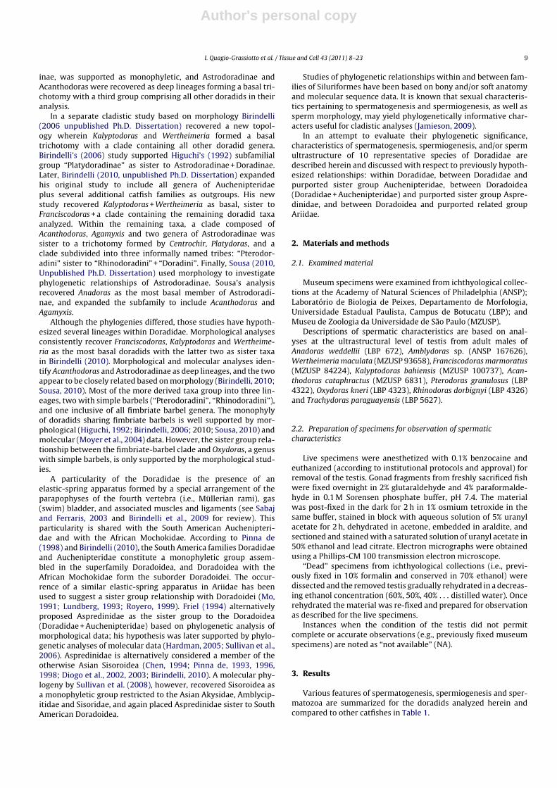
3.3.1. SpermatogenesisIn O. kneri, spermatogenesis occurs inside the cysts. At the end
of the differentiation process, spermatozoa are released into theluminal compartment of the testis (Fig. 6A).
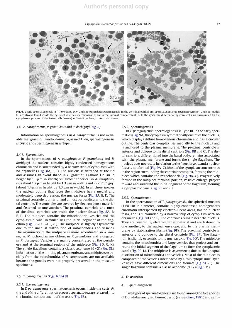
3.3.2. SpermiogenesisIn O. kneri spermiogenesis is a modification of Type I. In the
early spermatids (Fig. 7A) the cytoplasm symmetrically encirclesthe nucleus, which displays diffuse homogenous chromatin andhas a circular outline. The centriolar complex lies laterally to thenucleus and is anchored to the plasma membrane. The proximalcentriole is anterior and perpendicular to the distal centriole. Thedistal centriole differentiates into the basal body and forms thesingle flagellum. The nucleus rotates toward the centriolar com-plex (Fig. 7B) with nuclear rotation of 90◦ considered complete.A depression is newly formed in the nuclear outline at the levelof the centriolar complex that penetrates it (Fig. 7C). Simultane-

Author's personal copy
16 I. Quagio-Grassiotto et al. / Tissue and Cell 43 (2011) 8–23
Fig. 5. Spermatozoa of Wertheimeria maculata, Franciscodoras marmoratus and Kalyptodoras bahiensis (specimens recovered from zoological collections). (A)Medial/longitudinal section from the spermatozoon of W. maculata exposing the nuclear fossa (arrow), the slightly eccentric position of the flagellum (f) in relation tothe nucleus (n) and the also slightly asymmetric midpiece with few vesicles (v). c: centriolar complex; m: mitochondria. Note that the tip of the nucleus is flattened. (B andC) Cross sections of different levels of the midpiece. Note the cytoplasmic canal (asterisk) in which lies the initial segment of the flagellum (f), the ring shape and localizationof the mitochondria (m) and vesicles (v). (D) Medial/longitudinal section from the spermatozoon of F. marmoratus exposing the nuclear fossa (arrow), the slightly eccentricposition of the flagellum (f) in relation to the nucleus (n) and the also slightly asymmetric midpiece with few vesicles (v). c: centriolar complex. Note that the tip of thenucleus is flattened. (E–F) Cross sections at different levels of the midpiece. Only one of the centrioles (c) is visible and some vesicles (v). n: nucleus; arrow head: cytoplasmiccanal. (G) Medial/longitudinal section from the spermatozoon of K. bahiensis exposing the nuclear fossa (arrow) and the slightly eccentric position of the flagellum (f) inrelation to the nucleus (n). c: centriolar complex; i: midpiece. (H and I) Longitudinal and cross sections of the midpice (i) at the base of the nucleus (n), at its middle regionand at the terminal end. f: flagellum; m: mitochondria.
ous to nuclear rotation, the cytoplasm projects in the direction ofthe initial segment of the flagellum forming the cytoplasmic canaland midpiece (Fig. 7A–C). The midpiece contains the mitochondria,forming vesicles and cytoplasmic canal housing the initial segmentof the flagellum (Fig. 7B and C).
3.3.3. SpermatozoonIn the spermatozoon of O. kneri the spherical nucleus (about
1.5 �m in diameter) contains highly condensed homogeneouschromatin interspersed by electron-lucent areas, and is surroundedby a narrow strip of cytoplasm with no organelles (Fig. 7D and E).In the nuclear outline that faces the midpiece there is a medialand moderately deep depression, the nuclear fossa (Fig. 7D–F). Theproximal centriole, initially anterior and perpendicular to distal
one, attains an oblique acute angle to the distal centriole. The cen-trioles are covered by electron dense material and are fastened toone another, to the nuclear envelope at the nuclear fossa, and tothe plasma membrane by stabilization fibrils. The proximal centri-ole and most of the distal centriole are inside the nuclear fossa(Fig. 7F and G). The midpiece contains the mitochondria, abun-dant vesicles and the cytoplasmic canal in which lies the initialsegment of the flagellum. The midpiece is slightly asymmetricdue to the unequal distribution of mitochondria and vesicles. Themitochondria are elongated and mainly accumulated in the largerportion of the midpiece. Vesicles are elongated and mainly concen-trated at the periphery and at the terminal regions of the midpiece(Fig. 7H–K). The single flagellum contains a classic axoneme (9 + 2)(Fig. 7L).
Author's personal copy
I. Quagio-Grassiotto et al. / Tissue and Cell 43 (2011) 8–23 17
Fig. 6. Cystic spermatogenesis in (A) Oxydoras kneri and (B) Trachydoras paraguayensis. In the germinal epithelium, spermatogonia (g), spermatocytes (st) and spermatids(s) are always found inside the cysts (c) whereas spermatozoa (z) are in the luminal compartment (l). In the cysts, the differentiating germ cells are surrounded by thecytoplasmic process of the Sertoli cells (arrow). n: Sertoli nucleus; i: interstitial tissue.
3.4. A. cataphractus, P. granulosus and R. dorbignyi (Fig. 8)
Information on spermiogenesis in A. cataphractus is not avail-able. In P. granulosus and R. dorbignyi, as in O. kneri, spermatogenesisis cystic and spermiogenesis is Type I.
3.4.1. SpermatozoaIn the spermatozoa of A. cataphractus, P. granulosus and R.
dorbignyi the nucleus contains highly condensed homogeneouschromatin and is surrounded by a narrow strip of cytoplasm withno organelles (Fig. 8A, E, I). The nucleus is flattened at the tipand assumes an ovoid shape in P. granulosus (about 1.2 �m inheight by 1.8 �m in width) vs. almost spherical in A. cataphrac-tus (about 1.2 �m in height by 1.3 �m in width) and in R. dorbignyi(about 1.4 �m in height by 1.3 �m in width). In all three speciesthe nuclear outline that faces the midpiece has a medial andmoderately deep depression, the nuclear fossa (Fig. 8A, E, I). Theproximal centriole is anterior and almost perpendicular to the dis-tal centriole. The centrioles are covered by electron dense materialand fastened to one another. The proximal centriole and mostof the distal centriole are inside the nuclear fossa (Fig. 8A, B,E, I). The midpiece contains the mitochondria, vesicles and thecytoplasmic canal in which lies the initial segment of the flag-ellum (Fig. 8C–D, F–H, J–L). The midpiece is slightly asymmetricdue to the unequal distribution of mitochondria and vesicles.The asymmetry of the midpiece is more accentuated in R. dor-bignyi. Mitochondria are oblong in P. granulosus and elongatedin R. dorbignyi. Vesicles are mainly concentrated at the periph-ery and at the terminal regions of the midpiece (Fig. 8D, G, K).The single flagellum contains a classic axoneme (9 + 2) (Fig. 8L).Information on the limiting plasma membrane and midpiece, espe-cially from the mitochondria, of A. cataphractus are not availablebecause the gonads were not properly preserved in the museumspecimens.
3.5. T. paraguayensis (Figs. 6 and 9)
3.5.1. SpermatogenesisIn T. paraguayensis, spermatogenesis occurs inside the cysts. At
the end of the differentiation process spermatozoa are released intothe luminal compartment of the testis (Fig. 6B).
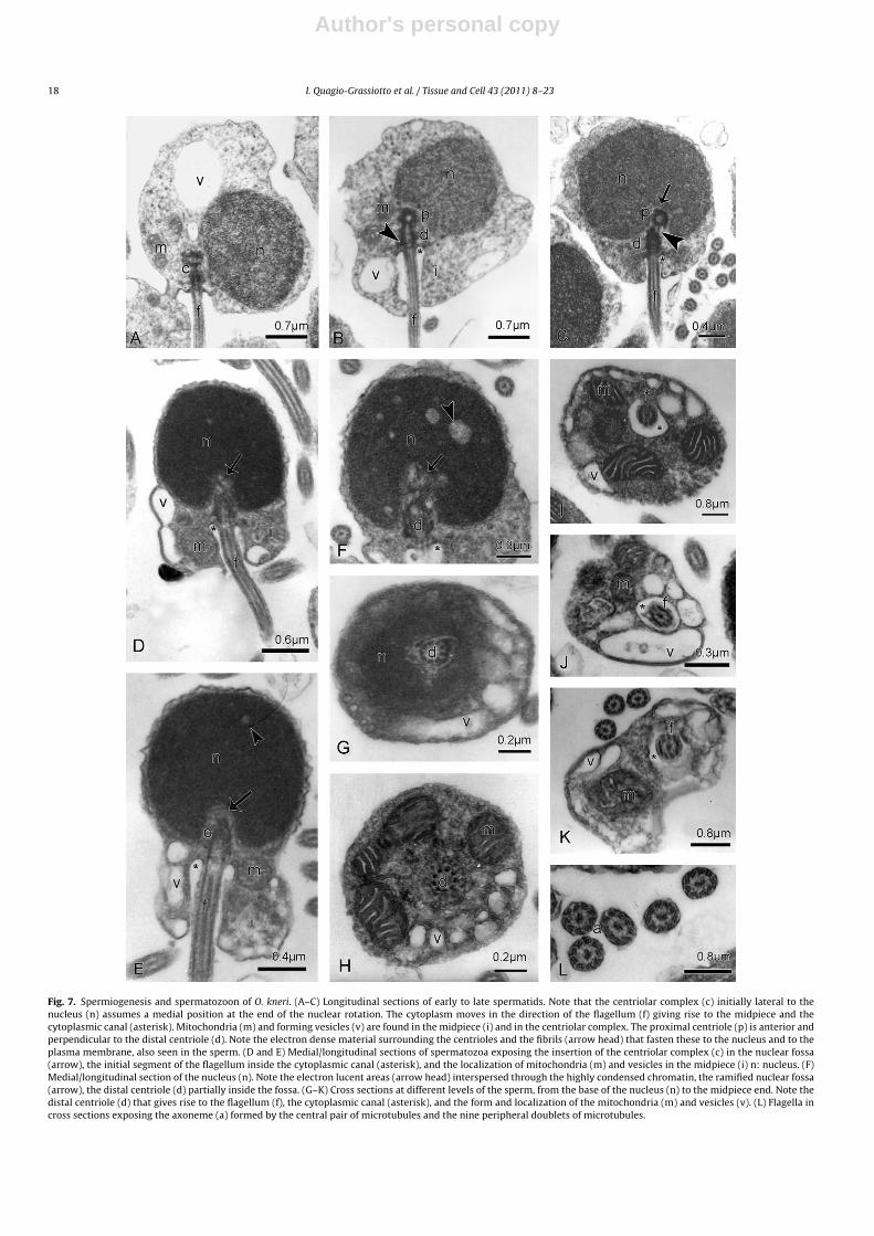
3.5.2. SpermiogenesisIn T. paraguayensis, spermiogenesis is Type III. In the early sper-
matids (Fig. 9A) the cytoplasm symmetrically encircles the nucleus,which displays diffuse homogenous chromatin and has a circularoutline. The centriolar complex lies medially to the nucleus andis anchored to the plasma membrane. The proximal centriole isanterior and oblique to the distal centriole (Fig. 9B and C). The dis-tal centriole, differentiated into the basal body, remains associatedwith the plasma membrane and forms the single flagellum. Thenucleus does not rotate in relation to the flagellar axis, and a nuclearfossa is not formed (Fig. 9A–C). Most of the cytoplasm concentratesin the region surrounding the centriolar complex, forming the mid-piece which contains the mitochondria (Fig. 9A–C). Progressivelyformed in the midpiece terminal portion, vesicles enlarge, projecttoward and surround the initial segment of the flagellum, forminga cytoplasmic canal (Fig. 9B and C).
3.5.3. SpermatozoonIn the spermatozoon of T. paraguayensis, the spherical nucleus
(1.68 �m in diameter) contains highly condensed homogeneouschromatin interspersed by electron-lucent areas, has no nuclearfossa, and is surrounded by a narrow strip of cytoplasm with noorganelles (Fig. 9D and E). The centrioles remain near the nucleus.They are covered by electron dense material and are fastened toone another, to the nuclear envelope, and to the plasma mem-brane by stabilization fibrils (Fig. 9F). The proximal centriole isanterior and oblique to the distal centriole (Fig. 9F). The flagel-lum is slightly eccentric to the nuclear axis (Fig. 9D). The midpiececontains the mitochondria and large vesicles that project and sur-round the initial segment of the flagellum to form the cytoplasmiccanal (Fig. 9F–L). The midpiece is asymmetric due to the unequaldistribution of mitochondria and vesicles. Most of the midpiece iscomposed of the vesicles interspaced by a thin cytoplasmic layer.Vesicles have different dimensions and formats (Fig. 9G–L). Thesingle flagellum contains a classic axoneme (9 + 2) (Fig. 9M).
4. Discussion
4.1. Spermatogenesis
Two types of spermatogenesis are found among the five speciesof Doradidae analyzed herein: cystic (sensu Grier, 1981) and semi-
Author's personal copy
18 I. Quagio-Grassiotto et al. / Tissue and Cell 43 (2011) 8–23
Fig. 7. Spermiogenesis and spermatozoon of O. kneri. (A–C) Longitudinal sections of early to late spermatids. Note that the centriolar complex (c) initially lateral to thenucleus (n) assumes a medial position at the end of the nuclear rotation. The cytoplasm moves in the direction of the flagellum (f) giving rise to the midpiece and thecytoplasmic canal (asterisk). Mitochondria (m) and forming vesicles (v) are found in the midpiece (i) and in the centriolar complex. The proximal centriole (p) is anterior andperpendicular to the distal centriole (d). Note the electron dense material surrounding the centrioles and the fibrils (arrow head) that fasten these to the nucleus and to theplasma membrane, also seen in the sperm. (D and E) Medial/longitudinal sections of spermatozoa exposing the insertion of the centriolar complex (c) in the nuclear fossa(arrow), the initial segment of the flagellum inside the cytoplasmic canal (asterisk), and the localization of mitochondria (m) and vesicles in the midpiece (i) n: nucleus. (F)Medial/longitudinal section of the nucleus (n). Note the electron lucent areas (arrow head) interspersed through the highly condensed chromatin, the ramified nuclear fossa(arrow), the distal centriole (d) partially inside the fossa. (G–K) Cross sections at different levels of the sperm, from the base of the nucleus (n) to the midpiece end. Note thedistal centriole (d) that gives rise to the flagellum (f), the cytoplasmic canal (asterisk), and the form and localization of the mitochondria (m) and vesicles (v). (L) Flagella incross sections exposing the axoneme (a) formed by the central pair of microtubules and the nine peripheral doublets of microtubules.

Author's personal copy
I. Quagio-Grassiotto et al. / Tissue and Cell 43 (2011) 8–23 19
Fig. 8. Spermatozoa of Acanthodoras cataphractus (specimen recovered from zoological collections), P. granulosus and R. dorbignyi. (A) Medial/longitudinal section from thespermatozoon of A. cataphractus exposing the nuclear fossa (arrow), the slightly eccentric position of the flagellum (f) in relation to the nucleus (n) and the also slightlyasymmetric midpiece. d: distal centriole; p: proximal centriole; asterisk: cytoplasmic canal. Note that the tip of the nucleus is slightly flattened. (B–D) Cross sections of thenucleus (n) and at different levels of the midpiece, on the base and under the nucleus. Note the distal centriole (d) inside the nuclear fossa (arrow), the cytoplasmic canal(asterisk) in which is the initial segment of the flagellum (f), the mitochondria and the vesicles (v). (E) Medial/longitudinal section from the spermatozoon of P. granulosusexposing the nuclear fossa (arrow head), the slightly eccentric position of the flagellum (f) in relation to the nucleus (n) and the also slightly asymmetric midpiece. d: distalcentriole; m: mitochondria; p: proximal centriole; vesicles (v). Note that the tip of the nucleus is flattened. (F–H) Cross sections at different levels of the midpiece. Notethe several oblong mitochondria (m) mainly accumulated around the distal centriole (d), the cytoplasmic canal (asterisk) in which is the initial segment of the flagellum (f),and the vesicles (v). (arrow head) fibrils. (I) Medial/longitudinal section from the spermatozoon of R. dorbignyi exposing the nuclear fossa (arrow) and the slightly eccentricposition of the flagellum (f) in relation to the nucleus (n). asterisk: cytoplasmic canal; d: distal centriole; p: proximal centriole. (J–L) Cross sections at different levels of themidpiece, from the base of the nucleus (n) to the midpiece end. Note the initial segment of the flagellum (f) inside the cytoplasmic canal (asterisk), and the localization ofthe elongate mitochondria (m) and the vesicles (v) in the midpiece. Note the flagellum in cross sections exposing the axoneme (a) formed by the central pair of microtubulesand the nine peripheral doublets of microtubules.
cystic (sensu Mattei, 1993). In the cystic type, the entire processfrom spermatogonia proliferation, through meiosis to spermatiddifferentiation, occurs totally inside the cysts, in the germinalepithelium. In semi-cystic spermatogenesis, spermatogonia pro-liferation and meiotic divisions occur inside the cysts, whereasspermatid differentiation occurs outside the cysts, in the luminal
compartment of the testis. Cystic spermatogenesis is characteristicof most Siluriformes (Burns et al., 2009), whereas the semi-cystictype of development has been previously documented only inAspredinidae and Cetopsidae (Spadella et al., 2006), Malapteruridae(Shahin, 2006), Callichthyidae (Spadella et al., 2007), and Ariidaeand Nematogenyidae (Burns et al., 2009). In Doradidae spermatoge-
Author's personal copy
20 I. Quagio-Grassiotto et al. / Tissue and Cell 43 (2011) 8–23
Fig. 9. Spermiogenesis and spermatozoon of T. paraguayensis. (A–C) Longitudinal sections of early to late spermatids. Note that the position of the centriolar complex (c)is always medial to the nucleus (n) and the nuclear rotation does not occur. The cytoplasm moves in the direction of the centriolar complex and the forming vesicles (v)enlarge and project in the direction of the flagellum (f) giving rise to the midpiece (i) and the cytoplasmic canal (asterisk). In the centriolar complex the proximal centriole(p) is anterior and oblique to the distal (d). (D) Medial/longitudinal section of spermatozoa exposing the slightly eccentric position of the flagellum (f) and the asymmetricmidpiece with abundant vesicles (v). d: distal centriole; n: nucleus; asterisk: cytoplasmic canal. (E) Cross section of the nucleus (n). Note the electron lucent areas (arrowhead) interspersed through the highly condensed chromatin. (F) Detail of the centriolar complex. Note that the proximal centriole (p) is anterior and oblique to the distal (d),these are fastened to one another, to the nuclear envelope and plasma membrane by fibrils and covered by electron dense material (arrow head). m: mitochondria. (G–K)Cross sections of different levels of the midpiece, from the base of the nucleus (n) to the midpiece end. Note the distal centriole (d) that gives rise to the flagellum (f), thecytoplasmic canal (asterisk), and form and localization of the mitochondria (m) and vesicles (v). (L) Detail of the elongated and ramified mitochondria (m). (M) Flagellum incross section exposing the axoneme (a) formed by the central pair of microtubules and the nine peripheral doublets of microtubules.

Author's personal copy
I. Quagio-Grassiotto et al. / Tissue and Cell 43 (2011) 8–23 21
nesis in A. weddellii, subfamily Astrodoradinae, is also semi-cystic.In species for which spermatogenesis is semi-cystic, the spermatidspresent centrioles parallel to each other. Each centriole gives rise toone axoneme resulting in a biflagellate sperm except in two knowncases. In Corydoras flaveolus (Callichthyidae: Corydoradinae) sper-matogenesis is semi-cystic, but sperm have only one axonemeand a single flagellum (Spadella et al., 2007). In the ariid Genidensgenidens sperm have two axonemes, but they share the same flag-ellar membrane and form a single flagellum (Burns et al., 2009). Theco-occurence of semi-cystic spermatogenesis and sperm with twoaxonemes in six families of Siluriformes suggests that the two char-acteristics are related (Burns et al., 2009). The four other species ofDoradidae analyzed herein, O. kneri, P. granulosus, R. dorbignyi andT. paraguayensis, all have cystic spermatogenesis.
4.2. Spermiogenesis
Spermiogenesis in Siluriformes may be of Type I (sensu Mattei,1970) or Type III (sensu Quagio-Grassiotto and Oliveira, 2008).Slight variations of these two types also are found. There is no reg-ister of Type II spermiogenesis in Siluriformes (Burns et al., 2009).In Type I spermiogenesis (Mattei, 1970) the centrioles that initiallyhave a lateral position migrate in the direction of the nucleus. Asthey are anchored at the plasma membrane, the migration pulls themembrane and forms an invagination that gives rise to the cyto-plasmic canal. The developing flagellum settles into the interior ofthe recently formed canal. The nucleus rotates 90◦ in relation tothe flagellar axis, and the flagellum from a lateral position staysmedial to the nucleus. In the region of the nucleus that faces thecentrioles a depression is formed, the nuclear fossa, which totally orpartially houses the centrioles. In Type III spermiogenesis (Quagio-Grassiotto et al., 2005; Quagio-Grassiotto and Oliveira, 2008), at thebeginning of the differentiation process, the centrioles are anchoredat the plasma membrane in a position medial to the nucleus. Thecentriolar migration does not occur and neither does the nuclearrotation. The cytoplasmic canal may or may not be formed. Whenit does occur, the formation of the cytoplasmic canal is due to themovement of the midpiece cytoplasm in the direction of the initialsegment of the flagellum. Alternatively, it may be due to the for-mation of vesicles at the midpiece terminal end that project in thedirection of the initial segment of the flagellum.
Variations in Type III spermiogenesis are found in Callichthyi-dae, subfamily Corydoradinae (Spadella et al., 2007). Here thecentriolar complex is strongly eccentric in relation to the nucleus.Consequently flagellum development also occurs in an eccentricposition. The centrioles do not migrate and the nuclear rotationdoes not occur. A shallow nuclear fossa is formed, but the centriolesstay outside.
T. paraguayensis has a classical spermiogenesis of Type III inwhich the nuclear fossa is never formed and the cytoplasmic canalresults from the projection of the midpiece vesicles in the directionof the initial segment of the flagellum. Spermiogenesis has peculiarcharacteristics in the two other doradids examined herein.
Spermiogenesis in A. weddellii is a variation of Type III (i.e., TypeIII modified). The initial position of the centrioles is medial to thenucleus, and the absence of nuclear rotation characterizes spermio-genesis as Type III. The formation of the nuclear fossa and thecytoplasmic canal are due to the simultaneous projection of thenucleus and cytoplasm toward the initial segments of the flagellaand to the migration of the centrioles forward towards the tip ofthe nucleus.
P. granulosus and R. dorbignyi have a classical spermiogenesis ofType I in which nuclear rotation is complete and centriolar migra-tion occurs. Spermiogenesis in O. kneri is a variation of Type I, inwhich the nuclear rotation is complete; however, the centrioles donot migrate. In O. kneri the nuclear fossa is formed by the projec-
tion of the nucleus toward the centrioles, whereas the cytoplasmiccanal results from the projection of the cytoplasm toward the initialsegment of the flagellum.
The different types of spermiogenesis, Types I and II (Mattei,1970) and Type III (Quagio-Grassiotto et al., 2005; Quagio-Grassiotto and Oliveira, 2008) characterize the extremes. Aspreviously noted by Mattei (1970), variations in these processesare conducive to the formation of intermediate types of sperm,mainly considering the orientation of the flagellum in relation tothe nucleus.
4.3. Sperm
Sperm of Doradidae analyzed herein can be separated into threemorphotypes on the basis of ultrastructural characteristics. Thespermatozoa of A. weddellii and Amblydoras represent the first mor-photype and differ from all others by having: a bell-shaped nucleuswith a deep nuclear fossa, centrioles parallel to one another, a longmidpiece, and, most interestingly, two flagella.
The second morphotype is represented by spermatozoa inAcanthodoras, Franciscodoras, Kalyptodoras, Wertheimeria, Oxydo-ras, Pterodoras and Rhinodoras, wherein the nucleus is spherical toovoid with flattened tip, nuclear fossa is present, centrioles are per-pendicular or nearly so, midpiece is relatively short, and a singleflagellum with one axoneme is present.
Although museum collections yield specimens that are inappro-priate for complete analysis of sperm formation and morphology,they do provide opportunities to make important observations inrare taxa such as Franciscodoras, Kalyptodoras and Wertheimeria.For example, the nuclear and flagellar characteristics remain suf-ficiently clear for morphological analysis, even though midpiecestructures, such as mitochondria and vesicles, do not. Preservationof specimens from museum collections (i.e., 70% alcohol) may resultin cell dehydration, which is detectable as a reduction in the dimen-sion of the cellular structures such as the nucleus. Thus, sperm ofWertheimeria and Franciscodoras, both from museum collections,share the same type of nucleus (i.e., ovoid, flattened at tip), format ofthe nuclear fossa (moderately deep), position of centrioles relativeto each other (nearly perpendicular), and apparently the generalaspect of the midpiece (short, asymmetric). The sperm of W. mac-ulata and F. marmoratus differ from that of A. cataphractus mainlyby having a shorter midpiece and more accentuated flatness of thenucleus. In the sperm of K. bahiensis, the nucleus is not remarkablyflattened and has an intermediate shape between distinctly flat-tened (e.g., W. maculata F. marmoratus, P. granulosus) and spherical(O. kneri, T. paraguayensis) or subspherical (A. cataphractus, R. dor-bignyi). Sperm of O. kneri and R. dorbignyi were very well preservedas they were collected fresh, and are quite similar, sharing nuclearcharacteristics and the same kinds of midpiece and organelles suchas mitochondria and vesicles.
The sperm of T. paraguayensis represents the third morpho-type and is relatively unique among doradids. It differs from allother uniflagellate doradid sperm by having a spherical nucleusthat lacks a nuclear fossa, centrioles obliquely oriented in relationto one another, and relatively large vesicles in the midpiece. Thesedifferences arise from their spermiogenesis, viz the ontogeny.
The spermatic characteristics of Doradidae are of interest whencompared to the separation of the family into two groups based onsimple vs. fimbriate maxillary barbels (see Sabaj and Ferraris, 2003and Birindelli and Sousa, 2010 for review). Substantial differencesin spermatic characteristics noted for doradids with simple bar-bels further corroborate the non-monophyly of this group (Higuchi,1992; Moyer et al., 2004; Birindelli, 2006, 2010). The unique spermmorphotype of T. paraguayensis, the only fimbriate-barbel dora-did examined, distinguishes it from doradids with simple barbels.Additional fimbriate-barbel taxa should be analyzed to deter-

Author's personal copy
22 I. Quagio-Grassiotto et al. / Tissue and Cell 43 (2011) 8–23
mine if the spermatic characteristics of T. paraguayensis are morewidespread in this group.
Spermatic patterns tend to be constant within families (Baccettiet al., 1984; Quagio-Grassiotto et al., 2003; Quagio-Grassiotto andOliveira, 2008; Burns et al., 2009) or subfamilies (Spadella et al.,2007, 2009). The types of spermatogenesis and spermiogenesisand the ultrastructural differences found in the sperm of theAstrodoradinae corroborate the distinctiveness of this subfam-ily as previously proposed by Higuchi (1992), Birindelli (2006),and Higuchi et al. (2007). Specifically semi-cystic spermatogen-esis and modified Type III spermiogenesis (both confirmed forAnadoras weddelii), and biflagellate sperm (confirmed for A. wed-dellii and Amblydoras) may be diagnostic characteristics uniquewithin Doradidae to Astrodoradinae. Spermatic characteristics of A.cataphractus (e.g., nucleus subspherical, centrioles perpendicular,single flagellum), however, do not corroborate its close relationshipwith Anadoras and Amblydoras (e.g., nucleus bell-shaped, centriolesparallel, two flagella) supported by phylogenetic analyses of bonyand soft anatomy (Birindelli, 2010; Sousa, 2010). Their morpholog-ical studies also recover Acanthodoras and Agamyxis as sister taxa,a relationship not supported by the molecular data (Moyer et al.,2004). Spermatic characteristics in Agamyxis should be analyzed tohelp resolve this conflict.
4.4. Doradoidea vs. Aspredinidae
Friel’s (1994) phylogenetic analysis of morphological datarecovered Aspredinidae as the sister group of Doradoidea (Dora-didae + Auchenipteridae), a relationship further corroborated bymolecular data (Hardman, 2005; Sullivan et al., 2006). The spermof the aspredinid, Bunocephalus amazonicus (Spadella et al., 2006)and of the doradids, A. weddellii and Amblydoras, subfamilyAstrodoradinae, are very similar, remarkably so with respectto the bell-shaped nucleus. Few differences include the patternof chromatin condensation (highly condensed and homogenousin A. weddellii and Amblydoras, vs. flocculent in B. amazoni-cus), mitochondrial shape (ovoid in A. weddellii and Amblydoras,vs. elongated in B. amazonicus), and details of midpiece struc-tures such as vesicles. In addition to sperm characteristics, A.weddellii and B. amazonicus share the same type of spermatoge-nesis (semi-cystic) and spermiogenesis (Type III modified withcentriole migration and formation of deep nuclear fossa). Thesimilarities in spermatogenesis, spermiogenesis and spermatozoashared among the Astrodoradinae (A. weddellii and Amblydoras)and the Aspredinidae (B. amazonicus) are consistent with thehypothesis that the two families are closely related and sug-gest that Astrodoradinae may occupy a basal position withinDoradidae.
4.5. Doradidae vs. Auchenipteridae
Various authors have long recognized Auchenipteridae asthe sister group of Doradidae (Pinna de, 1998; Sullivan et al.,2006; Birindelli, 2010), and together they form the superfam-ily Doradoidea. Auchenipteridae are inseminating (Meisner et al.,2000) and have highly modified sperm associated with their inter-nal mode of fertilization. Descriptions of sperm in Auchenipteridaeare restricted to the genus Trachelyopterus and species T. lucenai(Burns et al., 2002), T. galeatus (Parreira et al., 2009), and T. striat-ulus (Burns et al., 2009). The sperm of all three species are verysimilar to one another by having an elongated nucleus and peculiarmidpiece. As auchenipterid sperm are highly modified, they sharewith doradid sperm only a few characteristics such as the homo-geneous and highly condensed pattern of chromatin condensationand single flagellum (Astrodoradinae excluded). Auchenipteridaealso exhibits cystic spermatogenesis and Type I spermiogenesis
(Burns et al., 2009), conditions shared with several species of Dora-didae.
4.6. Doradidae vs. Ariidae
Early hypotheses of interfamilial relationships within Siluri-formes proposed Ariidae as closely related to Doradidae (Royero,1987; Mo, 1991; Lundberg, 1993; Pinna de, 1998). Comparison ofspermatozoa in the Doradidae analyzed herein and Ariidae (Burnset al., 2009: G. genidens) provide no compelling new evidence fortheir close relationship. Spermatic characteristics in the ariid G.genidens are most similar to that of Astrodoradinae as both sharesemi-cystic spermatogenesis and sperm with highly condensed,homogenous chromatin, deep nuclear fossa, parallel centrioles, andtwo axonemes (but forming only one flagellum in Genidens vs. twoin Astrodoradinae; flagellar fins lacking in both cases).
4.7. An end note
Spermatic characteristics have been little used in the cladisticanalysis of Teleostei. Available data show that the fine structureof the sperm in Ostariophysi is very conservative within gen-era and often similar among confamilial genera (see Burns et al.,2009 for review). Nevertheless, conspicuous intrafamilial differ-ences are apparent among the doradids analyzed herein (Table 1)and may prove a rich source of characteristics for diagnosing par-ticular taxa and subgroups within the family. More and morethe suspicion that spermatic characteristics are phylogeneticallyinformative has attracted the attention of systematists and sperma-tologists alike. Thus the co-occurrence of two axonemes (or of twoflagella) and semi-cystic spermatogenesis in many families of Sil-uriformes is thought to be a correlated feature of sperm formation(Burns et al., 2009). An intriguing question is whether semi-cysticspermatogenesis and two axonemes, as found in Ariidae, Aspre-dinidae, Cetopsidae, Doradidae (Astrodoradinae), Malapteruridaeand Nematogenyidae, represent ancestral spermatic characteristicsfor Siluriformes or has evolved independently in multiple lineages.
Acknowledgments
We thank the E.M. Laboratory of IBB-UNESP for allowing the useof their facilities. Study supported by the Brazilian Agency: FAPESP(Fundacão de Apoio à Pesquisa do Estado de São Paulo).
References
Baccetti, B., Burrini, A.G., Callaini, G., Gilbertini, G., Mazzini, M., Zerunian, S., 1984.Fish germinal cells. I. Comparative spermatology of seven cyprinid species.Gamete Res. 10, 373–396.
Birindelli, J.L.O., 2006. Revisão taxonômica e filogenia do gênero Rhinodoras Bleeker,1862 (Siluriformes: Doradidae), M.S. Dissertation, Universidade de São Paulo,São Paulo.
Birindelli, J.L.O., 2010. Relacões filogenéticas da superfamília Doradoidea (Ostario-physi, Siluriformes). Ph.D. Dissertation, Universidade de São Paulo, São Paulo.
Birindelli, J.L.O., Sousa, L.M., Sabaj Pérez, M.H., 2008. New species of thorny catfish,genus Leptodoras Boulenger (Siluriformes: Doradidae), from Tapajós and Xingubasins, Brazil. Neotrop. Ichthyol. 6, 465–480.
Birindelli, J.L.O., Sousa, L.M., Sabaj Pérez, M.H., 2009. Morphology of the gas bladde inthorny catfishes (Siluriformes: Doradidae). Proc. Acad. Nat. Sci. U.S.A. (Philadel-phia, PA) 158, 261–296.
Birindelli, J.L.O., Sousa, L.M., 2010. New species of thorny catfish, genus Lep-todoras (Siluriformes: Doradidae), from Rio Fresco, Xingu Basin, Brazil. Copeia2, 292–299.
Burns, J.R., Meisner, A.D., Weitzman, S.H., Malabarba, L.R., 2002. Sperm and sperma-tozeugma ultrastructure in the inseminating cathfish, Trachelyopterus lucenai(Ostariophysi: Siluriformes Auchenipteridae). Copeia 1, 173–179.
Burns, J.R., Quagio-Grassiotto, I., Jamieson, B.G.M., 2009. Ultrastructure of sper-matozoa: Ostariophysi. In: Jamieson, B.G.M. (Ed.), Reproductive Biology andPhylogeny of Fishes (Agnathans and Neotelestomi). Science Publishers, Enfield,pp. 287–387.
Chen, X., 1994. Phylogenetic studies of the amblycipitid catfish (Teleostei, Siluri-formes) with species accounts. Ph.D. Dissertation, Duke University, Durham.

Author's personal copy
I. Quagio-Grassiotto et al. / Tissue and Cell 43 (2011) 8–23 23
Diogo, R., Chardon, M., Vandewalle, P., 2002. Osteology and myology of the cephalicregion and pectoral girdle of Glypothorax fukiensis (Rendahl, 1925), compari-son with other sisorids, and comments on the synapomorphies of the Sisoridae(Teleostei:Siluriformes). Belg. J. Zool. 132, 95–103.
Diogo, R., Chardon, M., Vandewalle, P., 2003. On the osteology and myology ofthe cephalic region and pectoral girdle of Liobagrus reini Hilgendorf, 1878,with a discussion on the phylogenetic relationship of the Amblycipitidae(Teleostei:Siluriformes). Belg. J. Zool. 133, 77–84.
Friel, J.P., 1994. A phylogenetic study of the Neotropical banjo catfishes (Teleostei:Silurifomes: Aspredinidae). Ph.D. Theses, Duke University, Durham.
Grier, H., 1981. Cellular organization of the testis and spermatogenesis in fishes. Am.Zool. 21, 345–357.
Hardman, M., 2005. The phylogenetic relationships among non-diplomystid cat-fishes as inferred from mitochondrial cytochrome b sequences; the search forthe ictalurid sister taxon (Otophysi: Siluriformes). Mol. Phylogenet. Evol. 37,700–720.
Higuchi, H., 1992. A phylogeny of South American thorny catfishes (Osteichthyes:Siluriformes, Doradidae). Ph. D. Dissertation, Harvard University, Cambridge.
Higuchi, H., Birindelli, J.L., Sousa, L.M., Britski, H.A., 2007. Merodoras nheco, newgenus and species from Rio Paraguay basin, Brazil (Siluriformes, Doradidae),and nomination of the new subfamily Astrodoradinae. Zootaxa 1446, 31–42.
Jamieson, B.G.M., 2009. Reproductive Biology and Phylogeny of Fish (Agnatha andOsteichthyes). Science Publishers, Enfield.
Kner, R., 1853. Ueber einige sexual-Unterschiede bei der Gattung Callichthys und dieSchwimmblase bei Doras. C. Val. Sitzungsber. AKad. Wiss. Wein. 11, 138–146.
Lundberg, J.G., 1993. African–South American freshwater fish clades and continentaldrift: problems with a paradigm. In: Goldblat, P. (Ed.), Biological Relation-ships between Africa and South America. Yale University Press, New Haven,pp. 156–199.
Mattei, X., 1970. Spermiogenése comparé des poisson. In: Baccetti, B. (Ed.), Compar-ative Spermatology. Academic Press, New York, pp. 57–72.
Mattei, X., 1993. Peculiarities in the organization of testis of Ophidion sp. (Pisces:Teleostei). Evidence for two types of spermatogenesis in teleosts fish. J. FishBiol. 43, 931–937.
Meisner, A.D., Burns, J.R., Weitzman, S.H., Malabarba, L.R., 2000. Morphology andhistology of the male reproductive system in two species of internally inseminat-ing South American catfishes, Trachelyopterus lucenai and T. galeatus (Teleostei:Auchenipteridae). J. Morphol. 246, 131–141.
Mo, T., 1991. Anatomy, relationships and systematics of the Bagridae (Teleostei:Siluroidei) – with a hypotheses of siloroidei phylogeny. Theses Zoologicae 17,Koeltz Scientific Books, Königstein.
Moyer, G.R., Burr, B.M., Krajewski, C., 2004. Phylogenetic relantionships of thornycatfishes (Siluriformes: Doradidae) inferred from molecular and morphologicaldata. Zool. J. Linn. Soc. 140, 551–575.
Parreira, G.G., Chiarini-Garcia, H., Melo, R.C.N., Vieira, F.O., Godinho, H.P., 2009.Spermatozoon and its relationship with the ovarian lamellae in the internallyinseminating catfish Trachelyopterus galeatus. Microsc. Res. Tech. 72, 889–897.
Pinna de, M.C.C., 1993. Higher-level phylogeny of Siluriformes (Teleostei: Ostario-physi), with a new classification of the order. Ph.D. Dissertation, City Universityof New York. New York.
Pinna de, M.C., 1996. A phylogenetic analysis of the Asian catfish families Sisoridae,Akyidae, and Amblycipitidae, with a hypothesis on the relationships of Neotrop-
ical Aspredinidae (Teleostei: Ostariophysi): historical overview and synthesis ofhypotheses. Fieldiana (Zool.) 84, 1–83.
Pinna de, M.C., 1998. Phylogenetic relationships of Neotropical Siluriformes(Teleostei: Ostariophysi): historical overview and synthesis of hypotheses.In: Malabarba, L.R., Reis, R.E., Vari, R.P., Lucena, Z.M., Lucena, C.A.S. (Eds.),Phylogeny and Classification of Neotropical Fishes. Edipucrs, Porto Alegre,pp. 279–330.
Quagio-Grassiotto, I., Gameiro, M.C., Schneider, T., Malabarba, L.R., Oliveira, C., 2003.Spermiogenesis and spermatozoa ultrastructure in five species of curimatidaewith some considerations on spermatozoal ultrastructure in the Characiformes.Neotrop. Ichthyol. 1, 35–45.
Quagio-Grassiotto, I., Oliveira, C., Gosztonyi, A.E., 2001. The ultrastructure ofspermiogenesis and spermatozoa in Diplomystes mesembrinus. J. Fish Biol. 58,1623–1632.
Quagio-Grassiotto, I., Spadella, M.A., Carvalho, M., Oliveira, C., 2005. Comparativedescription and discussion of spermiogenesis and spermatozoal ultrastructurein some species of Heptapteridae and Pseudopimelodidae (Teleostei Siluri-formes). Neotrop. Ichthyol. 3, 401–410.
Quagio-Grassiotto, I., Oliveira, C., 2008. Sperm ultrastructure and a new type ofspermiogenesis in two species of Pimelodidae, with a comparative review onsperm ultrastructure in Siluriformes (Teleostei: Ostariophysi). Zool. Anz. 247,55–66.
Royero, R., 1987. Morfologia de la aleta dorsal en los bagres (Teleoste: Siluriformes),con especial referencia a las familias americanas. M.S. Dissertation, UniversidadCentral de Venezuela, Facultad de Ciencias, Escuela de Biologia, Caracas.
Royero, R., 1999. Studies on the systematics and the phylogeny of the catfish fam-ily Auchenipteridae (Teleostei: Siluriformes). Ph. D. Dissertation, University ofBristol, Bristol.
Sabaj, M.H., Ferraris Jr., C.J., 2003. Family Doradidae (thorny catfishes). In: Reis, R.E.,Kullander, S.O., Ferraris Jr, C.J. (Eds.), Check list of the freshwater fishes of Southand Central America. Edipucrs, Porto Alegre, pp. 456–469.
Shahin, A.A.B., 2006. Semicystic spermatogenesis and biflagellate spermatozoonultrastructure in the Nile eletric catfish Malapterurus electricus (Teleostei: Sil-uriformes: Malapteruridae). Acta Zool. 87, 215–227.
Sousa, L.M. 2010. Revisão taxonômica e filogenia de Astrodoradinae (Siluriformes,Doradidae). Ph.D. Dissertation, Universidade de São Paulo, São Paulo.
Spadella, M.A., Oliveira, C., Quagio-Grassiotto, I., 2006. Ocurrence of biflagel-late spermatozoa in Cetopsidae, Aspredinidae, and Nematogenidae (Teleostei:Ostariophysi: Siluriformes). Zoomorphol 125, 135–145.
Spadella, M.A., Oliveira, C., Quagio-Grassiotto, I., 2007. Comparative analysis ofspermiogenesis and sperm ultrastructure in Callichthyidae (Teleostei: Ostar-iophysi: Siluriformes). Neotrop. Ichthyol. 5, 337–350.
Spadella, M.A., Oliveira, C., Quagio-Grassiotto, I., 2009. Spermiogenesis andspermatozoal ultrastructure in Trichomycteridae (Teleostei: Ostariophysi: Silu-riformes). Acta Zool., doi:10.1111/j.1463-6395.2009.00412.x.
Sullivan, J.P., Lundberg, J.G., Hardman, M., 2006. A phylogenetic analysis of the majorgroups of catfishes (Teleostei: Siluriformes) using rag1 and rag2 nuclear genesequences. Mol. Phylogenet. Evol. 41, 636–662.
Sullivan, J.P., Peng, Z., Lundberg, J.G., Peng, J., He, S., 2008. Molecular evidence fordiphyly of the Asian catfish family Amblycipitidae (Teleostei: Siluriformes) andexclusion of the South American Aspredinidae from Sisoroidea. Proc. Acad. Nat.Sci. U.S.A. (Philadelphia, PA) 157, 51–65.