autofÁgia u kriticky chorÝch - akutne.cz€¦ · autofÁgia u kriticky chorÝch jozef kőppl...
TRANSCRIPT

AUTOFÁGIA U KRITICKY CHORÝCH
Jozef Kőppl
DKAIM NÚDCH Bratislava
21. Colours of Sepsis, Ostrava 2019
PŘEDBĚŽNÝ PROGRAM

Autofágia
• Bunkový systém určený na degradáciu intracelulárne nepotrebných látok - proteínov i organel, za účelom ich recyklácie
• Počas autofágie je cytoplazmatický materiál pohlcovaný novosyntetizovanými vezikulami zvanými fagofóry, uzatvorením ktorých vznikajú autofagozómy
•Historicky je tento proces známy ako fyziologická odpoveď na hladovanie a nedostatočný prívod živín, kedy si bunka prostredníctvom lyzozómov recyklovala esenciálne substráty pre svoj metabolizmus
Autofágia
to the pathogenesis of COPD (7). Althoughapoptosis was previously recognized as the soleform of programmed cell death, necrosis wasconsidered as an uncontrolled cell deathinduced by extreme physical or chemicalstress. However, emerging studies havedemonstrated the existence of a geneticallyprogrammed and regulated form of necrosis,termed necroptosis (8). Interestingly,necroptosis is regulated by mitophagy, whichmay contribute to the pathogenesis of COPD(3). Moreover, autophagy has been implicatedin the regulation of the inflammasomepathway (9). Inflammasomes represent aninflammatory signaling platform thatregulates the maturation and secretion ofproinflammatory cytokines (e.g., IL-1b andIL-18), which are implicated in sepsis (9, 10).The role of autophagy and selectiveautophagy processes, whether protectiveor deleterious, depend on the disease, and
sometimes differ between cell types. In thisreview, we examine the considerableemerging evidence for the contribution ofautophagy and selective autophagy in thepathogenesis of complex lung diseases. Abetter understanding of autophagy andselective autophagy as “double-edgedswords” in disease pathogenesis will helpdesign personalized therapies for thetreatment of lung diseases.
Autophagy: Regulation andFunction in Experimental andHuman Lung Diseases
Chronic Obstructive Pulmonary DiseaseThe World Health Organization reportedthat more than 3 million people died ofCOPD in 2012, which is equal to 6% of alldeaths globally that year; however, the
pathogenesis of this disease remainsincompletely understood. We previouslyanalyzed comprehensive gene expressionprofiles in Global Initiative for ChronicObstructive Lung Disease stage 2 versusstage 0 smokers, which revealed thatautophagy-related protein, ATG8/microtubule-associated protein-1 lightchain-3 (LC3), was a candidate gene thatmay serve as a potential molecular target inCOPD (11). Our further investigationdemonstrated a pivotal role for autophagyproteins in cigarette smoke (CS)-inducedemphysema (7, 12). We demonstrated thatautophagic vacuoles (autophagosomes/autolysosomes) were dramatically increasedin COPD lung tissues using electronmicroscopy, a gold-standard method forthe determination of autophagy, whereaslittle vacuole formation was evident incontrol tissues (12). The expression of the
NUCLEATION
ELONGATION
CARGOASSIMILATION
AUTOPHAGOSOMEMATURATION LYSOSOME
AUTOPHAGOSOMELYSOSOME FUSION
AUTOLYSOSOMALDEGRADATION
BECLIN 1Complex
mTORC1
ULK1Complex
LC3B-I LC3B-II
ATG5ATG12 ATG 5-12
AMPK
NUTRIENT SIGNALS
AKT
AMP
pro-LC3B
ATG-4BG-4B ATG-7G-7 ATG-3G-3
ATG-7G-7 ATG-10G-10
ATG 5-12ATG16
Mitochondria/OrganellesProteins
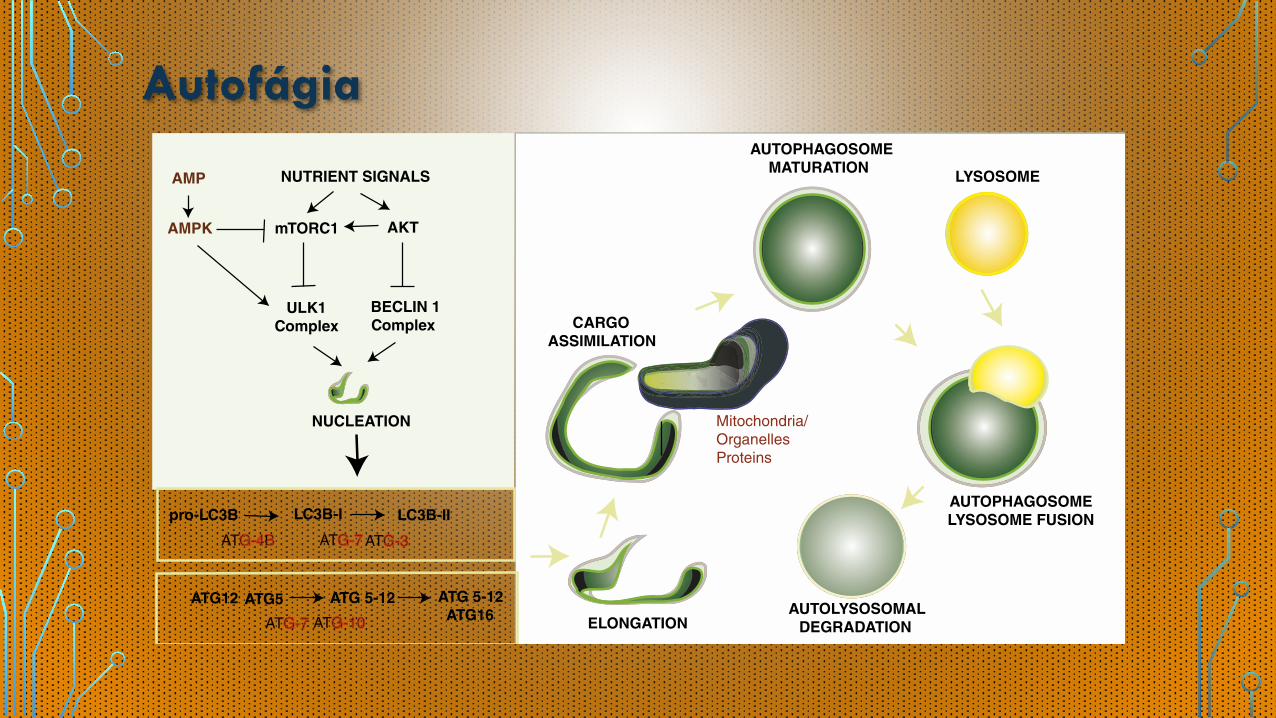
Figure 1. Autophagy pathway. Autophagy is a regulated process that responds to regulation by nutrient signals. Autophagy responds to negativeregulation by growth factor stimuli that activate the phosphatidylinositol-3-kinase (PI3K/AKT) pathway, which up-regulates the mTOR pathway and down-regulates the Beclin1 complex. Autophagy responds to up-regulation by depletion of cellular energy charge through the activation of the adenosinemonophosphate (AMP)-activated protein kinase (AMPK). In response to elevated AMP levels, AMPK can inhibit mTORC1 and directly phosphorylate ULK1,leading to activation of autophagy. The antibiotic rapamycin induces autophagy by inactivating mTORC1. The initiation of autophagosome formation is alsoregulated by the autophagy protein Beclin 1 (Atg6). Beclin 1 associates with a macromolecular complex that includes hVps34, a class IIIphosphatidylinositol-3 kinase. Autophagosome elongation requires two ubiquitin-like conjugation systems, the ATG5-12 conjugation system, and the ATG8(LC3) conjugation system, which are regulated by various ATG proteins. Autophagy protein LC3-II remains associated with the maturing autophagosome.The basic sequence of steps of autophagy include: (1 ) initiation and autophagosomal nucleation (formation of the phagophore); (2 ) elongation of the nascentautophagosomal membrane, to capture a cargo such as mitochondria; (3 ) maturation of the double-membraned autophagosomal structure with cargoassimilation; and (4 ) autophagosome-lysosome fusion, which is concluded by degradation of the autolysosomal contents.
STATE OF THE ART
Mizumura, Cloonan, Choi, et al.: Autophagy in Lung Disease S41

Autofágia
• Tento mechanizmus je súčasťou dôležitých evolúciou uchovávaných schopností adaptácie cicavcov na stres a hladovanie
• Rovnako tento mechanizmus pomáha čeliť mikrobiálnej invázii aktívnou elimináciou intracelulárnych mikróbov, zvýšením antigénovej odpovede, moduláciou imunitnej odpovede a odstraňovaním poškodených organel hostiteľských buniek (napr. mitochondria), za učelom obnovenie homeostázy, najmä v priebehu sepsy
Schmid et al., Immunity 2007

Autofágia a sepsa
• Sepsa je v súčastnosti chrakterizovaná ako stav systémovej hyperinflamácie a niekedy imunosupresie následokom infekcie. Napriek rôznym terapeutickým snahám zostáva signifikantnou príčinou vysokej mortality a morbidity
• Pritom publikované výsledky rozličných “nádejných” a odporučených terapeut ických postupov zostávajú kontroverzné
• Recentné štúdie zamerané na genómovú expresiu u kriticky chorých pacientov, poukazujú na simultánnu upreguláciu pro- i anti-infalmatórnych cytokínových génov
Xiao et al., J Exp Med 2011

Autofágia a sepsa
• Stúpa množstvo dôkazov, k toré naznačujú, že autofágia hrá protektívnu ú lohu p r i k r t i c ký ch stavoch a sepse - smer t e s t o v a n i a n o v ý c h terapeutických agens cielene modulujúcich atofágiu
FIGURE 2 | The intracellular signals of autophagy induction in immune cells. (A) Autophagy can be induced by bacteria, bacterial toxins such as LPS, and pro-inflammatory cytokines through TLR2 and TLR4. The activation of TLR4 and TLR2 initiate JNK, p38 MAPK, and ERK signaling in Myd88 and TRIF dependent ways, which further induce autophagy by inhibiting mTORC1 complex. NOD1 and NOD2 receptors, vital intracellular pattern recognition receptors, promote the formation of autophagosomes by enhancing the combination between ATG16L1 and invaded bacteria. HMGB1 induces autophagy through releasing Beclin-1 from Bcl2 after binding with Beclin-1. (B) Four major steps are required for autophagic process, including nucleation, elongation, closure, and fusion with lysosome for degradation, which are tightly regulated by autophagy associated proteins. (C) The function of autophagy associated proteins on immune system involves both canonical and non-canonical dependent ways. ATG16L1, autophagy-related 16-like 1 gene; Bcl2, B-cell lymphoma 2; ERK, extracellular signal-regulated kinase; HMGB1, high-mobility group box-1 protein; IL, interleukin; IRAK, interleukin-1 receptor-associated kinase; JNK, c-Jun N-terminal kinase; LAMP, lysosomal-associated membrane protein; LPS, lipopolysaccharide; MHC, major histocompatibility complex; mLST8, mammalian lethal with SEC13 protein; mTOR, mammalian target of rapamycin; mTORC1, mTOR complex 1; Myd88, myeloid differentiation factor 88; NOD, nucleotide-binding oligomerization domain-containing protein; PELI3, pellino E3 ubiquitin protein ligase family member 3; p38 MAPK, p38 mitogen-activated protein kinase; RIP, receptor interacting protein; ROS, reactive oxygen species; TNF, tumor necrosis factor; TRAF, TNF receptor-associated factor; TRIF, TIR-domain-containing adapter-inducing interferon-β.
4
Ren et al. Modulating Autophagy for Sepsis-Induced Immunosuppression
Frontiers in Immunology | www.frontiersin.org December 2017 | Volume 8 | Article 1832
neutrophil chemotaxis under sepsis exposure (45). Inappropriate infiltration of neutrophils resulted in the remote organ dysfunc-tion in sepsis, which was further identified as C-C chemokine receptor type 2 (CCR2) dependent (46). Targeting CCR2 with specific antagonist or gene blockade showed protective effects on lungs, heart, and kidneys and improved survival rate of septic animals by lowering infiltration of neutrophils (46).
Autophagic machinery has been documented to enhance the bactericidal activity of neutrophils from immunocompromised
patients, while it can be reversed by autophagy inhibition (47, 48). Both intracellular and extracellular mechanisms are involved in this effect, the former is characterized by killing invaded intracel-lular bacteria, while O2
− releases and NETs formation are major factors for the later, which show great benefits for septic cases (48, 49). Further studies suggested that phosphatase and tensin homolog on chromosome ten, a dual phosphatase that activated autophagic machinery by antagonizing Akt/mammalian target of rapamycin (mTOR) pathway, increased NETs formation upon
Sepsou indukovaná autofágia
•Autofágia je idukovaná po septickom inzulte väzbou špecifickej mikrobiálnej štruktúry rozoznanej tool-like receptorom
• To vedie k aktivácii rôznych intracelulárnych procesov zvyšujúcich autofágovú akt ivi tu buniek - väzba lipopolysacharidov z G- baktérii na TLR4
• Inými vplyvmi indukujúcimi autofágiu sú akcentácia endoplazmatického retikula a prítomnosť poškodených mitochondrií
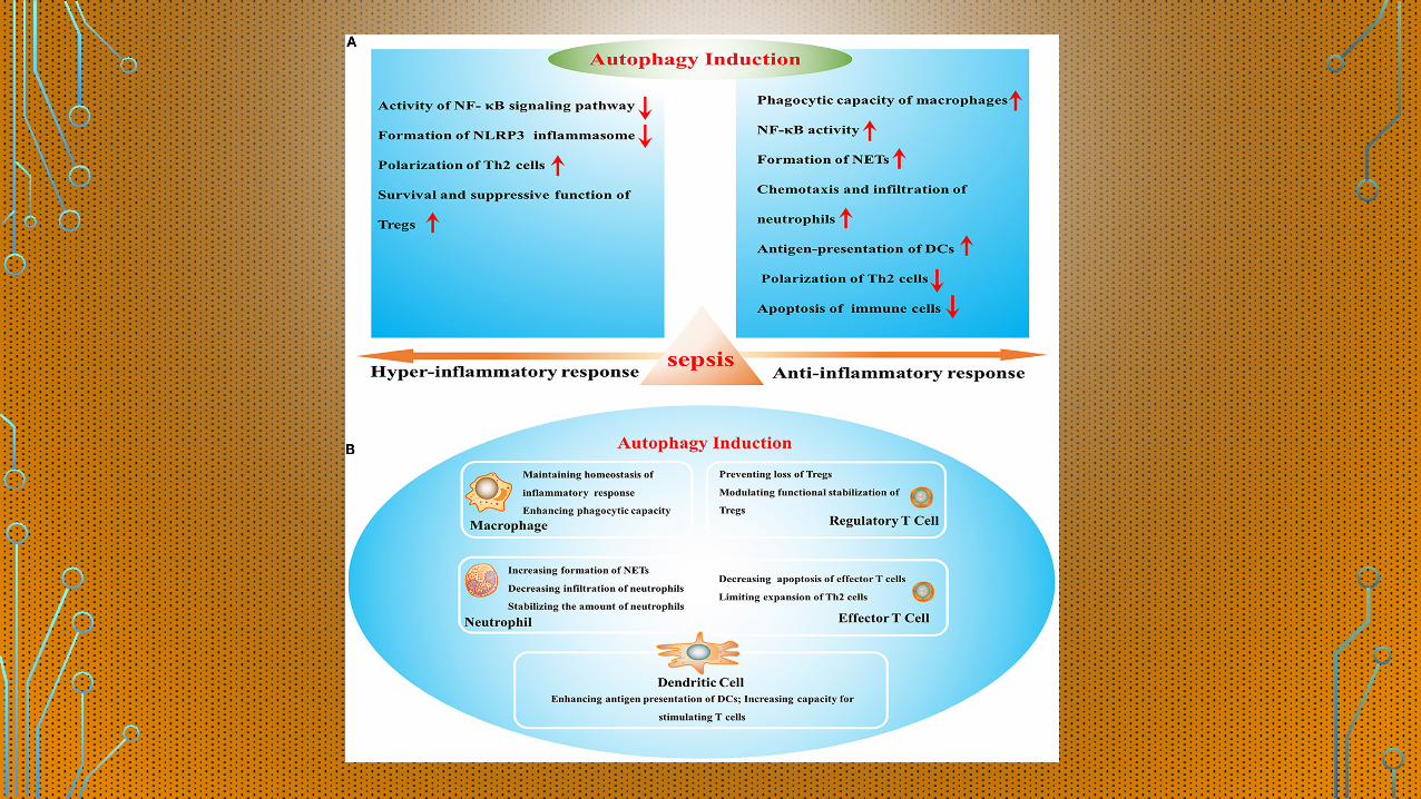
FIGURE 1 | The effects of autophagy induction on sepsis-induced immune dysfunction. (A) Effects of autophagy in various stages of sepsis. (B) The role of autophagy induction in the function of multiple immune cells. NF-κB, nuclear factor kappa-light-chain-enhancer of activated B cells; NLRP3, Nod-like receptor family pyrin domain-containing 3; Tregs, regulatory T cells; NETs, neutrophil extracellular traps; DCs, dendritic cells.
2
Ren et al. Modulating Autophagy for Sepsis-Induced Immunosuppression
Frontiers in Immunology | www.frontiersin.org December 2017 | Volume 8 | Article 1832
Autophagy is an important self-protective mechanism for cellular survival by controlling degradation of proteins and organelles, which involves the formation of double-membrane autophagosome and the proteolytic degradation after delivered to lysosome. Studies have shown that autophagy is mobilized early in sepsis and seen in various organs, manifested by increased accumulation of autophagic vacuoles and enhanced expression of autophagy-associated proteins (11). For example, microtubule-associated protein light chain 3 (LC3), one of key molecules for autophagy, showed enhanced expression at 6 h after cecal ligation and puncture (CLP) and released a cascade of benefits by eliminating invaded pathogens, avoiding over-production of stress proteins and modulating the function of multiple organelles (12). SQSTM1/p62, another specific marker for dynamic autophagic process that is also termed autophagic
flux, revealed rapid alteration after sepsis initiation and further contributed to functional stability of various cells (11). However, a host in recent studies found disturbed autophagic process in later stage of sepsis, which was considered as a major cause for sepsis-induced immune suppression (10). In this review, we will provide a detailed overview of the effects of autophagy on immune response and further therapeutic significance in sepsis.
THE PROTECTIVE ROLE OF AUTOPHAGY IN SEPSIS
Autophagy initiates early after the onset of sepsis and is induced by some kinds of bacteria, bacterial toxins such as lipopolysac-charide (LPS), and pro-inflammatory cytokines (13–15). The

Sepsou indukovaná autofágia
• Iniciácia autofágie idukovaná stretom s mikróbom je význomným typom vrodenej imunitnej odpovede podobne ako proteíny, ktoré túto odpoveď modulujú
•Na základe animálnych štúdii sa zistila orgánovo špecifická diferencia v aktivácii autofágie v jednotlivých vitálne dôležitých orgánoch
•Najvyššia úroveň aktivácie sa pozorovala v bunkách pečene, srdca a sleziny - hepatocyty a kardiomyocyty sa zdajú byť predominantými typmi buniek pre sepsou indukovanú autofágiu

Kinetika autofágie v sepse
• Sepsa je charakterizovaná iniciálne nadmernou produkciou pro-inflamatórnych cytokínov s následnou imunosupresiou
•Na základe animálnych štúdii sa hromadia dôkazy o tom, že autofágová aktivita sa zvyšuje v hyperdynamickej fáze sepsy nasledovaná jej poklesom
should focus on the mechanisms that may prevent this process.The use of molecular tools to determine lysosomal function inaddition to demonstrating their colocalization may be useful.
Lung
Respiratory failure usually occurs at the very early stage of sep-sis. Consistent with this, the expression of autophagy-relatedproteins (e.g. LC3-II, ATG2, RAB7) declines very early (at 4 h)and this continues up to 24 h. This decline is associated withincreased expression of pro-apoptotic proteins, such as FADD(Fas [TNFRSF6]-associated via death domain), BAX andcleaved CASP3 (caspase 3). Experimental induction of auto-phagy by rapamycin and activated PROC (protein C) mitigates
apoptosis and pro-inflammatory cytokines.52 In addition, miceoverexpressing LC3 ablate apoptosis, inflammation, and neu-trophil infiltration.53 Furthermore, the protective effect ofGAPDH (glyceraldehyde-3-phosphate dehydrogenase) againstsepsis-related lung injury is mediated by ATG12-dependentautophagy enhancement.54,55 Deletion of Atg4b in LPS-chal-lenged mice shows more prominent signs of pulmonary inflam-mation via modulation of ATF3 (activating transcriptionfactor 3).56
Kidney
Upon LPS challenge, young mice demonstrate a higher LC3-IIlevel than old mice, accompanied with lower CST3 (cystatin C)
Figure 1. Autophagy and its deregulation in sepsis. (A) The pathogen-autophagy interplay in relation to mitochondrial function and inflammatory response in sepsis. (B)Kinetics of autophagy and inflammatory response during sepsis. In addition to a hypoinflammatory response in protracted sepsis, the delayed autophagic depressionmay further contribute to mortality and morbidity by reduced microbial clearance, failure to sustain exotoxin tolerance and limited major histocompatibility complex II-mediated antigen presentation. IL, interleukin; LPS, lipopolysaccharide; LTA, lipoteichoic acid; mtDNA, mitochondrial DNA; MYD88, myeloid differentiation primaryresponse gene 88; NFKB, nuclear factor of kappa light polypeptide gene enhancer in B cells; NOD2, nucleotide-binding oligomerization domain containing 2; NLRP3, NLRfamily, pyrin domain containing 3; PGN, peptidoglycan; ROS, reactive oxygen species; SLR, sequestosome-like receptor; ssRNA, single-stranded ribonucleic acid; TNF,tumor necrosis factor; TLR, toll-like receptor;Cm, membrane potential.
1076 J. HO ET AL.
• Zvýšená aktivita hodnotená množstvom autofagozómov počas iniciálnych 4 - 6 hodín s poklesom od 8 hodiny a ďalších 24 hodín

Kinetika autofágie v sepse
• Práve tento postupný pokles aktivity autofágie môže byť oveľa dôležitejší ako akútna fáza pri determinovaní klinického výsledku pri sepse - chýbajú údaje
• Práve spomalenie autofágovej aktivity môže prispieť k apoptóze leukocytov a následne k zníženiu ich odpovede na mikrobiálne stimuly, či aktiváciu latentných vírusov - odstránenie autofágových génov signifikantne znížilo počet cirkulujúcich T a B lymfocytov
•Útlm autofágie môže viesť k vylúčeniu autofagozómov z bunky, proinflamatórnej reakcii a neschopnosti odstrániť dysfunkčné a poškodené mitochondrie

Autofágia a orgány
Srdce •Ovplyvnenie autofágie môže viesť k dysfunkcii kontraktility a
apoptotickej bunkovej smrti kardiomyocytov
• Zároveň je jedným z dôležitých faktorov sepsy vzostup NOS2 aktivity, ktorá inhibuje atofágiu a zároveň priamym účinkom vedie k atrofii buniek myokardu a redukcii kotraktility
•Koinhibícia autofágie spoločne s reaktívnymi kyslíkovými radikálmi indukuje kontraktilnú dysfunkciu

Autofágia a orgány
Imunitný systém •Náhla nadprodukcia pro-inflamatórnych cytokínov vedie k
edému tkanív a početnému orgánovému poškodeniu
• Interaguje s týmito sepsou navodeninými mechanizmami - redukuje produkciu niektorých so zápalom asociovaných interleukínov (NLRP3/NALP3), čím chráni mitochondrie pred poškodením
• Predpokladá sa, že jej ochranný vplyv môže byť nezávislý od cytokínov, samotným zapojením do regulácie aktivácie zápalu, uvoľnovania cytokínov ako aj iných zápalových látok

Autofágia a orgány
Pečeň •Aktivácia autofágie v hepatocytoch počas sepsy odstraňuje
poškodené mitochondrie a pôsobí tak preventívne voči tvorbe mitochondriálnych ROS a iniciácii poškodenou mitochondriou spusteného procesu apoptózy
•Vyšetrením post-motrem vzoriek pečene septických pacientov bolo dokázané významné nahromadenie autofágových vakuol

Autofágia a orgány
Pľúca •Respiračné zlyhanie sa obvykle objavuje vo veľmi skorých
fázach sepsy
•Zhodne s tým, expresia s autofágiou spojených proteínov klesá veľmi skoro (za 4 hodiny) a pokračuje kontinuálne do 24 hodín
• Tento pokles je spojený so zvýšením expresie pro-apoptoických prioteínov
• Experimentánla indukcia autofágie zmiernila apoptózu a produkciu pro-inflamatórnych cytokínov v pľúcach

Autofágia a orgányObličky • Zníženie autofágie súčasne zvýšilo apoptózu renálnych
tubulárnych buniek
Svalstvo • Počas sepsy sa v svalových bunkách pravidelne vyskytuje
poškodenie mitochondrií
• Experimentálne bol dokázaný negatívny vplyv endotoxémie na autofágiu skeletálneho svalstva
•Avšak, strata svalovej hmoty u kriticky chorých pacientov, spájaná s autofágiou vyžaduje ďalší výskum
Autofágia a orgány
Mozog • Encefalopatia je spojená so zvýšeným rizikom mortality a
morbidity pri sepse
• Je charakterizovaná poškodením hemato-encefalickej bariéry, zvýšením úrovne oxidatívneho stresu a zvýšením apoptózy
•V experimente bol dokázaný vzostup autofagozómových formácii a lyzozómovej aktivity v hipokampe pri navodenom septickom šoku

Mitofágie v sepse
•V experimente bolo dokázané poškodenie membrán mitochondrií spôsobené nárastom peroxidových radikálov a vzostup apoptózy
•Mitofágia hrá dôležitú úlohu pri eliminovaní poškodených mitochondrií z cytozólu, čím chráni hostiteľa proti oxidatívnemu poškodeniu rovnako ako pred zápalovou odpoveďou
•V prospektívnej štúdii mali pacienti s postraumatickou sepsou vyšie hladiny mitochondriálnej DNA v plazme, v porovnaní s pacientami bez septickej komplikácie

Škodlivosť supresie autofágie v sepse
• Bolo dokázané, že po krátkej transientnej iniciálnej aktivácii autofágie pri septickom inzulte dochádza k jej prolongovanej deklinácii, vplývajúcej na orgánovú dysfunkciu
•Možné mechanizmy môžu zahŕňať hyperaktivitu počas iniciálneho štádia stretu s mikrobiálnym patogénom, vedúcu k náhlemu uvoľneniu esenciálnych autofágových prekurzorov - overexpresia LC3 môže úspešne obnoviť autofágovú insuficienciu v spetických pľúcach
• Supresia procesu odstraňovania baktérií, toxínov, udržiavania mitochondriálnej integrity a kontroly uvoľnovania cytokínov by mohla byť práve príčinou zlého outcome pri sepse

Klinické implikácie
•Autofágia hrá protektívnu úlohu najmä ochranou integrity mitochondrie, prevenciou apoptózy a zvýšením antigénovej prezentácie
• Redukcia autofágovej aktivity je spojená s orgánovým poškodením
• To naznačuje, že modulácia neskorej aktivity autofágie počas sepsy môže byť sľubnou terapeutickou stratégiou
• Samozrejme, čaká nás ešte dlhá cesta vo vývoji látok, ktoré dokážu vhodne a v správny moment ovplyvniť autofágiu u kriticky chorého pacienta

Klinické implikácie
•Autofágia je historicky známy proces ako fyziologická odpoveď na hladovanie a nedostatočný prívod živín, kedy si bunka prostredníctvom lyzozómov recyklovala esenciálne substráty pre svoj metabolizmus
• Známe sú práce o pozitívnom vplyve stratégie permisívneho “underfeedingu” na outcome kriticky chorých pacientov
• To naznačuje, že udržiavanie stavu mierneho hladovania na udržanie autofágovej aktivity može redukovať morbiditu pri sepse

ĎAKUJEM VÁM ZA POZORNOSŤ