autoimmune disorders in the hands - pdh therapy · phsical therapists autoimmune disorders in the...
TRANSCRIPT
PHYSICAL THERAPISTS Autoimmune Disorders In The Hands | 1
AUTOIMMUNE DISORDERS IN THE HANDS: DIAGNOSIS, TREATMENT, AND THERAPEUTIC INTERVENTIONS
PDH Academy Course #PT-1702 | 3 CE HOURS
CONTINUING EDUCATIONfor Physical Therapists
Course AbstractThis course examines scleroderma, Raynaud’s phenomenon, and rheumatoid arthritis, three common autoimmune disorders that impact the hands. It discusses applicable definitions and terminology; their etiology, prevalence, diagnosis, and medical interventions; the normal joint anatomy and functional position of the hand; and the role of therapy as it pertains to their treatment.
Target audience: Physical Therapists, Physical Therapist Assistants, Occupational Therapists, Occupational Therapy Assistants (no prerequisites).
NOTE: Links provided within the course material are for informational purposes only. No endorsement of processes or products is intended or implied.
ApprovalsTo view the states that approve and accept our courses, visit https://pdhtherapy.com/physical-therapy/state-requirements/
Learning ObjectivesBy the end of this course, learners will be able to:
❏ Recognize definitions and terminology pertaining to autoimmune disorders in the hand
❏ Differentiate between the etiology and prevalence of scleroderma, Raynaud’s phenomenon, and rheumatoid arthritis
❏ Identify elements of medical diagnosis and treatment of autoimmune disorders in the hand
❏ Recognize the normal joint anatomy and functional position of the hand
❏ Identify roles of therapy as it pertains to autoimmune disorders in the hand
| Autoimmune Disorders In The Hands PHYSICAL THERAPISTS2
IntroductionMany patients present to our clinics with primary or secondary diagnoses of autoimmune disease, specifically Rheumatoid Arthritis, Scleroderma, and Raynaud’s phenomenon. Understanding these autoimmune disorders is paramount in providing good rehabilitative care to our patients who are afflicted with them. While these diseases can affect many organs in the body, we will focus on their impact on hand pain and hand function, and how we as therapists can most effectively intervene.
Physicians rely on occupational and physical therapists to educate their patients as to adaptive equipment needs, splinting, exercises, and appropriate modality use to reduce pain and increase function. This course provides an in-depth discussion of these concepts, as well as a thorough review of the disease processes for the treating therapist.
PLEASE NOTE: it is not within the scope of occupational therapy to prescribe. The information presented in this course is not intended nor is it implied to be a substitute for professional medical advice.
Amy’s note: My primary goal for readers of this course is to help them keep up with the latest research while learning practical information that they can use immediately in a clinic setting. In addition, occasionally I will toss in more personal musings, indicated by italicized writing. Text in italicized print is based in large part on my opinions, generated by 21 years of practical experience as a hand therapist – not solely science and research. This is what I like to call “evidence-based practice with a side dish of clinical experience (or old age).” For as we all know, and Albert Einstein so aptly stated, “In theory, theory and practice are the same. In practice, they are not.”
Timed Topic Outline I. Introduction, Definitions and Terminology (5 minutes) II. Autoimmune Disease Overview (10 minutes) III. Scleroderma (15 minutes) IV. Raynaud’s Phenomenon vs. Raynaud’s Disease (15 minutes) V. Rheumatoid Arthritis (15 minutes) VI. Anatomical Considerations for Autoimmune Disease in the Hands
(5 minutes) VII. The Role of Therapy (100 minutes)
History, Evaluation and Provocative Testing, Treatment VIII. Conclusion, Additional Resources, References, and Exam (15 minutes)
Delivery MethodCorrespondence/internet self-study with a provider-graded multiple choice final exam. To earn continuing education credit for this course, you must achieve a passing score of 80% on the final exam.
CancellationAs PDH Academy offers self-study courses only, provider cancellations due to inclement weather, instructor no-shows, and/or insufficient enrollment are not concerns. In the unlikely event that a self-study course is temporarily unavailable, already-enrolled participants will be notified by email. A notification will also be posted on the relevant pages of our website.
Customers who cancel orders within five business days of the order date receive a full refund. Cancellations can be made by phone at (888)564-9098 or email at [email protected].
Accessibility and/or Special Needs Concerns?Contact Customer Service by phone at (888)564-9098 or email at [email protected].
Course Author Bio & DisclosureAmy L. Paulson, OTR, CHT is a certified hand therapist with over 19 years of experience in outpatient upper extremity care. She has been a licensed occupational therapist for 20 years, has experience in home health, skilled nursing, and acute care, and is a member of the American Society of Hand Therapists.
Amy has led multiple community-based classes on arthritis care, energy conservation and work simplification, and she taught as an adjunct professor at Palm Beach Community College in the OTA program. She designs and instructs hands-on continuing education courses in splinting and upper extremity treatment, and enjoys providing clinical instruction to students interested in specializing in hand therapy. She is also involved in coordinating and overseeing therapy programs in Gulu, Uganda by volunteering with the Medical Missions Foundation of Overland Park, KS.
Amy currently owns and operates her private practice in outpatient hands, where she provides patient care, clinical instruction to Level II students, community education, marketing, and insurance billing.
DISCLOSURES: Financial – Amy Paulson received a stipend as the author of this course. Nonfinancial – No relevant nonfinancial relationship exists.
PHYSICAL THERAPISTS Autoimmune Disorders In The Hands | 3
Definitions and TerminologyCrucial components of treating autoimmune diseases effectively include understanding the disease processes, potential deformities, and the effects on a person’s activities of daily living. A conscientious therapist will not only understand these components and provide hands-on care, but will also be able to educate their patients on all three of these areas in a comprehensible way.
Acquired immune system: The part of the immune system that develops as a person grows in response to exposure to bacteria and toxins. The body develops antibodies and immune cells to fight harmful substances.
Antibodies: Special proteins produced by the body’s immune system that recognizes and helps fight infectious agents such as viruses and bacteria and other foreign substances such as chemicals and toxins that invade the body. The presence of certain antibodies in the blood can help to diagnose some diseases, including some forms of scleroderma.
Antigen: A foreign substance that triggers the production of antibodies when it is introduced into the body.
Biologics: Injection medications used to decrease the inflammation caused in the joints by rheumatoid arthritis. The most common biologics used are abatacept (Orencia), rituximab (Rituxan), tocilizumab (Actemra), anakinra (Kineret), adalimumab (Humira), etanercept (Enbrel), infliximab (Remicade), and certolizumab pegol (Cimzia).
Calcinosis: The formation of calcium deposits in the connective tissues, which can be detected by x ray. These deposits are typically found on the fingers, hands, face, and trunk and on the skin above elbows and knees. When the deposits break through the skin, painful ulcers can result.
CMC joint: Carpometacarpal joint of the thumb.
Collagen: A fabric-like material of fibrous threads that is a key component of the body’s connective tissues. In scleroderma, either too much collagen is produced or it is produced in the wrong places, causing stiff and inflamed skin, blood vessels, and internal organs.
Connective tissue: Tissues such as skin, tendons, and cartilage that support and hold body parts together. The chief component of connective tissue is collagen.
Connective tissue disease: Autoimmune diseases that affect the connective tissue include scleroderma, lupus, rheumatoid arthritis and Sjogren’s syndrome.
Corticosteroids: Anti-inflammatory hormones that are made naturally in the body or synthetically for use as drugs. They are also called glucocorticoids. Prednisone is the most commonly prescribed corticosteroid for
autoimmune disorders.
DIP joint: Distal interphalangeal joint, or the smallest joint of the finger.
Disease-modifying antirheumatic drugs (DMARDs): A class of medications used in the treatment of rheumatoid arthritis. DMARDs can ease the symptoms of rheumatoid arthritis and can sometimes slow or stop the course of the disease to help prevent joint damage. Some examples of commonly used DMARDs are hydroxychloroquine (Plaquenil), leflunomide (Arava), methotrexate (Trexall), sulfasalazine (Azulfidine), and minocycline (Minocin).
Fibroblast: A type of cell in connective tissue that secretes proteins, including collagen.
Fibrosis: A condition marked by increased fibrous tissue that develops between the cells of various organs or tissues. It is a common feature of scleroderma and some other diseases. Fibrosis causes hardening or stiffening of tissues in the skin, joints, and internal organs.
Flare: A period of heightened disease activity. In rheumatoid arthritis, a flare may be characterized by increased fatigue; fever; and painful, swollen, and tender joints.
Immune system: A complex network of specialized cells and organs that work together to defend the body against attacks by foreign invaders, such as bacteria and viruses.
Inflammation: A reaction of body tissues to injury or disease, typically marked by five signs: swelling, redness, heat, pain, and loss of function.
MCP joint: The metacarpophalangeal joint of the hand, also known as the MP joint.
Mixed connective tissue disorder: Diagnosis used for an autoimmune disorder when symptoms of lupus, systemic sclerosis, and polymyositis overlap.
Nonsteroidal anti-inflammatory drugs (NSAIDs): A class of medications available over the counter or with a prescription that ease pain and inflammation. The most frequently used NSAIDs are ibuprofen (Motrin, Advil) and naproxen (Aleve). Stronger NSAIDs are available through physicians (Mobic, Celebrex); however, many of them have side effects that discourage long-term use.
PIP joint: Proximal interphalangeal joint.
Psoriatic arthritis: A type of arthritis associated with psoriasis, a chronic skin disease that occurs when cells in the outer layer of the skin reproduce faster than normal.
Raynaud’s disease: Primary Raynaud’s disease is a condition in which the small blood vessels of the hands or feet contract in response to cold or anxiety.

| Autoimmune Disorders In The Hands PHYSICAL THERAPISTS4
As the vessels contract, the hands or feet turn white and cold, then blue. As blood flow returns, they become red. Fingertip tissues may suffer damage, leading to ulcers, scars, or gangrene. This is NOT due to an autoimmune disorder.
Raynaud’s phenomenon: Also called secondary Raynaud’s, this is a condition associated with scleroderma in which the small blood vessels of the hands or feet contract in response to cold or anxiety. As the vessels contract, the hands or feet turn white and cold, then blue. As blood flow returns, they become red. Fingertip tissues may suffer damage, leading to ulcers, scars, or gangrene.
Rheumatoid arthritis: A form of arthritis in which the immune system attacks the tissues of the joints, leading to pain, inflammation, and eventually joint damage and malformation. It causes swelling and redness in joints, and may make people feel sick, tired, and feverish. Rheumatoid arthritis may also affect skin tissue, the lungs, the eyes, or the blood vessels.
Rheumatoid factor (RF): An antibody that is present eventually in the blood of most people with rheumatoid arthritis. Not all people with rheumatoid arthritis test positive for rheumatoid factor, and some people test positive for rheumatoid factor, yet never develop the disease. Rheumatoid factor also can be positive in some other diseases.
Scleroderma/systemic sclerosis: An autoimmune disease characterized by abnormal growth of connective tissue in the skin and blood vessels. In more severe forms, connective tissue can build up in the kidneys, lungs, heart, and gastrointestinal tract, leading in some cases to organ failure.
Sclerodactyly: Thick and tight skin on the fingers, resulting from deposits of excess collagen within skin layers. The condition makes it harder to bend or straighten the fingers. The skin may also appear shiny and darkened, with hair loss.
Systemic condition: A condition involving the body as a whole, as opposed to limited conditions that affect particular parts of the body.
Synovial fluid: The viscous fluid that fills a synovial joint and acts as a shock absorber and reduces friction between the articular surfaces of the joint. It is produced by the synovial lining of the joint.
Synovial joint: Also known as a diarthrosis, a synovial joint is any joint in the body where two bones meet, the ends are covered in cartilage, the joint is connected by a fibrous capsule, and the area is filled with synovial fluid for ease in movement. Some amount of movement is possible at all synovial joints.
Synovial membrane/Synovium: The inside lining of the fibrous capsule of a synovial joint. This combines with the fibrous capsule to form the articular capsule. The synovial membrane produces synovial fluid. In
rheumatoid arthritis, the synovium is attacked by the immune system.
T-cells: Lymphocytes that develop in the thymus that fight infected or malignant cells. There are two types of t-cells. “Killer t-cells” attack infected cells, and “suppressor t-cells” inactivate the killer t-cells to return the immune system back to normal. An elevated number of t-cells found in the blood suggest that the body is fighting infection or malignancy.
Autoimmune Disease OverviewThe immune system is a complex network of cells and organs that defend our bodies against germs (viruses and bacteria) and other foreign invaders (toxins and drugs). The immune system’s function is to tell the body the difference between its own cells and foreign matter. When the immune system is not functioning properly, the body produces autoantibodies that attack normally functioning cells by mistake, and the regulatory T-cells that are supposed to fight foreign matter fail to engage, which leads to autoimmune disease.
There are two components to the immune system. The first component is the innate immune system that activates white blood cells to destroy invaders without using any antibodies. We are born with this system in place. The second is the acquired immune system which develops as a person grows and becomes exposed to foreign matter. Together, they allow the working immune system to remember invaders so that it can build up antibodies that will recognize and attach to the invaders if they return. This is the basis of inoculations – exposing the body to a very small amount of a toxin/bacteria/virus in order for the body to build up defense for the future.
There are two classifications of autoimmune disorders – organ-specific disorders and non-organ-specific types. Some examples of organ-specific disorders include Hashimoto’s thyroiditis (thyroid gland), pernicious anemia (stomach), Addison’s disease (adrenal glands), and type-1 diabetes (pancreas). In these diseases, there is a slow destruction or proliferation of specific tissues or cells, causing the organ to malfunction. In non-organ-specific disorders, the autoimmune response is spread throughout the body, affecting many body parts and systems simultaneously. Examples of these include rheumatoid arthritis, lupus, and systemic sclerosis (or scleroderma).
Etiology
Lymphocytes are one of the two main types of immune cells available to our bodies to combat infection: they produce antibodies to fight foreign invaders, and are programmed to recognize, remember, and respond to antigens. All human beings have some lymphocytes that are capable of reacting against the

PHYSICAL THERAPISTS Autoimmune Disorders In The Hands | 5
body; however, in most of us, those lymphocytes are suppressed and do not result in disease. For reasons we don’t quite understand fully, in some individuals there is an interruption in the control process over these lymphocytes, and they attack the body systems. It appears that some people have a genetic predisposition to develop autoimmune responses to invading matter.
Occurrence
According to the American Autoimmune Related Diseases Association (AARDA), over 50 million Americans are affected by autoimmune disease. This is approximately 20% of the population. Women are more likely than men to be affected: almost three to one, according to the statistics provided by the AARDA. While there is a strong genetic component to developing an autoimmune disease, this is not the only factor involved. A person who is genetically predispositioned to an autoimmune disease must have other factors present (most commonly a virus, bacteria, toxin, or drug) in order to initiate the disease process.
Pathology
There are over 80 known types of autoimmune disease. They can affect almost any area of the body, including the heart, brain, nerves, muscles, skin, eyes, joints, lungs, kidneys, glands, the digestive tract, and blood vessels. The classic sign of an autoimmune disease is inflammation, which can cause redness, heat, pain, and swelling in the body part affected.
Medical Diagnostics
Receiving a diagnosis of an autoimmune disorder can oftentimes be a long and arduous process. Extensive bloodwork can identify elevations in white blood cell count (suggesting that the body is fighting an infection or invader of some sort), elevated erythrocyte sedimentation rates and elevated c-reactive protein levels (both suggesting increased inflammation present in the body), and the presence of autoantibodies and/or auto-nuclear antibodies (which attack certain tissues and cells of the body). While the simple presence of one or more of these factors in the blood does not automatically lead to a diagnosis of an autoimmune disorder, when these markers are found in conjunction with certain patient complaints (lethargy, unexplainable pain, discomfort, numbness, etc.), the physician can begin to suspect that an autoimmune response may be the culprit.
Again, our focus is the autoimmune diseases that most drastically affect the hands (rheumatoid arthritis, scleroderma, and Raynaud’s phenomenon). However, please be aware that there are several other autoimmune diseases that can cause changes in the upper extremities. Hemolytic anemia, which attacks and destroys the body’s red blood cells, causes decreased oxygen in the blood which can result in hands feeling cold for unexplained reasons. Multiple sclerosis is a disease process by which the body attacks
the myelin sheath of the nerves, which can cause numbness and tingling to the hands. Type-1 Diabetes, AIDS, Guillain-Barré syndrome, and many other forms of autoimmune disorders can cause peripheral neuropathies, leading to pain, numbness, and tingling to the hands.
Likewise, it is extremely important for the physician to obtain a thorough medical history in order to rule out any acute exposures to dangerous toxins, as toxicity will mimic many of the signs and symptoms of autoimmunity as the healthy body tries to fight the invading toxins.. For example, high quantities of artificial sweeteners such as aspartame (like Equal and Nutrasweet), saccharin (Sweet’N Low), and sucralose (Splenda) in the system can mimic signs and symptoms of Multiple Sclerosis (MS). Although the FDA classifies artificial sweeteners as “generally recognized as safe” to consume, many people who have been tested for MS have instead been found to use excessive quantities of these artificial sweeteners, which can be found in diet sodas, gum, and low-calorie yogurts (Mayo Clinic). The research is sparse in this area, but the link to numbness and tingling in the extremities that mimics MS at least anecdotally is something that a neurologist testing for this disease process should consider.
Amy’s note: I have actually experienced these symptoms myself and am now very vocal against the consistent use of artificial sweeteners. I was having progressive numbness starting on my left big toe that traveled up into my shin, and then I noticed the same thing was happening on my right foot. I also had blurry vision and muscle cramping, which are all clinical complaints seen with patient with Multiple Sclerosis and Guillain-Barre syndrome. I went through a battery of tests with a local neurologist who stated that my results were inconclusive and he couldn’t make any sense of my complaints. I’d read some research by happenstance about artificial sweetener toxicity and immediately gave up my diet soda habit (about 6 a day, I’m ashamed to admit). Within two weeks, I had no bouts of blurry vision, absolutely no muscle cramping, and my numbness was 80% resolved. Since then I’ve researched it quite a bit and found that many people go through unnecessary testing for neurological and autoimmune disorders and have discovered that artificial sweeteners are to blame. Clinical research in this area is hard to find, but it’s food for thought.
Medical Intervention
The treatment depends on the disease, but in most cases one important goal is to reduce inflammation systemically. Sometimes doctors prescribe corticosteroids or immunosuppressive drugs, which will be discussed in more depth when talking individually about scleroderma and rheumatoid arthritis. Patients with autoimmune disease are typically followed long-term by a rheumatologist to manage their medications and also provide consultation when another medical diagnosis arises. These patients also have special considerations when
| Autoimmune Disorders In The Hands PHYSICAL THERAPISTS6
undergoing surgery due to the nature of their disease and the strong medications that they take to manage it; rheumatologists are typically the health care provider to oversee these considerations as well.
SclerodermaScleroderma, also known as systemic sclerosis, is an autoimmunity that attacks the body’s connective tissues, including the skin, blood vessels, muscles, and organs, causing them to harden. It is considered a rheumatic disease because of the inflammation and pain in the muscles, joints, and fibrous tissues that it causes. It’s also considered a connective tissue disease because it affects the skin, tendons, and cartilage.
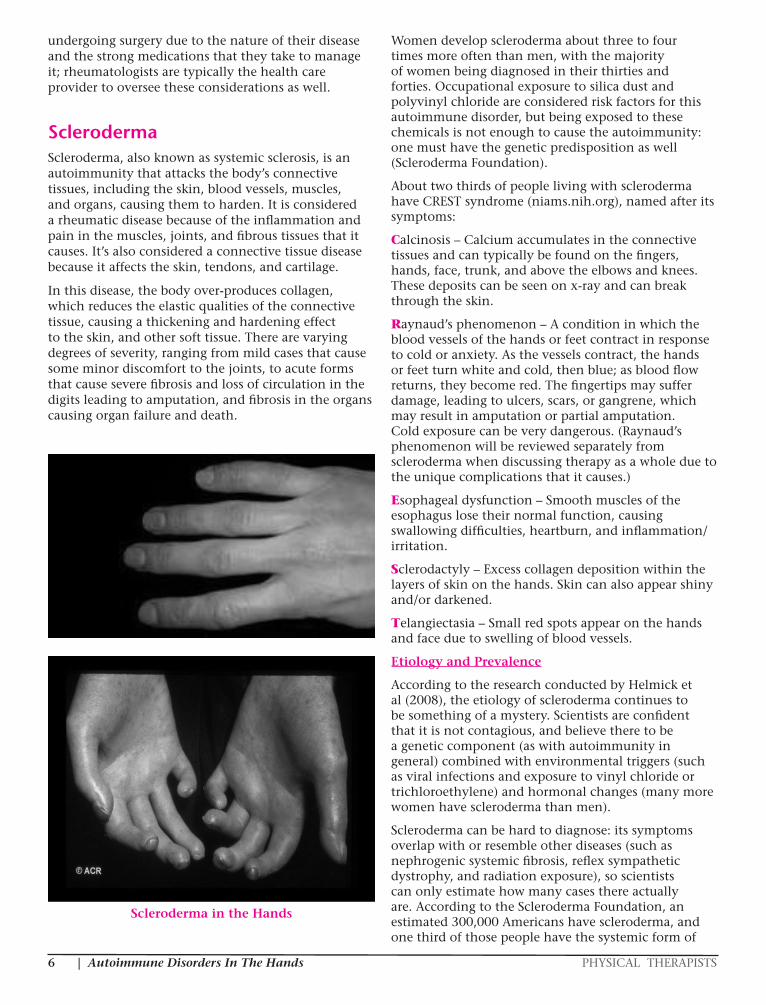
In this disease, the body over-produces collagen, which reduces the elastic qualities of the connective tissue, causing a thickening and hardening effect to the skin, and other soft tissue. There are varying degrees of severity, ranging from mild cases that cause some minor discomfort to the joints, to acute forms that cause severe fibrosis and loss of circulation in the digits leading to amputation, and fibrosis in the organs causing organ failure and death.
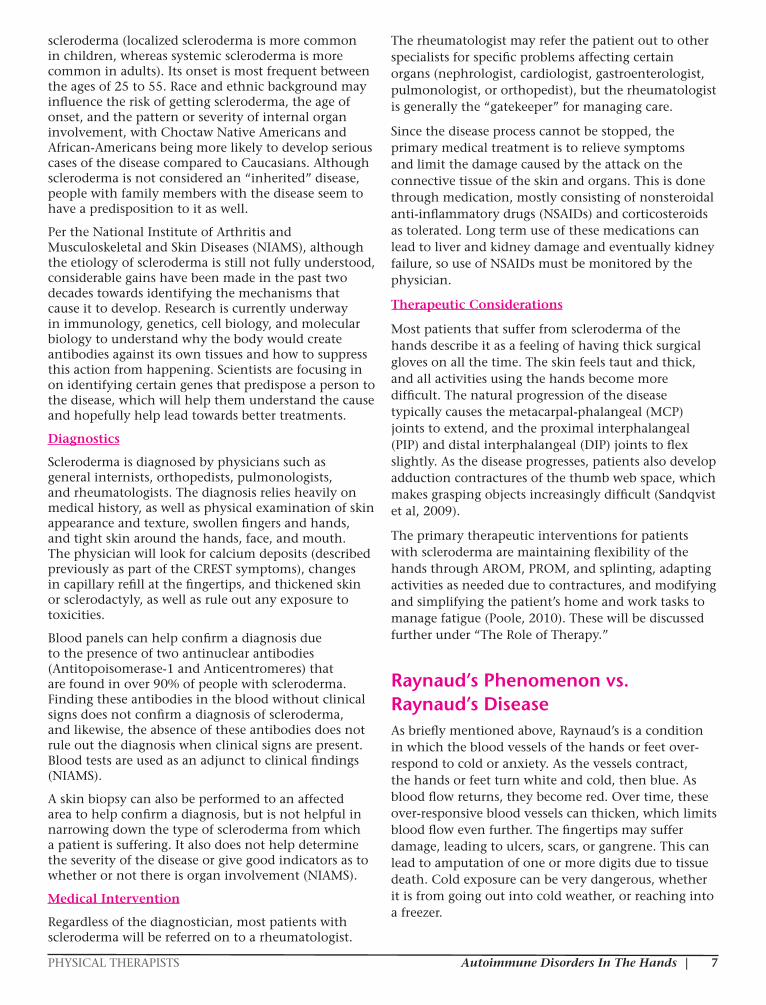
Scleroderma in the Hands
Women develop scleroderma about three to four times more often than men, with the majority of women being diagnosed in their thirties and forties. Occupational exposure to silica dust and polyvinyl chloride are considered risk factors for this autoimmune disorder, but being exposed to these chemicals is not enough to cause the autoimmunity: one must have the genetic predisposition as well (Scleroderma Foundation).
About two thirds of people living with scleroderma have CREST syndrome (niams.nih.org), named after its symptoms:
Calcinosis – Calcium accumulates in the connective tissues and can typically be found on the fingers, hands, face, trunk, and above the elbows and knees. These deposits can be seen on x-ray and can break through the skin.
Raynaud’s phenomenon – A condition in which the blood vessels of the hands or feet contract in response to cold or anxiety. As the vessels contract, the hands or feet turn white and cold, then blue; as blood flow returns, they become red. The fingertips may suffer damage, leading to ulcers, scars, or gangrene, which may result in amputation or partial amputation. Cold exposure can be very dangerous. (Raynaud’s phenomenon will be reviewed separately from scleroderma when discussing therapy as a whole due to the unique complications that it causes.)
Esophageal dysfunction – Smooth muscles of the esophagus lose their normal function, causing swallowing difficulties, heartburn, and inflammation/irritation.
Sclerodactyly – Excess collagen deposition within the layers of skin on the hands. Skin can also appear shiny and/or darkened.
Telangiectasia – Small red spots appear on the hands and face due to swelling of blood vessels.
Etiology and Prevalence
According to the research conducted by Helmick et al (2008), the etiology of scleroderma continues to be something of a mystery. Scientists are confident that it is not contagious, and believe there to be a genetic component (as with autoimmunity in general) combined with environmental triggers (such as viral infections and exposure to vinyl chloride or trichloroethylene) and hormonal changes (many more women have scleroderma than men).
Scleroderma can be hard to diagnose: its symptoms overlap with or resemble other diseases (such as nephrogenic systemic fibrosis, reflex sympathetic dystrophy, and radiation exposure), so scientists can only estimate how many cases there actually are. According to the Scleroderma Foundation, an estimated 300,000 Americans have scleroderma, and one third of those people have the systemic form of
PHYSICAL THERAPISTS Autoimmune Disorders In The Hands | 7
scleroderma (localized scleroderma is more common in children, whereas systemic scleroderma is more common in adults). Its onset is most frequent between the ages of 25 to 55. Race and ethnic background may influence the risk of getting scleroderma, the age of onset, and the pattern or severity of internal organ involvement, with Choctaw Native Americans and African-Americans being more likely to develop serious cases of the disease compared to Caucasians. Although scleroderma is not considered an “inherited” disease, people with family members with the disease seem to have a predisposition to it as well.
Per the National Institute of Arthritis and Musculoskeletal and Skin Diseases (NIAMS), although the etiology of scleroderma is still not fully understood, considerable gains have been made in the past two decades towards identifying the mechanisms that cause it to develop. Research is currently underway in immunology, genetics, cell biology, and molecular biology to understand why the body would create antibodies against its own tissues and how to suppress this action from happening. Scientists are focusing in on identifying certain genes that predispose a person to the disease, which will help them understand the cause and hopefully help lead towards better treatments.
Diagnostics
Scleroderma is diagnosed by physicians such as general internists, orthopedists, pulmonologists, and rheumatologists. The diagnosis relies heavily on medical history, as well as physical examination of skin appearance and texture, swollen fingers and hands, and tight skin around the hands, face, and mouth. The physician will look for calcium deposits (described previously as part of the CREST symptoms), changes in capillary refill at the fingertips, and thickened skin or sclerodactyly, as well as rule out any exposure to toxicities.
Blood panels can help confirm a diagnosis due to the presence of two antinuclear antibodies (Antitopoisomerase-1 and Anticentromeres) that are found in over 90% of people with scleroderma. Finding these antibodies in the blood without clinical signs does not confirm a diagnosis of scleroderma, and likewise, the absence of these antibodies does not rule out the diagnosis when clinical signs are present. Blood tests are used as an adjunct to clinical findings (NIAMS).
A skin biopsy can also be performed to an affected area to help confirm a diagnosis, but is not helpful in narrowing down the type of scleroderma from which a patient is suffering. It also does not help determine the severity of the disease or give good indicators as to whether or not there is organ involvement (NIAMS).
Medical Intervention
Regardless of the diagnostician, most patients with scleroderma will be referred on to a rheumatologist.
The rheumatologist may refer the patient out to other specialists for specific problems affecting certain organs (nephrologist, cardiologist, gastroenterologist, pulmonologist, or orthopedist), but the rheumatologist is generally the “gatekeeper” for managing care.
Since the disease process cannot be stopped, the primary medical treatment is to relieve symptoms and limit the damage caused by the attack on the connective tissue of the skin and organs. This is done through medication, mostly consisting of nonsteroidal anti-inflammatory drugs (NSAIDs) and corticosteroids as tolerated. Long term use of these medications can lead to liver and kidney damage and eventually kidney failure, so use of NSAIDs must be monitored by the physician.
Therapeutic Considerations
Most patients that suffer from scleroderma of the hands describe it as a feeling of having thick surgical gloves on all the time. The skin feels taut and thick, and all activities using the hands become more difficult. The natural progression of the disease typically causes the metacarpal-phalangeal (MCP) joints to extend, and the proximal interphalangeal (PIP) and distal interphalangeal (DIP) joints to flex slightly. As the disease progresses, patients also develop adduction contractures of the thumb web space, which makes grasping objects increasingly difficult (Sandqvist et al, 2009).
The primary therapeutic interventions for patients with scleroderma are maintaining flexibility of the hands through AROM, PROM, and splinting, adapting activities as needed due to contractures, and modifying and simplifying the patient’s home and work tasks to manage fatigue (Poole, 2010). These will be discussed further under “The Role of Therapy.”
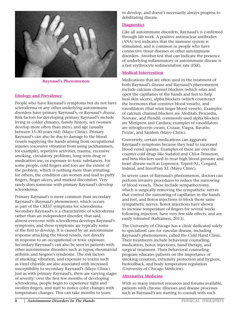
Raynaud’s Phenomenon vs. Raynaud’s DiseaseAs briefly mentioned above, Raynaud’s is a condition in which the blood vessels of the hands or feet over-respond to cold or anxiety. As the vessels contract, the hands or feet turn white and cold, then blue. As blood flow returns, they become red. Over time, these over-responsive blood vessels can thicken, which limits blood flow even further. The fingertips may suffer damage, leading to ulcers, scars, or gangrene. This can lead to amputation of one or more digits due to tissue death. Cold exposure can be very dangerous, whether it is from going out into cold weather, or reaching into a freezer.
| Autoimmune Disorders In The Hands PHYSICAL THERAPISTS8
Raynaud’s Phenomenon
Etiology and Prevalence
People who have Raynaud’s symptoms but do not have scleroderma or any other underlying autoimmune disorders have primary Raynaud’s, or Raynaud’s disease. Risk factors for developing primary Raynaud’s include living in colder climates, family history, sex (women develop more often than men), and age (usually between 15-30 years old) (Mayo Clinic). Primary Raynaud’s can also be due to damage to the blood vessels supplying the hands arising from occupational injuries (excessive vibration from using jackhammers, for example), repetitive activities, trauma, excessive smoking, circulatory problems, long term drug or medication use, or exposure to toxic substances. For some people, cold fingers and toes are the extent of the problem, which is nothing more than irritating; for others, the condition can worsen and lead to puffy fingers, finger ulcers, and marked discomfort. Very rarely does someone with primary Raynaud’s develop scleroderma.
Primary Raynaud’s is more common than secondary Raynaud’s (Raynaud’s phenomenon), which occurs as part of the CREST symptoms for scleroderma. Secondary Raynaud’s is a characteristic of scleroderma rather than an independent disorder; that said, almost everyone with scleroderma develops Raynaud’s symptoms, and these symptoms are typically some of the first to develop. It is caused by an autoimmune response attacking the blood vessels, not directly in response to an occupational or toxic exposure. Secondary Raynaud’s can also be seen in patients with other autoimmune disorders such as lupus, rheumatoid arthritis and Sjogren’s syndrome. The risk factors of smoking, vibration, and exposure to toxins such as vinyl chloride are also associated with increased susceptibility to secondary Raynaud’s (Mayo Clinic). Just as with primary Raynaud’s, there are varying stages of severity: over the first few months of developing scleroderma, people begin to experience tight and swollen fingers, and start to notice color changes with temperature changes. This can take months to years
to develop, and doesn’t necessarily always progress to debilitating disease.
Diagnostics
Like all autoimmune disorders, Raynaud’s is confirmed through lab work. A positive antinuclear antibodies (ANA) test indicates that the immune system is stimulated, and is common in people who have connective tissue diseases or other autoimmune disorders. Another test that can indicate the presence of underlying inflammatory or autoimmune disease is a fast erythrocyte sedimentation rate (ESR).
Medical Intervention
Medications that are often used in the treatment of both Raynaud’s disease and Raynaud’s phenomenon include calcium channel blockers (which relax and open the capillaries of the hands and feet to help heal skin ulcers), alpha blockers (which counteract the hormones that constrict blood vessels), and vasodilators (that relax larger blood vessels). Examples of calcium channel blockers are Afeditab, Procardia, Norvasc, and Plendil; commonly used alpha blockers are Minipress and Cardura; examples of vasodilators are nitroglycerin cream, Cozaar, Viagra, Ravatio, Prozac, and Sarafem (Mayo Clinic).
Conversely, certain medications can aggravate Raynaud’s symptoms because they lead to increased blood vessel spasms. Examples of these are over the counter cold drugs like Sudafed and Chlor-Trimeton, and beta blockers used to treat high blood pressure and heart disease such as Lopressor, Toprol-XL, Corgard, Inderal, and InnoPran XL (Mayo Clinic).
In severe cases of Raynaud’s phenomenon, doctors can perform invasive procedures to reduce the narrowing of blood vessels. These include sympathectomy, which is surgically removing the sympathetic nerves that control the narrowing of capillaries in the hands and feet, and Botox injections to block those same sympathetic nerves. Botox injections have shown to increase temperature of fingers for several weeks following injection, have very few side effects, and are easily tolerated (Kalliainen, 2013).
The University of Chicago has a clinic dedicated solely to specialized care for vascular disease, including Raynaud’s phenomenon, called the Cold Hand Clinic. Their treatments include behavioral counseling, medication, botox injections, hand therapy, and surgical treatment. Their behavioral counseling program educates patients on the importance of smoking cessation, extremity protection and hygiene, biofeedback, and body temperature regulation (University of Chicago Medicine).
Alternative Medicine
With so many internet resources and forums available, patients with chronic illnesses and disease processes such as Raynaud’s are starting to consult with each
PHYSICAL THERAPISTS Autoimmune Disorders In The Hands | 9
other to compare treatment regimens. Many patients resist being on medication long-term, and are also resistant to more invasive procedures, so particularly in milder cases, some patients may attempt to manage their symptoms with alternative remedies (Malenfant et al, 2009).
Some common alternative remedies currently endorsed by patients (but not necessarily by the AMA or FDA) are:
• Fish oils: Said to increase a person’s tolerance to cold by delaying the constriction of the blood vessels in response to cold that triggers a Raynaud’s attack.
• Gingko: Users state that gingko has helped reduce the number of Raynaud’s attacks that they have. (Obviously, this is very user specific and one must be aware that there is no research to back up these claims.)
• Acupuncture: Increases blood flow to treatment areas, so can potentially reduce discomfort of an attack.
• Biofeedback: Includes guided imagery, relaxation, and breathing techniques to increase the temperature regulation to the hands and feet which can decrease the severity and frequency of attacks. This is a technique that can be learned through DVDs or through a live therapist. Medical doctors have very limited knowledge about biofeedback techniques, but patients that implement it are adamant that it is helpful.
Therapeutic Intervention
A comprehensive therapy plan of care for a patient with Raynaud’s phenomenon can include pain management, edema management, scar tissue mobilization/management, wound care as needed, desensitization, strengthening, flexibility, splinting, and education to activity modification, energy conservation, and work simplification techniques. Behavioral modification (desensitizing the cold response) and hand care instructions are also therapeutic interventions (Kalliainen, 2013). Researchers are also working on a classical conditioning protocol that involves exposure to cold to desensitize the blood vessels which shows some promise (Vaskvik et al, 2016). We will review these interventions in detail under “The Role of Therapy.”
Amy’s note: In my 21 year career, I have only seen two patients that were sent to me because of their diagnosis of Raynaud’s phenomenon and both of them were being seen for extensive wound care (due to slow closure). Every other patient that I’ve seen that has this disorder was being seen for an unrelated condition or trauma. The most important factor that has come into play for me is being aware that patients with Raynaud’s phenomenon don’t heal at the same rate as our other patients. We shouldn’t expect to see our post-operative patients heal in 10-14 days like we
see with our “typical” patients. Poor circulation causes slow healing. Take that into account when educating your patient and creating your plan of care.
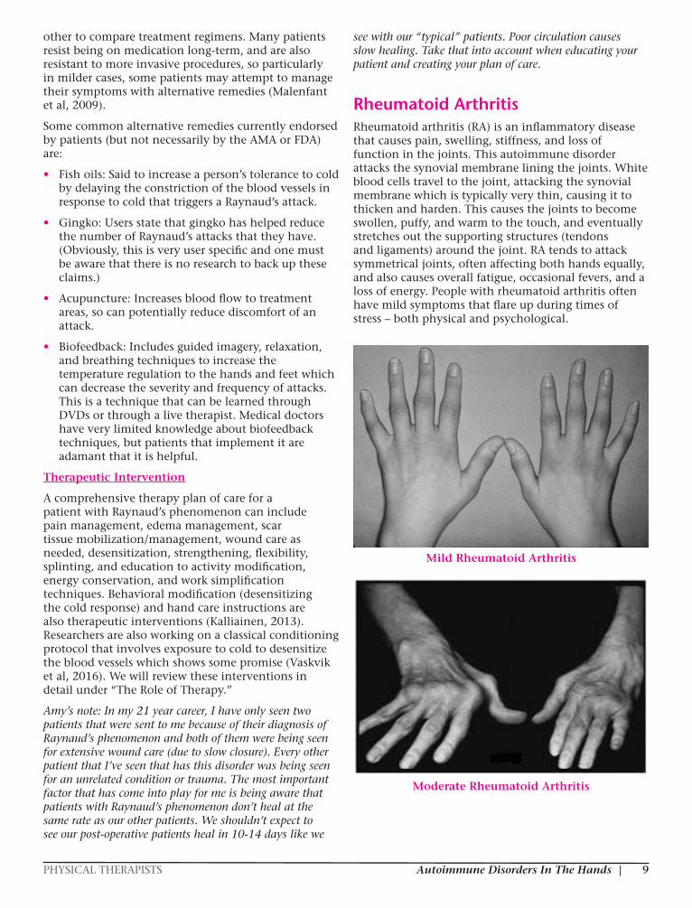
Rheumatoid ArthritisRheumatoid arthritis (RA) is an inflammatory disease that causes pain, swelling, stiffness, and loss of function in the joints. This autoimmune disorder attacks the synovial membrane lining the joints. White blood cells travel to the joint, attacking the synovial membrane which is typically very thin, causing it to thicken and harden. This causes the joints to become swollen, puffy, and warm to the touch, and eventually stretches out the supporting structures (tendons and ligaments) around the joint. RA tends to attack symmetrical joints, often affecting both hands equally, and also causes overall fatigue, occasional fevers, and a loss of energy. People with rheumatoid arthritis often have mild symptoms that flare up during times of stress – both physical and psychological.
Mild Rheumatoid Arthritis
Moderate Rheumatoid Arthritis
| Autoimmune Disorders In The Hands PHYSICAL THERAPISTS10
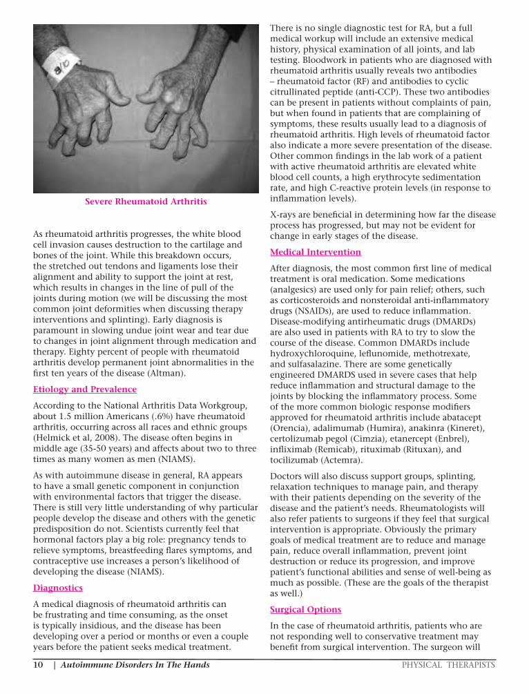
Severe Rheumatoid Arthritis
As rheumatoid arthritis progresses, the white blood cell invasion causes destruction to the cartilage and bones of the joint. While this breakdown occurs, the stretched out tendons and ligaments lose their alignment and ability to support the joint at rest, which results in changes in the line of pull of the joints during motion (we will be discussing the most common joint deformities when discussing therapy interventions and splinting). Early diagnosis is paramount in slowing undue joint wear and tear due to changes in joint alignment through medication and therapy. Eighty percent of people with rheumatoid arthritis develop permanent joint abnormalities in the first ten years of the disease (Altman).
Etiology and Prevalence
According to the National Arthritis Data Workgroup, about 1.5 million Americans (.6%) have rheumatoid arthritis, occurring across all races and ethnic groups (Helmick et al, 2008). The disease often begins in middle age (35-50 years) and affects about two to three times as many women as men (NIAMS).
As with autoimmune disease in general, RA appears to have a small genetic component in conjunction with environmental factors that trigger the disease. There is still very little understanding of why particular people develop the disease and others with the genetic predisposition do not. Scientists currently feel that hormonal factors play a big role: pregnancy tends to relieve symptoms, breastfeeding flares symptoms, and contraceptive use increases a person’s likelihood of developing the disease (NIAMS).
Diagnostics
A medical diagnosis of rheumatoid arthritis can be frustrating and time consuming, as the onset is typically insidious, and the disease has been developing over a period or months or even a couple years before the patient seeks medical treatment.
There is no single diagnostic test for RA, but a full medical workup will include an extensive medical history, physical examination of all joints, and lab testing. Bloodwork in patients who are diagnosed with rheumatoid arthritis usually reveals two antibodies – rheumatoid factor (RF) and antibodies to cyclic citrullinated peptide (anti-CCP). These two antibodies can be present in patients without complaints of pain, but when found in patients that are complaining of symptoms, these results usually lead to a diagnosis of rheumatoid arthritis. High levels of rheumatoid factor also indicate a more severe presentation of the disease. Other common findings in the lab work of a patient with active rheumatoid arthritis are elevated white blood cell counts, a high erythrocyte sedimentation rate, and high C-reactive protein levels (in response to inflammation levels).
X-rays are beneficial in determining how far the disease process has progressed, but may not be evident for change in early stages of the disease.
Medical Intervention
After diagnosis, the most common first line of medical treatment is oral medication. Some medications (analgesics) are used only for pain relief; others, such as corticosteroids and nonsteroidal anti-inflammatory drugs (NSAIDs), are used to reduce inflammation. Disease-modifying antirheumatic drugs (DMARDs) are also used in patients with RA to try to slow the course of the disease. Common DMARDs include hydroxychloroquine, leflunomide, methotrexate, and sulfasalazine. There are some genetically engineered DMARDS used in severe cases that help reduce inflammation and structural damage to the joints by blocking the inflammatory process. Some of the more common biologic response modifiers approved for rheumatoid arthritis include abatacept (Orencia), adalimumab (Humira), anakinra (Kineret), certolizumab pegol (Cimzia), etanercept (Enbrel), infliximab (Remicab), rituximab (Rituxan), and tocilizumab (Actemra).
Doctors will also discuss support groups, splinting, relaxation techniques to manage pain, and therapy with their patients depending on the severity of the disease and the patient’s needs. Rheumatologists will also refer patients to surgeons if they feel that surgical intervention is appropriate. Obviously the primary goals of medical treatment are to reduce and manage pain, reduce overall inflammation, prevent joint destruction or reduce its progression, and improve patient’s functional abilities and sense of well-being as much as possible. (These are the goals of the therapist as well.)
Surgical Options
In the case of rheumatoid arthritis, patients who are not responding well to conservative treatment may benefit from surgical intervention. The surgeon will
PHYSICAL THERAPISTS Autoimmune Disorders In The Hands | 11
determine the indication for surgery of the hands by weighing not only the severity of joint deformity and the patient’s pain levels, but also the age of the patient, their health status, and their bone density (Alderman et al, 2003).
Depending on the patient’s complaints, the status of the joints, and the stability of the hand and wrist, the doctor has several surgical options that can be indicated. There are two categories of surgical intervention: prophylactic and therapeutic surgeries. Prophylactic procedures such as synovectomy, tenosynovectomy, and osteotomy can improve joint function and prevent tendon rupture. Joint fusions, joint replacements, and tendon repairs are considered therapeutic interventions, as they improve function and reduce pain in the hands (Chim et al, 2014).
Synovectomy – Removal of the irritated synovial lining during the early stages of the disease may relieve some of the pain and discomfort in the joints. It may also relieve pressure to the median nerve that could lead to carpal tunnel syndrome.
Tenosynovectomy – Excision of the inflamed tendon sheath/synovial sheath to reduce pain. This procedure is typically performed on the extensor tendons as they go under the extensor retinaculum due to pain with active extension.
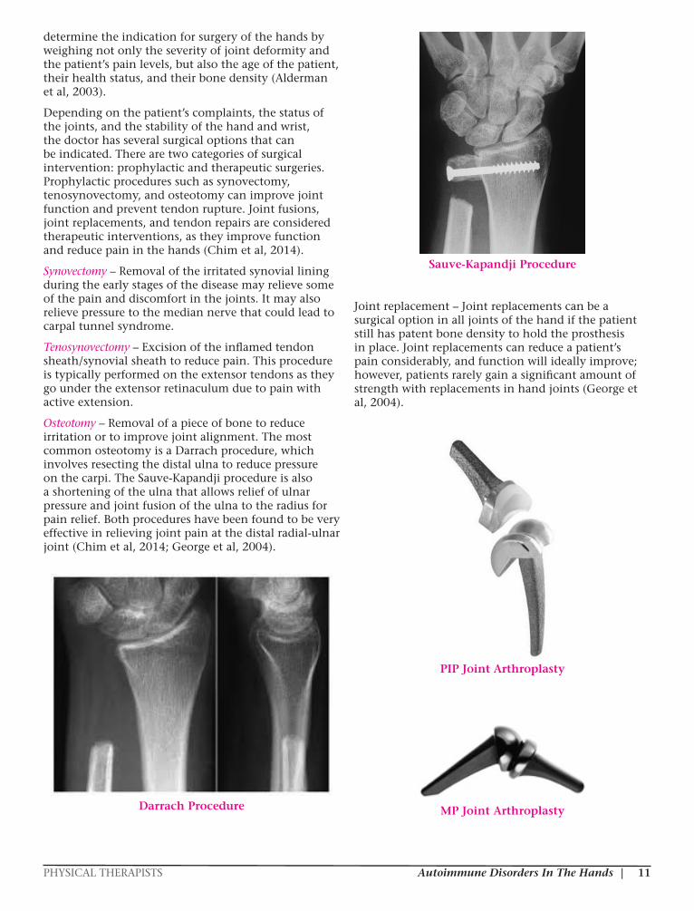
Osteotomy – Removal of a piece of bone to reduce irritation or to improve joint alignment. The most common osteotomy is a Darrach procedure, which involves resecting the distal ulna to reduce pressure on the carpi. The Sauve-Kapandji procedure is also a shortening of the ulna that allows relief of ulnar pressure and joint fusion of the ulna to the radius for pain relief. Both procedures have been found to be very effective in relieving joint pain at the distal radial-ulnar joint (Chim et al, 2014; George et al, 2004).
Darrach Procedure
Sauve-Kapandji Procedure
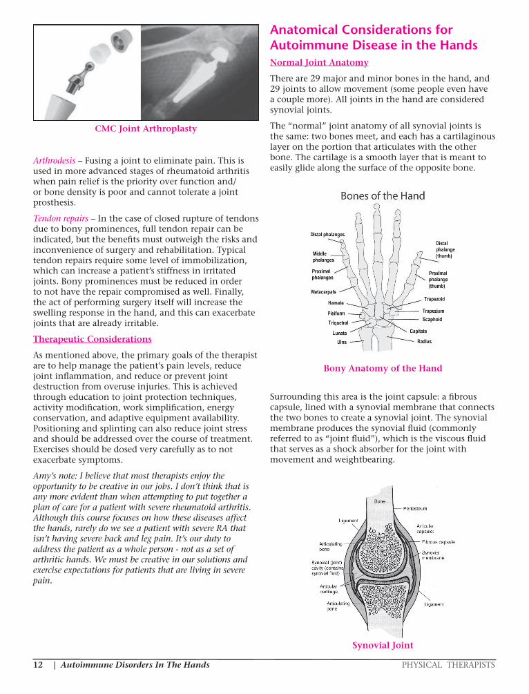
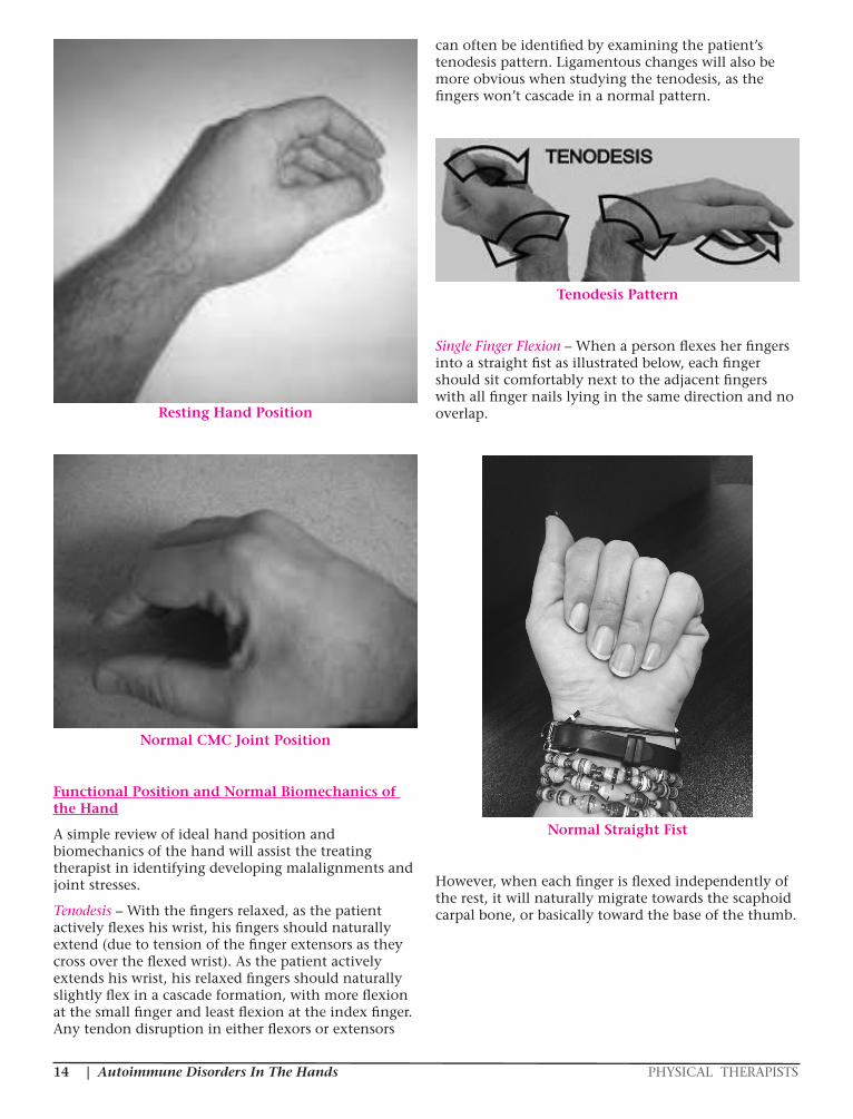
Joint replacement – Joint replacements can be a surgical option in all joints of the hand if the patient still has patent bone density to hold the prosthesis in place. Joint replacements can reduce a patient’s pain considerably, and function will ideally improve; however, patients rarely gain a significant amount of strength with replacements in hand joints (George et al, 2004).
PIP Joint Arthroplasty
MP Joint Arthroplasty
| Autoimmune Disorders In The Hands PHYSICAL THERAPISTS12
CMC Joint Arthroplasty
Arthrodesis – Fusing a joint to eliminate pain. This is used in more advanced stages of rheumatoid arthritis when pain relief is the priority over function and/or bone density is poor and cannot tolerate a joint prosthesis.
Tendon repairs – In the case of closed rupture of tendons due to bony prominences, full tendon repair can be indicated, but the benefits must outweigh the risks and inconvenience of surgery and rehabilitation. Typical tendon repairs require some level of immobilization, which can increase a patient’s stiffness in irritated joints. Bony prominences must be reduced in order to not have the repair compromised as well. Finally, the act of performing surgery itself will increase the swelling response in the hand, and this can exacerbate joints that are already irritable.
Therapeutic Considerations
As mentioned above, the primary goals of the therapist are to help manage the patient’s pain levels, reduce joint inflammation, and reduce or prevent joint destruction from overuse injuries. This is achieved through education to joint protection techniques, activity modification, work simplification, energy conservation, and adaptive equipment availability. Positioning and splinting can also reduce joint stress and should be addressed over the course of treatment. Exercises should be dosed very carefully as to not exacerbate symptoms.
Amy’s note: I believe that most therapists enjoy the opportunity to be creative in our jobs. I don’t think that is any more evident than when attempting to put together a plan of care for a patient with severe rheumatoid arthritis. Although this course focuses on how these diseases affect the hands, rarely do we see a patient with severe RA that isn’t having severe back and leg pain. It’s our duty to address the patient as a whole person - not as a set of arthritic hands. We must be creative in our solutions and exercise expectations for patients that are living in severe pain.
Anatomical Considerations for Autoimmune Disease in the HandsNormal Joint Anatomy
There are 29 major and minor bones in the hand, and 29 joints to allow movement (some people even have a couple more). All joints in the hand are considered synovial joints.
The “normal” joint anatomy of all synovial joints is the same: two bones meet, and each has a cartilaginous layer on the portion that articulates with the other bone. The cartilage is a smooth layer that is meant to easily glide along the surface of the opposite bone.
Bony Anatomy of the Hand
Surrounding this area is the joint capsule: a fibrous capsule, lined with a synovial membrane that connects the two bones to create a synovial joint. The synovial membrane produces the synovial fluid (commonly referred to as “joint fluid”), which is the viscous fluid that serves as a shock absorber for the joint with movement and weightbearing.
Synovial Joint

PHYSICAL THERAPISTS Autoimmune Disorders In The Hands | 13
Outside of the capsule are the ligaments that surround the joint and provide it with stability during movement and at rest. Each joint can have several ligaments around it which provide stability in various positions. There are actually 123 named ligaments just in the hand and wrist! The volar plate of the PIP joints is actually a thickened ligament specifically at the PIP on the volar surface that does not allow PIP hyperextension. This is important to note when we discuss joint deformity patterns with disease.
Ligamentous Anatomy of the Hand
Volar Plate Anatomy
All joints have a “close-packed” position and a “loose-packed” position, which refers to the position of the joint at rest due to ligamentous stability and the joint capsule surrounding it. Close-packed positions are the positions of most stability with the most joint congruity because the ligaments and joint capsule are on full stretch, essentially tightening around the joint. In the hand, a close-packed position is an intrinsic plus position – that is full MP flexion and full PIP and DIP extension. A close-packed wrist would either sit in full flexion or full extension.
Intrinsic Plus Position
Loose-packed positions provide the most play in the joint, as the ligaments (and capsule) are in their most relaxed position. The loose packed position is the “resting hand position.” Relax your hand. Observe the angle of your finger joints and the position of your thumb. Your hand will naturally relax into a loose-packed position at all joints. In general, the hand rests with the MP joints in a neutral position of about 10-15 degrees of flexion, and the IP joints rest in slightly more flexion (35 degrees at the PIP, and 5-10 degrees at the DIP). In a normal resting position, the thumb should sit in about 10-15 degrees of radial abduction at the CMC, and halfway between volar (or palmar) abduction and thumb radial abduction. The CMC joint is not typically “visible” to the naked eye, as the base of the first metacarpal is securely seated in the trapezium. With the joint in normal alignment, the tendons will rest in such a way that the thumb MP joint sits comfortably in about 30 degrees of flexion, and the IP is fairly neutral. The wrist will also sit in relative neutral – that is at 0-10 degrees of extension.
Understanding and observing “normal” resting hand position is the basis for understanding and effectively treating deformity (or potential deformity). As joints break down, as is the case with rheumatoid arthritis, malalignment occurs. With scleroderma, the hands will “default” to a resting hand position and the skin will harden in this pattern. It is crucial to understand the ideal position for hand function in order to assist your patient to return to their highest level of pain free mobility and activity.
| Autoimmune Disorders In The Hands PHYSICAL THERAPISTS14
Resting Hand Position
Normal CMC Joint Position
Functional Position and Normal Biomechanics of the Hand
A simple review of ideal hand position and biomechanics of the hand will assist the treating therapist in identifying developing malalignments and joint stresses.
Tenodesis – With the fingers relaxed, as the patient actively flexes his wrist, his fingers should naturally extend (due to tension of the finger extensors as they cross over the flexed wrist). As the patient actively extends his wrist, his relaxed fingers should naturally slightly flex in a cascade formation, with more flexion at the small finger and least flexion at the index finger. Any tendon disruption in either flexors or extensors
can often be identified by examining the patient’s tenodesis pattern. Ligamentous changes will also be more obvious when studying the tenodesis, as the fingers won’t cascade in a normal pattern.
Tenodesis Pattern
Single Finger Flexion – When a person flexes her fingers into a straight fist as illustrated below, each finger should sit comfortably next to the adjacent fingers with all finger nails lying in the same direction and no overlap.
Normal Straight Fist
However, when each finger is flexed independently of the rest, it will naturally migrate towards the scaphoid carpal bone, or basically toward the base of the thumb.

PHYSICAL THERAPISTS Autoimmune Disorders In The Hands | 15
Normal Single Finger Flexion
Extensor Hood Mechanism – The extensor hood mechanism for the fingers is a system of fibers which allows the MPs, PIPs, and DIPs of the fingers to extend. It arises from the extensor tendons as they travel distally across the dorsal surface of the hand and over the MP joints to each digit. As you can see when you review the anatomy, the central slip of the extensor tendon travels distally to insert at the base of the middle phalanx, which is what allows for active PIP extension. Proximal to that, the tendon splits into lateral bands that travel laterally on each side of the finger, going around the PIP joint, and eventually coming back together just distal to the insertion of the central slip to insert as one tendon again on the base of the distal phalanx. This terminal tendon is what allows for active DIP extension.
This is a complicated system, as there are oblique fibers that give the extensor hood support across the dorsum of the finger, and the intrinsic muscles and oblique retinacular ligaments also insert into the extensor hood mechanism. This simple overview of the system is important to have a basic understanding as it often comes into play with chronic swelling of the joints.
Extensor Hood Mechanism
Power vs. Fine Motor Coordination – The ulnar two digits of the hand (ring and small fingers) are used primarily for power grip (such as holding a hammer or heavy object), and the radial two digits (index and middle) and thumb are used for fine motor dexterity.
Wrist Position – Slight wrist extension (15-20 degrees) combined with about 10 degrees of radial deviation is the position of function for most daily activities. This slight wrist extension puts the finger extensors on slack, allowing full use of the finger flexors. Full pronation is needed for most functional activities, including typing, writing, typing shoes, and preparing food.
The Role of TherapyBoth occupational and physical therapists treat patients with joint changes due to autoimmune disease in their hands. Whether it is the primary diagnosis that led them to our clinic for corrective or protective splinting or a secondary diagnosis that affects a patient’s ability to heal in a timely manner, our understanding of how autoimmune deficiencies affect hand function is paramount in proper exercise dosing, use of therapeutic modalities, and appropriate manual techniques. Thorough evaluation of the joint health of the hands begins on day one of treatment.
History
Depending on whether a patient is coming with a primary diagnosis of an autoimmune disorder or the therapist is suspicious that the patient has some underlying autoimmune symptoms that may affect treatment, some questions to consider and review

| Autoimmune Disorders In The Hands PHYSICAL THERAPISTS16
during our interview with the patient can include:
• Which joints are hurting the most? Are any other joints bothersome as well?
• Is the pain sharp/dull/throbbing/aching/zinging, etc? (Describe in their words)
• How long has the patient been experiencing pain in the joint?
• Have they recently had an accident/surgery/life event which has exacerbated symptoms?
• Have they been exposed to any unusual toxins recently?
• Are they noticing any crepitus in the joints?
• What activities irritate the symptoms?
• Are the symptoms worse with rest or activity?
• Does the patient wake up with stiffness in the affected joint?
• What relieves the pain?
• Have they tried heat or ice to eliminate symptoms?
• Has the patient had a medical workup including x-rays?
• Have they been started on any medications by their physician for joint pain or related issues?
• Are they taking any over-the-counter supplements?
• Have they tried any splinting or immobilization devices? Were those helpful?
• Have they used any alternative remedies such as acupuncture, copper bracelets, magnet therapy, or chiropractic to reduce pain? Has any of this been helpful?
• Has the patient had any other hand, wrist, shoulder, or cervical surgeries to date?
• Does the patient engage in activities that require excessive or repetitive gripping or weight-bearing to the hands? (such as gardening, knitting, working on cars)
• Does the patient exercise regularly? Describe exercise regimen.
Typically by the time a patient with autoimmune diseases like rheumatoid arthritis and scleroderma reaches us for evaluation, they have had symptoms for quite some time – several months to even a couple of years. They are having difficulty doing activities such as opening jars, turning keys to open a door or start the car, gardening, and mechanic work. They may even complain of difficulties with self care such as buttoning, zipping, or pulling up their pants due to pain or deformity.
Amy’s note: One of the most interesting sidebars about patients with autoimmune disease is that they will
commonly downplay their symptoms and self care difficulties. I find that it is much more useful to ask a patient in this situation more direct, objective questions, such as “How long does it take you to get dressed in the morning?” or “How long can you spend typing at the computer before you need to take a break due to pain?” Patients who have been living with progressive deformity tend to forget what is “normal” – they’ve continued to be functional and independent by taking rest breaks, adapting how they perform their activities, or just abandoning certain activities altogether. In general, I’ve found that patients in this subgroup hold very fiercely to their independence by minimizing their complaints.
They also have a very different pain scale than the rest of us. So when asking them to rate a pain on a scale from 0-10, make sure you get an understanding of what a “10” means to them. I’ve heard such descriptions as “I got my leg run over by a truck. That was a 10.” and “I woke up during surgery once and could feel everything. That was a 10.” Patients with rheumatoid arthritis (specifically) have an unbelievable tolerance and response to pain, so something you or I would rate as an 8/10 might be a 2/10 to your patient. This is important to appreciate when you are dosing exercises or asking a patient to tolerate a splint.
Evaluation and Provocative Testing
Assessing Joint Deformity
After a thorough history is taken, the therapist can now assess the severity of joint changes in the patient both at rest and with movement. When at rest, what positions do the fingers lie in? When the hands are placed in a relaxed position on the table, do all the fingernails line up and face the same direction? Is one arthritic finger over-supinated or over-pronated? Are there obvious joint deformities at rest?
Rheumatoid Arthritis – The most common patterns of deformity in the fingers are Swan Neck deformities and Boutonniere deformities, which both occur due to changes in the extensor hood mechanism and its line of pull. They can occur at any or all of the fingers and the thumb, and can present at all levels of severity.
A Swan Neck deformity is a hyperextension at the PIP joint with subsequent flexion at the DIP joint. The mechanism of injury with regards to rheumatoid arthritis is that the PIP joint can stay chronically swollen, which eventually will stretch out the joint capsule, including the thickened volar plate which is designed to prevent PIP hyperextension. This leads to joint ligament laxity, and the central slip of the extensor hood has an unfair advantage, pulling too hard on the dorsal surface of the joint, causing the PIP joint to hyperextend. As a result of this collapse of the joint, as the extensor tendons pull the PIP joint into extension, the lateral bands and terminal extensor tendon are thrown off balance, pulling the DIP into flexion. A swan neck deformity can diminish
PHYSICAL THERAPISTS Autoimmune Disorders In The Hands | 17
a patient’s ability to grasp and to pinch and will need to be addressed with supportive splinting, as we will discuss later.
Swan Neck Deformity Anatomy
Swan Neck Deformity Image
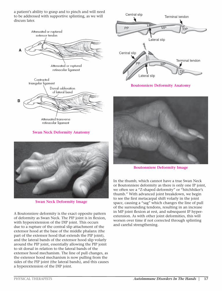
A Boutonniere deformity is the exact opposite pattern of deformity as Swan Neck. The PIP joint is in flexion, with hyperextension of the DIP joint. This occurs due to a rupture of the central slip attachment of the extensor hood at the base of the middle phalanx (the part of the extensor hood that extends the PIP joint), and the lateral bands of the extensor hood slip volarly around the PIP joint, essentially allowing the PIP joint to sit dorsal in relation to the lateral bands of the extensor hood mechanism. The line of pull changes, as the extensor hood mechanism is now pulling from the sides of the PIP joint (the lateral bands), and this causes a hyperextension of the DIP joint.
Boutonniere Deformity Anatomy
Boutonniere Deformity Image
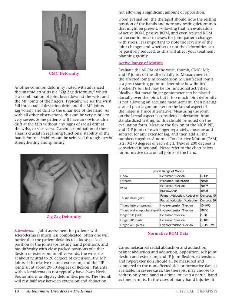
In the thumb, which cannot have a true Swan Neck or Boutonniere deformity as there is only one IP joint, we often see a “Z-shaped deformity” or “hitchhiker’s thumb.” With advanced joint breakdown, we begin to see the first metacarpal shift volarly in the joint space, causing a “sag” which changes the line of pull of the surrounding tendons, resulting in an increase in MP joint flexion at rest, and subsequent IP hyper-extension. As with other joint deformities, this will worsen over time if not corrected through splinting and careful strengthening.
| Autoimmune Disorders In The Hands PHYSICAL THERAPISTS18
CMC Deformity
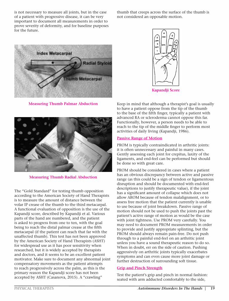
Another common deformity noted with advanced rheumatoid arthritis is a “Zig Zag deformity,” which is a combination of joint breakdown at the wrist and the MP joints of the fingers. Typically, we see the wrist fall into a radial deviation drift, and the MP joints sag volarly and drift to the ulnar side of the hand. As with all other observations, this can be very subtle to very severe. Some patients will have an obvious ulnar drift at the MPs without any signs of radial drift at the wrist, or vice versa. Careful examination of these areas is crucial in regaining functional stability of the hands for use. Stability can be achieved through careful strengthening and splinting.
Zig Zag Deformity
Scleroderma – Joint assessment for patients with scleroderma is much less complicated: often one will notice that the patient defaults to a loose-packed position of the joints (or resting hand position), and has difficulty with close packed positions of either flexion or extension. In other words, the wrist sits at about neutral to 20 degrees of extension, the MP joints sit in relative neutral extension, and the PIP joints sit at about 20-30 degrees of flexion). Patients with scleroderma do not typically have Swan Neck, Boutonniere, or Zig Zag deformities per se. The thumb will rest half way between extension and abduction,
not allowing a significant amount of opposition.
Upon evaluation, the therapist should note the resting position of the hands and note any resting deformities that might be present. Following that, an evaluation of active ROM, passive ROM, and even resisted ROM can occur in order to assess for joint pattern changes with stress. It is important to note the severity of the joint changes and whether or not the deformities can be passively reduced, as this will affect your treatment planning greatly.
Active Range of Motion
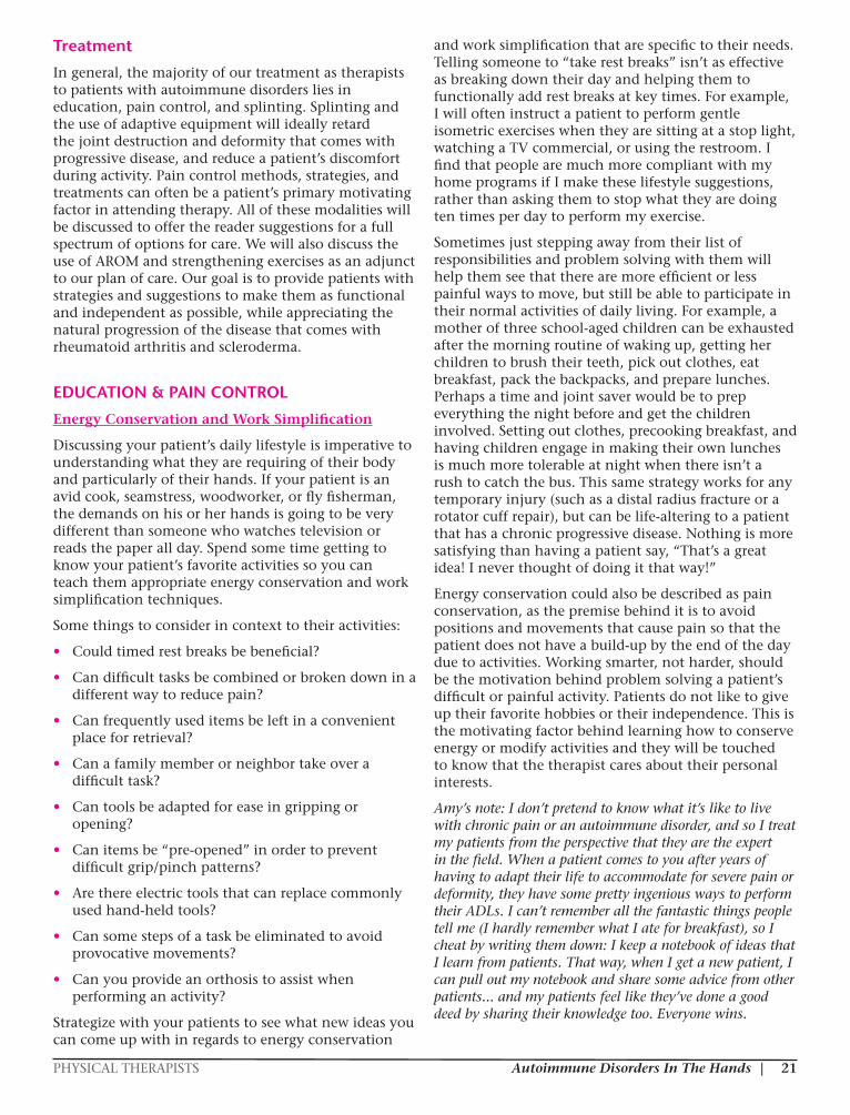
Evaluate the AROM of the wrist, thumb, CMC, MP, and IP joints of the affected digits. Measurement of the affected joints in comparison to unaffected joints is a great starting point to determine how limited a patient’s full fist may be for functional activities. Ideally a flat metal finger goniometer can be placed dorsally over the joint, but if too much joint deformity is not allowing an accurate measurement, then placing a small plastic goniometer on the lateral aspect of the finger is a nice alternative. Measuring the joint on the lateral aspect is considered a deviation from standardized testing, so this should be noted on the evaluation form. Measure the flexion of the MCP, PIP, and DIP joints of each finger separately, measure and subtract for any extensor lag, and then add all the numbers together. A normal Total Active Motion (TAM) is 250-270 degrees of each digit. TAM of 200 degrees is considered functional. Please refer to the chart below for normative data on all joints of the hand.
Normative ROM Data
Carpometacarpal radial abduction and adduction, palmar abduction and adduction, opposition, MP joint flexion and extension, and IP joint flexion, extension, and hyperextension should all be measured and compared to the non-affected side or normative data as available. In severe cases, the therapist may choose to address only one hand at a time, or even a partial hand as time permits. In the cases of many hand injuries, it
PHYSICAL THERAPISTS Autoimmune Disorders In The Hands | 19
is not necessary to measure all joints, but in the case of a patient with progressive disease, it can be very important to document all measurements in order to prove severity of deformity, and for baseline purposes for the future.
Measuring Thumb Palmar Abduction
Measuring Thumb Radial Abduction
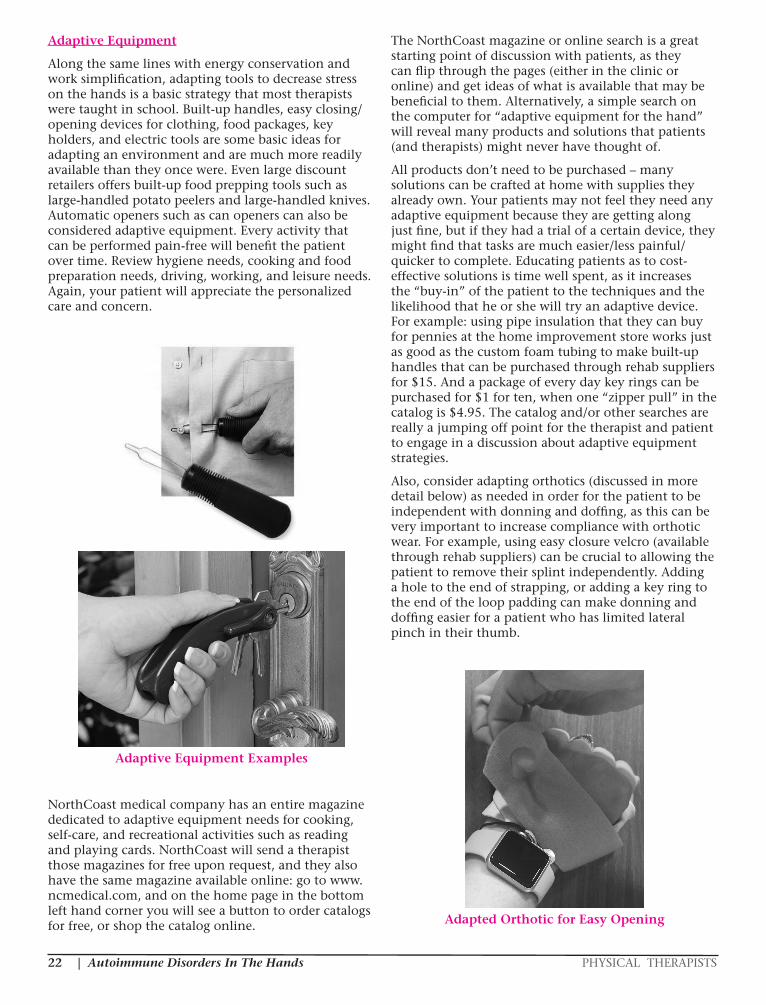
The “Gold Standard” for testing thumb opposition according to the American Society of Hand Therapists is to measure the amount of distance between the volar IP crease of the thumb to the third metacarpal. A functional evaluation of opposition is the use of the Kapandji score, described by Kapandji et al. Various parts of the hand are numbered, and the patient is asked to progress from one to ten, with the goal being to reach the distal palmar crease at the fifth metacarpal (if the patient can reach that far with the unaffected thumb). This test has not been approved by the American Society of Hand Therapists (ASHT) for widespread use as it has poor sensitivity when researched, but it is widely accepted by therapists and doctors, and it seems to be an excellent patient motivator. Make sure to document any abnormal joint compensatory movements as the patient attempts to reach progressively across the palm, as this is the primary reason the Kapandji score has not been accepted by ASHT (Casanova, 2015). A “crawling”
thumb that creeps across the surface of the thumb is not considered an opposable motion.
Kapandji Score
Keep in mind that although a therapist’s goal is usually to have a patient oppose from the tip of the thumb to the base of the fifth finger, typically a patient with advanced RA or scleroderma cannot oppose this far. Functionally, however, a person needs to be able to reach to the tip of the middle finger to perform most activities of daily living (Kapandji, 1986).
Passive Range of Motion
PROM is typically contraindicated in arthritic joints: it is often unnecessary and painful in many cases. Gently assessing each joint for crepitus, laxity of the ligaments, and end-feel can be performed but should be done so with great care.
PROM should be considered in cases where a patient has an obvious discrepancy between active and passive range (as this could be a sign of tendon or ligamentous disruption and should be documented with end-feel descriptions to justify therapeutic value), if the joint has a significant amount of collapse which does not allow AROM because of tendon malalignment, or to assess free motion that the patient currently is unable to use because of joint breakdown. Passive range of motion should not be used to push the joints past the patient’s active range of motion as would be the case with joint tightness. Use PROM very carefully. You may need to document PROM measurements in order to provide and justify appropriate splinting, but the PROM should always remain pain-free. Do not push through to a painful end-feel on an arthritic joint unless you have a sound therapeutic reason to do so. When in doubt, err on the side of caution. Pushing aggressively on arthritic joints typically exacerbates symptoms and can even cause more joint damage or further destruction of surrounding soft tissue.
Grip and Pinch Strength
Test the patient’s grip and pinch in normal fashion: seated with arm adducted comfortably to the side,

| Autoimmune Disorders In The Hands PHYSICAL THERAPISTS20
elbow at 90 degrees, forearm and wrist in neutral. Compare to the unaffected side. Document any complaints related to testing such as a sharp pain, difficulty with grasp, or inability to hold the testing equipment comfortably. Performing a pinch test will typically accentuate any deformities that might be forming in collapsing thumb or finger joints, but is good information to justify treatment to attempt to increase a patient’s functional grasp. Normative data can be found online and in many textbooks but should be used with caution. Compare both sides and discuss the patient’s expectations for strength in relation to their ADLs, job duties, and recreational goals.
Grip strength can be measured using a dynamometer, and a pinch meter can be used to test lateral or key pinch, 3-jaw chuck, and tip-to-tip pinch. A trial of three reps can give the therapist an average strength number to document, as well as feedback as to pain levels or substitutions with grasping in different patterns. If a pinch meter is not available, the therapist can use the Jamar dynamometer on grip level one to assess pinch, but these numbers cannot be used against normative data. If a patient’s joint deformities are extreme, using a Jamar dynamometer may not be possible. Consider having the patient squeeze excess water out of a washcloth and make notes of substitution patterns, pain levels, and other modifications for your written documentation.
Grip Strength Using Jamar Dynamometer
Pinch Strength Showing Increased Deformity
Edema/Volume
Circumferential measurements of the joints can be performed with a standard
retractable tape measure or volumeter if available. Volumeters give very accurate measurement readings but there is no normative data for this. The therapist must rely on measurements between treatments to get feedback with regards to the treatment’s effect on edema levels. Volumeters are also not readily available in all clinics, and can be time-consuming and messy. Circumferential measurements are a good alternative to the use of volumeters, as they are readily available and easily transportable. There is no normative data for circumferential measurements either, but measurements can be taken from visit to visit to assess edema fluctuations. Circumferential measurements are also necessary when ordering prefabricated orthoses such as Oval-8 splints to realign finger joints. Edema can fluctuate due to skin temperature, food (sodium) intake, time of day, and activity levels.
Volumeter
Tape Measure
PHYSICAL THERAPISTS Autoimmune Disorders In The Hands | 21
Treatment
In general, the majority of our treatment as therapists to patients with autoimmune disorders lies in education, pain control, and splinting. Splinting and the use of adaptive equipment will ideally retard the joint destruction and deformity that comes with progressive disease, and reduce a patient’s discomfort during activity. Pain control methods, strategies, and treatments can often be a patient’s primary motivating factor in attending therapy. All of these modalities will be discussed to offer the reader suggestions for a full spectrum of options for care. We will also discuss the use of AROM and strengthening exercises as an adjunct to our plan of care. Our goal is to provide patients with strategies and suggestions to make them as functional and independent as possible, while appreciating the natural progression of the disease that comes with rheumatoid arthritis and scleroderma.
EDUCATION & PAIN CONTROL
Energy Conservation and Work Simplification
Discussing your patient’s daily lifestyle is imperative to understanding what they are requiring of their body and particularly of their hands. If your patient is an avid cook, seamstress, woodworker, or fly fisherman, the demands on his or her hands is going to be very different than someone who watches television or reads the paper all day. Spend some time getting to know your patient’s favorite activities so you can teach them appropriate energy conservation and work simplification techniques.
Some things to consider in context to their activities:
• Could timed rest breaks be beneficial?
• Can difficult tasks be combined or broken down in a different way to reduce pain?
• Can frequently used items be left in a convenient place for retrieval?
• Can a family member or neighbor take over a difficult task?
• Can tools be adapted for ease in gripping or opening?
• Can items be “pre-opened” in order to prevent difficult grip/pinch patterns?
• Are there electric tools that can replace commonly used hand-held tools?
• Can some steps of a task be eliminated to avoid provocative movements?
• Can you provide an orthosis to assist when performing an activity?
Strategize with your patients to see what new ideas you can come up with in regards to energy conservation
and work simplification that are specific to their needs. Telling someone to “take rest breaks” isn’t as effective as breaking down their day and helping them to functionally add rest breaks at key times. For example, I will often instruct a patient to perform gentle isometric exercises when they are sitting at a stop light, watching a TV commercial, or using the restroom. I find that people are much more compliant with my home programs if I make these lifestyle suggestions, rather than asking them to stop what they are doing ten times per day to perform my exercise.
Sometimes just stepping away from their list of responsibilities and problem solving with them will help them see that there are more efficient or less painful ways to move, but still be able to participate in their normal activities of daily living. For example, a mother of three school-aged children can be exhausted after the morning routine of waking up, getting her children to brush their teeth, pick out clothes, eat breakfast, pack the backpacks, and prepare lunches. Perhaps a time and joint saver would be to prep everything the night before and get the children involved. Setting out clothes, precooking breakfast, and having children engage in making their own lunches is much more tolerable at night when there isn’t a rush to catch the bus. This same strategy works for any temporary injury (such as a distal radius fracture or a rotator cuff repair), but can be life-altering to a patient that has a chronic progressive disease. Nothing is more satisfying than having a patient say, “That’s a great idea! I never thought of doing it that way!”
Energy conservation could also be described as pain conservation, as the premise behind it is to avoid positions and movements that cause pain so that the patient does not have a build-up by the end of the day due to activities. Working smarter, not harder, should be the motivation behind problem solving a patient’s difficult or painful activity. Patients do not like to give up their favorite hobbies or their independence. This is the motivating factor behind learning how to conserve energy or modify activities and they will be touched to know that the therapist cares about their personal interests.
Amy’s note: I don’t pretend to know what it’s like to live with chronic pain or an autoimmune disorder, and so I treat my patients from the perspective that they are the expert in the field. When a patient comes to you after years of having to adapt their life to accommodate for severe pain or deformity, they have some pretty ingenious ways to perform their ADLs. I can’t remember all the fantastic things people tell me (I hardly remember what I ate for breakfast), so I cheat by writing them down: I keep a notebook of ideas that I learn from patients. That way, when I get a new patient, I can pull out my notebook and share some advice from other patients... and my patients feel like they’ve done a good deed by sharing their knowledge too. Everyone wins.
| Autoimmune Disorders In The Hands PHYSICAL THERAPISTS22
Adaptive Equipment
Along the same lines with energy conservation and work simplification, adapting tools to decrease stress on the hands is a basic strategy that most therapists were taught in school. Built-up handles, easy closing/opening devices for clothing, food packages, key holders, and electric tools are some basic ideas for adapting an environment and are much more readily available than they once were. Even large discount retailers offers built-up food prepping tools such as large-handled potato peelers and large-handled knives. Automatic openers such as can openers can also be considered adaptive equipment. Every activity that can be performed pain-free will benefit the patient over time. Review hygiene needs, cooking and food preparation needs, driving, working, and leisure needs. Again, your patient will appreciate the personalized care and concern.
Adaptive Equipment Examples
NorthCoast medical company has an entire magazine dedicated to adaptive equipment needs for cooking, self-care, and recreational activities such as reading and playing cards. NorthCoast will send a therapist those magazines for free upon request, and they also have the same magazine available online: go to www.ncmedical.com, and on the home page in the bottom left hand corner you will see a button to order catalogs for free, or shop the catalog online.
The NorthCoast magazine or online search is a great starting point of discussion with patients, as they can flip through the pages (either in the clinic or online) and get ideas of what is available that may be beneficial to them. Alternatively, a simple search on the computer for “adaptive equipment for the hand” will reveal many products and solutions that patients (and therapists) might never have thought of.
All products don’t need to be purchased – many solutions can be crafted at home with supplies they already own. Your patients may not feel they need any adaptive equipment because they are getting along just fine, but if they had a trial of a certain device, they might find that tasks are much easier/less painful/quicker to complete. Educating patients as to cost-effective solutions is time well spent, as it increases the “buy-in” of the patient to the techniques and the likelihood that he or she will try an adaptive device. For example: using pipe insulation that they can buy for pennies at the home improvement store works just as good as the custom foam tubing to make built-up handles that can be purchased through rehab suppliers for $15. And a package of every day key rings can be purchased for $1 for ten, when one “zipper pull” in the catalog is $4.95. The catalog and/or other searches are really a jumping off point for the therapist and patient to engage in a discussion about adaptive equipment strategies.
Also, consider adapting orthotics (discussed in more detail below) as needed in order for the patient to be independent with donning and doffing, as this can be very important to increase compliance with orthotic wear. For example, using easy closure velcro (available through rehab suppliers) can be crucial to allowing the patient to remove their splint independently. Adding a hole to the end of strapping, or adding a key ring to the end of the loop padding can make donning and doffing easier for a patient who has limited lateral pinch in their thumb.
Adapted Orthotic for Easy Opening
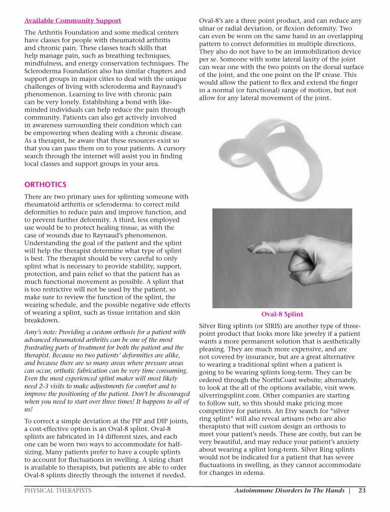
PHYSICAL THERAPISTS Autoimmune Disorders In The Hands | 23
Available Community Support
The Arthritis Foundation and some medical centers have classes for people with rheumatoid arthritis and chronic pain. These classes teach skills that help manage pain, such as breathing techniques, mindfulness, and energy conservation techniques. The Scleroderma Foundation also has similar chapters and support groups in major cities to deal with the unique challenges of living with scleroderma and Raynaud’s phenomenon. Learning to live with chronic pain can be very lonely. Establishing a bond with like-minded individuals can help reduce the pain through community. Patients can also get actively involved in awareness surrounding their condition which can be empowering when dealing with a chronic disease. As a therapist, be aware that these resources exist so that you can pass them on to your patients. A cursory search through the internet will assist you in finding local classes and support groups in your area.
ORTHOTICS
There are two primary uses for splinting someone with rheumatoid arthritis or scleroderma: to correct mild deformities to reduce pain and improve function, and to prevent further deformity. A third, less employed use would be to protect healing tissue, as with the case of wounds due to Raynaud’s phenomenon. Understanding the goal of the patient and the splint will help the therapist determine what type of splint is best. The therapist should be very careful to only splint what is necessary to provide stability, support, protection, and pain relief so that the patient has as much functional movement as possible. A splint that is too restrictive will not be used by the patient, so make sure to review the function of the splint, the wearing schedule, and the possible negative side effects of wearing a splint, such as tissue irritation and skin breakdown.
Amy’s note: Providing a custom orthosis for a patient with advanced rheumatoid arthritis can be one of the most frustrating parts of treatment for both the patient and the therapist. Because no two patients’ deformities are alike, and because there are so many areas where pressure areas can occur, orthotic fabrication can be very time consuming. Even the most experienced splint maker will most likely need 2-3 visits to make adjustments for comfort and to improve the positioning of the patient. Don’t be discouraged when you need to start over three times! It happens to all of us!
To correct a simple deviation at the PIP and DIP joints, a cost-effective option is an Oval-8 splint. Oval-8 splints are fabricated in 14 different sizes, and each one can be worn two ways to accommodate for half-sizing. Many patients prefer to have a couple splints to account for fluctuations in swelling. A sizing chart is available to therapists, but patients are able to order Oval-8 splints directly through the internet if needed.
Oval-8’s are a three point product, and can reduce any ulnar or radial deviation, or flexion deformity. Two can even be worn on the same hand in an overlapping pattern to correct deformities in multiple directions. They also do not have to be an immobilization device per se. Someone with some lateral laxity of the joint can wear one with the two points on the dorsal surface of the joint, and the one point on the IP crease. This would allow the patient to flex and extend the finger in a normal (or functional) range of motion, but not allow for any lateral movement of the joint.
Oval-8 Splint
Silver Ring splints (or SIRIS) are another type of three-point product that looks more like jewelry if a patient wants a more permanent solution that is aesthetically pleasing. They are much more expensive, and are not covered by insurance, but are a great alternative to wearing a traditional splint when a patient is going to be wearing splints long-term. They can be ordered through the NorthCoast website; alternately, to look at the all of the options available, visit www.silverringsplint.com. Other companies are starting to follow suit, so this should make pricing more competitive for patients. An Etsy search for “silver ring splint” will also reveal artisans (who are also therapists) that will custom design an orthosis to meet your patient’s needs. These are costly, but can be very beautiful, and may reduce your patient’s anxiety about wearing a splint long-term. Silver Ring splints would not be indicated for a patient that has severe fluctuations in swelling, as they cannot accommodate for changes in edema.
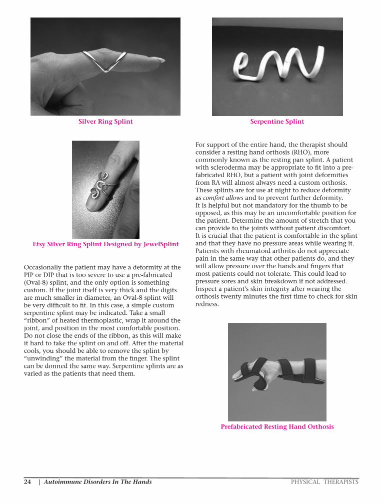
| Autoimmune Disorders In The Hands PHYSICAL THERAPISTS24
Silver Ring Splint
Etsy Silver Ring Splint Designed by JewelSplint
Occasionally the patient may have a deformity at the PIP or DIP that is too severe to use a pre-fabricated (Oval-8) splint, and the only option is something custom. If the joint itself is very thick and the digits are much smaller in diameter, an Oval-8 splint will be very difficult to fit. In this case, a simple custom serpentine splint may be indicated. Take a small “ribbon” of heated thermoplastic, wrap it around the joint, and position in the most comfortable position. Do not close the ends of the ribbon, as this will make it hard to take the splint on and off. After the material cools, you should be able to remove the splint by “unwinding” the material from the finger. The splint can be donned the same way. Serpentine splints are as varied as the patients that need them.
Serpentine Splint
For support of the entire hand, the therapist should consider a resting hand orthosis (RHO), more commonly known as the resting pan splint. A patient with scleroderma may be appropriate to fit into a pre-fabricated RHO, but a patient with joint deformities from RA will almost always need a custom orthosis. These splints are for use at night to reduce deformity as comfort allows and to prevent further deformity. It is helpful but not mandatory for the thumb to be opposed, as this may be an uncomfortable position for the patient. Determine the amount of stretch that you can provide to the joints without patient discomfort. It is crucial that the patient is comfortable in the splint and that they have no pressure areas while wearing it. Patients with rheumatoid arthritis do not appreciate pain in the same way that other patients do, and they will allow pressure over the hands and fingers that most patients could not tolerate. This could lead to pressure sores and skin breakdown if not addressed. Inspect a patient’s skin integrity after wearing the orthosis twenty minutes the first time to check for skin redness.
Prefabricated Resting Hand Orthosis
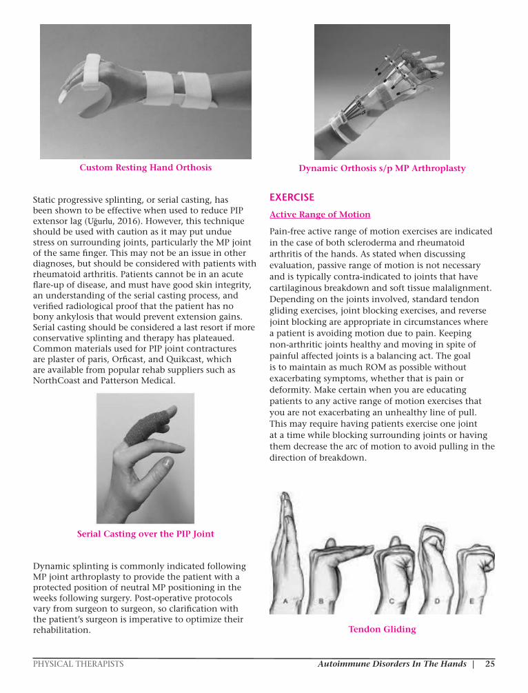
PHYSICAL THERAPISTS Autoimmune Disorders In The Hands | 25
Custom Resting Hand Orthosis
Static progressive splinting, or serial casting, has been shown to be effective when used to reduce PIP extensor lag (Uğurlu, 2016). However, this technique should be used with caution as it may put undue stress on surrounding joints, particularly the MP joint of the same finger. This may not be an issue in other diagnoses, but should be considered with patients with rheumatoid arthritis. Patients cannot be in an acute flare-up of disease, and must have good skin integrity, an understanding of the serial casting process, and verified radiological proof that the patient has no bony ankylosis that would prevent extension gains. Serial casting should be considered a last resort if more conservative splinting and therapy has plateaued. Common materials used for PIP joint contractures are plaster of paris, Orficast, and Quikcast, which are available from popular rehab suppliers such as NorthCoast and Patterson Medical.
Serial Casting over the PIP Joint
Dynamic splinting is commonly indicated following MP joint arthroplasty to provide the patient with a protected position of neutral MP positioning in the weeks following surgery. Post-operative protocols vary from surgeon to surgeon, so clarification with the patient’s surgeon is imperative to optimize their rehabilitation.
Dynamic Orthosis s/p MP Arthroplasty
EXERCISE
Active Range of Motion
Pain-free active range of motion exercises are indicated in the case of both scleroderma and rheumatoid arthritis of the hands. As stated when discussing evaluation, passive range of motion is not necessary and is typically contra-indicated to joints that have cartilaginous breakdown and soft tissue malalignment. Depending on the joints involved, standard tendon gliding exercises, joint blocking exercises, and reverse joint blocking are appropriate in circumstances where a patient is avoiding motion due to pain. Keeping non-arthritic joints healthy and moving in spite of painful affected joints is a balancing act. The goal is to maintain as much ROM as possible without exacerbating symptoms, whether that is pain or deformity. Make certain when you are educating patients to any active range of motion exercises that you are not exacerbating an unhealthy line of pull. This may require having patients exercise one joint at a time while blocking surrounding joints or having them decrease the arc of motion to avoid pulling in the direction of breakdown.
Tendon Gliding
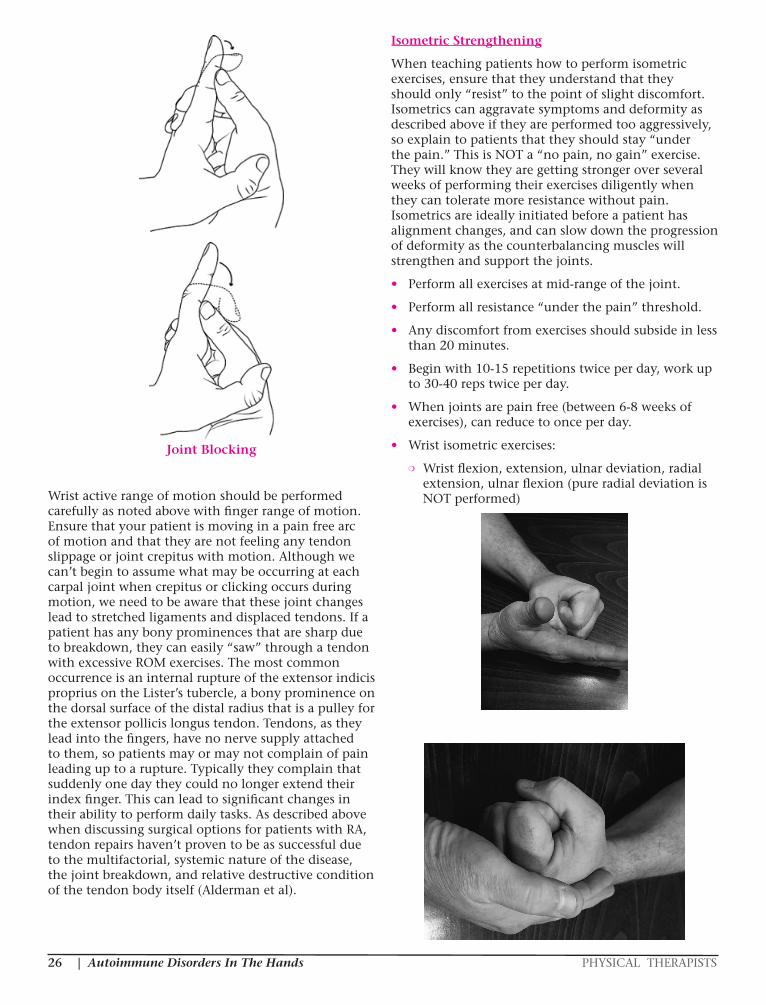
| Autoimmune Disorders In The Hands PHYSICAL THERAPISTS26
Joint Blocking
Wrist active range of motion should be performed carefully as noted above with finger range of motion. Ensure that your patient is moving in a pain free arc of motion and that they are not feeling any tendon slippage or joint crepitus with motion. Although we can’t begin to assume what may be occurring at each carpal joint when crepitus or clicking occurs during motion, we need to be aware that these joint changes lead to stretched ligaments and displaced tendons. If a patient has any bony prominences that are sharp due to breakdown, they can easily “saw” through a tendon with excessive ROM exercises. The most common occurrence is an internal rupture of the extensor indicis proprius on the Lister’s tubercle, a bony prominence on the dorsal surface of the distal radius that is a pulley for the extensor pollicis longus tendon. Tendons, as they lead into the fingers, have no nerve supply attached to them, so patients may or may not complain of pain leading up to a rupture. Typically they complain that suddenly one day they could no longer extend their index finger. This can lead to significant changes in their ability to perform daily tasks. As described above when discussing surgical options for patients with RA, tendon repairs haven’t proven to be as successful due to the multifactorial, systemic nature of the disease, the joint breakdown, and relative destructive condition of the tendon body itself (Alderman et al).
Isometric Strengthening
When teaching patients how to perform isometric exercises, ensure that they understand that they should only “resist” to the point of slight discomfort. Isometrics can aggravate symptoms and deformity as described above if they are performed too aggressively, so explain to patients that they should stay “under the pain.” This is NOT a “no pain, no gain” exercise. They will know they are getting stronger over several weeks of performing their exercises diligently when they can tolerate more resistance without pain. Isometrics are ideally initiated before a patient has alignment changes, and can slow down the progression of deformity as the counterbalancing muscles will strengthen and support the joints.
• Perform all exercises at mid-range of the joint.
• Perform all resistance “under the pain” threshold.
• Any discomfort from exercises should subside in less than 20 minutes.
• Begin with 10-15 repetitions twice per day, work up to 30-40 reps twice per day.
• When joints are pain free (between 6-8 weeks of exercises), can reduce to once per day.
• Wrist isometric exercises:
❍ Wrist flexion, extension, ulnar deviation, radial extension, ulnar flexion (pure radial deviation is NOT performed)
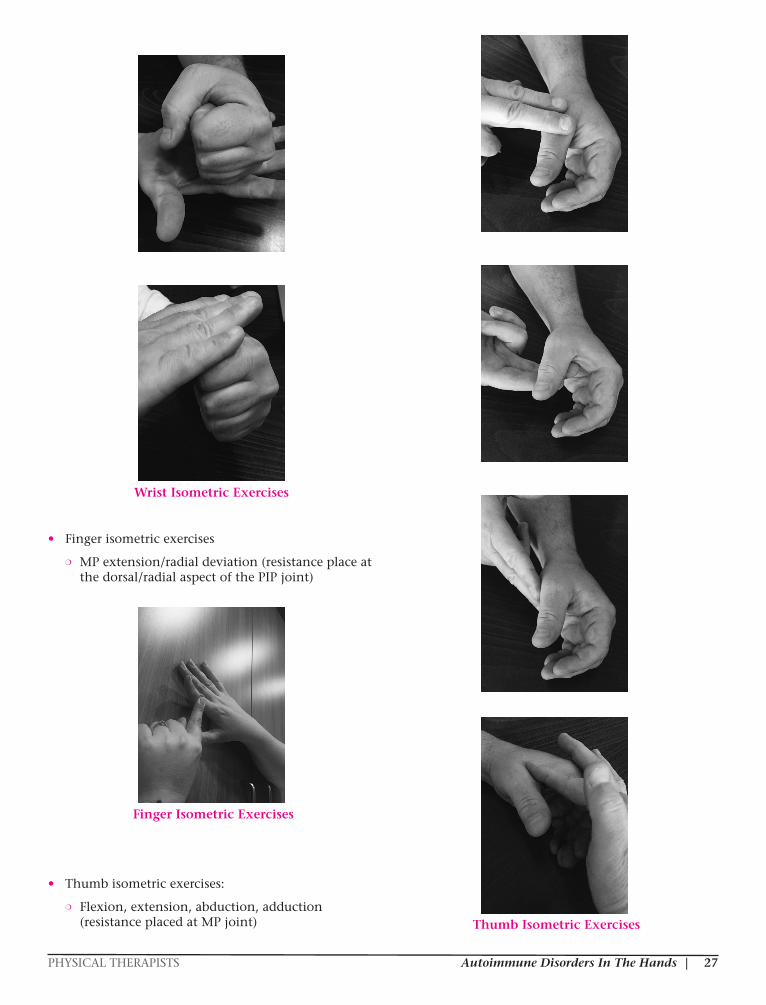
PHYSICAL THERAPISTS Autoimmune Disorders In The Hands | 27
Wrist Isometric Exercises
• Finger isometric exercises
❍ MP extension/radial deviation (resistance place at the dorsal/radial aspect of the PIP joint)
Finger Isometric Exercises
• Thumb isometric exercises:
❍ Flexion, extension, abduction, adduction (resistance placed at MP joint)
Thumb Isometric Exercises
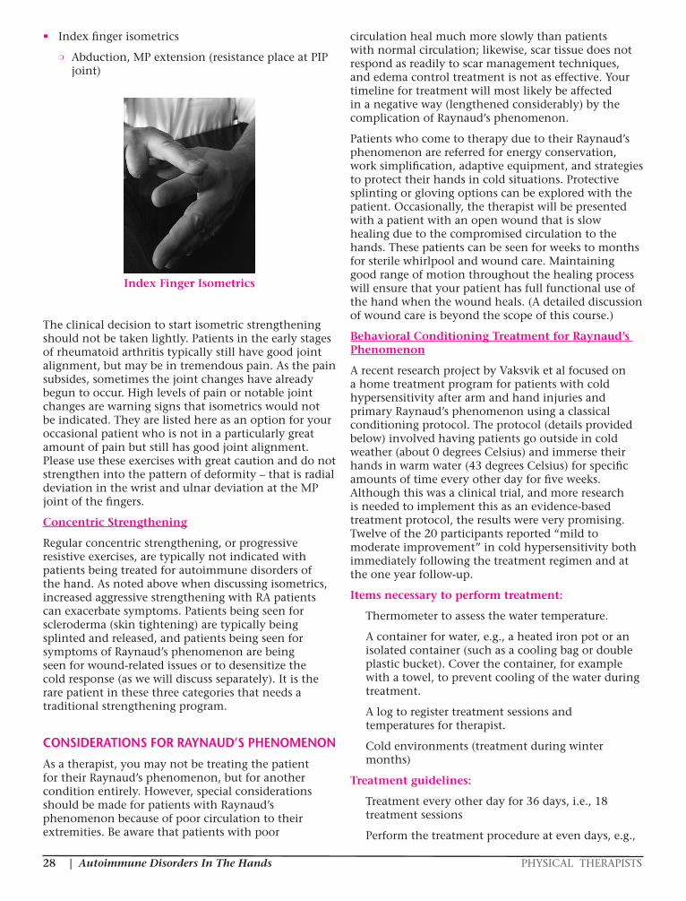
| Autoimmune Disorders In The Hands PHYSICAL THERAPISTS28
• Index finger isometrics
❍ Abduction, MP extension (resistance place at PIP joint)
Index Finger Isometrics
The clinical decision to start isometric strengthening should not be taken lightly. Patients in the early stages of rheumatoid arthritis typically still have good joint alignment, but may be in tremendous pain. As the pain subsides, sometimes the joint changes have already begun to occur. High levels of pain or notable joint changes are warning signs that isometrics would not be indicated. They are listed here as an option for your occasional patient who is not in a particularly great amount of pain but still has good joint alignment. Please use these exercises with great caution and do not strengthen into the pattern of deformity – that is radial deviation in the wrist and ulnar deviation at the MP joint of the fingers.
Concentric Strengthening
Regular concentric strengthening, or progressive resistive exercises, are typically not indicated with patients being treated for autoimmune disorders of the hand. As noted above when discussing isometrics, increased aggressive strengthening with RA patients can exacerbate symptoms. Patients being seen for scleroderma (skin tightening) are typically being splinted and released, and patients being seen for symptoms of Raynaud’s phenomenon are being seen for wound-related issues or to desensitize the cold response (as we will discuss separately). It is the rare patient in these three categories that needs a traditional strengthening program.
CONSIDERATIONS FOR RAYNAUD’S PHENOMENON
As a therapist, you may not be treating the patient for their Raynaud’s phenomenon, but for another condition entirely. However, special considerations should be made for patients with Raynaud’s phenomenon because of poor circulation to their extremities. Be aware that patients with poor
circulation heal much more slowly than patients with normal circulation; likewise, scar tissue does not respond as readily to scar management techniques, and edema control treatment is not as effective. Your timeline for treatment will most likely be affected in a negative way (lengthened considerably) by the complication of Raynaud’s phenomenon.
Patients who come to therapy due to their Raynaud’s phenomenon are referred for energy conservation, work simplification, adaptive equipment, and strategies to protect their hands in cold situations. Protective splinting or gloving options can be explored with the patient. Occasionally, the therapist will be presented with a patient with an open wound that is slow healing due to the compromised circulation to the hands. These patients can be seen for weeks to months for sterile whirlpool and wound care. Maintaining good range of motion throughout the healing process will ensure that your patient has full functional use of the hand when the wound heals. (A detailed discussion of wound care is beyond the scope of this course.)
Behavioral Conditioning Treatment for Raynaud’s Phenomenon
A recent research project by Vaksvik et al focused on a home treatment program for patients with cold hypersensitivity after arm and hand injuries and primary Raynaud’s phenomenon using a classical conditioning protocol. The protocol (details provided below) involved having patients go outside in cold weather (about 0 degrees Celsius) and immerse their hands in warm water (43 degrees Celsius) for specific amounts of time every other day for five weeks. Although this was a clinical trial, and more research is needed to implement this as an evidence-based treatment protocol, the results were very promising. Twelve of the 20 participants reported “mild to moderate improvement” in cold hypersensitivity both immediately following the treatment regimen and at the one year follow-up.
Items necessary to perform treatment:
Thermometer to assess the water temperature.
A container for water, e.g., a heated iron pot or an isolated container (such as a cooling bag or double plastic bucket). Cover the container, for example with a towel, to prevent cooling of the water during treatment.
A log to register treatment sessions and temperatures for therapist.
Cold environments (treatment during winter months)
Treatment guidelines:
Treatment every other day for 36 days, i.e., 18 treatment sessions
Perform the treatment procedure at even days, e.g.,
PHYSICAL THERAPISTS Autoimmune Disorders In The Hands | 29
January 10, 12, 14 etc. If you forget the treatment one day, continue the treatment on odd days. The procedure should be performed for a total of 18 days during five weeks. Try to keep your hands warm during the treatment period.
Ensure that the body is warm and avoid large meals before treatment. Dress in regular indoor clothing, such a long-sleeved shirt and trousers. If the weather is very cold, you may need a sweater and underpants, but it is important that you have a chilly feeling during treatment.
Log the date, time and outdoor temperature for every treatment day.
Procedure:
Fill a container with warm water from the tap. The water temperature should be about 43 C to give a pleasant warm feeling. Warmer water may elicit discomfort or pain. Record the water temperature in the log.
Bring the container to a cold garage, balcony, or another cold place.
Immerse both the hands and the wrists in the water for 8e10 min. Then, quickly dry the hands carefully with a towel. Return indoors and bring the container. Warm your body (5-10 min, until warm). Rewarm the water and ensure a temperature of 43 C.
Go out for a second time and immerse both hands in the water for 8-10 min.
Repeat the procedure a third time.
Perform the program including three hand immersions of 8-10 min every other day until you have completed 18 treatment sessions.
The research leading up to this study shows that the central nervous system can be strengthened or weakened by environmental stressors through experience and learning (Schacter, 2013). Vaksvik’s research supports the theory that vasomotor responses can be modified by classical conditioning, and if further researched, could be the basis for an effective treatment program for patients with Raynaud’s phenomenon.
CASE STUDY
MW is a 48 year old man who was referred to hand therapy following a spider bite to his left index finger.
He was seen by a primary physician two days after the bite, and then seen again a week later when he had significant progression of tissue loss, swelling, and pain. He was referred to a hand surgeon for further evaluation.
MW was taken into surgery for a debridement of the infected tissue and to attempt to irrigate any remaining venom from the spider. He was referred to therapy one
week following the debridement to regain his ROM and functional use of the left index finger.
History revealed that MW also has mild rheumatoid arthritis, Sjogren’s disease, scleroderma, and moderate Raynaud’s phenomenon. He is taking Humira to manage his RA, and a vasodilator to manage his Raynaud’s symptoms. He did not have any noted joint deformities or wrist instability at the time of evaluation.
MW is a business owner, and very motivated to return to full function. Upon evaluation, he had 40 degrees of MP flexion, 40 degrees of PIP flexion, and 10 degrees of DIP flexion of the index finger. He had full extension. His wound was located on the dorsal surface of the proximal phalanx, and MW had severe tissue restrictions forming from the healing response, tissue loss, and chronic swelling. His total active motion was 90 degrees (250-270 is normal) and his PROM was 120 degrees.
Therapist recommended therapy three times per week to address open wound and ROM deficits.
Following several weeks of sterile whirlpool and dressings, eventually the wound was closed and treatment focused more on scar management, regaining ROM, reducing soft tissue restrictions, and regaining strength. Wound closure took at least twice as long as usual (about 6 weeks vs 2-3 weeks for the size of wound) due to the poor circulation that is seen with patients with Raynaud’s phenomenon.
Due to MW’s rheumatoid arthritis, the therapist was very discriminatory with PROM exercises as to not exacerbate joint pain at the MP joint. AROM and very gentle PROM was used, taking special care to educate patient that any increase in joint pain is an indicator to reduce the intensity of exercises. Tendon gliding exercises, joint blocking exercises, and eventually strengthening with putty, digiflex, and flexbar (this is one of those rare occasions that very controlled concentircs are appropriate) were employed.
MW completed his therapy program in 36 visits over a period of 4.5 months with total active motion of the index finger of 230 degrees, and a grip strength within functional limits compared to his right hand.
We also reviewed energy conservation, work simplification, and adaptive equipment needs with regards to his business and his hobbies of playing baseball with his sons. We reviewed the progression of joint disease over time and MW was educated on what patterns of deformity to look for as his disease progresses. This is all information the MW was not aware of with his diagnosis, and he was very appreciative for the preparation.
This plan of care took approximately triple the amount of time that would typically be necessary following a spider bite. Because MW has several autoimmune disorders, our timeline for therapy was greatly increased; however, he eventually made a functional recovery and is pleased with his results.
| Autoimmune Disorders In The Hands PHYSICAL THERAPISTS30
ConclusionTherapeutic strategies for autoimmune disorders are unique in that we must take into account that these disease processes are chronic, complicated, and often progressive in nature. Until the medical community finds a cure for these challenging diseases, patients will rely on medication, surgery, and therapeutic management to address their pain and functional needs.
Patients with chronic diseases such as rheumatoid arthritis, scleroderma, and Raynaud’s phenomenon may need to follow-up with a therapist every 6-12 months to “tweak” their exercise programs, orthoses, and adaptive equipment needs. An alternative for patients being seen on a regular basis in the clinic is participation in an ongoing program such as “SMART” program used at the Hand Therapy Center at the Hospital for Special Surgery in New York City, which involves evaluation and establishment of a home program and subsequent through technology to remotely monitor and assess patients at their home (Wolff, 2014). Regardless of the therapy program designed, it is paramount to customize your plan of care to the patient’s individual needs.
Additional ResourcesAmerican Society for Surgery of the Hand (ASSH): www.assh.org
American Society of Hand Therapists (ASHT): www.asht.org
Arthritis Foundation: www.arthritis.org
Cold Hand Clinic: www.uchospitals.edu/specialties/plastic-surgery/cold-hands
E-Hand.Com: www.eatonhand.com
Mayo Clinic: www.mayoclinic.org
National Board for Certification in Occupational Therapy (NBCOT): www.nbcot.org
National Institute of Arthritis and Musculoskeletal and Skin Diseases (NIAMS): www.niams.nih.gov
North Coast: www.northcoastmedical.com
Orthobullets: www.orthobullets.com
Raynaud’s Association: www.raynauds.org
Scleroderma Foundation: www.scleroderma.org
World Health Organization: www.who.int
ReferencesAlderman AK, Chung KC, Kim HM, Fox DA, Ubel PA. Effectiveness of rheumatoid hand surgery: contrasting perceptions of hand surgeons and rheumatologists. J Hand Surg Am. 2003 Jan; 28(1):3-11.
Altman, Roy D. Merck Manuals <http://www.merckmanuals.com/home/bone,-joint,-and-muscle-disorders/joint-disorders/rheumatoid-arthritis-ra> N.p., n.d. Web. October 2016.
American Autoimmune Related Diseases Association <https://www.aarda.org/autoimmune-information/> N.p., n.d. Web. October 2016.
Casanova JS, ed. Clinical Assessment Recommendations. ed 3. Chicago, 2015, American Society of Hand Therapists.
Chim H, Reese A, Toomey S, Moran S. Update on the surgical treatment for rheumatoid arthritis of the wrist and hand. Journal of Hand Therapy 27 (2014) 134-142.
George M, Kiefhaber TR, Stern PJ. The Sauve–Kapandji Procedure and the Darrach Procedure for Distal Radio–Ulnar Joint Dysfunction after Colles’ Fracture J Hand Surg [Br] December 2004 29: 608-613.
Helmick CG, Felson DT, Lawrence RC, Gabriel S, Hirsch R, Kwoh CK, Liang MH, Kremers M, Mayes MD, Merkel PA, Pillemer SR, Reveille JD, Stone JH; National Arthritis Data Workgroup. Estimates of the prevalence of arthritis and other rheumatic conditions in the United States. Part I. Arthritis Rheum. 2008 Jan;58(1):15-25.
Kalliainen L, O’Brien V. Current uses of botulinum toxin A as an adjunct to hand therapy interventions of hand conditions. J Hand Ther 2013; 27: 85-95.
Kapandji A. Clinical test of apposition and counter-apposition of the thumb. Ann Chir Main 1986 (1): 67–73.
Malenfant D, Catton M, Pope JE. The efficacy of complementary and alternative medicine in the treatment of Raynaud’s phenomenon: a literature review and meta-analysis. Rheumatology (Oxford). 2009 Jul;48(7):791-5.
Mayo Clinic http://www.mayoclinic.org/healthy-lifestyle/nutrition-and-healthy-eating/in-depth/artificial-sweeteners/art-20046936, <http://www.mayoclinic.org/diseases-conditions/raynauds-disease/basics/risk-factors/con-20022916>, <http://www.mayoclinic.org/diseases-conditions/raynauds-disease/basics/treatment/con-20022916> N.p., n.d. Web. October 2016.
National Institute of Arthritis and Musculoskeletal and Skin Diseases (NIAMS) <http://www.niams.nih.gov/Health_Info/Scleroderma/default.asp>, <http://www.niams.nih.gov/Health_Info/Rheumatic_Disease/default.asp> N.p., n.d. Web. October 2016.

PHYSICAL THERAPISTS Autoimmune Disorders In The Hands | 31
Poole, JL. Musculoskeletal rehabilitation in the person with scleroderma. Curr Opin Rheumatol. 2010;22(2):205.
Sandqvist G, Hesslestrand R, Eberhardt K. A longitudinal follow-up of hand involvement and activities of daily living in early systemic sclerosis. Scand J Rheumatol 2009; 38:304-310.
Schachter DL, Wagner AD. Learning and memory. In: Kandel ER, Mack S, eds. Principles of Neural Science. 5th ed. New York: McGraw-Hill Medical; 2013: 1455e1456.
Scleroderma Foundation <http://www.scleroderma.org/site/PageServer?pagename=patients_whatis#.WBd5HC0rKUk> N.p., n.d. Web. October 2016.
Uğurlu U, Özdoğan H. Effects of serial casting in the treatment of flexion contractures of proximal interphalangeal joints in patients with rheumatoid arthritis and juvenile idiopathic arthritis: A retrospective study. J Hand Ther 2016; 29:41-50.
University of Chicago Medicine <http://www.uchospitals.edu/specialties/plastic-surgery/cold-hands> N.p., n.d. Web. October 2016.
Vaksvik T, Ruijs A, Rokkum M, Holm I. Evaluation of a home treatment program for cold hypersensitivity using a classical conditioning procedure in patients with hand and arm injuries. J Hand Ther 2016; 29:14-22.
Wolff A, Weinstock-Zlotnick G, Gordon, J. SSc management – In person appointments and remote therapy (SMART): A framework for management of chronic hand conditions. J of Hand Ther 2014; 27, 143-151.
| Autoimmune Disorders In The Hands PHYSICAL THERAPISTS32
Autoimmune Disorders in the Hands: Diagnosis, Treatment, and Therapeutic Interventions
Final Exam (3 CE Hours)
1. A period of heightened disease activity. In rheumatoid arthritis, it may be characterized by increased fatigue; fever; and painful, swollen, and tender joints.
a. Calcinosis b. Fibrosis c. Flare d. Mixed connective tissue disorder
2. A fabric-like material of fibrous threads that is a key component of the body’s connective tissues. In scleroderma, either too much is produced or it is produced in the wrong places, causing stiff and inflamed skin, blood vessels, and internal organs.
a. Antigen b. Collagen c. Sclerodactyly d. Synovium
3. In non-organ-specific disorders, the autoimmune response is spread throughout the body, affecting many body parts and systems simultaneously. Examples of these include ________.
a. Addison’s disease b. Hashimoto’s condition c. Lupus d. Pernicious anemia
4. The physician can consider a diagnosis of an autoimmune disorder when ________.
a. Bloodwork identifies diminished white blood cell count
b. Bloodwork indicates that autoantibodies and/or auto-nuclear antibodies are not present
c. Certain patient complaints (lethargy, unexplainable pain, discomfort, numbness, etc. are present
d. One or more specific factors in the blood, in conjunction with certain patient complaints (lethargy, unexplainable pain, discomfort, numbness, etc.), are present
5. The diagnosis of scleroderma relies heavily on ________.
a. Bloodwork, which usually reveals two antibodies – rheumatoid factor (RF) and antibodies to cyclic citrullinated peptide (anti-CCP)
b. Physical examination of skin appearance and texture, swollen fingers and hands, and tight skin around the hands, face, and mouth
c. X-rays, particularly in the early stages of the disease
d. All of the above
6. Secondary Reynaud’s ________. a. Can be due to damage to the blood vessels
supplying the hands arising from occupational injuries
b. Is an independent disorder, not a characteristic of scleroderma
c. Is caused by an autoimmune response attacking the blood vessels
d. Occurs directly in response to an occupational or toxic exposure
7. Rheumatoid arthritis (RA) is an inflammatory disease that causes ________.
a. A thickening and hardening effect to the skin and other soft tissue
b. Cold fingers and toes c. Pain, swelling, stiffness, and loss of function in
the joints d. Puffy fingers, finger ulcers, and marked
discomfort
8. As ________ progresses, the white blood cell invasion causes destruction to the cartilage and bones of the joint. While this breakdown occurs, the stretched out tendons and ligaments lose their alignment and ability to support the joint at rest, which results in changes in the line of pull of the joints during motion.
a. Raynaud’s disease b. Raynaud’s phenomenon c. Rheumatoid arthritis d. Scleroderma

PHYSICAL THERAPISTS Autoimmune Disorders In The Hands | 33
9. Most hand patients that suffer from ________ describe it as a feeling of having thick surgical gloves on all the time. The skin feels taut and thick, and all activities using the hands become more difficult.
a. Raynaud’s disease b. Raynaud’s phenomenon c. Rheumatoid arthritis d. Scleroderma
10. ________ occurs as part of the CREST symptoms. a. Raynaud’s disease b. Raynaud’s phenomenon c. Rheumatoid arthritis d. Scleroderma
11. In general, the hand rests with the MP joints in a neutral position of about ________ degrees of flexion, and the IP joints rest in slightly more flexion (35 degrees at the PIP, and 5-10 degrees at the DIP).
a. 2-5 b. 5-10 c. 10-15 d. 15-22
12. Commonly seen with rheumatoid arthritis , a ________ is a hyperextension at the PIP joint with subsequent flexion at the DIP joint.
a. Boutonniere deformity b. “Hitchhiker’s thumb” c. Swan Neck deformity d. Zig Zag deformity
13. A functional evaluation of thumb opposition is the use of the ________, which has not been approved by the American Society of Hand Therapists (ASHT) for widespread use as it has poor sensitivity when researched, but is widely accepted by therapists and doctors.
a. Finklestein maneuver b. Kapandji score c. Phalen test d. Tinel test
14. In arthritic joints, PROM ________. a. Is typically contraindicated b. Should be avoided in cases where a patient has
an obvious discrepancy between active and passive range
c. Should be used to push the joints past the patient’s active range of motion
d. Should push through to a painful end-feel
15. For support of the entire hand, the therapist should consider ________.
a. A resting hand orthosis (RHO) b. An Oval-8 splint c. A serpentine splint d. Silver Ring splints (SIRIS)
16. ________ are indicated in the case of both scleroderma and rheumatoid arthritis of the hands.
a. Isometrics, ideally initiated in response to alignment changes
b. Pain-free active range of motion exercises c. Passive range of motion exercises d. Regular concentric strengthening, or
progressive resistive exercises
17. When teaching patients how to perform isometric exercises, ________.
a. Encourage them to “resist” past the point of severe discomfort.
b. Ensure that they understand that these are “no pain, no gain” exercises
c. Explain that any discomfort should subside in around 45 minutes
d. Explain that they should stay “under the pain”
18. Vaksvik et al’s research ________. a. Could be the basis for an effective treatment
program for patients with Raynaud’s phenomenon
b. Disproves the theory that vasomotor responses can be modified by classical conditioning
c. Suggests that the central nervous system is not impacted by environmental stressors
d. None of the above
| Autoimmune Disorders In The Hands PHYSICAL THERAPISTS34
ANSWER SHEET
First Name: ______________________________ Last Name: _____________________________ Date: _______________
Address: _______________________________________________________City: __________________________________
State: ___________________________________ ZIP: __________________Country ________________________________
Phone: _________________________________ Email: _______________________________________________________
License/certification # and issuing state/organization ______________________________________________________
Clinical Fellow: Supervisor name and license/certification # ________________________________________________
Graduate Student: University name and expected graduation date __________________________________________
** See instructions on the cover page to submit your exams and pay for your course.
By submitting this final exam for grading, I hereby certify that I have spent the required time to study this course material and that I have personally completed each module/session of instruction.
Accessibility and/or special needs concerns? Contact customer service by phone at (888) 564-9098
or email at [email protected].
Refund and general policies are available online at http://pdhacademy.com/policies/
Autoimmune Disorders in the Hands: Diagnosis, Treatment, and Therapeutic Interventions Final Exam
1. A B C D
2. A B C D
3. A B C D
4. A B C D
5. A B C D
6. A B C D
7. A B C D
8. A B C D
9. A B C D
10. A B C D
11. A B C D
12. A B C D
13. A B C D
14. A B C D
15. A B C D
16. A B C D
17. A B C D
18. A B C D
PHYSICAL THERAPISTS Autoimmune Disorders In The Hands | 35
COURSE EVALUATION
Learner Name: ____________________________________________ Completion Date: _____________________________
❑ PT ❑ PTA ❑ OT ❑ OTA ❑ SLP ❑ SLPA Other: _______________________________________
Disagree Agree
Orientation was thorough and clear 1 2 3 4 5
Instructional personnel disclosures were readily available and clearly stated 1 2 3 4 5
Learning objectives were clearly stated 1 2 3 4 5
Completion requirements were clearly stated 1 2 3 4 5
Content was well-organized 1 2 3 4 5
Content was at or above entry-level knowledge 1 2 3 4 5
Content was substantiated through use of references, footnotes, etc. 1 2 3 4 5
Content reflected stated learning objectives 1 2 3 4 5
Exam assessed stated learning objectives 1 2 3 4 5
Exam was graded promptly 1 2 3 4 5
Satisfied with learning experience 1 2 3 4 5
Satisfied with customer service (if applicable) 1 2 3 4 5 n/a
What suggestions do you have to improve this program, if any?
_________________________________________________________________________________________________________
_________________________________________________________________________________________________________
What educational needs do you currently have?
_________________________________________________________________________________________________________
_________________________________________________________________________________________________________
What other courses or topics are of interest to you?
_________________________________________________________________________________________________________
_________________________________________________________________________________________________________
AUTOIMMUNE DISORDERS IN THE HANDS: DIAGNOSIS, TREATMENT, AND THERAPEUTIC INTERVENTIONS
(3 CE HOURS)