automated high-throughput sirna transfection in raw - hal - riip
TRANSCRIPT

HAL Id: pasteur-00683854https://hal-riip.archives-ouvertes.fr/pasteur-00683854
Submitted on 30 Mar 2012
HAL is a multi-disciplinary open accessarchive for the deposit and dissemination of sci-entific research documents, whether they are pub-lished or not. The documents may come fromteaching and research institutions in France orabroad, or from public or private research centers.
L’archive ouverte pluridisciplinaire HAL, estdestinée au dépôt et à la diffusion de documentsscientifiques de niveau recherche, publiés ou non,émanant des établissements d’enseignement et derecherche français ou étrangers, des laboratoirespublics ou privés.
Automated high-throughput siRNA transfection in raw264.7 macrophages: a case study for optimization
procedure.Jean-Philippe Carralot, Tae-Kyu Kim, Boris Lenseigne, Annette S Boese,
Peter Sommer, Auguste Genovesio, Priscille Brodin
To cite this version:Jean-Philippe Carralot, Tae-Kyu Kim, Boris Lenseigne, Annette S Boese, Peter Sommer, et al.. Au-tomated high-throughput siRNA transfection in raw 264.7 macrophages: a case study for optimiza-tion procedure.. Journal of Biomolecular Screening, SAGE Publications, 2009, 14 (2), pp.151-60.<10.1177/1087057108328762>. <pasteur-00683854>

Automated High-Throughput siRNA Transfection in Raw 264.7 macrophages: A Case Study for Optimization Procedure
JEAN-PHILIPPE CARRALOT,1 TAE-KYU KIM,1 BORIS LENSEIGNE,2 ANNETTE S. BOESE,3 PETER SOMMER,3 AUGUSTE GENOVESIO,2 and PRISCILLE BRODIN1
RNAi using siRNA is a very powerful tool for functional genomics to identify new drug targets and biological pathways. Although their use in epithelial cells is relatively easy and straightforward, transfection in other cell types is still challenging. The authors report the optimization of transfection conditions for Raw 267.4 macrophage cells. The herein described proce-dure makes use of automated confocal microscopy, enhanced green fluorescent protein (EGFP)–expressing macrophages, and fluorescently labeled siRNAs to simultaneously quantify both siRNA uptake and silencing efficiency. A comparison of 10 commercial transfectants was performed, leading to the selection of the transfectant giving the highest reproducible knock-down effect without inducing cell toxicity or cell activation. Several buffers used for siRNA/lipid complex assembly were tested, and such a study revealed the crucial importance of this parameter. In addition, a kinetics study led to the deter-mination of the optimal siRNA concentration and the best time window for the assay. In an original approach aimed at simultaneously optimizing both the high-throughput screening process and biological factors, optimal reagent volumes and a process flowchart were defined to ensure robust silencing efficiencies during screening. Such an account should pave the way for future genome-wide RNAi research in macrophages and present an optimization procedure for other “hard-to-transfect” cell lines. (Journal of Biomolecular Screening XXXX:xx-xx)
Key words: siRNA, transfection, macrophages, automation, optimization procedure
© 2009 Society for Biomolecular Sciences www.sbsonline.org 1
INTRODUCTION
O vER ThE PAST yEARS, RNA INTERFERENcE (RNAi) using short interfering RNA (siRNA) has been adopted as the
method of choice for loss-of-function studies in a variety of mammalian cells. Indeed, as the introduction of long double-stranded RNA induces innate immune responses,1 RNAi in mammalian cells is achieved using siRNA.2 The emergence of commercially available genome-wide libraries of chemically synthesized siRNAs has enabled the rapid development of high-throughput screening (hTS) of siRNA. however, one major drawback for successful siRNA screening resides in an efficient delivery of siRNA into cells. cell transfection through cationic lipid-based reagents has become one of the most popu-lar methods as it enables protocol automation and yields effi-cient knock-down in many cell types.2-4
For various cell lines such as heLa, a direct adaptation of the manufacturer’s single-tube protocol to the hTS format is sufficient to achieve efficient siRNA delivery. Therefore, reports on the optimization of lipid-mediated transfection are typically limited to transfectant screening and the optimization of siRNA and lipofectant concentrations.5 however, for other cell types, transfection still remains a challenge,6 and such classical opti-mization approaches do not lead to levels of knock-down suf-ficient for loss-of-function studies. For “hard-to-transfect” cell lines, viral vector-mediated expression of short hairpin RNAs (shRNAs) offers a possible alternative to lipid-mediated siRNA delivery.7,8 Nevertheless, several studies have reported higher variability in silencing efficacy exhibited by shRNAs compared with siRNAs.9 In addition, viral-mediated transduction might increase off-target effect risk due to an excessive presence of siRNA.10,11
Macrophages are key players of the immune system as they constitute the first cellular mediators of innate immune defense together with neutrophils and dendritic cells (Dcs). They are involved in many inflammatory diseases12 and are the target for many intracellular pathogens such as Listeria monocytogenes, Mycobacterium tuberculosis, or Leishmania spp.13 Despite such a central role in many diseases, the only genome-wide siRNA screenings in macrophage-like cells, to our knowledge, were
1Biology of Intracellular Pathogens Équipe Avenir Inserm, 2Image Mining group, and 3cell Biology of Retroviruses, Institut Pasteur Korea, Seoul, South Korea.
Received Jul 4, 2008 and in revised form Oct 12, 2008. Accepted for publica-tion Oct 16, 2008.
Journal of Biomolecular Screening XX(X); XXXX DOI: 10.1177/1087057108328762
J Biomol Screen OnlineFirst, published on February 4, 2009 as doi:10.1177/1087057108328762

Perrin et al.
2 www.sbsonline.org Journal of Biomolecular Screening XX(X); XXXX
performed in Drosophila S2 cells,14,15 and to date, no report exists on a siRNA screen using mammalian macrophages. The lack of such report might be explained by the fact that accepted models of macrophage-like cell lines, such as J774.1, Raw 264.7, and ThP-1, are particularly difficult to transfect.
In the present study, several parameters were tested to increase silencing efficacy in Raw 264.7 cells. In addition, method adaptation to the hTS format was embedded in bio-logical parameter optimization to ensure consistent knock-down levels during hTS. This original procedure led to the delivery of an automated high-throughput siRNA transfection protocol in murine macrophage-like Raw 264.7 cells.
MATERIALS AND METHODS
Generation of EGFP-expressing Raw 264.7 cell using the lentiviral system
vector particles were produced by transient cotransfection of hEK 293T/17 cells (ATcc cRL-11268) with a pTRIP-LTR-EGFP plasmid, an encapsidation plasmid (p8.71), and a vSv-g envelope expression plasmid (pvSv-G) using calcium phos-phate coprecipitation as described elsewhere.16,17 cell culture supernatants were harvested 48 h after transfection. Retroviral particles were recovered from the cell supernatant and concen-trated by ultracentrifugation (48,000 g/1h45/4 °c) on a Sorvall Rc-6 Plus Superspeed centrifuge (Thermo Fisher Scientific, Waltham, MA). After resuspension of the pellet in pure Dulbecco’s modified Eagle’s medium (DMEM) media (Gibco, carlsbad, cA), virus was stored at –80 °c.
Raw 264.7 (ATcc TIB-71) cells expressing enhanced green fluorescent protein (EGFP) were generated by Lentiviral trans-duction. Briefly, Raw 264.7 cells were plated on 96-well plates (Falcon) and incubated with a half dilution of thawed viral stock. Plates were centrifuged twice (300 g, 2 × 30 min) to increase infection efficiency and then placed at 37 °c. At 72 h posttransduction, more than 90% of the Raw 264.7 cells were expressing EGFP, as confirmed by fluorescence microscopy.
EGFP-expressing Raw 264.7 cells were cultured at 37 °c/5% cO2 in RPMI 1640 medium (Gibco) supplemented with 10% of fetal bovine serum (FBS; Gibco).
siRNA stocks
All siRNAs were labeled with Dy647 fluorochrome and were purchased from Dharmacon Thermofisher (Boulder, cO). Four labeled siRNAs targeting EGFP (target sequences: 5′-GGc TAc GTc cAG GAG cGc Acc-3′, 5′-GcA AGc TGA ccc TGA AGT Tc-3′, 5′-GGc AAG cTG Acc cTG AAG TTc-3′, 5′- GcG AcG TAA AcG Gcc AcA AGT Tc-3′) were resuspended at 20 µM in resuspension buffer (Dharmacon Thermofisher) and pooled together. Dy647-labeled nontargeting
siRNA (target sequence: 5′-U AAG GcU AUG AAG AGA UAc-3′) was resuspended at 20 µM and used as a control.
Transfectants
To determine the best transfectant for Raw 264.7 cells, 10 different lipofectants were tested. Dharmafect 1 (cat. T2001-02), Dharmafect 2 (cat. T2002-02), Dharmafect 3 (cat. T2003-02), and Dharmafect 4 (cat. T2004-02) were purchased from Dharmacon. hiPerFect (cat. 301704) and Effectene (cat. 301425) were purchased from QIAGEN Gmbh (hilden, Germany). INTERFERin (cat. 409-10) was purchased from Polyplus transfection SA (Illkirch, France). Lipofectamine 2000 (cat. 11668-027), Lipofectamine RNAiMax (cat. 13778-075), and Lipofectamine LTX (cat. 15338-100) were purchased from Invitrogen (carlsbad, cA). All transfectants were stored at 4 °c.
siRNA transfection
Dy647-labeled siRNAs were diluted in OPTIMEM medium (Gibco), and 5 µL of the diluted siRNA was distributed into each well of 384-well EvOTEc plates (cat. 781058) from Greiner Bio-One (Frickenhausen, Germany). Transfectant was gently mixed and diluted in OPTIMEM medium, and 5 µL of the diluted lipofectant was distributed onto the siRNA in each well. Lipid/siRNA complexes were allowed to form during 30 min at room temperature.
EGFP-expressing Raw 264.7 cells at 60% to 80% confluence were detached from culture vessels, washed with RPMI 1640 medium, and counted. cells were resuspended at 8.3 × 10⁴ cells/mL, and 30 µL (2500 cells) of the resulting cell suspension was dispensed onto the siRNA/lipid complexes. For siRNA transfec-tion, cells were always maintained at less than 5 passages.
Cell viability assay
On indicated day posttransfection, cell viability was evalu-ated with 100 µg/mL of Resazurin (cat. R7017-5G) purchased from Sigma (St. Louis, MO). After 2 h of incubation at 37 °c/5% cO2, resofurin fluorescence was measured at ex 544/em 595 nm on a spectraMax M5 microplate reader from Molecular Devices (Sunnyvale, cA). Relative cell viability was expressed as a percentage, taking nontransfected cells as a reference.
Automated fluorescence microscopy
Fluorescence images of transfected EGFP-expressing Raw 264.7 cells in 384-well plates were acquired using an OPERA automated fluorescence microscope from EvOTEc AG (hamburg, Germany) equipped with a 20×/0.7 NA water immersion lens. Of the 4 available light sources, 488- and
Carralot et al.
Caliper Nanofluidics Technology
Journal of Biomolecular Screening XX(X); XXXX www.sbsonline.org 3
635-nm confocal excitation lines were used to excite EGFP and Dy647, respectively. Emission was detected simultaneously using 2 ccD cameras in spectral bands at 535 and 690 nm. Five fields within each well were collected.
Image analysis
An appropriate threshold value set by the user led to an implicit cell/background separation and produced images that could be used for binary morphologic operations. Under a given value, all pixels were set to 0, whereas all pixels with a higher intensity were set to 1. In the same way, a second threshold on the red channel allowed for the identification of Dy647-labeled siRNAs. EGFP fluorescence of the cells was measured by con-sidering the global intensity of the green signal above threshold. The global fluorescence within the well was therefore considered rather than the fluorescence of the transfected cell subpopulation only. The percentage of EGFP expression was calculated taking EGFP fluorescence in nontransfected wells as a reference.
A morphological reconstruction was then performed using the masked red channel to reconstruct the threshold green chan-nel. This operation selected in the reconstructed channel all objects whose intersection with the mask was not empty and enabled the selection of transfected cells only. The percentage of transfected cells was determined as the ratio of transfected cells to total cells [(red signal-containing cells/(cells without red signal + red signal-containing cells))*100].
Finally, the average fluorescence of objects in the red chan-nel was used to quantify the amount of siRNA inside the trans-fected cells.
RESULTS
Transfectant screening
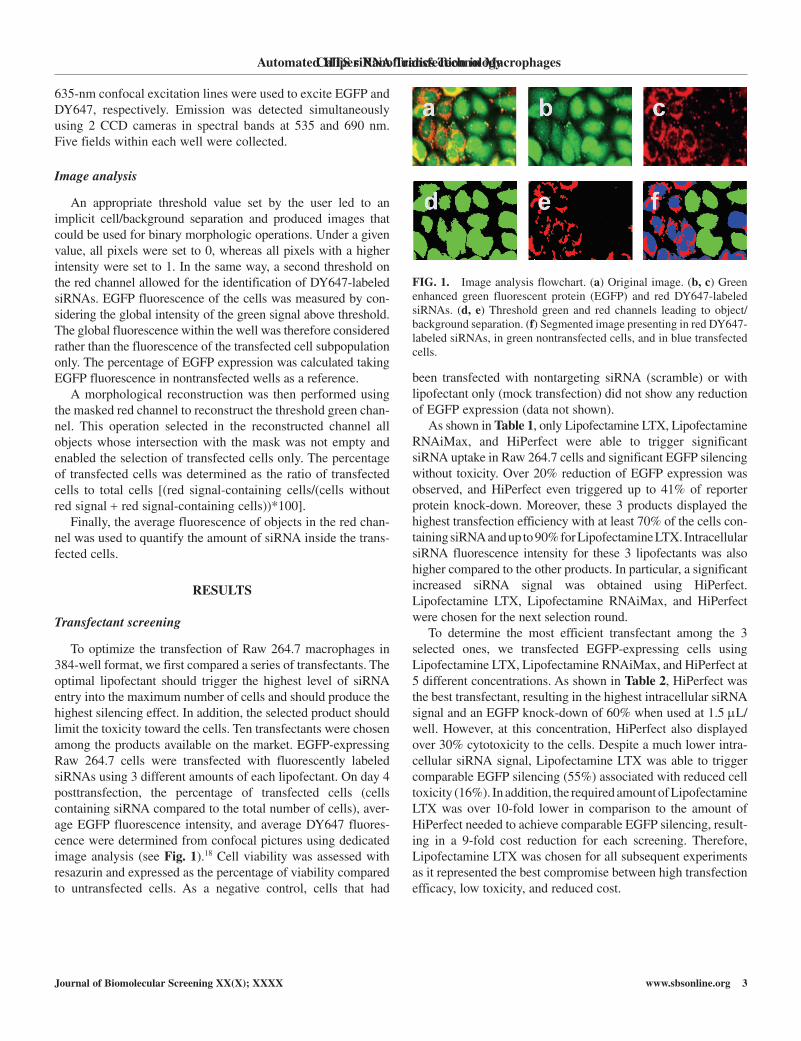
To optimize the transfection of Raw 264.7 macrophages in 384-well format, we first compared a series of transfectants. The optimal lipofectant should trigger the highest level of siRNA entry into the maximum number of cells and should produce the highest silencing effect. In addition, the selected product should limit the toxicity toward the cells. Ten transfectants were chosen among the products available on the market. EGFP-expressing Raw 264.7 cells were transfected with fluorescently labeled siRNAs using 3 different amounts of each lipofectant. On day 4 posttransfection, the percentage of transfected cells (cells containing siRNA compared to the total number of cells), aver-age EGFP fluorescence intensity, and average Dy647 fluores-cence were determined from confocal pictures using dedicated image analysis (see Fig. 1).18 cell viability was assessed with resazurin and expressed as the percentage of viability compared to untransfected cells. As a negative control, cells that had
been transfected with nontargeting siRNA (scramble) or with lipofectant only (mock transfection) did not show any reduction of EGFP expression (data not shown).
As shown in Table 1, only Lipofectamine LTX, Lipofectamine RNAiMax, and hiPerfect were able to trigger significant siRNA uptake in Raw 264.7 cells and significant EGFP silencing without toxicity. Over 20% reduction of EGFP expression was observed, and hiPerfect even triggered up to 41% of reporter protein knock-down. Moreover, these 3 products displayed the highest transfection efficiency with at least 70% of the cells con-taining siRNA and up to 90% for Lipofectamine LTX. Intracellular siRNA fluorescence intensity for these 3 lipofectants was also higher compared to the other products. In particular, a significant increased siRNA signal was obtained using hiPerfect. Lipofectamine LTX, Lipofectamine RNAiMax, and hiPerfect were chosen for the next selection round.
To determine the most efficient transfectant among the 3 selected ones, we transfected EGFP-expressing cells using Lipofectamine LTX, Lipofectamine RNAiMax, and hiPerfect at 5 different concentrations. As shown in Table 2, hiPerfect was the best transfectant, resulting in the highest intracellular siRNA signal and an EGFP knock-down of 60% when used at 1.5 µL/well. however, at this concentration, hiPerfect also displayed over 30% cytotoxicity to the cells. Despite a much lower intra-cellular siRNA signal, Lipofectamine LTX was able to trigger comparable EGFP silencing (55%) associated with reduced cell toxicity (16%). In addition, the required amount of Lipofectamine LTX was over 10-fold lower in comparison to the amount of hiPerfect needed to achieve comparable EGFP silencing, result-ing in a 9-fold cost reduction for each screening. Therefore, Lipofectamine LTX was chosen for all subsequent experiments as it represented the best compromise between high transfection efficacy, low toxicity, and reduced cost.
Automated HTS siRNA Transfection in Macrophages
FIG. 1. Image analysis flowchart. (a) Original image. (b, c) Green enhanced green fluorescent protein (EGFP) and red Dy647-labeled siRNAs. (d, e) Threshold green and red channels leading to object/ background separation. (f) Segmented image presenting in red Dy647-labeled siRNAs, in green nontransfected cells, and in blue transfected cells.

Perrin et al.
4 www.sbsonline.org Journal of Biomolecular Screening XX(X); XXXX
Transfectant buffer selection
Using Lipofectamine LTX, silencing typically ranged from 40% to 60%. To increase the knock-down efficacy, we then investigated the effect of the buffer used for the preparation of siRNA/lipofectant complexes. EGFP-expressing Raw 264.7 cells were transfected with Dy647-labeled siRNA using 0.1 µL
of Lipofectamine LTX diluted in different media or buffers. Percent EGFP silencing, viability, and intracellular siRNA sig-nal obtained with the different media/buffers are summarized in Figure 2. Dulbecco’s phosphate-buffered saline (DPBS) was the only buffer that showed increased EGFP knock-down (73% vs. 59%) compared to OPTIMEM, which was used in the first
Carralot et al.
Table 1. Lipid Reagents Primary Screening
Reagent Volume, µL/Well % of Transfected Cells % of Silencing siRNA (DY647) Fluorescence % of Viability
Dharmafect 1 0.1 57 ± 3 — 217 ± 6 114 ± 1 0.075 61 ± 2 — 223 ± 8 107 ± 1 0.05 67 ± 3 6 ± 2 264 ± 21 100 ± 2Dharmafect 2 0.1 67 ± 4 — 264 ± 27 111 ± 2 0.075 85 ± 4 — 277 ± 10 102 ± 1 0.05 71 ± 9 1 ± 7 261 ± 19 97 ± 2Dharmafect 3 0.1 81 ± 4 2 ± 7 284 ± 13 109 ± 0 0.075 78 ± 2 8 ± 3 270 ± 12 102 ± 4 0.05 69 ± 3 — 262 ± 12 97 ± 5Dharmafect 4 0.075 71 ± 4 — 266 ± 15 108 ± 3 0.05 86 ± 4 — 275 ± 3 102 ± 1 0.025 76 ± 12 — 270 ± 15 97 ± 53Lipofectamine RNAiMax 0.075 80 ± 9 32 ± 12 264 ± 5 106 ± 2 0.05 91 ± 4 33 ± 5 276 ± 5 100 ± 2 0.025 86 ± 4 10 ± 8 267 ± 8 95 ± 1Lipofectamine LTX 0.075 94 ± 2 26 ± 4 285 ± 1 108 ± 1 0.05 93 ± 8 23 ± 1 297 ± 1 102 ± 2 0.025 92 ± 7 13 ± 7 285 ± 9 98 ± 2Lipofectamine 2000 0.075 78 ± 4 — 285 ± 5 111 ± 5 0.05 75 ± 7 — 269 ± 13 105 ± 3 0.025 75 ± 3 3 ± 9 267 ± 13 98 ± 1InterferIN 0.25 24 ± 10 5 ± 7 173 ± 21 94 ± 5 0.1 37 ± 12 5 ± 6 208 ± 14 88 ± 6 0.075 57 ± 6 — 251 ± 9 92 ± 3hiPerfect 1 68 ± 13 39 ± 7 314 ± 23 97 ± 1 0.75 78 ± 21 41 ± 6 332 ± 18 94 ± 1 0.5 67 ± 12 30 ± 5 281 ± 12 96 ± 0Effectene 1 35 ± 12 — 226 ± 4 106 ± 1 0.75 67 ± 23 — 256 ± 34 99 ± 2 0.5 82 ± 9 1 ± 13 265 ± 16 96 ± 1
Raw 264.7 cells expressing enhanced green fluorescent protein (EGFP) were transfected with 75 nM of Dy647-labeled siRNA targeting EGFP using different lipofectants at the indicated concentrations. On day 4 posttransfection, pictures were taken at 488 and 635 nm on an OPERA automated confocal microscope. cells and siRNA fluorescence were analyzed using in-house developed IM software. cells were stained with 100 µg/mL of resazurin, and after 8 h at 37 °c, resofurin fluorescence was measured at em 544/ex 595 nm. Percentage of silenc-ing and viability were calculated using nontransfected cells as a reference. For each condition, average and standard deviation (± SD) values were determined for 3 replicate wells.
Table 2. Lipid Reagents Secondary Screening
Lipofectamine LTX Lipofectamine RNAiMax HiPerfect
µL/Well 0.15 0.125 0.1 0.075 0.05 0.15 0.125 0.1 0.075 0.05 2 1.5 1 0.75 0.5
% of silencing 70 ± 5 52 ± 18 55 ± 0 24 ± 0 35 ± 8 59 ± 3 31 ± 0 33 ± 6 — 10 ± 18 58 ± 12 60 ± 5 52 ± 17 50 ± 24 51 ± 6% of viability 73 ± 5 86 ± 30 84 ± 1 100 ± 22 91 ± 1 77 ± 1 88 ± 34 88 ± 3 99 ± 26 69 ± 27 48 ± 33 69 ± 7 92 ± 17 70 ± 7 75 ± 3siRNA 206 ± 28 197 ± 13 217 ± 2 210 ± 19 210 ± 27 223 ± 1 241 ± 12 241 ± 2 245 ± 11 225 ± 1 624 ± 37 840 ± 51 426 ± 23 203 ± 21 147 ± 13
Raw 264.7 cells expressing enhanced green fluorescent protein (EGFP) were transfected with 50 nM of Dy647-labeled siRNA targeting EGFP using Lipofectamine LTX, Lipofectamine RNAiMax, and hiPerfect at the indicated concentrations. On day 4 after transfection, pictures were taken at 488 and 635 nm on an OPERA automated confocal microscope. cells and siRNA fluorescence were analyzed using in-house developed IM software. On day 5 posttransfection, cells were stained with 100 µg/mL of resazurin, and after 8 h at 37 °c, resofurin fluorescence was measured at em 544/ex 595 nm. Percentages of silencing and cell viability were calculated using nontransfected cells as a reference. For each condition, average and standard deviation (± SD) values were determined for 2 replicate wells.

Caliper Nanofluidics Technology
Journal of Biomolecular Screening XX(X); XXXX www.sbsonline.org 5
set of experiments. This difference was likely due to a much more efficient siRNA uptake triggered by DPBS as an over 3-fold increase in Dy647 fluorescence was observed.
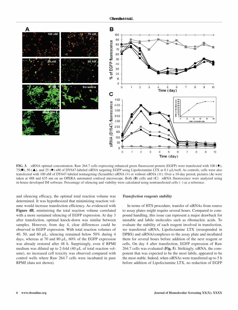
siRNA optimal concentration and silencing kinetics
To determine the optimal siRNA concentration, we trans-fected EGFP-expressing cells with different amounts of Dy647-labeled siRNA (Fig. 3). On day 4 posttransfection, no significant difference in EGFP silencing was observed between samples (Fig. 3B). Although knock-down efficiency reached ~75% at all siRNA concentrations, intracellular Dy647-labeled siRNA signals varied significantly. Entry of siRNA increased propor-tionally to the concentration used until 75 nM, when the intra-cellular siRNA signal reached a plateau (Fig. 3C, small box). Differential ribonucleic acid uptake influenced the kinetics of silencing as siRNA clearance from cells correlated with the end of gene expression repression. As shown in Figure 3B, maxi-mal silencing reached at day 4 was comparable for all concen-trations. however, when lower siRNA concentrations were applied (25 and 50 nM), EGFP expression began again from day 5 while knock-down was still maintained with higher siRNA concentrations (75 and 100 nM).
Optimal reagent volumes
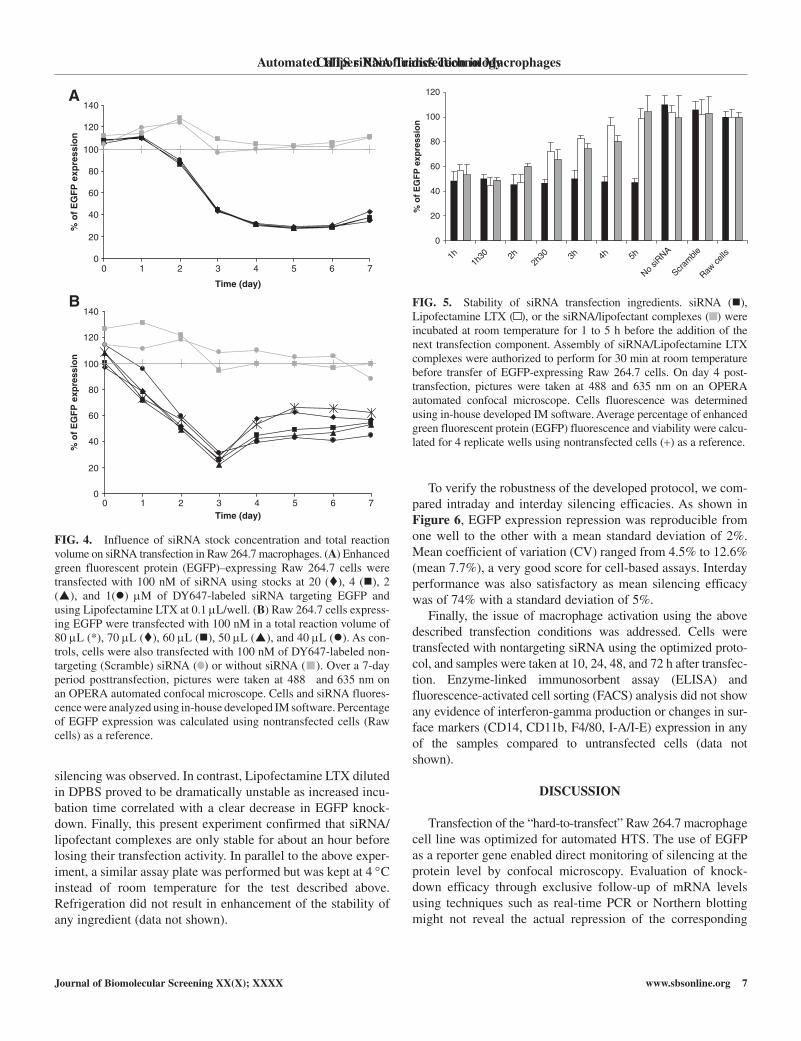
The effect of reagent volumes on the silencing efficacy was then investigated with the aim of adapting the method to hTS genome-wide siRNA screening. Optimal buffer volume for the assembly of siRNA/lipofectant complexes was first studied and did not have any noticeable effect on siRNA transfection effi-ciency (data not shown).
To determine the optimal volume to resuspend the genome-wide siRNA library, we studied the influence of siRNA stock concentration. Indeed, resuspension at low concentration could facilitate siRNA transfer through the handling of larger volumes. On the other hand, a larger volume of siRNA (resuspended in siRNA resuspension buffer) could dilute the buffer used for the assembly of siRNA/Lipofectant, a parameter that was shown to be essential (Fig. 2). To determine the optimal volume for siRNA, we transfected cells with 0.2, 1, 2, and 4 µL of siRNA solution at 20, 4, 2, and 1 µM, respectively. After cell addition (resuspended in 20 µL), the siRNA final concentration was 100 nM in all wells. As shown in Figure 4A, no significant difference was observed in EGFP knock-down efficiency for any of the volumes tested.
Finally, in terms of automation and with the objective of determining the best compromise between handling comfort
Automated HTS siRNA Transfection in Macrophages
FIG. 2. Screening of transfection mix buffer. Raw 264.7 cells expressing enhanced green fluorescent protein (EGFP) were transfected with 75 nM of Dy647-labeled siRNA targeting EGFP using Lipofectamine LTX at 0.1 µL/well. On day 4 posttransfection, pictures were taken at 488 and 635 nm on an OPERA automated confocal microscope. cells and siRNA fluorescence were analyzed using in-house developed IM software. On day 5 posttransfection, cells were stained with 100 µg/mL of resazurin, and after 8 h at 37 °c, resofurin fluorescence was measured at em 544/ex 595 nm. Percentage of silencing and viability were calculated using nontransfected cells as a reference. For each condition, average and standard deviation (± SD) values were determined for 2 replicate wells. MEM, minimal essential medium; DMEM, Dulbecco’s modified Eagle’s medium; PBS, phosphate-buffered saline; DPBS, Dulbecco’s phosphate-buffered saline.
Perrin et al.
6 www.sbsonline.org Journal of Biomolecular Screening XX(X); XXXX
and silencing efficacy, the optimal total reaction volume was determined. It was hypothesized that minimizing reaction vol-ume would increase transfection efficiency. As evidenced with Figure 4B, minimizing the total reaction volume correlated with a more sustained silencing of EGFP expression. At day 3 after transfection, optimal knock-down was similar between samples. however, from day 4, clear differences could be observed in EGFP expression. With total reaction volumes of 40, 50, and 60 µL, silencing remained below 50% during 4 days, whereas at 70 and 80 µL, 60% of the EGFP expression was already restored after 48 h. Surprisingly, even if RPMI medium was diluted up to 2-fold (40 µL of total reaction vol-ume), no increased cell toxicity was observed compared with control wells where Raw 264.7 cells were incubated in pure RPMI (data not shown).
Transfection reagents stability
In terms of hTS procedure, transfer of siRNAs from source to assay plates might require several hours. compared to com-pound handling, this issue can represent a major drawback for unstable and labile molecules such as ribonucleic acids. To evaluate the stability of each reagent involved in transfection, we transferred siRNA, Lipofectamine LTX (resuspended in DPBS) and siRNA/complexes to the assay plate and incubated them for several hours before addition of the next reagent or cells. On day 4 after transfection, EGFP expression of Raw 264.7 cells was evaluated (Fig. 5). Strikingly, siRNA, the com-ponent that was expected to be the most labile, appeared to be the most stable. Indeed, when siRNAs were transferred up to 5 h before addition of Lipofectamine LTX, no reduction of EGFP
Carralot et al.
FIG. 3. siRNA optimal concentration. Raw 264.7 cells expressing enhanced green fluorescent protein (EGFP) were transfected with 100 (), 75(), 50 (), and 25 () nM of Dy647-labeled siRNA targeting EGFP using Lipofectamine LTX at 0.1 µL/well. As controls, cells were also transfected with 100 nM of Dy647-labeled nontargeting (Scramble) siRNA ( ) or without siRNA ( ). Over a 10-day period, pictures (A) were taken at 488 and 635 nm on an OPERA automated confocal microscope. Both (B) cells and (C) siRNA fluorescence were analyzed using in-house developed IM software. Percentage of silencing and viability were calculated using nontransfected cells (+) as a reference.
Caliper Nanofluidics Technology
Journal of Biomolecular Screening XX(X); XXXX www.sbsonline.org 7
silencing was observed. In contrast, Lipofectamine LTX diluted in DPBS proved to be dramatically unstable as increased incu-bation time correlated with a clear decrease in EGFP knock-down. Finally, this present experiment confirmed that siRNA/ lipofectant complexes are only stable for about an hour before losing their transfection activity. In parallel to the above exper-iment, a similar assay plate was performed but was kept at 4 °c instead of room temperature for the test described above. Refrigeration did not result in enhancement of the stability of any ingredient (data not shown).
To verify the robustness of the developed protocol, we com-pared intraday and interday silencing efficacies. As shown in Figure 6, EGFP expression repression was reproducible from one well to the other with a mean standard deviation of 2%. Mean coefficient of variation (cv) ranged from 4.5% to 12.6% (mean 7.7%), a very good score for cell-based assays. Interday performance was also satisfactory as mean silencing efficacy was of 74% with a standard deviation of 5%.
Finally, the issue of macrophage activation using the above described transfection conditions was addressed. cells were transfected with nontargeting siRNA using the optimized proto-col, and samples were taken at 10, 24, 48, and 72 h after transfec-tion. Enzyme-linked immunosorbent assay (ELISA) and fluorescence-activated cell sorting (FAcS) analysis did not show any evidence of interferon-gamma production or changes in sur-face markers (cD14, cD11b, F4/80, I-A/I-E) expression in any of the samples compared to untransfected cells (data not shown).
DISCUSSION
Transfection of the “hard-to-transfect” Raw 264.7 macrophage cell line was optimized for automated hTS. The use of EGFP as a reporter gene enabled direct monitoring of silencing at the protein level by confocal microscopy. Evaluation of knock-down efficacy through exclusive follow-up of mRNA levels using techniques such as real-time PcR or Northern blotting might not reveal the actual repression of the corresponding
Automated HTS siRNA Transfection in Macrophages
0
20
40
60
80
100
120
140
0 1 2 3 4 5 6 7
Time (day)
% o
f E
GF
P e
xpre
ssio
n
0
20
40
60
80
100
120
140
0 1 2 3 4 5 6 7Time (day)
% o
f E
GF
P e
xpre
ssio
n
B
A
FIG. 4. Influence of siRNA stock concentration and total reaction volume on siRNA transfection in Raw 264.7 macrophages. (A) Enhanced green fluorescent protein (EGFP)–expressing Raw 264.7 cells were transfected with 100 nM of siRNA using stocks at 20 (), 4 (), 2 (), and 1() µM of Dy647-labeled siRNA targeting EGFP and using Lipofectamine LTX at 0.1 µL/well. (B) Raw 264.7 cells express-ing EGFP were transfected with 100 nM in a total reaction volume of 80 µL (*), 70 µL (), 60 µL (), 50 µL (), and 40 µL (). As con-trols, cells were also transfected with 100 nM of Dy647-labeled non-targeting (Scramble) siRNA ( ) or without siRNA ( ). Over a 7-day period posttransfection, pictures were taken at 488 and 635 nm on an OPERA automated confocal microscope. cells and siRNA fluores-cence were analyzed using in-house developed IM software. Percentage of EGFP expression was calculated using nontransfected cells (Raw cells) as a reference.
0
20
40
60
80
100
120
1h
1h30 2h
2h30 3h 4h 5h
No siR
NA
Scram
ble
Raw ce
lls
% o
f E
GF
P e
xpre
ssio
n
FIG. 5. Stability of siRNA transfection ingredients. siRNA (), Lipofectamine LTX ( ), or the siRNA/lipofectant complexes ( ) were incubated at room temperature for 1 to 5 h before the addition of the next transfection component. Assembly of siRNA/Lipofectamine LTX complexes were authorized to perform for 30 min at room temperature before transfer of EGFP-expressing Raw 264.7 cells. On day 4 post-transfection, pictures were taken at 488 and 635 nm on an OPERA automated confocal microscope. cells fluorescence was determined using in-house developed IM software. Average percentage of enhanced green fluorescent protein (EGFP) fluorescence and viability were calcu-lated for 4 replicate wells using nontransfected cells (+) as a reference.

Perrin et al.
8 www.sbsonline.org Journal of Biomolecular Screening XX(X); XXXX
protein. Several recent reports have indeed indicated that pro-tein expression levels only weakly correlate with correspond-ing mRNA levels,19-21 and consequently, mRNA levels are an inappropriate surrogate for protein expression evaluation.22 In addition, the model used for the optimization procedure was particularly challenging as transduced Raw 264.7 cells expressed relatively high levels of EGFP, a protein known for its long half-life.23,24 Therefore, optimization of silencing of this reporter protein should probably guarantee an efficient knock-down of endogenous proteins in follow-up phenotypic assays.
Dy647-labeled siRNA enabled the visualization of the inter-nalization of siRNA into the cells and the discrimination of trans-fected from nontransfected cells. Toxicity to the cells could be determined through cell number monitoring and through resaz-urin assay. The latter was the method of choice as it measures the actual metabolic activity of the cells compared to cell number, which does not provide any information on cell viability.
In a first step, 10 commercially available transfectants were screened based on their ability to transfect the highest number of cells, produce the maximum amount of siRNA entry into the cells, induce the greatest knock-down of EGFP expression, and minimize cell toxicity (Table 1). Of 3 compounds able to fulfill these criteria, Lipofectamine LTX was selected as the best can-didate for its capacity to induce 40% to 60% silencing with less than 20% cell mortality (Table 2). The optimal amount of Lipofectamine LTX amount was subsequently determined to be 0.1 µL/well; this was the maximum amount in which this reagent was triggering gene expression knock-down without major cyto-toxicity. Although high levels of intracellular siRNA were
obtained using Lipofectamine 2000 and Dharmafect 2, 3, and 4, no significant EGFP silencing was observed. This confirmed that an efficient transfectant should not only shuttle the siRNA into the cells but also ensure its delivery into the right subcellular compartment.25 Such a consideration is of particular importance when dealing with macrophages that display an enhanced phago-cytic activity. Indeed, siRNA/lipid complexes that uptake through the phagocytosis pathway rather than through endocytosis might dramatically change the siRNA intracellular fate.26
To further increase transfection efficacy, we tested different media or buffers for their effect on the assembly of siRNA/Lipofectamine LTX complexes (Fig. 2). Of the 8 solutions compared to the gold-standard OPTIMEM, DPBS was the only buffer that improved transfection efficacy. DPBS enhanced by 3-fold the siRNA uptake, which resulted in an increased EGFP silencing above 70%. This experiment confirmed that the physicochemical properties of buffer can greatly influence the formation of active siRNA/transfectant complexes. Similarly, several reports have demonstrated that helper molecules such as serum27 or DOPE28 can increase transfection activity of lipo-fectants. Such helper molecules can directly act on the siRNA/lipid complex formation, on its size, or in the intracellular delivery of the nucleic acid.29,30 We introduce here a novel critical parameter that should be tested for transfection optimi-zation in any cell line.
Direct visualization of reporter protein and siRNA allowed for the simultaneous monitoring of silencing and siRNA entry (Fig. 3A). These 2 parameters were of major importance in assessing siRNA optimal concentration and phenotypic assay schedule. At the different siRNA concentrations tested, EGFP knock-down was similar. however, silencing kinetics and intra-cellular siRNA follow-up revealed that only high siRNA con-centrations guaranteed sustained gene expression inhibition (Fig. 3B,C). Therefore, the siRNA concentration should be adjusted based on the planned phenotypic assay. For a “short-term” assay involving a readout within the same day, a low siRNA concentration of 25 or 50 nM can be employed to reduce the risk of an off-target effect.31 On the contrary, for “long-term” phenotypic assays, such as intracellular bacterial growth, the siRNA concentration should be raised to 75 or 100 nM to ensure gene silencing throughout the entire experiment.
To apply this transfection method to automated hTS, we optimized several parameters to match biological activity with robotized pippetting handling requirements. handling of large volumes generally results in reliable and accurate transfers from robotic systems. however, maximizing the different reagent vol-umes might also reduce the biological activity. The volume used for siRNA/lipofectant complex formation was found to have no effect on transfection efficiency (data not shown) and was there-fore set to 20 µL to facilitate component transfer. Similarly, several volume ratios of DPBS and siRNA resuspension buffer were tested to verify that DPBS buffer dilution would not impair
Carralot et al.
0
20
40
60
80
100
120
Exp 1 Exp 2 Exp 3 Exp 4
% o
f E
GF
P e
xpre
ssio
n
FIG. 6. Intraday and interday performance. Enhanced green fluores-cent protein (EGFP)–expressing Raw 264.7 cells were transfected with 100 nM of Dy647-labeled siRNA targeting EGFP using Lipofectamine LTX at 0.1 µL/well (). As controls, cells were also transfected with 100 nM of Dy647-labeled nontargeting (Scramble) siRNA ( ) or without siRNA ( ). On day 4 posttransfection, pictures w e r e taken at 488 and 635 nm on an OPERA automated confocal microscope. cells and siRNA fluorescences were analyzed using in-house developed IM software. Percentage of EGFP expression was calculated using nontransfected cells (+) as a reference.

Caliper Nanofluidics Technology
Journal of Biomolecular Screening XX(X); XXXX www.sbsonline.org 9
transfection efficiency. Raw 264.7 cell transfection with different siRNA volumes showed that DPBS could be diluted up to 1.25-fold without any loss of function (Fig. 4A). Finally, the optimal cell resuspension volume was determined. When total volume was above 60 µL, EGFP knock-down was reduced (Fig. 4B). Therefore, the cell resuspension volume was set to 30 µL as no increased cytotoxicity was observed under this condition.
For the objective of developing a method suitable for auto-mated hTS, the stability of the different products involved in siRNA transfection was tested because siRNA transfer from source to assay plates (~30 plates/day) might require several hours. Surprisingly, siRNA was found to be the most stable component, with no reduction in silencing efficacy for up to 5 h, whereas Lipofectamine LTX (resuspended in DPBS) and siRNA/ Lipofectamine LTX complexes were only stable for a couple of hours (Fig. 5). This biological result was crucial for hTS assay design, and consequently, the transfection protocol had to be adapted to match siRNA stability and Lipofectamine LTX lability.
In summary, to improve siRNA transfection, we tested sev-eral parameters close to both biological activity and the hTS process. New factors such as the buffer used for siRNA/lipo-fectant assembly or the stability of the different reagents were analyzed. This new, optimized procedure was successfully applied for the optimization of lipid-mediated siRNA transfec-tion of the macrophage-like Raw 264.7 cell line. This study led to the establishment of a robust method (Fig. 6) for the trans-fection of these “hard-to-transfect” cells in an hTS format.
ACKNOWLEDGMENTS
Reagents were kindly provided by Pierre charneau (Institut Pasteur Paris). We thank Lisa cechetto, and Fanny Ewann for critical reading of the manuscript.
REFERENCES
1. Gil J, Esteban M: Induction of apoptosis by the dsRNA-dependent pro-tein kinase (PKR): mechanism of action. Apoptosis 2000;5:107-114.
2. cullen LM, Arndt GM: Genome-wide screening for gene function using RNAi in mammalian cells. Immunol Cell Biol 2005;83:217-223.
3. Echeverri cJ, Perrimon N: high-throughput RNAi screening in cultured cells: a user’s guide. Nat Rev Genet 2006;7:373-384.
4. Zhang S, Zhao B, Jiang h, Wang B, Ma B: cationic lipids and polymers mediated vectors for delivery of siRNA. J Control Release 2007;123:1-10.
5. Borawski J, Lindeman A, Buxton F, Labow M, Gaither LA: Optimization procedure for small interfering RNA transfection in a 384-well format. J Biomol Screen 2007;12:546-559.
6. Rozema DB, Lewis DL: siRNA delivery technologies for mammalian systems. Targets 2003;2:253-260.
7. Arts G-J, Langemeijer E, Tissingh R, Ma L, Pavliska h, Dokic K, et al: Adenoviral vectors expressing siRNAs for discovery and validation of gene function. Genome Res 2003;13:2325-2332.
8. Rubinson DA, Dillon cP, Kwiatkowski Av, Sievers c, yang L, Kopinja J, et al: A lentivirus-based system to functionally silence genes in primary mammalian cells, stem cells and transgenic mice by RNA interference. Nat Genet 2003;33:401-406.
9. Sachse c, Echeverri cJ: Oncology studies using siRNA libraries: the
dawn of RNAi-based genomics. Oncogene 2004;23:8384-8391.
10. Persengiev SP, Zhu X, Green MR: Nonspecific, concentration-dependent
stimulation and repression of mammalian gene expression by small inter-
fering RNAs (siRNAs). RNA 2004;10:12-18.
11. Sledz cA, holko M, de veer MJ, Silverman Rh, Williams BRG:
Activation of the interferon system by short-interfering RNAs. Nat Cell
Biol 2003;5:834-839.
12. Sasaguri y, Tanimoto A: Role of macrophage-derived histamine in
atherosclerosis-chronic participation in the inflammatory response. J
Atheroscler Thromb 2004;11:122-130.13. christian B: Mechanisms and consequences of persistence of intracellular
pathogens: leishmaniasis as an example. Cell Microbiol 2008;10: 1221-1234.
14. Agaisse h, Burrack LS, Philips JA, Rubin EJ, Perrimon N, higgins DE: Genome-wide RNAi screen for host factors required for intracellular bacterial infection. Science 2005;309:1248-1251.
15. Philips JA, Rubin EJ, Perrimon N: Drosophila RNAi screen reveals cD36 family member required for mycobacterial infection. Science 2005;309: 1251-1253.
16. Boese A, Sommer P, Gaussin A, Reimann A, Nehrbass U: Ini1/hSNF5 is dispensable for retrovirus-induced cytoplasmic accumulation of PML and does not interfere with integration. FEBS Lett 2004;578:291-296.
17. Naldini L, Blomer U, Gallay P, Ory D, Mulligan R, Gage Fh, et al: In vivo gene delivery and stable transduction of nondividing cells by a len-tiviral vector. Science 1996;272:263-267.
18. Moon h, Genovesio A: IM, a computing approach for image mining of high throughput-high content screening. Presented at the 9th IEEE/AcM International conference on Grid computing 2008; September 29 to October 1, 2008; Tsukuba, Japan.
19. Tian Q, Stepaniants SB, Mao M, Weng L, Feetham Mc, Doyle MJ, et al: Integrated genomic and proteomic analyses of gene expression in mam-malian cells. Mol Cell Proteomics 2004;3:960-969.
20. Nie L, Wu G, Zhang W: correlation between mRNA and protein abun-dance in Desulfovibrio vulgaris: a multiple regression to identify sources of variations. Biochem Biophys Res Commun 2006;339:603-610.
21. chen G, Gharib TG, huang cc, Taylor JM, Misek DE, Kardia SL, et al: Discordant protein and mRNA expression in lung adenocarcinomas. Mol
Cell Proteomics 2002;1:304-313.22. Pascal L, True L, campbell D, Deutsch E, Risk M, coleman I, et al:
correlation of mRNA and protein levels: cell type-specific gene expression of cluster designation antigens in the prostate. BMC Genomics 2008;9:246.
23. Li X, Zhao X, Fang y, Jiang X, Duong T, Fan c, et al: Generation of destabilized green fluorescent protein as a transcription reporter. J Biol
Chem 1998;273:34970-34975.24. choi I, cho B-R, Kim D, Miyagawa S, Kubo T, Kim Jy, et al: choice of
the adequate detection time for the accurate evaluation of the efficiency of siRNA-induced gene silencing. J Biotechnol 2005;120:251-261.
25. Zuhorn I, Engberts J, hoekstra D: Gene delivery by cationic lipid vectors: overcoming cellular barriers. Eur Biophys J 2007;36:349-362.
26. Khalil IA, Kogure K, Akita h, harashima h: Uptake pathways and sub-sequent intracellular trafficking in nonviral gene delivery. Pharmacol Rev
2006;58:32-45.
Automated HTS siRNA Transfection in Macrophages

Perrin et al.
10 www.sbsonline.org Journal of Biomolecular Screening XX(X); XXXX
27. Zuhorn IS, visser Wh, Bakowsky U, Engberts JBFN, hoekstra D: Interference of serum with lipoplex-cell interaction: modulation of intracellular processing. Biochimica et Biophysica Acta (BBA)—Biomembranes 2002;1560:25-36.
28. Ma Bc, Zhang SB, Jiang hM, Zhao BD, Lv hT: Lipoplex morphologies and their influences on transfection efficiency in gene delivery. J Control Release 2007;123:184-194.
29. Wasungu L, hoekstra D: cationic lipids, lipoplexes and intracellular delivery of genes. J Control Release 2006;116:255-264.
30. Elouahabi A, Ruysschaert J-M: Formation and intracellular trafficking of lipoplexes and polyplexes. Mol Ther 2005;11:336-347.
31. Svoboda P: Off-targeting and other non-specific effects of RNAi experi-ments in mammalian cells. Curr Opin Mol Ther 2007;9:248-257.
Address correspondence to:Jean-Philippe Carralot
Institut Pasteur Korea39-1 Hawolgok-dong
Seongbuk-gu, Seoul 136-791, South Korea
E-mail: [email protected]
Carralot et al.