autophagy plays a protective role in free cholesterol … xu , yi yang , ming yan , jianan zhan ,...
TRANSCRIPT

This article is available online at http://www.jlr.org Journal of Lipid Research Volume 51, 2010 2581
Copyright © 2010 by the American Society for Biochemistry and Molecular Biology, Inc.
tose lipoproteins accumulate in the intimal layer of arte-rial wall and develop into “foam cells.” It is proposed that typical apoptosis of macrophages’ original foam cell is caused by the intracellular accumulation of unesterifi ed cholesterol ( 2 ). However, SMCs are more resistant to the accumulation of free cholesterol (FC) than macro-phages ( 3 ). Signifi cant increase of apoptotic cell death is not observed until SMCs are incubated with a higher concentration of cholesterol ( 4 ). Moreover, the studies using TUNEL technique reported that lipid-laden SMCs rarely undergo apoptosis in early atherosclerotic plaques ( 5 ). The investigation of human carotid plaques and plaques from cholesterol-fed rabbits also showed that dying SMCs exhibit typical ultrastructures of autophagic and necrotic cell death ( 6 ). These results indicate that SMCs may undergo a complex cell death mode after FC stimulation and that autophagy may provide an alterna-tive mechanism of cell death in lipid-laden SMCs.
Autophagy (specifi cally macroautophagy) is an evolu-tionarily conserved catabolic process for subcellular degra-dation of proteins, organelles, and other cytoplasmic components ( 7 ). As the degradation of cellular organelles generates free fatty acids, amino acids, and nucleotides that can be reused to fuel mitochondrial ATP production and maintain protein synthesis, autophagy is considered primarily a cell survival mechanism, especially during nu-trient deprivation ( 8 ). However, it has also been argued that autophagy may act as an initiator of cell death ( 9 ). Excessive autophagic activity may directly destroy a major proportion of the cytosol and organelles, or alternatively, it may trigger apoptosis rather than play a direct role in
Abstract Smooth muscle cells (SMC) make up most of the vascular system. In advanced atherosclerotic plaques, dying SMCs undergo a complex death mode. In the present study, we examined the activation of autophagy in SMCs over-loaded with excess free cholesterol (FC) and investigated the possible role which autophagy plays during the FC-induced cell death. After incubation with excess FC, a robust expression of autophagic vacuoles (AV) was detected using both fl uorescence microscopy and transmission electron mi-croscopy (TEM). The results revealed that FC induced a time-dependent upregulation of microtubule-associated protein-1 light chain 3-II (LC3-II). Inhibition of autophagy by 3-methyladenine (3-MA) enhanced both cell apoptosis and necrosis, while on the contrary, rapamycin inhibited cell death following cholesterol application. Furthermore, the impact of the colocalization of fragmented mitochondria with AVs was observed after cholesterol treatment. Our re-sults also revealed that the modulation of autophagy directly infl uenced the cellular organellar stress. In conclusion, our fi ndings demonstrated that excess FC induced the activation of autophagy in SMCs as a cellular defense mechanism, possibly through the degradation of dysfunc-tional organelles such as mitochondria and endoplasmic reticulum. —Xu, K., Y. Yang, M. Yan, J. Zhan, X. Fu, and X. Zheng. Autophagy plays a protective role in free cholesterol overload-induced death of smooth muscle cells. J. Lipid Res. 2010. 51: 2581–2590.
Supplementary key words cholesterol • cell death • organellar stress • smooth muscle cell
Smooth muscle cells (SMCs) are the major components of the vascular system. The loss of SMCs in advanced ath-erosclerotic lesions promotes lesional necrosis, compro-mises plaque stability, and ultimately triggers thrombotic vascular occlusion ( 1 ). In the late stage of atherosclerosis, a great number of SMCs and macrophages that phagocy-
This work was supported by the Key Laboratory of Biomedical Engineering of the Ministry of China and by the Key Laboratory of Chinese Medicine Screening, Exploitation and Medicinal Effectiveness Appraisal for Cardio-cerebral Vascu-lar and Nervous System of Zhejiang Province.
Manuscript received 23 January 2010 and in revised form 19 May 2010
Published, JLR Papers in Press, May 19, 2010 DOI 10.1194/jlr.M005702
Autophagy plays a protective role in free cholesterol overload-induced death of smooth muscle cells
Kedi Xu , Yi Yang , Ming Yan , Jianan Zhan , Xiao Fu, and Xiaoxiang Zheng 1
Department of Biomedical Engineering, Key Laboratory of Biomedical Engineering of Ministry of Education , Zhejiang University, Hangzhou, China
Abbreviations: ATG6, autophagy protein-6; AV, autophagic vacuole; Chol:M � CD, cholesterol-cyclodextrin complex; DAPK, death-associated protein kinase; � � m, mitochondrial membrane potential; ER, endo-plasmic reticulum; FC, free cholesterol; GSH, y-L-glutamyl-L-cysteinyl-glycine; 4-HNE, 4-hydroxynonenal; LC3, microtubule-associated protein-1 light chain 3; 3-MA, 3-methyladenine; NAC, N-acetyl-cysteine; PI, propidium iodide; ROS, reactive oxygen species; SMC, smooth muscle cell; TEM, transmission electron mic roscopy; mTOR, mammalian target of rapamycin; UPR, unfolded protein response.
1 To whom correspondence should be addressed. e-mail: [email protected]
by guest, on June 2, 2018w
ww
.jlr.orgD
ownloaded from

2582 Journal of Lipid Research Volume 51, 2010
the death process. For instance, stress in certain organ-elles, such as mitochondria and endoplasmic reticulum (ER), were indicated as stimulators of autophagic response because autophagy could mediate the specifi c removal of dysfunctional or damaged cytoplasmic organelles ( 10, 11 ). Several pathways that link the apoptotic and autophagic mechanisms also have been described at the molecular level ( 12 ). Thus, the result of a common stimulation may be decided by the crosstalk between autophagy and apo-ptosis or as a result of a cellular “decision” between these two responses.
In our present study, we found that overloading FC tri ggered autophagy in SMCs. This was confi rmed by the conversion of microtubule-associated protein-1 light chain 3-I (LC3-I) to a phosphatidylethanolamine-conjugated form (LC3-II) and the accumulation of autophagic vacu-oles (AV) under excess FC conditions. We further discov-ered that 3-methyladenine (3-MA) suppressed autophagy induced by FC overloading and led to large-scale cell death, whereas preincubation of rapamycin, an autophagy stimulator that inactivates mammalian target of rapamycin (mTOR), protected SMCs from death induced by excess FC. Moreover, the modulation of autophagy infl uenced the cellular organellar function, suggesting that au-tophagy played an important role in cell survival after FC overloading.
MATERIALS AND METHODS
Reagents and cell culture Unless otherwise noted, all reagents and chemicals were
purchased from Sigma. Annexin V/propidium iodide (PI), H2DCFDA, and JC-1 were from Invitrogen (CA). Antibody against KDEL (anti-GRP78/GRP94 SPA-827) was purchased from Stressgen (Victoria, Canada). The other antibodies were from Santa Cruz Biotechnology (Santa Cruz, CA).
Primary SMCs were obtained from the aortic media of a male Sprague-Dawley rat with the explants technique. Briefl y, tho-racic aorta was isolated and cleaned of adventitia and endothe-lium. The aorta was then cut into 1 mm sections, transferred to a culture dish, and maintained in growth medium of DMEM supplemented with 10% FBS, 2 mM L-glutamine, 4 mM sodium pyruvate, 100 U/ml streptomycin, and 100 U/ml penicillin. The initial migration of SMCs was observed within the fi rst 6 days. After 9 days of migration, these cells were transferred to a new culture dish. All experiments were conducted using SMCs be-tween passage 2 and 5. SMCs were identifi ed by immunocy-tochemical analysis with anti- � -actin antibody. The purity of the cultures was over 90%.
FC overloading of rat SMCs Cholesterol was delivered to SMCs by a cholesterol-cyclodex-
trin complex (Chol:M � CD) that contained approximately 50 mg of cholesterol/g solid (C4951, Sigma). All treatment concentra-tions were calculated based on cholesterol weight. SMCs were plated on the culture dish until the cells reached 90% confl u-ence. To load excess FC, the monolayer was then incubated with 30 µg/ml Chol:M � CD and the acyl-CoA:cholesterol acyltrans-ferase (ACAT) inhibitor sandoz58035 (58035) (10 µg/ml) in the culture medium for 24 h ( 4, 13 ). In some experiments, SMCs were preincubated with 10 mM 3-MA or 1 µM rapamycin for 4 h,
followed by the incubation with FC in the presence of 3-MA or rapamycin to modulate the autophagy activity.
Assessment of cell death and cellular ROS concentration with fl uorescent dyes
SMCs were collected using a brief trypsin treatment and labeled for 15 min with 5 µg/ml Alexa 488-conjugated Annexin V and 10 µg/ml PI according to the manufacture’s protocol. Labeled cells were analyzed with FACScan fl ow cytometry (Becton-Dickinson, Bedford, MA) using CellQuest software. The percentages of Annexin V- and PI-positive cells were calculated and presented as apoptosis and death percentage, respectively.
To evaluate the concentration of cellular ROS, SMCs were collected and stained with 5 µM H2DCFDA at 37°C for 20 min according to the manufacture’s protocol. The mean fl uorescence (FL1 channel, excitation 488 nm, emission 530 nm) of labeled cells was determined by fl ow cytometry.
Visualization of cellular organelles The pEGFP-LC3 expression vector was a gift from Dr. Yoshimori.
Mitochondria were visualized by expression of pDsRed2-mito (Clontech) in SMCs. Transfection was performed one day after subculture with Lipofectamine 2000 (Invitrogen) according to the manufacturer’s protocol. Fluorescence images were obtained using an inverted fl uorescent microscope (Olympus IX81; 60×; NA 1.45).
Western blot analysis SMCs were lysed in an appropriate volume of ice-cold radioim-
mune precipitation assay buffer containing 1% Triton X-100, 50 mM Tris/HCl, pH 7.4, 300 mM NaCl, 1:100 protease inhibitor cocktail, and 1:100 phosphatase inhibitor cocktail. Equal amounts of protein were loaded to 10% to 15% SDS-polyacrylamide gel. After electrophoresis, proteins were electrotransferred onto poly-vinylidene difl uoride membrane (Millipore). Membranes were blocked in 5% (w/v) nonfat dry milk for 1 h and probed over-night at 4°C with each primary antibody, followed by incubation with the secondary antibody for 2 h at room temperature. Anti-body detection was developed using 3,3,5,5-Tetramethylbenzi-dine Liquid Substrate kit (Amresco).
Transmission electron microscopy (TEM) analysis Samples were fi xed in 0.1 M sodium cacodylatebuffered (pH
7.4) for 2 h and postfi xed in 0.1 M sodium cacodylatebuffered (pH 7.4) 1% OsO 4 solution for 1 h. After dehydration in an etha-nol gradient [70% (v/v) ethanol (15 min), 80% (v/v) ethanol (15 min), 90% (v/v) ethanol (15 min), 100% ethanol (2 × 20 min)], samples were incubated with propylenoxid (2 × 10 min), impregnated with a mixture of propylenoid/LX-112 (Ladd Research Industries, 1:1), and embedded in LX-112. Ultrathin sections were stained with uranyl acetate and lead citrate. Sec-tions were examined in a Jeol-100 CX II TEM.
Detection and quantifi cation of mitochondrial membrane potential ( � � m)
To evaluate the depolarization of mitochondria, SMCs were collected and stained with 5 µM JC-1 at 37°C for 20 min accord-ing to the manufacture’s protocol. JC-1 undergoes aggregation within the mitochondrial matrix above a critical threshold con-centration. JC-1 aggregations appear as red fl uorescence (the FL2 channel of fl ow cytometry, excitation 488 nm, emission 585 nm), whereas the monomer of JC-1 appears as green fl uores-cence (FL1 channel). Mitochondrial depolarization will, there-fore, decrease the concentration of red JC-1 aggregates and increase the green fl uorescence ( 14 ). Thus, mean red (FL2) and
by guest, on June 2, 2018w
ww
.jlr.orgD
ownloaded from

Free cholesterol overloading activates autophagy 2583
green (FL1) fl uorescence were determined by fl ow cytometry for subsequent calculation of mean FL2:FL1 ratio, which was used as a semi-quantitative monitor of � � m.
Statistical analysis The data were plotted as mean ± SEM from three independent
experiments. Statistical signifi cant differences were carried out by one-way ANOVA followed by Tukey’s posthoc tests. P < 0.01 was considered statistically signifi cant.
RESULTS
FC loading induced complex SMC death In our previous work, treatment of SMCs with excess
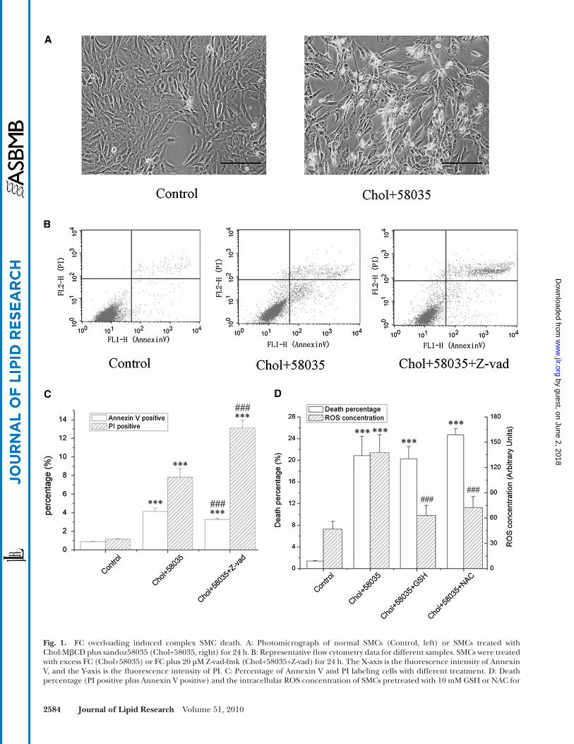
FC rapidly induced cell death ( Fig. 1A ) via the mitochon-drial and ER-dependent death pathway with widespread cellular organelle dysfunction ( 4 ). To further explore the mechanism of FC overload-induced cell death, SMCs were pretreated with Z-vad-fmk, a pan-caspase inhibitor that blocks cell apoptosis, and incubated with FC in the pres-ence of Z-vad-fmk for 24 h. As shown in Fig. 1B and C , the caspase inhibitor had no effect on the protection of SMCs under the lipid-laden condition as it led to a slight de-crease in the percentage of apoptotic (Annexin V positive) cells but signifi cant increase in the necrotic percentage (PI positive). Furthermore, FC overload-induced SMC death was accompanied with an upregulation of cellular reactive oxygen species (ROS), which we previously be-lieved to be a major inducer of cell death. Two widely used antioxidants, y-L-glutamyl-L-cysteinyl-glycine (GSH) and N-acetylcysteine (NAC), were used to reduce the produc-tion of cellular ROS. Unexpectedly, neither GSH nor NAC could protect SMCs from FC-induced cell death, though both antioxidants recovered the cellular ROS level ( Fig. 1D ). These results suggested that apoptosis might not be the only pathway involved in FC overload-induced SMC death and that other mechanisms, such as autophagy and necrosis, might contribute to this complex mode of cell death.
Autophagy was activated in FC-overloaded SMCs Since the formation of AVs is by far the most important
feature of autophagy, demonstration of these structures by electron microscopy is considered the gold standard for documenting autophagy. Hence, we examined the ultra-structural changes in SMCs upon cholesterol treatment. AVs were rarely detected in either normal SMCs ( Fig. 2A , a, b) or SMCs incubated with Chol:M � CD alone ( Fig. 2A , c, d). After overloading SMCs with FC for 24 h, a large number of AVs formed and were distributed throughout the whole cytoplasm ( Fig. 2A , e–h, and 2B ). Most of the AVs contained the remnants of cellular organelles (black arrows in Fig. 2A , f). Mild to extensive autophagic vacu-olizations were also observed in FC-overloaded SMCs ( Fig. 2A , e–h). Note that no pronounced chromatin condensa-tion in the nucleus was presented in any specimens.
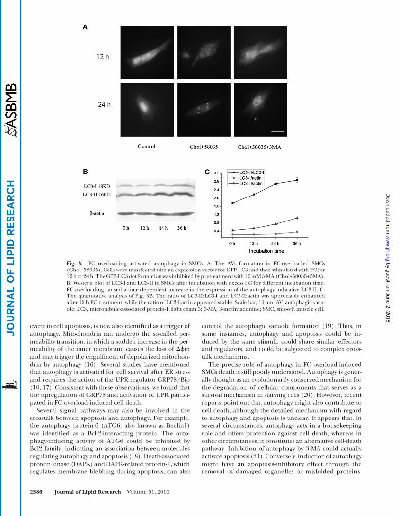
The AV formation in living cells was determined by fl uo-rescence microscopy. As shown in Fig. 3A , in normal SMCs, most GFP-LC3 protein was distributed diffusely through-
out the whole cell, although a small part of normal SMCs contained low-level punctual fl uorescence dots (especially in SMCs after long-term culture). After treatment with ex-cess FC, diffuse cytoplasmic forms were redistributed to discrete vesicular structures of GFP-LC3, suggesting the activation of autophagy. The formation of vesicular struc-tures was absent in SMCs pretreated with the specifi c au-tophagy inhibitor 3-MA. Autophagy induction was further confi rmed by Western blotting analysis with anti-LC3 antibody. Overloading SMCs with FC caused a signifi cant and time-dependent increase in the expression of the autophagy-indicative LC3-II. The ratio of LC3-II:LC3-I was enhanced appreciably after a 12 h treatment, and a robust production of LC3-II was observed after a 24 h treatment ( Fig. 3B, C ). These results further strengthened the hy-pothesis that autophagy was activated in FC-treated SMCs.
Autophagy provided protection from FC overload-induced cell death
To further clarify the function of autophagy as a pro-death or pro-survival pathway in FC-overloaded SMCs, both autophagy inhibitor and inducer were applied. 3-MA was reported to suppress autophagic activity by preventing the formation of AVs ( 15 ). In our experiments, the expres-sion of LC3-II was dramatically low in SMCs pretreated with 10 mM 3-MA for 4 h followed by 24 h incubation with FC in the presence of 3-MA ( Fig. 4B , C ). Meanwhile, 3-MA signifi cantly triggered cell apoptosis and death in FC-over-loaded SMCs ( Fig. 4A , 16.4 ± 2.0% for annexin V-positive and 28.5 ± 3.7% for PI-positive, both upregulated about three times compared with the model group). Next, we investigated whether induction of autophagy with rapa-mycin could have a protective effect from FC overload-induced cell death( Fig. 4B, C ). SMCs treated with rapamycin exhibited a reduced susceptibility to the excess FC treat-ment, as demonstrated by a lower annexin V- and PI-posi-tive cell percentage. However, this protective effect was not complete, and it was entirely reversed when the FC-overloaded cells were simultaneously treated with 3-MA plus rapamycin, suggesting that the protective effect of rapamycin was blocked when autophagy was inhibited ( Fig. 4A ).
Enhanced autophagy activity reduced mitochondrial and ER stress
A possible mechanism for the protective effect of autophagy was that autophagy accelerated the clearance of impaired cellular organelles. To verify this hypothesis, SMCs were cotransfected with DsRed2-Mitochondria and GFP-LC3 plasmids. As illustrated in Fig. 5A , in normal SMCs, most LC3 protein was distributed diffusely through-out the whole cell without colocalization of DsRed2-labeled mitochondria and GFP-LC3-labeled AVs. The FC overload induced the formation of punctual AVs and the fragmentation of mitochondria ( Fig. 5A , middle line). The colocalization of AVs and mitochondria were more frequently detected after 24 h treatment with FC ( Fig. 5A , white arrows in the last line). We then measured the � � m to estimate the mitochondrial function. The inhibition of
by guest, on June 2, 2018w
ww
.jlr.orgD
ownloaded from
2584 Journal of Lipid Research Volume 51, 2010
Fig. 1. FC overloading induced complex SMC death. A: Photomicrograph of normal SMCs (Control, left) or SMCs treated with Chol:M � CD plus sandoz58035 (Chol+58035, right) for 24 h. B: Representative fl ow cytometry data for different samples. SMCs were treated with excess FC (Chol+58035) or FC plus 20 µM Z-vad-fmk (Chol+58035+Z-vad) for 24 h. The X-axis is the fl uorescence intensity of Annexin V, and the Y-axis is the fl uorescence intensity of PI. C: Percentage of Annexin V and PI labeling cells with different treatment. D: Death percentage (PI positive plus Annexin V positive) and the intracellular ROS concentration of SMCs pretreated with 10 mM GSH or NAC for
by guest, on June 2, 2018w
ww
.jlr.orgD
ownloaded from

Free cholesterol overloading activates autophagy 2585
trast to the FC overload-induced macrophage death, only a small increase of annexin V labeling was noticed after excess FC treatment, and the caspase inhibitor z-vad-fmk could not rescue SMCs from cell death. Although the FC-overloaded SMCs presented a high level of cellular ROS accompanied by the loss of � � m, antioxidants such as GSH and NAC failed to extenuate cell death. On the other hand, autophagy was also involved in FC overload-induced cell death. The activation of autophagy was ob-served from the formation of punctual AVs and increasing LC3-II conversion and was further confi rmed by TEM analysis. Large-scale damage of cellular organelles, includ-ing mitochondria and ER, was observed after SMCs were incubated with excess FC. Most of the AVs in the FC-over-loaded SMCs contained the remnants of these cellular or-ganelles, which indicated that excess FC induced severe cellular organelle damage and that these cellular organ-elles might be cleared through autophagy. Thus, the pres-ent study illustrated that, after overloaded with FC, SMCs underwent a complex mode of cell death that involved apoptosis, autophagy, and to some extent, necrosis.
Emerging lines of evidence have revealed the potential link between autophagy and apoptosis ( 12 ). Autophagy and apoptosis can be elicited by common upstream regu-lators, such as impaired cellular organelles. The dysfunc-tion of mitochondria, formerly recognized as an initial
autophagy by 3-MA resulted in an apparent reduction in the ratios of red (FL2) to green (FL1) fl uorescence, indi-cating a large-scale mitochondrial depolarization. Con-versely, SMCs incubated with FC plus rapamycin maintained a better � � m condition than those overloaded with FC ( Fig. 5B ). Furthermore, both GRP78 and GRP94 protein levels, a positive symbol of unfolded protein response (UPR) activation and ER stress, declined after rapamycin treatment and rose with 3-MA treatment ( Fig. 5C, D ). These results illustrated that autophagy acted as a cellular defense mechanism and promoted cell survival in FC-over-loaded SMCs, possibly by facilitating the clearance of im-paired cellular organelles.
DISCUSSION
FC overload-induced vascular cell death is considered an important event in progression of atherosclerosis. Recently, Feng reported that excess intracellular FC tri ggered macrophage death with the characteristics of apoptosis ( 2 ). In our previous study, FC was also found to be a potent inducer of rat aortic SMC death; both mito-chondrial- and ER-dependent apoptotic signal pathways were involved in FC-induced SMC death ( 4 ). However, the present study demonstrated that apoptosis may not be the only manner of FC overload-induced SMC death. In con-
Fig. 2. TEM micrographs of autophagic vacuoles. A: Representative electron micrographs of normal SMCs (a, b), SMCs incubated with Chol:M � CD for 24 h (c, d), and SMCs treated with Chol:M � CD plus sandoz58035 (e–h). AVs contain the remnants of cellular organelles, such as mitochondria and ER (indicated by black arrows). Myelin fi gure-like structures are indicated by white arrows. N = nucleus. Scale bar = 2 µm or 0.5 µm. B: The average number of AVs present in normal SMCs, Chol:M � CD-treated SMCs (Cholesterol), and FC-overloaded SMCs (Chol+58035). *** P < 0.001 compared with control group. AV, autophagic vacuole; Chol:M � CD, cholesterol-cyclodextrin complex; ER, endoplasmic reticulum; FC, free cholesterol; SMC, smooth muscle cell.
4 h followed by 24 h treatment of FC plus GSH or NAC. Data correspond to mean ± SEM, n = 6, *** P < 0.001 compared with control group; ## P < 0.01, ### P < 0.001 compared with Chol+58035 group. Scale bar, 50 µm. Chol:M � CD, cholesterol-cyclodextrin complex; FC, free cho-lesterol; GSH, y-L-glutamyl-L-cysteinyl-glycine; PI, propidium iodide; ROS, reactive oxygen species; SMC, smooth muscle cell.
by guest, on June 2, 2018w
ww
.jlr.orgD
ownloaded from
2586 Journal of Lipid Research Volume 51, 2010
control the autophagic vacuole formation ( 19 ). Thus, in some instances, autophagy and apoptosis could be in-duced by the same stimuli, could share similar effectors and regulators, and could be subjected to complex cross-talk mechanisms.
The precise role of autophagy in FC overload-induced SMCs death is still poorly understood. Autophagy is gener-ally thought as an evolutionarily conserved mechanism for the degradation of cellular components that serves as a survival mechanism in starving cells ( 20 ). However, recent reports point out that autophagy might also contribute to cell death, although the detailed mechanism with regard to autophagy and apoptosis is unclear. It appears that, in several circumstances, autophagy acts in a housekeeping role and offers protection against cell death, whereas in other circumstances, it constitutes an alternative cell-death pathway. Inhibition of autophagy by 3-MA could actually activate apoptosis ( 21 ). Conversely, induction of autophagy might have an apoptosis-inhibitory effect through the removal of damaged organelles or misfolded proteins.
event in cell apoptosis, is now also identifi ed as a trigger of autophagy. Mitochondria can undergo the so-called per-meability transition, in which a sudden increase in the per-meability of the inner membrane causes the loss of � � m and may trigger the engulfment of depolarized mitochon-dria by autophagy ( 16 ). Several studies have mentioned that autophagy is activated for cell survival after ER stress and requires the action of the UPR regulator GRP78/Bip ( 10, 17 ). Consistent with these observations, we found that the upregulation of GRP78 and activation of UPR partici-pated in FC overload-induced cell death.
Several signal pathways may also be involved in the crosstalk between apoptosis and autophagy. For example, the autophagy protein-6 (ATG6, also known as Beclin1) was identifi ed as a Bcl-2-interacting protein. The auto-phagy-inducing activity of ATG6 could be inhibited by Bcl2 family, indicating an association between molecules regulating autophagy and apoptosis ( 18 ). Death-associated protein kinase (DAPK) and DAPK-related protein-1, which regulates membrane blebbing during apoptosis, can also
Fig. 3. FC overloading activated autophagy in SMCs. A: The AVs formation in FC-overloaded SMCs (Chol+58035). Cells were transfected with an expression vector for GFP-LC3 and then stimulated with FC for 12 h or 24 h. The GFP-LC3 dot formation was inhibited by pretreatment with 10 mM 3-MA (Chol+58035+3MA). B: Western blot of LC3-I and LC3-II in SMCs after incubation with excess FC for different incubation time. FC overloading caused a time-dependent increase in the expression of the autophagy-indicative LC3-II. C: The quantitative analysis of Fig. 3B . The ratio of LC3-II:LC3-I and LC3-II:actin was appreciably enhanced after 12 h FC treatment, while the ratio of LC3-I:actin appeared stable. Scale bar, 10 µm. AV, autophagic vacu-ole; LC3, microtubule-associated protein-1 light chain 3; 3-MA, 3-methyladenine; SMC, smooth muscle cell.
by guest, on June 2, 2018w
ww
.jlr.orgD
ownloaded from

Free cholesterol overloading activates autophagy 2587
SMCs. Pretreatment with rapamycin stabilized � � m and decreased FC overload-induced ER stress. However, we cannot exclude the possibility that autophagy may also play an active role in FC overload-induced SMC death. Autophagy may provide intrinsic degradation machinery
Pretreatment of cells with rapamycin can reduce mito-chondria load to � 50% while having a protective effect against pro-apoptotic insults ( 22 ). In our present experi-ments, it seems more likely that autophagy served a self-protective role that promoted cell survival in FC-overloaded
Fig. 4. Modulation of autophagy-impacted cell death in FC-overloaded SMCs. A: SMCs were incubated with FC in the presence of 10 mM 3-MA and/or 1µM rapamycin for 24 h. Both 3-MA and rapamycin were added 4 h before FC treatment. The percentage of apoptosis and necrosis were then assessed. B: Western blot of LC3-I and LC3-II expression in SMCs after incubation with excess FC plus 3MA or rapamycin. C: The ratio of LC3-II:LC3-I in 3MA- or rapamycin-treated SMCs. Data correspond to mean ± SEM, n = 6, * P < 0.05, ** P < 0.01, *** P < 0.001 compared with control group; # P < 0.05, ## P < 0.01, ### P < 0.001 compared with Chol+58035 group; †† P < 0.01, ††† P < 0.001 compared with Chol+58035+Rapa group. FC, free cholesterol; LC3, microtubule-associated protein-1 light chain 3; 3-MA, 3-methyladenine; SMC, smooth muscle cell.
by guest, on June 2, 2018w
ww
.jlr.orgD
ownloaded from

2588 Journal of Lipid Research Volume 51, 2010
Fig. 5. Modulation of the autophagy activity impaired the clearance of dysfunctional cellular organelles. A: Effect of FC on the colocaliza-tion of mitochondria and AVs. SMCs were transfected with pDsRed2-mito and pEGFP-LC3, followed with (Chol+58035) or without (Con-trol) FC incubation. White arrows illustrated the colocalization of AVs and mitochondria. B: Estimation of mitochondrial membrane potential by JC-1 after modulation of autophagy activity in FC-overloaded SMCs. The ratio of mean red (FL2) and green (FL1) fl uorescence is presented in the right panel. C: Pretreatment with rapamycin reduced the expression of the UPR-upregulated target proteins GRP78 and GRP94. D: ER stress was upregulated in SMCs pretreated with 3-MA. Data correspond to mean ± SEM, n = 3; * P < 0.05, ** P < 0.01, *** P < 0.001 compared with control group; # P < 0.05, ## P < 0.01, ### P < 0.001 compared with Chol+58035 group. Scale bar, 10 µm. AV, auto-phagic vacuole; FC, free cholesterol; SMC, smooth muscle cell; UPR, unfolded protein response.
by guest, on June 2, 2018w
ww
.jlr.orgD
ownloaded from
Free cholesterol overloading activates autophagy 2589
endoplasmic reticulum is the site of cholesterol-induced cytotoxic-ity in macrophages. Nat. Cell Biol. 5 : 781 – 792 .
3 . Rong , J. X. , J. Kusunoki , P. Oelkers , S. L. Sturley , and E. A. Fisher . 2005 . Acyl-coenzymeA (CoA):cholesterol acyltransferase inhibition in rat and human aortic smooth muscle cells is nontoxic and retards foam cell formation. Arterioscler. Thromb. Vasc. Biol. 25 : 122 – 127 .
4 . Kedi , X. , Y. Ming , W. Yongping , Y. Yi , and Z. Xiaoxiang . 2009 . Free cholesterol overloading induced smooth muscle cells death and activated both ER- and mitochondrial-dependent death pathway. Atherosclerosis . 207 : 123 – 130 .
5 . Hegyi , L. , J. N. Skepper , N. R. Cary , and M. J. Mitchinson . 1996 . Foam cell apoptosis and the development of the lipid core of human atherosclerosis. J. Pathol. 180 : 423 – 429 .
6 . Kockx , M. M. , G. R. De Meyer , N. Buyssens , M. W. Knaapen , H. Bult , and A. G. Herman . 1998 . Cell composition, replication, and apoptosis in atherosclerotic plaques after 6 months of cholesterol withdrawal. Circ. Res. 83 : 378 – 387 .
7 . Baehrecke , E. H. 2005 . Autophagy: dual roles in life and death? Nat. Rev. Mol. Cell Biol. 6 : 505 – 510 .
8 . Yang , Y. , K. Fukui , T. Koike , and X. Zheng . 2007 . Induction of autophagy in neurite degeneration of mouse superior cervical ganglion neurons. Eur. J. Neurosci. 26 : 2979 – 2988 .
9 . Levine , B. , and J. Yuan . 2005 . Autophagy in cell death: an innocent convict? J. Clin. Invest. 115 : 2679 – 2688 .
10 . Yorimitsu , T. , U. Nair , Z. Yang , and D. J. Klionsky . 2006 . Endoplasmic reticulum stress triggers autophagy. J. Biol. Chem. 281 : 30299 – 30304 .
11 . Kiffi n , R. , U. Bandyopadhyay , and A. M. Cuervo . 2006 . Oxidative stress and autophagy. Antioxid. Redox Signal. 8 : 152 – 162 .
12 . Maiuri , M. C. , E. Zalckvar , A. Kimchi , and G. Kroemer . 2007 . Self-eating and self-killing: crosstalk between autophagy and apoptosis. Nat. Rev. Mol. Cell Biol. 8 : 741 – 752 .
13 . Rong , J. X. , M. Shapiro , E. Trogan , and E. A. Fisher . 2003 . Transdifferentiation of mouse aortic smooth muscle cells to a mac-rophage-like state after cholesterol loading. Proc. Natl. Acad. Sci. USA . 100 : 13531 – 13536 .
14 . Nicholls , D. G. , and M. W. Ward . 2000 . Mitochondrial membrane potential and neuronal glutamate excitotoxicity: mortality and millivolts. Trends Neurosci. 23 : 166 – 174 .
15 . Lum , J. J. , D. E. Bauer , M. Kong , M. H. Harris , C. Li , T. Lindsten , and C. B. Thompson . 2005 . Growth factor regulation of autophagy and cell survival in the absence of apoptosis. Cell . 120 : 237 – 248 .
16 . Rodriguez-Enriquez , S. , I. Kim , R. T. Currin , and J. J. Lemasters . 2006 . Tracker dyes to probe mitochondrial autophagy (mitophagy) in rat hepatocytes. Autophagy . 2 : 39 – 46 .
17 . Li , J. , M. Ni , B. Lee , E. Barron , D. R. Hinton , and A. S. Lee . 2008 . The unfolded protein response regulator GRP78/BiP is required for endoplasmic reticulum integrity and stress-induced autophagy in mammalian cells. Cell Death Differ. 15 : 1460 – 1471 .
18 . Maiuri , M. C. , G. Le Toumelin , A. Criollo , J. C. Rain , F. Gautier , P. Juin , E. Tasdemir , G. Pierron , K. Troulinaki , N. Tavernarakis , et al . 2007 . Functional and physical interaction between Bcl-X(L) and a BH3-like domain in Beclin-1. EMBO J. 26 : 2527 – 2539 .
19 . Inbal , B. , S. Bialik , I. Sabanay , G. Shani , and A. Kimchi . 2002 . DAP kinase and DRP-1 mediate membrane blebbing and the formation of autophagic vesicles during programmed cell death. J. Cell Biol. 157 : 455 – 468 .
20 . Wu , Y. T. , H. L. Tan , Q. Huang , Y. S. Kim , N. Pan , W. Y. Ong , Z. G. Liu , C. N. Ong , and H. M. Shen . 2008 . Autophagy plays a protec-tive role during zVAD-induced necrotic cell death. Autophagy . 4 : 457 – 466 .
21 . Boya , P. , R. A. Gonzalez-Polo , N. Casares , J. L. Perfettini , P. Dessen , N. Larochette , D. Metivier , D. Meley , S. Souquere , T. Yoshimori , et al . 2005 . Inhibition of macroautophagy triggers apoptosis. Mol. Cell. Biol. 25 : 1025 – 1040 .
22 . Ravikumar , B. , Z. Berger , C. Vacher , C. J. O’Kane , and D. C. Rubinsztein . 2006 . Rapamycin pre-treatment protects against apo-ptosis. Hum. Mol. Genet. 15 : 1209 – 1216 .
23 . Martinet , W. , and G. R. De Meyer . 2008 . Autophagy in atheroscle-rosis. Curr. Atheroscler. Rep. 10 : 216 – 223 .
24 . Hill , B. G. , P. Haberzettl , Y. Ahmed , S. Srivastava , and A. Bhatnagar . 2008 . Unsaturated lipid peroxidation-derived aldehydes activate autophagy in vascular smooth-muscle cells. Biochem. J. 410 : 525 – 534 .
25 . Martinet , W. , M. De Bie , D. M. Schrijvers , G. R. De Meyer , A. G. Herman , and M. M. Kockx . 2004 . 7-ketocholesterol induces protein
under certain cellular stress. However, when beyond a cer-tain threshold, it could also cause irreversible cellular atro-phy and, fi nally, the collapse of whole cellular function. In our study, large-scale formation of AVs was observed by TEM analysis; in some cases, most of the intracellular volume was occupied by AVs. Therefore, this excessive autophagic activity is capable of destroying major propor-tions of cellular organelles and may fi nally lead to cellular demise.
A large amount of evidence indicates that SMCs in ad-vanced human atherosclerotic plaques show signs of apo-ptotic and nonapoptotic cell death. Accumulation of FC can be observed in foam-cell like macrophages or SMCs in advanced plaques, but these cells usually do not undergo apoptotic cell death. Similar to our fi nding, activation of au-tophagy in SMCs was also reported in several other in vitro experiments ( 23 ). Exposure to the products of lipid peroxi-dation, such as 4-hydroxynonenal (4-HNE), activated au-tophagy in cultured rat aortic SMCs, and inhibition of autophagy by 3-MA caused 4-HNE-induced cell death ( 24 ). 7-ketocholesterol, one of the major oxysterols present in ox-LDL, triggered not only oxidative damage but also extensive vacuolization, depletion of cellular organelles, and LC3-II conversion in SMCs ( 25 ). Moreover, lipid-laden SMCs in hu-man plaques and cultured SMCs upregulated DAPK, a posi-tive mediator of AV formation ( 19 ). On the other hand, Singh reported that autophagy played a critical role in lipid metabolism and could have important implications for hu-man diseases ( 26 ). Several pharmacologic studies have been tested for the therapeutic application of autophagy in ath-erosclerosis ( 27 ). SMCs apoptosis induced by statins was attenuated by the autophagy inducer 7-ketocholesterol ( 28 ). Inhibition of mTOR by rapamycin or its analogs led to au-tophagic cell death and might be used for selective clear-ance of macrophages in atherosclerotic plaques ( 29, 30 ). All these results support the hypothesis that understanding au-tophagy might ultimately allow scientists to harness this pro-cess in the treatment for atherosclerosis ( 31, 32 ).
CONCLUSION
Our fi ndings demonstrated that FC-overloaded SMCs underwent a complex mode of cell death. The excess in-tracellular FC led to large-scale cellular organelles dam-age, which further activated the formation of AVs and LC3 processing. During this process, enhanced autophagy be-came involved in a prosurvival mechanism that prevented cell death in FC-overloaded SMCs. One possible explana-tion for this observation is that the induction of autophagy resulted in the clearance of impaired cellular organelles and reduced organellar stress that fi nally protected SMCs from the death stimuli.
REFERENCES
1 . Kockx , M. M. 1998 . Apoptosis in the atherosclerotic plaque: quan-titative and qualitative aspects. Arterioscler. Thromb. Vasc. Biol. 18 : 1519 – 1522 .
2 . Feng , B. , P. M. Yao , Y. Li , C. M. Devlin , D. Zhang , H. P. Harding , M. Sweeney , J. X. Rong , G. Kuriakose , E. A. Fisher , et al . 2003 . The
by guest, on June 2, 2018w
ww
.jlr.orgD
ownloaded from

2590 Journal of Lipid Research Volume 51, 2010
ubiquitination, myelin fi gure formation, and light chain 3 process-ing in vascular smooth muscle cells. Arterioscler. Thromb. Vasc. Biol. 24 : 2296 – 2301 .
26 . Singh , R. , S. Kaushik , Y. Wang , Y. Xiang , I. Novak , M. Komatsu , K. Tanaka , A. M. Cuervo , and M. J. Czaja . 2009 . Autophagy regulates lipid metabolism. Nature . 458 : 1131 – 1135 .
27 . Martinet , W. , and G. R. De Meyer . 2009 . Autophagy in athero-sclerosis: a cell survival and death phenomenon with therapeutic potential. Circ. Res. 104 : 304 – 317 .
28 . Martinet , W. , D. M. Schrijvers , J. P. Timmermans , and H. Bult . 2008 . Interactions between cell death induced by statins and 7-ketocholesterol in rabbit aorta smooth muscle cells. Br. J. Pharmacol. 154 : 1236 – 1246 .
29 . Martinet , W. , S. Verheye , and G. R. De Meyer . 2007 . Everolimus-induced mTOR inhibition selectively depletes macrophages in atherosclerotic plaques by autophagy. Autophagy . 3 : 241 – 244 .
30 . Verheye , S. , W. Martinet , M. M. Kockx , M. W. Knaapen , K. Salu , J. P. Timmermans , J. T. Ellis , D. L. Kilpatrick , and G. R. De Meyer . 2007 . Selective clearance of macrophages in atherosclerotic plaques by autophagy. J. Am. Coll. Cardiol. 49 : 706 – 715 .
31 . Mizushima , N. , B. Levine , A. M. Cuervo , and D. J. Klionsky . 2008 . Autophagy fi ghts disease through cellular self-digestion. Nature . 451 : 1069 – 1075 .
32 . Rubinsztein , D. C. , J. E. Gestwicki , L. O. Murphy , and D. J. Klionsky . 2007 . Potential therapeutic applications of autophagy. Nat. Rev. Drug Discov. 6 : 304 – 312 .
by guest, on June 2, 2018w
ww
.jlr.orgD
ownloaded from