bali med j p-issn.2089-1180, e-issn.2302-2914 a case
TRANSCRIPT

IBL CONFERENCE 2017 - PROCEEDINGSBali Medical Journal (Bali Med J) 2017, Volume 3, Number 3 (IBL Conference 2017 Special Issue): S93-S96
P-ISSN.2089-1180, E-ISSN.2302-2914
S93Open access: www.balimedicaljournal.org and ojs.unud.ac.id/index.php/bmj
CrossMark
Published by DiscoverSys
ABSTRACT
Background: All diabetic patients have 15-20% risk of foot ulcer during a lifetime. Approximately 70% diabetic ulcers heal within five years. However, the healing is often slow, and the ulcer may become a chronic wound. Proper treatment can improve the healing process. It includes autolytic debridement. It is a process in which the body removes the necrotic tissue. Case: A female, 45-year-old complained wound on her right foot since 1.5 months ago. The wound did not heal and became larger with bad
odor and pus. She had type 2 diabetes mellitus since five years ago with uncontrolled blood sugar. We performed surgical debridement to extend the wound and to drain the pus. We used a combination of hydrogel and hydrocellular foam to treat h wound. Conclusion: The overall performance of a combination of hydrogel and hydrocellular foam was shown to have clinical advantages such as autolytic debridement. We observed an increase of wound granulation and epithelialization and a decrease of slough and exudates.
Keywords: autolytic debridement, diabetic foot, wound dressingCite This Article: Irawan, H., Yasa, K.P., 2017. A case report of diabetic foot ulcer underwent an autolytic debridement using hydrogel and hydrocellular foam combination. Bali Medical Journal 3(3): S93-S96. DOI:10.15562/bmj.v3i3.411
A case report of diabetic foot ulcer underwent an autolytic debridement using hydrogel
and hydrocellular foam combination
Hendry Irawan,1 Ketut Putu Yasa2
INTRODUCTION
All diabetic patients have 15-20% risk of foot ulcer during a lifetime.1,2 Approximately 70% of ulcers heal within five years.1 The main risk factors are peripheral vascular disease, peripheral neuropa-thy, abnormal plantar pressure load, and infection. All diabetic foot ulcers (DFUs) may develop into necrotic tissue, and it leads to amputation of toes, foot, or limb.1,2 The risk of amputation in DFU is more than 15%.2 The DFU is often difficult to heal and become a chronic wound. The wound care is challenging. Wound care and wound healing are complex processes. However, a proper treatment includes debridement, and topical regimen can improve the healing process.2
A debridement involves removal of dead, infected, or damaged tissue to promote the healing process of the healthy tissues. Autolytic debride-ment is a process in which the body removes the necrotic tissue. A necrotic tissue liquefies in a moist wound environment.2,3 In this case, we performed DFU wound care using a combination of hydrogel and hydrocellular foam.
CASE REPORT
A 45-year-old female came with a wound on her right foot which appeared 1.5 months ago. She already had her first toe of the right foot amputated one month ago
because the first toe was necrotic and the surrounding skin was blackish. After the amputation, the wound did not heal and became larger with bad odor, and pus. She had had type 2 diabetes mellitus since five years ago, but the blood sugar had been uncontrolled because she did not consume the diabetic medicine routinely. Her vital sign was within the normal limit. The physical examination of her right foot revealed a missing first toe, slough, no necrotic tissue, positive for pus, exudates, and bad odor (Figure 1). The distal vascular perfusion was good.
We performed a surgical debridement to extend the wound and drain the pus (Figure 2). We used a combination of hydrogel (Figure 3) and hydro-cellular foam (Figure 4) to treat the wound every two days. We observed a granulation, epitheliali-zation, slough presentation, and pus. We found the exudates absorbed in the hydrocellular foam and hydrogel was liquefied with slough and necrotic tissue (Figure 5A). After we had cleaned the wound with normal saline, the wound had a healthy gran-ulation, less slough, and more epithelialization on the edge of the wound (Figure 5B).
We continued treating the wound, and we found an excellent response of autolytic debridement (Figure 6A, Figure 6B, Figure 7A, Figure 7B). We changed the frequency of the wound treatment to every three days at day six because the wound
1General Surgery Resident, 2Cardiothoracic and Vascular SurgeonDepartment of Surgery, Udayana University School of Medicine/Sanglah General Hospital Denpasar Bali
*Corresponding Author: Hendry Irawan, MD Department of Surgery, Udayana University School of Medicine/Sanglah General Hospital Denpasar Bali [email protected]
Received: 2017-07-11 Accepted: 2017-07-15 Published: 2017-07-17
Volume No.: 3
Issue: 3
First page No.: S93
P-ISSN.2089-1180
E-ISSN.2302-2914
Doi: http://dx.doi.org/10.15562/bmj.v3i3.411
IBL Conference 2017 - Proceedings

S94 Published by DiscoverSys | Bali Med J 2017; 3(3): S93-S96 | doi: 10.15562/bmj.v3i3.411
IBL CONFERENCE 2017 - PROCEEDINGS
Figure 1 The clinical picture of the foot in her first visit
Figure 2 The clinical picture after a surgical debridement (Day 0)
Figure 3 Hydrogel wound dressing (Intrasite Gel)
Figure 4 Hydrocellular Foam wound dressing. (A) Allevyn Non-Adhesive. (B) Wound contact layer. (C) Outer polyurethane top film.
Figure 5A The foot after the wound dressing was removed (Day 2)
Figure 5B The foot after the wound was cleaned (Day 2)
S95Published by DiscoverSys | Bali Med J 2017; 3(3): S93-S96 | doi: 10.15562/bmj.v3i3.411
IBL CONFERENCE 2017 - PROCEEDINGS
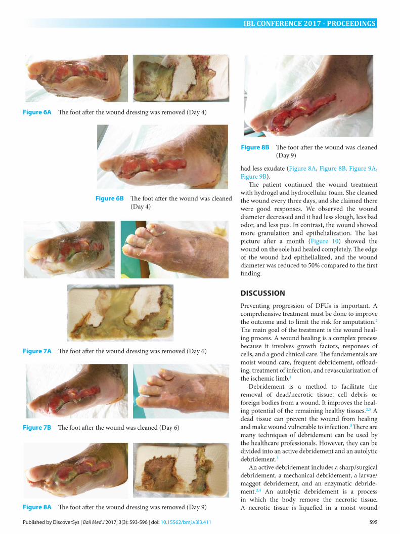
had less exudate (Figure 8A, Figure 8B, Figure 9A, Figure 9B).
The patient continued the wound treatment with hydrogel and hydrocellular foam. She cleaned the wound every three days, and she claimed there were good responses. We observed the wound diameter decreased and it had less slough, less bad odor, and less pus. In contrast, the wound showed more granulation and epithelialization. The last picture after a month (Figure 10) showed the wound on the sole had healed completely. The edge of the wound had epithelialized, and the wound diameter was reduced to 50% compared to the first finding.
DISCUSSION
Preventing progression of DFUs is important. A comprehensive treatment must be done to improve the outcome and to limit the risk for amputation.2 The main goal of the treatment is the wound heal-ing process. A wound healing is a complex process because it involves growth factors, responses of cells, and a good clinical care. The fundamentals are moist wound care, frequent debridement, offload-ing, treatment of infection, and revascularization of the ischemic limb.2
Debridement is a method to facilitate the removal of dead/necrotic tissue, cell debris or foreign bodies from a wound. It improves the heal-ing potential of the remaining healthy tissues.2,3 A dead tissue can prevent the wound from healing and make wound vulnerable to infection.3 There are many techniques of debridement can be used by the healthcare professionals. However, they can be divided into an active debridement and an autolytic debridement.3
An active debridement includes a sharp/surgical debridement, a mechanical debridement, a larvae/maggot debridement, and an enzymatic debride-ment.2,4 An autolytic debridement is a process in which the body remove the necrotic tissue. A necrotic tissue is liquefied in a moist wound
Figure 6A The foot after the wound dressing was removed (Day 4)
Figure 6B The foot after the wound was cleaned (Day 4)
Figure 7A The foot after the wound dressing was removed (Day 6)
Figure 7B The foot after the wound was cleaned (Day 6)
Figure 8A The foot after the wound dressing was removed (Day 9)
Figure 8B The foot after the wound was cleaned (Day 9)

S96 Published by DiscoverSys | Bali Med J 2017; 3(3): S93-S96 | doi: 10.15562/bmj.v3i3.411
IBL CONFERENCE 2017 - PROCEEDINGS
environment.2,3 A moist environment will naturally degrade and remove a slough from the healthy tissue. This process is helped by the presence of enzyme matrix metalloproteinases (MMPs). The enzyme is produced by the injured tissue. The liquefying process can be enhanced by a moist wound environment.3,4A moist environment can use products produce moisture such as hydrocol-loids and hydrogels and absorb excess moisture such as alginates, cellulose dressings or foams.3,5 Hydrogel consist of carboxymethylcellulose poly-mers or insoluble starch, and 96% water.2 Hydrogel can promote autolysis, wound hydration, cool the wound, wound healing, increased granulation tissue, maintains clean wound bed, and provide an analgesic effect.2,3
Foam dressing is used to absorb excess fluid from the wound and to avoid the exudates from damaging its surrounding skin which causing a
maceration.3,6 Allevyn Non-Adhesive dressing combines hydrocellular pad with non-adherent wound contact layer, and a breathable top film contain polyurethane.7 Foam dressing with poly-urethane film provides barrier to bacteria, prevents strikethrough, and reduces risk of bacterial contamination.6 The overall performance of hydro-gel and hydrocellular foam combination was shown to function as autolytic debridement. We observe increased wound granulation and epithelialization and decreased slough and exudates.
CONCLUSION
Foot ulcer in a diabetic patient is often difficult to heal and may become a chronic wound. Proper treatment can improve healing process includes autolytic debridement. The use of hydrogel and hydrocellular foam combination was shown to have a clinical advantage as autolytic debridement. We observed increased wound granulation and epithe-lialization and decreased slough and exudates.
ADDITIONAL INFORMATION
The patients gave a written permission that the picture of the wound will be used for a scientific forum and used as a learning material. We do not have any conflict of interest of the products utilized in this case report.
REFERENCES1. Beckert S, Witte M, Wicke C, Königsrainer A, Coerper S.
A New Wound-Based Severity Score for Diabetic Foot Ulcers: A prospective analysis of 1,000 patients. Diabetes Care. 2006; 29:988-92.
2. Kavitha KV, Tiwari S, Purandare VB, Khedkar S, Bhosale SS, Unnikrishnan AG. Choice of wound care in diabetic foot ulcer: A practical approach. World J Diabetes. 2014; 5:546-56.
3. Atkin L, Stephenson J, Bateman S. Foam dressings: a review of the literature and evaluation of fluid-handling capacity of four leading foam dressings. Wound UK. 2015; 11:75-81.
4. Smith & Nephew. ALLEVYN Non-Adhesive. (online). 2016 [cited 2016 Oct 28] Available from: http://www.smith-nephew.com/key-products/advanced-wound-management/allevyn/allevyn-non-adhesive.
5. Frykberg RG, Banks J. Challenges in the Treatment of Chronic Wounds. Advances in Wound Care. 2015; 4:560-82.
6. Parks WC, Wilson CL, López-Boado YS. Matrix metal-loproteinases as modulators of inflammation and innate immunity. Nature Reviews Immunology. 2004; 4: 617-29.
This work is licensed under a Creative Commons Attribution
Figure 9A The foot after the wound dressing was removed (Day 12)
Figure 9B The foot after the wound was cleaned (Day 12)
Figure 10 The foot after a month treatment