bandara, h. m. h. n., herpin, m. j., kolacny, d., harb, a ... · incorporation of farnesol...
TRANSCRIPT

Bandara, H. M. H. N., Herpin, M. J., Kolacny, D., Harb, A., Romanovicz, D.,& Smyth, H. D. C. (2016). Incorporation of Farnesol Significantly Increasesthe Efficacy of Liposomal Ciprofloxacin against Pseudomonas aeruginosaBiofilms in Vitro. Molecular Pharmaceutics, 13(8), 2760-70.https://doi.org/10.1021/acs.molpharmaceut.6b00360
Peer reviewed version
License (if available):Other
Link to published version (if available):10.1021/acs.molpharmaceut.6b00360
Link to publication record in Explore Bristol ResearchPDF-document
This is the author accepted manuscript (AAM). The final published version (version of record) is available onlinevia ACS publications at http://pubs.acs.org/doi/abs/10.1021/acs.molpharmaceut.6b00360. Please refer to anyapplicable terms of use of the publisher.
University of Bristol - Explore Bristol ResearchGeneral rights
This document is made available in accordance with publisher policies. Please cite only the publishedversion using the reference above. Full terms of use are available:http://www.bristol.ac.uk/pure/about/ebr-terms

This document is confidential and is proprietary to the American Chemical Society and its authors. Do not copy or disclose without written permission. If you have received this item in error, notify the sender and delete all copies.
Incorporation of farnesol significantly increases the efficacy
of liposomal ciprofloxacin against Pseudomonas aeruginosa biofilms in vitro
Journal: Molecular Pharmaceutics
Manuscript ID mp-2016-00360p.R1
Manuscript Type: Article
Date Submitted by the Author: n/a
Complete List of Authors: Bandara, H.M.H.N.; The University of Texas at Austin, College of Pharmacy; The University of Queensland, School of Dentistry Herpin, Matthew; The University of Texas at Austin, College of Pharmacy Kolacny, David; The University of Texas at Austin, College of Pharmacy Harb, Alejandro; The University of Texas at Austin, College of Pharmacy Romanovicz, Dwight; Louise and James Robert Moffett Molecular Biology Building, Smyth, Hugh; The University of Texas at Austin,
ACS Paragon Plus Environment
Molecular Pharmaceutics

Incorporation of farnesol significantly increases the efficacy of liposomal
ciprofloxacin against Pseudomonas aeruginosa biofilms in vitro
H.M.H.N. Bandara1,3, M.J. Herpin1, D. Kolacny Jr.1, A. Harb1, D. Romanovicz2, & H.D.C. Smyth1*
1Division of Pharmaceutics, College of Pharmacy, The University of Texas at Austin, Austin,
Texas, USA.
2Institute of Cellular and Molecular Biology, College of Natural Sciences, The University of
Texas at Austin, Austin, Texas, USA.
3 Current address: School of Dentistry, The University of Queensland, Australia
* Corresponding author:
Dr. Hugh D. C. Smyth,
College of Pharmacy, 2409 West University Avenue, PHR 4.214,
Austin, TX, USA 78712
Phone: +1 (512) 471 3383
Fax: +1 (512) 471 7474
E mail: [email protected]
Page 1 of 33
ACS Paragon Plus Environment
Molecular Pharmaceutics
123456789101112131415161718192021222324252627282930313233343536373839404142434445464748495051525354555657585960
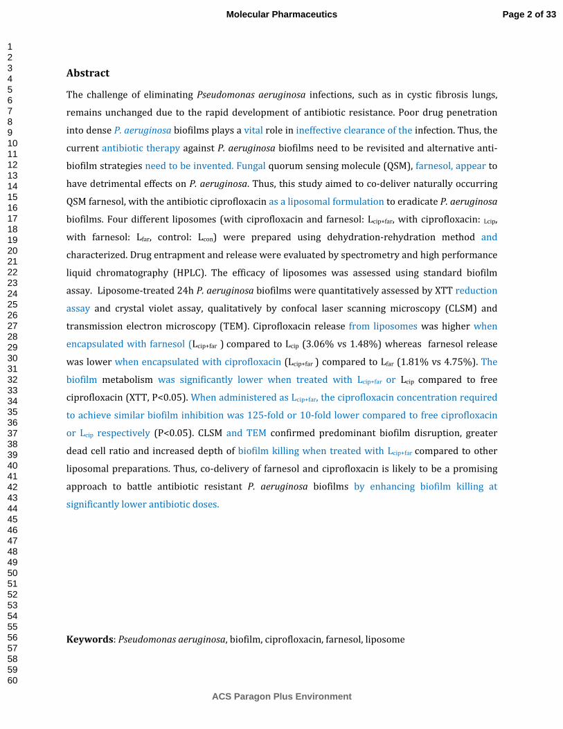
Abstract
The challenge of eliminating Pseudomonas aeruginosa infections, such as in cystic fibrosis lungs,
remains unchanged due to the rapid development of antibiotic resistance. Poor drug penetration
into dense P. aeruginosa biofilms plays a vital role in ineffective clearance of the infection. Thus, the
current antibiotic therapy against P. aeruginosa biofilms need to be revisited and alternative anti-
biofilm strategies need to be invented. Fungal quorum sensing molecule (QSM), farnesol, appear to
have detrimental effects on P. aeruginosa. Thus, this study aimed to co-deliver naturally occurring
QSM farnesol, with the antibiotic ciprofloxacin as a liposomal formulation to eradicate P. aeruginosa
biofilms. Four different liposomes (with ciprofloxacin and farnesol: Lcip+far, with ciprofloxacin: Lcip,
with farnesol: Lfar, control: Lcon) were prepared using dehydration-rehydration method and
characterized. Drug entrapment and release were evaluated by spectrometry and high performance
liquid chromatography (HPLC). The efficacy of liposomes was assessed using standard biofilm
assay. Liposome-treated 24h P. aeruginosa biofilms were quantitatively assessed by XTT reduction
assay and crystal violet assay, qualitatively by confocal laser scanning microscopy (CLSM) and
transmission electron microscopy (TEM). Ciprofloxacin release from liposomes was higher when
encapsulated with farnesol (Lcip+far ) compared to Lcip (3.06% vs 1.48%) whereas farnesol release
was lower when encapsulated with ciprofloxacin (Lcip+far ) compared to Lfar (1.81% vs 4.75%). The
biofilm metabolism was significantly lower when treated with Lcip+far or Lcip compared to free
ciprofloxacin (XTT, P<0.05). When administered as Lcip+far, the ciprofloxacin concentration required
to achieve similar biofilm inhibition was 125-fold or 10-fold lower compared to free ciprofloxacin
or Lcip respectively (P<0.05). CLSM and TEM confirmed predominant biofilm disruption, greater
dead cell ratio and increased depth of biofilm killing when treated with Lcip+far compared to other
liposomal preparations. Thus, co-delivery of farnesol and ciprofloxacin is likely to be a promising
approach to battle antibiotic resistant P. aeruginosa biofilms by enhancing biofilm killing at
significantly lower antibiotic doses.
Keywords: Pseudomonas aeruginosa, biofilm, ciprofloxacin, farnesol, liposome
Page 2 of 33
ACS Paragon Plus Environment
Molecular Pharmaceutics
123456789101112131415161718192021222324252627282930313233343536373839404142434445464748495051525354555657585960

Introduction
The opportunistic bacterial pathogen, Pseudomonas aeruginosa is one of the leading causes of
nosocomial infections worldwide and ranked the second most prevalent among the Gram-negative
pathogens reported to the National Nosocomial Infection Surveillance System 1. In the USA,
approximately 51,000 healthcare-associated P. aeruginosa infections occur annually and
alarmingly, over 6,000 (13%) of these infections are caused by multidrug-resistant variant of the
pathogen 2. P. aeruginosa is the most commonly isolated pathogen in lung infections in cystic
fibrosis patients and is considered as the leading cause for morbidity and mortality in such
patients3. Despite aggressive therapeutic approach, current antibiotic treatments could only
accomplish an adjournment of the spread of the pathogen as well as the destruction of the lung
tissues; mucus-embedded biofilms persist for decades and cannot be completely eradicated. Thus,
the applications of current armory of antimicrobial compounds against P. aeruginosa biofilm
infections are needed to be revisited and alternative anti-biofilm drugs and strategies are ought to
be sought 4.
P. aeruginosa biofilms are multicellular surface-attached and spatially oriented, bacterial
communities encased in an extracellular matrix that display characteristic and significant
resistance to antimicrobial agents and environmental stresses. Limited drug penetration through
the biofilm matrix 5, nutrients and oxygen-based heterogeneity of the bacterial cell populations 6,
biofilm specific bacterial phenotypes6, subpopulations of multidrug resistant persister cells 7, and
bacterial communications via quorum sensing and signal transduction systems 8 are suggested to
contribute the drug resistance associated with P. aeruginosa biofilms. As a result of significant
antibiotic tolerance, individual antibiotic therapy often fails to eliminate P. aeruginosa infections in
CF patients and combined antibiotic therapies at high concentrations are required in managing
acute CF related infections 9.
A variety of antibiotic treatment strategies such as systemic and nebulized antibiotics have been
employed to treat P. aeruginosa biofilms in CF lungs 10-12. However, to date, no therapeutic approach
has achieved complete eradication of P. aeruginosa from the chronic lung infections most likely due
to biofilm-specific intrinsic antibiotic resistance. In particular, the efficacy of aminoglycosides,
widely used first-line therapy in the management of CF infections, is limited due to their chemical
interactions with the biofilm matrix resulting slow and incomplete drug penetration into the
bacterial biofilm core 13. Conversely, Low pH in the biofilm matrix facilitates antibacterial agents
such as ciprofloxacin to strongly bind to the alginates within the biofilm causing significant
Page 3 of 33
ACS Paragon Plus Environment
Molecular Pharmaceutics
123456789101112131415161718192021222324252627282930313233343536373839404142434445464748495051525354555657585960

reduction in the effective drug concentrations at the target site. Alarmingly, antibiotic doses below
Minimum Inhibitory Concentrations (MIC) i.e. sub-MICs of tobramycin have recently been found to
promote P. aeruginosa biofilm formation, and to generate drug resistant bacterial strains 14, 15.
Encapsulation of the drugs in to liposomes was recently introduced in overcoming nonspecific
drug-binding to extracellular matrix and the metabolic protection in the biofilm and appears greatly
to aid in delivering the drug of interest to the target site. For instance, liposomal amikacin
demonstrated a high drug loading, long term stability, slow and sustained drug release, and a
greater potential for in vitro penetration through mucus to reach the biofilm cells of P. aeruginosa
16. Furthermore, drug delivery via liposomes potentially aids persister cell eradication due to slow
and sustained release of the drug providing long term localized high concentrations of the antibiotic
compared to free form of the drug in order to kill persister cells 17, 18. .
In recent studies, a 12-carbon sesquiterpene; farnesol, a known virulence factor and repressor of
yeast to hyphae morphological transition in Candida albicans 19-25 , was shown to inhibit the
synthesis of P. aeruginosa quorum sensing molecule (QSM), 2-heptyl-3-hydroxy-4-quinolone
(Pseudomonas quinolone signal: PQS), PQS-regulated virulence factor pyocyanin 26, and bacterial
swarming motility 27. Importantly, PQS is shown to play an important role in the biofilm formation
of P. aeruginosa 28. Hence, we speculate that farnesol possesses inhibitory effects on P. aeruginosa
biofilm development.
Quorum sensing has not been targeted before in the management of CF lung infections, and co-
delivery of antibiotics and QSMs has never been attempted. Thus, the aim of this study was to co-
deliver a naturally occurring fungal QSM (farnesol), and a well-established antipseudomonal
antibiotic (ciprofloxacin), in the form of liposomes to P. aeruginosa biofilms. We hypothesized that a
liposomal delivery system of farnesol with ciprofloxacin will efficiently disrupt and eliminate P.
aeruginosa biofilms by targeting multiple mechanisms of biofilm development and maintenance
compared to antibiotic treatment alone.
Material and methods
Preparation of liposomes
For each experiment, 4 different formulations of liposomes were freshly prepared. These included:
liposome only (Lcon), Liposome with ciprofloxacin (Lcip), liposomes with farnesol (Lfar) and liposome
Page 4 of 33
ACS Paragon Plus Environment
Molecular Pharmaceutics
123456789101112131415161718192021222324252627282930313233343536373839404142434445464748495051525354555657585960

with ciprofloxacin and farnesol (Lcip+far). Liposomes were prepared as described by Lagace et al with
modifications 29. DPPC (1,2-Dipalmitoyl-sn-glycero-3-phosphocholine, Genzyme Pharmaceuticals,
Switzerland) and cholesterol were added 4:1 molar ratio and dissolved in chloroform (HPLC grade,
Fisher Scientific, USA) in a 1:1 w/v ratio. Farnesol 2μl (0.879g/ml density) was added to the
dissolved lipid solution and mixed thoroughly for Lfar and Lcip+far preparations. The lipid solutions
were evaporated to form a thin layer of lipid using a rotary evaporator (Buchi R-210 rotovapor,
Buchi Labortechnik, Switzerland). The flasks were stored under a vacuum for an hour to remove
any residual chloroform.
Ten milliliters of unbuffered saline (for Lcon and Lfar) or 10ml of 3mg/ml ciprofloxacin dissolved in
unbuffered saline (for Lcip and Lcip+far , Ciprofloxacin hydrochloride USP grade, Letco Medical,
Decatur, USA, Catalog No. 690953) were added to the flasks and sonicated in a water bath for 5 min.
The formed multi lamellar liposomes were collected, centrifuged at 15000rpm for 10min at 40C.
The supernatant was collected and the pellet was resuspended in unbuffered saline, washed and
washes were collected. The resulting liposome pellet was resuspended in unbuffered saline as
necessary and used for liposome characterization and microbiological studies.
Determination of drug entrapment efficiency
Ciprofloxacin entrapment
To determine the entrapped ciprofloxacin, the optical density of the supernatant and washes
collected during the preparation of liposomes was read in a spectrophotometer (Infinite M200,
Tecan Systems Inc., CA, USA) at 272nm. The entrapment efficiency (EE) was calculated using
following formula;
�Totalquantityofciprofloxacinadded − thequantityofunboundciprofloxacin�×100
�Totalquantityofciprofloxacinadded�
Farnesol entrapment
The entrapment of farnesol was quantified using High Performance Liquid Chromatography (HPLC)
as described by Chen et al 2009 30. Supernatant and wash were filtered through 0.22μm sterile
filters before performing HPLC. The assay was performed on a Waters 2495 HPLC system with a
C18 reverse phase column (μBondapak®, 3.9×300mrn column, Waters Corporation, MA, USA). The
Page 5 of 33
ACS Paragon Plus Environment
Molecular Pharmaceutics
123456789101112131415161718192021222324252627282930313233343536373839404142434445464748495051525354555657585960

following conditions were used in the HPLC assay: mobile phase Acetonitrile: water (80:20 V/V);
the flow rate 1ml/min; UV detection at 210nm; retention time 6min. All samples were analyzed in
duplicates.
Farnesol entrapment was calculated as follows;
�Totalquantityoffarnesoladded − thequantityofunboundfarnesol�×100
�Totalquantityoffarnesoladded�
Characterization of liposomes
Particle size and zeta potential
The size, polydispersity index and the zeta potential of liposomes were determined by Dynamic
Light Scattering Zetasizer Nano ZS (Malvern instruments Ltd, UK).
Morphology of liposomes
The morphology of liposomes was visualized by transmission light microscope (TLM, Leica TCS
SP5, Leica Microsystems, IL) fluorescent microscopy (FM, Stained with propidium iodide, Leica TCS
SP5, Leica Microsystems, IL) and Scanning electron microscopy (SEM). In order to prepare
liposomes for the latter, liposomes were placed on a coverslip and coated with Platinum/Palladium
(Cressington sputter coater 208 HR, Cressington Scientific Instruments Ltd, UK). The surface
topography of liposomes was visualized with scanning electron microscope (Zeiss Supra 40VP, CA,
USA) in high-vacuum mode.
Drug release
Ciprofloxacin release from the liposomes were studied as described by Halwani M. et al 2008 with
modifications 31. The modified assay used in this study is similar to agar diffusion assay. However,
UV absorbance at 272 nm was used to estimate ciprofloxacin concentration instead of the zone of
inhibition of microbial growth due to the potential variations of antibiotic penetration efficiencies
through agar, asymmetry of zone of inhibition and variations among the microorganism used in
agar diffusion.
Page 6 of 33
ACS Paragon Plus Environment
Molecular Pharmaceutics
123456789101112131415161718192021222324252627282930313233343536373839404142434445464748495051525354555657585960

Known volumes of liposomes were added to 10ml of saline and incubated at 370C for 24h in a
shaker (250rpm). The liposomal suspensions were withdrawn in defined time intervals,
centrifuged (15000rpm, 10 min) and the absorbance of the supernatant was measured at 272nm
(Infinite M200, Tecan Systems Inc., CA, USA) to estimate ciprofloxacin release . The liposome pellet
was re-suspended in fresh saline (10ml) after each time interval to prevent possible antibiotic
saturation.
After 24h of incubation, a sample was prepared as mentioned above and filtered (0.22μm). The
quantification of farnesol released was assessed by HPLC assay as described above.
Microorganisms and growth conditions
Pseudomonas aeruginosa PAO1 was used throughout the study. The identity of the bacteria was
confirmed with the commercially available API 20 E kit (Biomérieux, Mercy I’Etoile, France). All
isolates were stored in multiple aliquots at -200C, after confirming their purity. Blood Agar (Sigma
Aldrich, USA) and Brain Heart Infusion (BHI, Sigma Aldrich, USA) solutions were used for culturing
P. aeruginosa.
Prior to each experiment, P. aeruginosa was subcultured on blood agar for 18 h at 370C. A loopful of
the overnight bacterial growth was inoculated into BHI medium, and, incubated for 18h in an
orbital shaker (80 rpm) at 370C. The resultant growth was harvested, washed twice in Phosphate
Buffered Saline (PBS, pH 7.2) and resuspended. The concentration of P. aeruginosa was adjusted
1×10⁷ cells/ml by spectrophotometry and confirmed by hemocytometric counting.
Biofilm Formation
P. aeruginosa biofilms were developed as described by Bandara et al 32 with some modifications.
Commercially available pre-sterilized, polystyrene, flat bottom 96-well microtiter plates (BD
Biosciences, California, USA) were used. Hundred µl of a standard cell suspension of bacteria
(107organisms/ml) was prepared and transferred into the wells of a microtiter plate, and the plate
was incubated for 1.5h (370C, 80 rpm) to promote microbial adherence to surface of the wells. After
the initial adhesion phase, the cell suspensions were aspirated and each well was washed twice
with PBS to remove loosely adherent cells. A total of 200µl of BHI was transferred to each well and
the plate was incubated for 24 h (370C, 80 rpm), and wells washed twice with PBS to eliminate
traces of the medium. The resultant biofilms were considered ready for experimental use.
Determination of minimum inhibitory concentration (MIC)
Page 7 of 33
ACS Paragon Plus Environment
Molecular Pharmaceutics
123456789101112131415161718192021222324252627282930313233343536373839404142434445464748495051525354555657585960

Planktonic phase
MIC was determined by a broth microdilution assay in accordance with the CLSI guidelines 33.
Briefly, bacterial cell suspensions (5×10⁵ Cells/ml) were treated with ciprofloxacin or farnesol in a
concentration gradient (two-fold) and incubated in a 96 well microtiter plate for 24h at 350C. At the
end of the incubation, the optical density was measured by a spectrophotometer at 595 nm. The
lowest concentration of the antibiotic or farnesol at which the bacteria demonstrated visible
growth inhibition compared to the solvent control was considered the MIC of the antibiotic or
farnesol against P. aeruginosa. The assay was performed in quadruplicates at three times.
Biofilm phase
P. aeruginosa biofilms were grown in sterile 96 well plates (BD Biosciences, USA) as described
above. Biofilms were washed twice with PBS and ciprofloxacin or farnesol was administered in a
concentration gradient (two fold). The plates were incubated for 24h at 370C and 80 rpm.
At the end of the incubation period, a XTT (sodium 2,3,-bis(2-methoxy-4-nitro-5-sulfophenyl)-5-
[(phenylamino)-carbonyl]-2H-tetrazolium inner salt) reduction assay was performed to quantify
the viability of biofilms as described in following sections. The lowest concentration of the
antibiotic or farnesol at which the bacteria demonstrate 80% of reduction of the viability compared
to the solvent control is considered as the Minimum Biofilm Inhibitory Concentration (MBIC) of the
antibiotic or farnesol against P. aeruginosa. The assay was performed quadruplicates three separate
times.
In addition, as described in following sections, a crystal violet assay was performed at the end of the
incubation to estimate the effect of farnesol on the biomass of the mature P. aeruginosa biofilms.
The assay was performed in quadruplicates at three separate times.
Treatment of biofilms with liposomes
P. aeruginosa biofilms were developed in 96 well plates as described above. Freshly prepared
liposomes (Lcon, Lcip, Lfar and Lcip+far) were added in a concentration gradient to the biofilms with BHI
and incubated for another 24h (370C, 80rpm). At the end of this incubation, biofilms were washed
twice with PBS and a XTT reduction assay was performed. Each experiment was conducted in
quadruplicates on three different occasions.
XTT reduction assay
Page 8 of 33
ACS Paragon Plus Environment
Molecular Pharmaceutics
123456789101112131415161718192021222324252627282930313233343536373839404142434445464748495051525354555657585960

At the end of incubation of both test and control biofilms, a standard XTT (sodium 2,3,-bis(2-
methoxy-4-nitro-5-sulfophenyl)-5-[(phenylamino)-carbonyl]-2H-tetrazolium inner salt) reduction
assay was performed as described by Bandara et al 34 to measure the viability of biofilms by means
of bacterial cell metabolic activity. In brief, commercially available XTT powder (Sigma, MO, USA)
was dissolved in PBS to a final concentration of 1 mg/ml. Then the solution was filter-sterilized
(0.22 μm pore size filter) and stored at -700C. Freshly prepared 0.4 mM menadione solution was
used for XTT reduction assay. Thawed XTT solution was mixed with menadione solution in a 20:1
(v/v) ratio immediately before the assay. Thereafter, PBS: XTT: Menadione in a 79:20:1 ratio were
added into each well containing biofilms and incubated in the dark for 5 h at 370C. The color
changes were measured with a microtiter plate reader (Infinite M200 microplate reader, TECAN US
Inc, NC, USA) at 492nm.
Crystal violet assay
At the end of incubation of both test and control biofilms, a crystal violet assay was performed to
quantify biofilm biomass. Biofilms were carefully washed twice with PBS and stained with a 1%
crystal violet solution for 15 min at 250C without shaking. Wells were carefully washed three times
with PBS to remove excess stain and air dried at room temperature. Thirty percent acetic acid was
added to the wells containing stained biofilms and incubated for 20 min at 250C. The solution was
transferred to a new well plate and the optical density was measured at 570nm.
Confocal Laser Scanning Microscopy (CLSM)
Biofilms were prepared on cover slips placed in flat bottom six well plates (Nunclon, Nunc, thermo
Fisher scientific, USA) as described above. Pre-formed 24h biofilms were exposed to all 4 different
liposomal preparations and incubated for another 24h at 370C in a shaker (80rpm). At the end of
incubation, the prewashed films were stained with Live and Dead stain (Live/Dead BacLight
Bacterial Viability kit, Invitrogen, Eugene, USA) 32. The biofilm was then analyzed using confocal
laser scanning microscopy
Transmission electron microscopy (TEM)
P. aeruginosa biofilms were prepared on ACLAR® film (Electron Microscopy Sciences, PA, USA) and
treated with various liposomal preparations as described above. At the end of the incubation,
biofilms were washed twice with PBS and fixed with an aldehyde mixture for 2-4h on ice (4%
gluteraldehyde, 2% paraformaldehyde, 0.1M cacodylate, 2mM Ca and 4mM Mg). The samples were
Page 9 of 33
ACS Paragon Plus Environment
Molecular Pharmaceutics
123456789101112131415161718192021222324252627282930313233343536373839404142434445464748495051525354555657585960

then washed three times with 0.1M sodium cacodylate buffer (pH7.4, Electron Microscopy Sciences,
PA, USA) for 15 min. Subsequently, the biofilms were fixed with reduced osmium (1:1 4%
potassium ferrocyanide in 0.2M cacodylate buffer: 4% osmium tetroxide) in a microwave (100w, 3
sessions of 2 min on, 2 min off, two times). After fixation, the samples were washed 5 times with
water for 10 min and dehydrated with ethanol (15 min each, 50%, 70%, and 95%, and 2×100%
ethanol) and 100% acetone (15 min, two times). The samples were placed in a polymerization tray
and infiltrated with resin (30%, 66% and 2×100% resin in 100% acetone) and polymerized for 2
days at 600C. The samples were then sectioned using a Leica UltraCut Ultramicrotome® (Leica
Microsystems Inc. IL, USA), placed on grids and observed under an FEI Tecnai Transmission
Electron Microscope® (FEI, Oregon, USA) at 80kV.
Statistical analysis
Statistical analysis was performed using SPSS software (version 16.0). Mann—Whitney U-test was
performed to compare the significant differences between corresponding controls and test samples
of the P. aeruginosa biofilms. A P-value of less than 0.05 was considered statistically significant.
Results
Characteristics of liposomes
The entrapment efficiencies of ciprofloxacin were 60% in Lcip+far and 61% in Lcip. Farnesol
entrapment efficiencies were 91% in Lcip+far and 93% in Lfar. The diameters of Lcon, Lcip, Lfar and Lcip+far
were 2808.0±529.6nm, 677.8±46.6nm, 684.0±43.8nm and 536.8±21nm respectively. Lcon were
significantly larger than all three test liposomes and the difference in the diameters of test
liposomes were insignificant (P<0.05). The zeta potentials of respective liposomes were -1.66mV, -
3.27mV, +0.09mV and -0.11mV (Table 1) and polydispersity indices of respective liposomes were
0.086, 0.281, 0.391 and 0.213.
Morphology of liposomes
FM, TLM and SEM confirmed that the liposomes were spherical in shape and varied somewhat in
size but were consistent with DLS measurements (control liposomes; Lcon, Figure 1). FM images
exhibited a fluorescently stained liposomal membrane and an unstained dark core (Figure 1).
Drug release
Page 10 of 33
ACS Paragon Plus Environment
Molecular Pharmaceutics
123456789101112131415161718192021222324252627282930313233343536373839404142434445464748495051525354555657585960

After 24h of drug release, 3.06% (18.04μg) of total encapsulated ciprofloxacin (589.14μg) was
released from Lcip+far, however, only 1.48% (13.27μg) of the total encapsulated ciprofloxacin
(897.09μg) was released from Lcip. During a period of 24h, there was a continuous and a greater
release of ciprofloxacin from Lcip+far compared to Lcip (Figure 2).
After 24h, 1.81% (13.08μg) of total farnesol encapsulated (723.14μg) was released from Lcip+far. In
contrast, 4.75% (35.13μg) of total farnesol encapsulated (738.91μg) was released from Lfar.
Minimum inhibitory concentrations (MIC)
The MIC of ciprofloxacin was 0.125μg/ml for planktonic P. aeruginosa and MBIC was 16μg/ml for
24h developed P. aeruginosa biofilms.
Farnesol did not have any significant effects on the P. aeruginosa planktonic phase, even up to
175μg/ml. In contrast, the maximum reduction of metabolic activity as indicated by XTT readings
(40% reduction compared to solvent controls, P<0.05) were observed at farnesol concentrations of
90μg/ml in established P. aeruginosa biofilms.
A crystal violet assay showed that concentrations of farnesol equal to or greater than 2.8μg/ml
resulted in the greatest reduction in the biofilm biomass (45%, P<0.05) compared to its controls.
Effect of liposomes on the metabolism of established P. aeruginosa biofilms- XTT reduction
assay
At the end of the 24h treatment of P. aeruginosa biofilms with various liposomal formulations,
significant treatment effects were observed. The biofilms treated with Lcip+far showed a significantly
lower metabolic activity compared to Lcip and free ciprofloxacin. Only ≥0.128μg/ml (P<0.05) of
ciprofloxacin concentrations delivered from Lcip+far resulted in bacterial metabolic activity (80%
reduction compared to control) equal to those observed in Lcip treated biofilms at ≥1.310μg
(P<0.05) and, in contrast, 16μg/ml of free ciprofloxacin treated biofilms (P<0.05, Figure 3).
Treatment with Lfar and Lcon did not exhibit any reduction of the P. aeruginosa biofilm metabolism
compared to controls, as manifested by the XTT readings.
Effect of liposomes on P. aeruginosa biofilms - Confocal laser scanning microscopy
CLSM imaging also demonstrated the significant effect of the drug loaded liposomal preparations on
P. aeruginosa pre-formed biofilms. Biofilms treated with liposomes containing either ciprofloxacin
Page 11 of 33
ACS Paragon Plus Environment
Molecular Pharmaceutics
123456789101112131415161718192021222324252627282930313233343536373839404142434445464748495051525354555657585960

(Lcip), farnesol (Lfar), or both (Lcip+far, Figure 4D, E, and F) exhibited a greater degree of structural
disruption and a lower live: dead cell ratio compared to the dense, well-organized, undisturbed
biofilm (Figure 4A) and biofilms treated with control liposomes (Lcon, Figure 4C). It was clearly
visible that the biofilms treated with Lcip+far revealed the most significant disruption, exhibiting
lessened biofilm mass/architecture and a significant proportion of dead cells compared to all other
treatment combinations tested (Figure 4F). Interestingly, the biofilms treated with free
ciprofloxacin demonstrated only isolated areas of cell death and were relatively undisrupted and
appeared to be viable (Figure 4B) compared to Lcip and Lcip+far treated biofilms (Figure 4E and F).
Figure 5 shows the longitudinal and horizontal sections of P. aeruginosa biofilms treated with
various liposomal formulations. The biofilms treated with liposomal control, Lcon, showed preserved
dense biofilm architecture and the viability deeper bacterial cell layers. The dead and dying cells
appeared only in the superficial cell layers of the biofilm (Figure 5A). Similarly, the Lfar treated
biofilm also exhibited a spatially oriented biofilm structure; however, regions of bacterial death
were also noted (Figure 5B). The thickness of the dead cell layers in the Lfar treated biofilm was
greater than that of Lcon treated biofilm and in some areas,the full thickness of the biofilm consisted
of dead cells (Figure 5B). Compared to the Lcon and Lfar treated biofilms, the Lcip treated biofilms
exhibited a greater disruption of the biofilm architecture and higher proportions of dead cells
(Figure 5C). However, live cells were still existed in the deeper layers of the biofilm (Figure 5C). In
contrast, the Lcip+far treated biofilm showed the most significant quantities of dead bacteria
compared to any of the other liposome treated biofilms. More importantly, the full thickness of the
majority of the areas of the biofilm was filled with dead cells and the entire biofilm appeared dead
(figure 5D).
Effect of liposomes on P. aeruginosa biofilms – Transmission electron microscopy
TEM imaging corroborated with the CSLM findings of the biofilms treated with various liposomal
preparations. P. aeruginosa cells in the control, untreated biofilms appeared to be healthy with a
rod shaped cell structure and dark intracellular materials. Cells were loosely packed and some
clearly showed intact cell membranes and slimy secretions in their immediate environment. There
were few dead cells (observed as clear cells), while some cells showed disintegration of cell
membrane and cellular detritus (Figure 6A). Similarly, Lcon treated biofilms also showed healthy
heterogeneous bacteria with some dividing cells. Cell membranes were intact and no dead cells
were visible (Figure 6B). Similar to the Lcon treated biofilm, Lfar treated biofilm also consisted of
healthy bacteria with some pale colored/less condensed intracellular materials (Figure 6C). In
Page 12 of 33
ACS Paragon Plus Environment
Molecular Pharmaceutics
123456789101112131415161718192021222324252627282930313233343536373839404142434445464748495051525354555657585960

contrast, Lcip treated biofilms had higher numbers of dead cells/clear cells. The cell membranes of
the majority of the cells were disintegrated and the cellular contents appear to be leaked out. Small
vesicles were also present surrounding dead cells (Figure 6D). Most importantly, the Lcip+far treated
biofilms also exhibited the highest proportion of clear and partially ruptured cells. Some
disintegrated cells contained cytoplasm filled vesicles. Cell debris of dead cells was distributed
throughout the microscopic field (Figure 6E).
Discussion
The biofilm antibiotic resistance and the synthesis of various virulence determinants During P.
aeruginosa biofilm formation are mediated by two major and chemically distinct quorum sensing
systems; N-acetyl homoserine lactones and the 4-quinolones. Among all quinolones, only PQS (2-
heptyl-3-hydroxy-4-quinolone, Pseudomonas Quinolone Signal, PQS) is isolated in broncho-alveolar
lavage fluid in CF lungs suggesting the potential association of PQS with P. aeruginosa infections and
subsequent inflammatory damage to host respiratory tissues in CF patients 26, 35, 36.
The role of PQS in P. aeruginosa biofilm development is well-known. PQS enhances P. aeruginosa
biofilm formation by stimulating two known regulators of biofilm development; RhlR/C4-HSL and
RpoS 37. Also, PQS regulates the synthesis of extracellular DNA (eDNA) in P. aeruginosa biofilms.
eDNA acts as a cell-cell interconnecting compound that predominantly maintain the 3D structure
and architecture of P. aeruginosa biofilms 38. eDNA is also isolated at very high concentrations in
sputum samples of CF lungs (up to 20mg/ml), suggesting the existence of eDNA rich micro colonies
of P. aeruginosa in CF lungs 39. Alarmingly, eDNA enriched P. aeruginosa biofilms showed up to 640-
fold more antibiotic resistance than planktonic cultures 40. Importantly, PQS regulates the synthesis
of various other extracellular proteases such as hydrogen cyanide and redox active phenazines like
pyocyanin in P. aeruginosa26, 41. Pyocyanin, in turn, generates the reactive oxygen species (ROS)
superoxide and hydrogen peroxide 42. These ROS favor bacterial colonization by damaging various
host cells by neutrophil apoptosis, induction of IL-8 and inhibition of the dual-oxidase based
antimicrobial system in airway epithelia 43, 44. Thus, pyocyanin producing P. aeruginosa strains are
more difficult to clear from the lung by host immune reactions than non-pyocyanin producing P.
aeruginosa strains 45. In addition, PQS induces P. aeruginosa to enter into a metabolically less active
state to cope external stresses such as antibiotics 46. Due to its prominent role in P. aeruginosa
biofilm development and maintenance, PQS appears as an attractive target for novel therapeutic
strategies in pathogenic biofilm elimination by improving drug delivery.
Page 13 of 33
ACS Paragon Plus Environment
Molecular Pharmaceutics
123456789101112131415161718192021222324252627282930313233343536373839404142434445464748495051525354555657585960

A major QSM secreted by Candida albicans, E,E-farnesol, has been identified to significantly reduce
PQS synthesis in P. aeruginosa 26. In addition, farnesol is known to inhibit swarming of P.
aeruginosa, most likely through inhibiting PQS synthesis 27. Interestingly, using The Lubbock
Chronic Wound Biofilm model, recent research revealed that farnesol at a concentration of
1,000μg/ml could completely suppress the development of P. aeruginosa biofilms 47. Moreover, a
role of farnesol in potentiating bacterial susceptibility to antibiotics has been recently unveiled 48.
Hence, we designed a novel drug delivery system, exploiting aforementioned anti-pseudomonas
properties of farnesol, using well-established liposomes to co-deliver ciprofloxacin and farnesol
simultaneously. The latter delivery system was expected to achieve superior elimination of mature
P. aeruginosa biofilms through farnesol-mediated inhibition of PQS synthesis and ciprofloxacin-
mediated killing of the bacteria. Furthermore, farnesol has very low aqueous solubility and is
lipophilic, attributes making formulation in liposomes an attractive option.
Liposomes were originally developed for use in intravenous drug delivery systems. For localized
target release of the antibiotic such as in managing lung infections, liposomes must be intact until
reaching the desired site of infection and have slow and sustained drug release. The therapeutic use
of liposomes for inhaled drug delivery has been recently studied 49-51. Liposomes are expected to
deliver antibiotics sufficiently and sustainably to the site of infection in the lung. In effect, when
tobramycin is administered intratracheally in the form of liposomes, drug retention and
antimicrobial activity in the lung are significantly enhanced 52-54. To synthesize liposomes with
substantial encapsulation of both ciprofloxacin and farnesol that exhibit a slow and sustained
release, we experimented with various combinations of DPPC and cholesterol. The optimal ratio of
these lipids for drug encapsulation and release was 4:1 DPPC: Cholesterol (data not shown). The
resultant, drug-loaded multi lamellar liposome populations were heterogeneous as indicated by the
polydispersity indices and ranged between 500-700nm in diameter. Zeta potential is a valuable
physicochemical parameter that determines the stability of any nanosuspensions such as
liposomes. The repulsion between liposomes with similar electric charges ensures easy
redispersion of the particles and prevents their aggregation. Hence, extremely positive or negative
zeta potential values are preferred55, 56, i.e. a minimum zeta potential of ± 20 mV is desirable for
combined electrostatic and steric stabilization of a nanosuspension 57 . However, all the multi
lamellar liposomes prepared in our study demonstrated weak zeta potentials (between 0±5mV)
and the tendency toward liposomal aggregation was confirmed through TLM imaging, where some
liposomal aggregates were visible among individually dispersed liposomes. Thus, weak charge of
liposomes may have influenced its stability, drug encapsulation and their adsorption to pathogens
Page 14 of 33
ACS Paragon Plus Environment
Molecular Pharmaceutics
123456789101112131415161718192021222324252627282930313233343536373839404142434445464748495051525354555657585960

58. Addition of surfactant may have added an advantage of dissolution of farnesol and increase the
stability of liposomes. However, due to potential cytotoxic effects of various surfactants on P.
aeruginosa biofilms and to simplify the liposomal composition, surfactants were not used 59.
Nevertheless, the liposomes reported here will be further optimized for a desirable zeta potential
and stability in the future.
The relevance of the liposomal charge and its capability of penetration to the biofilms have been
recently studied 60. John et al showed that the penetration of oral and topical bacterial biofilms by
charged (+ or -) liposomes was significantly higher than that of free drug 61-63. In contrast,
uncharged liposomes demonstrated more favorable interactions with planktonic P. aeruginosa 64. In
concurrence with these findings, our liposomes were weakly charged (+ or -) and thus expected to
penetrate P. aeruginosa biofilms more effectively than free drug. The CLSM images presented in this
study further supported the enhanced penetration of liposome into P. aeruginosa biofilms.
Compared to free ciprofloxacin treated biofilms, Lcip and Lcip+far exhibited significantly higher ratio of
biofilm death, likely due to enhanced penetration. In particular, Lcip+far treated biofilms showed
bacterial cell death throughout the full thickness of the biofilms compared to other tested liposomal
formulation exhibiting its properties of superior biofilm killing. TEM images were also consistent
with these observations and Lcip and Lcip+far killed more bacteria in P. aeruginosa biofilms compared
to other liposomal preparations.
In the design of liposomes, farnesol was expected to be entrapped in the lipid bilayer due to its lipid
solubility, whereas the water soluble ciprofloxacin was expected to be encapsulated in the core of
the liposome. Farnesol demonstrated a very high efficiency of entrapment in both Lfar and Lcip+far,
(over a 90% entrapment). However, the release of farnesol from Lcip+far was 40% lower than that
from Lfar during a 24h period, suggesting that the presence of ciprofloxacin affects the release of
farnesol. Drug release from liposomes is known to be influenced by the membrane composition and
the biochemical properties of the encapsulated drug (charge, pKas, drug stability, pH, etc.). Though
the exact reason is yet to be unraveled, the presence of ciprofloxacin may have altered the
liposomal stability resulting reduced farnesol release65-67. Similar to farnesol entrapment,
ciprofloxacin was also entrapped in liposomes efficiently regardless of the presence of farnesol.
However, ciprofloxacin showed constantly higher rates of release from Lcip+far compared to Lcip
throughout a 24h period, indicating that the presence of farnesol in the liposome increased the
release of the antibiotic by two fold. Hence, from a drug release point of view, Lcip+far was a preferred
Page 15 of 33
ACS Paragon Plus Environment
Molecular Pharmaceutics
123456789101112131415161718192021222324252627282930313233343536373839404142434445464748495051525354555657585960

antibiotic release system despite possessing similar entrapment efficiencies of farnesol and
ciprofloxacin compared to Lcip and Lfar.
The most important and unique features of these dually loaded liposomes were noted when
delivered to mature P. aeruginosa biofilms. A significant reduction of the biofilm viability was noted
at 0.128μg/ml ciprofloxacin released by Lcip+far. However, to achieve a similar efficiency in biofilm
viability reduction, ten-fold more ciprofloxacin was needed in the form of Lcip. These results
strongly indicate that Lcip+far possesses an enhanced functional capability of killing established P.
aeruginosa biofilms compared to liposomal ciprofloxacin (Lcip). It further confirms that the addition
of farnesol to the liposomes significantly reduced the antibiotic dose needed to effectively eliminate
biofilms. Furthermore, over 16μg/ml of free ciprofloxacin was needed to achieve a similar biofilm
inhibition to the liposomal formulations indicating liposomal formulations offer superior anti-
biofilm properties in comparison to free ciprofloxacin. Similarly, previous studies also showed
superior biofilm inhibitory properties when active agents were co-delivered. For instance,
adsorption of antibiotic loaded liposomes to zinc citrate particles produced solid supported vesicles
and provided an additional inhibitory effect against S. oralis biofilms compared to liposomal
encapsulated antibiotics without zinc citrate particles 68.
Co-delivery of more than one antimicrobial agent to target the site of infection via liposomes was
hypothesized to provide a wide range of antibacterial effects with low drug toxicity as well as
efficient drug release, even at sub-minimal concentrations For example, it was recently shown that
co-encapsulation of bismuth with tobramycin increases the potential of P. aeruginosa killing,
secretion of homoserine lactones, and reduces the bacterial adhesion 69. In addition, bismuth EDT-
tobramycin combination has been shown to disturb bacterial membrane integrity and biofilm
formation 70, 71. Interestingly, co-encapsulation decreased the toxic effect of bismuth to lung
epithelium 31, 72. In contrast, farnesol, the molecule used in our study, is known to be non-toxic for
humans and possesses anticancer properties 73. In fact, farnesol is a common ingredient used in
colognes and fragrances 74, thus direct contact with skin is well-tolerable. Farnesol is well-
metabolized by liver cells. Thus, the safety margin of liposomes loaded with farnesol and
ciprofloxacin is expected to be greater compared to previously reported liposomal formulations.
The proposed mechanisms of action of farnesol and ciprofloxacin loaded liposomes are presented
in Figure 7. Further studies on the toxicity of these liposomal formulations need to be conducted.
Nevertheless, a combination therapy of ciprofloxacin with farnesol in the form of liposomes, appear
Page 16 of 33
ACS Paragon Plus Environment
Molecular Pharmaceutics
123456789101112131415161718192021222324252627282930313233343536373839404142434445464748495051525354555657585960

to be a promising approach in eliminating devastating infections of P. aeruginosa such as in CF
patients.
Summary
This study gives an important insight into a novel antipseudomonal biofilm strategy that
synergistically combines microbial quorum sensing and bactericidal agents in a rationally designed
delivery system for biofilms. The findings of this study confirmed that co-delivering farnesol and
ciprofloxacin in liposomes allows for superior biofilm killing at significantly lower antimicrobial
doses. The findings of this study support further investigation into this novel drug delivery system
in managing P. aeruginosa infections in CF models.
Acknowledgement
Authors would like to acknowledge Dr. Kristin Fathe for her editorial assistance.
References
1. Carmeli, Y.; Troillet, N.; Eliopoulos, G. M.; Samore, M. H. Emergence of antibiotic-resistant
Pseudomonas aeruginosa: comparison of risks associated with different antipseudomonal agents.
Antimicrobial agents and chemotherapy 1999, 43, (6), 1379-82.
2. CDC Antibiotic resistance threats in the United States 2013; Centers for Disease Control and
Prevention: Georgia, USA, 2013; p 69.
3. Rajan, S.; Saiman, L. Pulmonary infections in patients with cystic fibrosis. Seminars in respiratory
infections 2002, 17, (1), 47-56.
4. Tolker-Nielsen, T. Pseudomonas aeruginosa biofilm infections: from molecular biofilm biology
to new treatment possibilities. APMIS. Supplementum 2014, (138), 1-51.
5. Costerton, J. W.; Stewart, P. S.; Greenberg, E. P. Bacterial biofilms: a common cause of
persistent infections. Science 1999, 284, (5418), 1318-22.
6. Drenkard, E. Antimicrobial resistance of Pseudomonas aeruginosa biofilms. Microbes and
infection / Institut Pasteur 2003, 5, (13), 1213-9.
7. Keren, I.; Kaldalu, N.; Spoering, A.; Wang, Y.; Lewis, K. Persister cells and tolerance to
antimicrobials. FEMS microbiology letters 2004, 230, (1), 13-8.
8. Mulcahy, H.; Charron-Mazenod, L.; Lewenza, S. Extracellular DNA chelates cations and induces
antibiotic resistance in Pseudomonas aeruginosa biofilms. PLoS pathogens 2008, 4, (11), e1000213.
9. Smyth, A.; Elborn, J. S. Exacerbations in cystic fibrosis: 3--Management. Thorax 2008, 63, (2),
180-4.
10. Banerjee, D.; Stableforth, D. The treatment of respiratory Pseudomonas infection in cystic
fibrosis: what drug and which way? Drugs 2000, 60, (5), 1053-64.
11. Hoiby, N. Recent advances in the treatment of Pseudomonas aeruginosa infections in cystic
fibrosis. BMC medicine 2011, 9, 32.
12. Hassett, D. J.; Korfhagen, T. R.; Irvin, R. T.; Schurr, M. J.; Sauer, K.; Lau, G. W.; Sutton, M. D.; Yu,
H.; Hoiby, N. Pseudomonas aeruginosa biofilm infections in cystic fibrosis: insights into pathogenic
processes and treatment strategies. Expert opinion on therapeutic targets 2010, 14, (2), 117-30.
Page 17 of 33
ACS Paragon Plus Environment
Molecular Pharmaceutics
123456789101112131415161718192021222324252627282930313233343536373839404142434445464748495051525354555657585960

13. Ramphal, R.; Lhermitte, M.; Filliat, M.; Roussel, P. The binding of anti-pseudomonal antibiotics
to macromolecules from cystic fibrosis sputum. The Journal of antimicrobial chemotherapy 1988, 22, (4),
483-90.
14. Drenkard, E.; Ausubel, F. M. Pseudomonas biofilm formation and antibiotic resistance are linked
to phenotypic variation. Nature 2002, 416, (6882), 740-3.
15. Hoffman, L. R.; D'Argenio, D. A.; MacCoss, M. J.; Zhang, Z.; Jones, R. A.; Miller, S. I.
Aminoglycoside antibiotics induce bacterial biofilm formation. Nature 2005, 436, (7054), 1171-5.
16. Meers, P.; Neville, M.; Malinin, V.; Scotto, A. W.; Sardaryan, G.; Kurumunda, R.; Mackinson, C.;
James, G.; Fisher, S.; Perkins, W. R. Biofilm penetration, triggered release and in vivo activity of inhaled
liposomal amikacin in chronic Pseudomonas aeruginosa lung infections. The Journal of antimicrobial
chemotherapy 2008, 61, (4), 859-68.
17. Chambless, J. D.; Hunt, S. M.; Stewart, P. S. A three-dimensional computer model of four
hypothetical mechanisms protecting biofilms from antimicrobials. Applied and environmental
microbiology 2006, 72, (3), 2005-13.
18. Cogan, N. G.; Cortez, R.; Fauci, L. Modelling physiological resistance in bacterial biofilms. . Bull
Math Biol 2005, 67, 831-833.
19. Enjalbert, B.; Whiteway, M. Release from quorum-sensing molecules triggers hyphal formation
during Candida albicans resumption of growth. Eukaryotic cell 2005, 4, (7), 1203-10.
20. Hogan, D. A. Quorum sensing: alcohols in a social situation. Current biology : CB 2006, 16, (12),
R457-8.
21. Hogan, D. A. Talking to themselves: autoregulation and quorum sensing in fungi. Eukaryotic cell
2006, 5, (4), 613-9.
22. Hornby, J. M.; Jensen, E. C.; Lisec, A. D.; Tasto, J. J.; Jahnke, B.; Shoemaker, R.; Dussault, P.;
Nickerson, K. W. Quorum sensing in the dimorphic fungus Candida albicans is mediated by farnesol.
Applied and environmental microbiology 2001, 67, (7), 2982-92.
23. Navarathna, D. H.; Hornby, J. M.; Krishnan, N.; Parkhurst, A.; Duhamel, G. E.; Nickerson, K. W.
Effect of farnesol on a mouse model of systemic candidiasis, determined by use of a DPP3 knockout
mutant of Candida albicans. Infection and immunity 2007, 75, (4), 1609-18.
24. Navarathna, D. H.; Nickerson, K. W.; Duhamel, G. E.; Jerrels, T. R.; Petro, T. M. Exogenous
farnesol interferes with the normal progression of cytokine expression during candidiasis in a mouse
model. Infection and immunity 2007, 75, (8), 4006-11.
25. Nickerson, K. W.; Atkin, A. L.; Hornby, J. M. Quorum sensing in dimorphic fungi: farnesol and
beyond. Applied and environmental microbiology 2006, 72, (6), 3805-13.
26. Cugini, C.; Calfee, M. W.; Farrow, J. M., 3rd; Morales, D. K.; Pesci, E. C.; Hogan, D. A. Farnesol, a
common sesquiterpene, inhibits PQS production in Pseudomonas aeruginosa. Molecular microbiology
2007, 65, (4), 896-906.
27. McAlester, G.; O'Gara, F.; Morrissey, J. P. Signal-mediated interactions between Pseudomonas
aeruginosa and Candida albicans. Journal of medical microbiology 2008, 57, (Pt 5), 563-9.
28. Yang, L.; Nilsson, M.; Gjermansen, M.; Givskov, M.; Tolker-Nielsen, T. Pyoverdine and PQS
mediated subpopulation interactions involved in Pseudomonas aeruginosa biofilm formation. Molecular
microbiology 2009, 74, (6), 1380-92.
29. Lagace, J.; Beaulac, C.; Clement-Major, S., Therapeutic liposomal formulation. Google Patents:
1997.
30. Chen, F.; Liu, X. M.; Rice, K. C.; Li, X.; Yu, F.; Reinhardt, R. A.; Bayles, K. W.; Wang, D. Tooth-
binding micelles for dental caries prevention. Antimicrobial agents and chemotherapy 2009, 53, (11),
4898-902.
Page 18 of 33
ACS Paragon Plus Environment
Molecular Pharmaceutics
123456789101112131415161718192021222324252627282930313233343536373839404142434445464748495051525354555657585960

31. Halwani, M.; Blomme, S.; Suntres, Z. E.; Alipour, M.; Azghani, A. O.; Kumar, A.; Omri, A.
Liposomal bismuth-ethanedithiol formulation enhances antimicrobial activity of tobramycin.
International journal of pharmaceutics 2008, 358, (1-2), 278-84.
32. Bandara, H. M.; Yau, J. Y.; Watt, R. M.; Jin, L. J.; Samaranayake, L. P. Pseudomonas aeruginosa
inhibits in-vitro Candida biofilm development. BMC microbiology 2010, 10, 125.
33. CLSI, Performance standards for antimicrobial susceptibility testing; seventeenth informational
supplement. CLSI documant M100-S17 Clinical and Laboratory Standards Institute: Pennsylvania, 2007.
34. Bandara, H. M.; Lam, O. L.; Watt, R. M.; Jin, L. J.; Samaranayake, L. P. Bacterial
lipopolysaccharides variably modulate in vitro biofilm formation of Candida species. Journal of medical
microbiology 2010, 59, (Pt 10), 1225-34.
35. Collier, D. N.; Anderson, L.; McKnight, S. L.; Noah, T. L.; Knowles, M.; Boucher, R.; Schwab, U.;
Gilligan, P.; Pesci, E. C. A bacterial cell to cell signal in the lungs of cystic fibrosis patients. FEMS
microbiology letters 2002, 215, (1), 41-6.
36. Pesci, E. C.; Milbank, J. B.; Pearson, J. P.; McKnight, S.; Kende, A. S.; Greenberg, E. P.; Iglewski, B.
H. Quinolone signaling in the cell-to-cell communication system of Pseudomonas aeruginosa.
Proceedings of the National Academy of Sciences of the United States of America 1999, 96, (20), 11229-
34.
37. Diggle, S. P.; Winzer, K.; Chhabra, S. R.; Worrall, K. E.; Camara, M.; Williams, P. The
Pseudomonas aeruginosa quinolone signal molecule overcomes the cell density-dependency of the
quorum sensing hierarchy, regulates rhl-dependent genes at the onset of stationary phase and can be
produced in the absence of LasR. Molecular microbiology 2003, 50, (1), 29-43.
38. Whitchurch, C. B.; Tolker-Nielsen, T.; Ragas, P. C.; Mattick, J. S. Extracellular DNA required for
bacterial biofilm formation. Science 2002, 295, (5559), 1487.
39. Ranasinha, C.; Assoufi, B.; Shak, S.; Christiansen, D.; Fuchs, H.; Empey, D.; Geddes, D.; Hodson,
M. Efficacy and safety of short-term administration of aerosolised recombinant human DNase I in adults
with stable stage cystic fibrosis. Lancet 1993, 342, (8865), 199-202.
40. Mulcahy, L. R.; Burns, J. L.; Lory, S.; Lewis, K. Emergence of Pseudomonas aeruginosa strains
producing high levels of persister cells in patients with cystic fibrosis. Journal of bacteriology 2010, 192,
(23), 6191-9.
41. Diggle, S. P.; Matthijs, S.; Wright, V. J.; Fletcher, M. P.; Chhabra, S. R.; Lamont, I. L.; Kong, X.;
Hider, R. C.; Cornelis, P.; Camara, M.; Williams, P. The Pseudomonas aeruginosa 4-quinolone signal
molecules HHQ and PQS play multifunctional roles in quorum sensing and iron entrapment. Chemistry &
biology 2007, 14, (1), 87-96.
42. Hassan, H. M.; Fridovich, I. Mechanism of the antibiotic action pyocyanine. Journal of
bacteriology 1980, 141, (1), 156-63.
43. Denning, G. M.; Wollenweber, L. A.; Railsback, M. A.; Cox, C. D.; Stoll, L. L.; Britigan, B. E.
Pseudomonas pyocyanin increases interleukin-8 expression by human airway epithelial cells. Infection
and immunity 1998, 66, (12), 5777-84.
44. Prince, L. R.; Bianchi, S. M.; Vaughan, K. M.; Bewley, M. A.; Marriott, H. M.; Walmsley, S. R.;
Taylor, G. W.; Buttle, D. J.; Sabroe, I.; Dockrell, D. H.; Whyte, M. K. Subversion of a lysosomal pathway
regulating neutrophil apoptosis by a major bacterial toxin, pyocyanin. J Immunol 2008, 180, (5), 3502-11.
45. Lau, G. W.; Ran, H.; Kong, F.; Hassett, D. J.; Mavrodi, D. Pseudomonas aeruginosa pyocyanin is
critical for lung infection in mice. Infection and immunity 2004, 72, (7), 4275-8.
46. Haussler, S.; Becker, T. The Pseudomonas quinolone signal (PQS) balances life and death in
Pseudomonas aeruginosa populations. PLoS pathogens 2008, 4, (9), e1000166.
47. Dowd, S. E.; Sun, Y.; Smith, E.; Kennedy, J. P.; Jones, C. E.; Wolcott, R. Effects of biofilm
treatments on the multi-species Lubbock chronic wound biofilm model. Journal of wound care 2009, 18,
(12), 508, 510-12.
Page 19 of 33
ACS Paragon Plus Environment
Molecular Pharmaceutics
123456789101112131415161718192021222324252627282930313233343536373839404142434445464748495051525354555657585960

48. Jabra-Rizk, M. A.; Meiller, T. F.; James, C. E.; Shirtliff, M. E. Effect of farnesol on Staphylococcus
aureus biofilm formation and antimicrobial susceptibility. Antimicrobial agents and chemotherapy 2006,
50, (4), 1463-9.
49. Salem, II; Flasher, D. L.; Duzgunes, N. Liposome-encapsulated antibiotics. Methods in
enzymology 2005, 391, 261-91.
50. Conley, J.; Yang, H.; Wilson, T.; Blasetti, K.; Di Ninno, V.; Schnell, G.; Wong, J. P. Aerosol delivery
of liposome-encapsulated ciprofloxacin: aerosol characterization and efficacy against Francisella
tularensis infection in mice. Antimicrobial agents and chemotherapy 1997, 41, (6), 1288-92.
51. Knight, V.; Koshkina, N. V.; Waldrep, J. C.; Giovanella, B. C.; Gilbert, B. E. Anticancer effect of 9-
nitrocamptothecin liposome aerosol on human cancer xenografts in nude mice. Cancer chemotherapy
and pharmacology 1999, 44, (3), 177-86.
52. Beaulac, C.; Sachetelli, S.; Lagace, J. Aerosolization of low phase transition temperature
liposomal tobramycin as a dry powder in an animal model of chronic pulmonary infection caused by
Pseudomonas aeruginosa. Journal of drug targeting 1999, 7, (1), 33-41.
53. Beaulac, C.; Clement-Major, S.; Hawari, J.; Lagace, J. Eradication of mucoid Pseudomonas
aeruginosa with fluid liposome-encapsulated tobramycin in an animal model of chronic pulmonary
infection. Antimicrobial agents and chemotherapy 1996, 40, (3), 665-9.
54. Marier, J. F.; Brazier, J. L.; Lavigne, J.; Ducharme, M. P. Liposomal tobramycin against pulmonary
infections of Pseudomonas aeruginosa: a pharmacokinetic and efficacy study following single and
multiple intratracheal administrations in rats. The Journal of antimicrobial chemotherapy 2003, 52, (2),
247-52.
55. Patil, S.; Sandberg, A.; Heckert, E.; Self, W.; Seal, S. Protein adsorption and cellular uptake of
cerium oxide nanoparticles as a function of zeta potential. Biomaterials 2007, 28, (31), 4600-7.
56. Puttipipatkhachorn, S.; Nunthanid, J.; Yamamoto, K.; Peck, G. E. Drug physical state and drug-
polymer interaction on drug release from chitosan matrix films. Journal of controlled release : official
journal of the Controlled Release Society 2001, 75, (1-2), 143-53.
57. Honary, S.; Zahir, F. Effect of zeta potential on the properties of nano-drug delivery systems - A
review (Part 2). Tropical Journal of Pharmaceutical Research 2013, 12, (2), 265-273.
58. Jones, M. N.; Song, Y. H.; Kaszuba, M.; Reboiras, M. D. The interaction of phospholipid
liposomes with bacteria and their use in the delivery of bactericides. Journal of drug targeting 1997, 5,
(1), 25-34.
59. Bozzuto, G.; Molinari, A. Liposomes as nanomedical devices. International journal of
nanomedicine 2015, 10, 975-99.
60. Forier, K.; Messiaen, A. S.; Coenye, T.; Nelis, H.; Celen, S.; Van Calenbergh, S.; De Smedt, S.;
Demeester, J.; Braeckmans, K. Transport and delivery of antimicrobial agents in Burkholderia biofilms.
Journal of controlled release : official journal of the Controlled Release Society 2010, 148, (1), e35-6.
61. Sanders, N. N.; Van Rompaey, E.; De Smedt, S. C.; Demeester, J. On the transport of lipoplexes
through cystic fibrosis sputum. Pharmaceutical research 2002, 19, (4), 451-6.
62. Ahmed, K.; Gribbon, P. N.; Jones, M. N. The application of confocal microscopy to the study of
liposome adsorption onto bacterial biofilms. Journal of liposome research 2002, 12, (4), 285-300.
63. Kim, H. J.; Jones, M. N. The delivery of benzyl penicillin to Staphylococcus aureus biofilms by use
of liposomes. Journal of liposome research 2004, 14, (3-4), 123-39.
64. Mugabe, C.; Halwani, M.; Azghani, A. O.; Lafrenie, R. M.; Omri, A. Mechanism of enhanced
activity of liposome-entrapped aminoglycosides against resistant strains of Pseudomonas aeruginosa.
Antimicrobial agents and chemotherapy 2006, 50, (6), 2016-22.
65. Ait-Oudhia, S.; Mager, D. E.; Straubinger, R. M. Application of pharmacokinetic and
pharmacodynamic analysis to the development of liposomal formulations for oncology. Pharmaceutics
2014, 6, (1), 137-74.
Page 20 of 33
ACS Paragon Plus Environment
Molecular Pharmaceutics
123456789101112131415161718192021222324252627282930313233343536373839404142434445464748495051525354555657585960

66. Lindner, L. H.; Hossann, M. Factors affecting drug release from liposomes. Curr Opin Drug Discov
Devel 2010, 13, (1), 111-23.
67. Waterhouse, D. A.; Madden, T. D.; Cullis, P. R.; Bally, M. B.; Mayer, L. D.; Webb, M. S.,
Preparation, characterization and biological analysis of liposomal formulations of vincristine. In
Liposomes, part E, duzgunes, N., Ed. Elsevier Academic Press: California, USA, 2005; Vol. 391, pp 40-57.
68. Catuogno, C.; Jones, M. N. The antibacterial properties of solid supported liposomes on
Streptococcus oralis biofilms. International journal of pharmaceutics 2003, 257, (1-2), 125-40.
69. Alipour, M.; Suntres, Z. E.; Lafrenie, R. M.; Omri, A. Attenuation of Pseudomonas aeruginosa
virulence factors and biofilms by co-encapsulation of bismuth-ethanedithiol with tobramycin in
liposomes. The Journal of antimicrobial chemotherapy 2010, 65, (4), 684-93.
70. Wu, C. L.; Domenico, P.; Hassett, D. J.; Beveridge, T. J.; Hauser, A. R.; Kazzaz, J. A. Subinhibitory
bismuth-thiols reduce virulence of Pseudomonas aeruginosa. American journal of respiratory cell and
molecular biology 2002, 26, (6), 731-8.
71. Huang, C. T.; Stewart, P. S. Reduction of polysaccharide production in Pseudomonas aeruginosa
biofilms by bismuth dimercaprol (BisBAL) treatment. The Journal of antimicrobial chemotherapy 1999,
44, (5), 601-5.
72. Halwani, M.; Hebert, S.; Suntres, Z. E.; Lafrenie, R. M.; Azghani, A. O.; Omri, A. Bismuth-thiol
incorporation enhances biological activities of liposomal tobramycin against bacterial biofilm and
quorum sensing molecules production by Pseudomonas aeruginosa. International journal of
pharmaceutics 2009, 373, (1-2), 141-6.
73. Staines, A. G.; Sindelar, P.; Coughtrie, M. W.; Burchell, B. Farnesol is glucuronidated in human
liver, kidney and intestine in vitro, and is a novel substrate for UGT2B7 and UGT1A1. The Biochemical
journal 2004, 384, (Pt 3), 637-45.
74. Lapczynski, A.; Bhatia, S. P.; Letizia, C. S.; Api, A. M. Fragrance material review on farnesol. Food
and chemical toxicology : an international journal published for the British Industrial Biological Research
Association 2008, 46 Suppl 11, S149-56.
Page 21 of 33
ACS Paragon Plus Environment
Molecular Pharmaceutics
123456789101112131415161718192021222324252627282930313233343536373839404142434445464748495051525354555657585960

Legends
Table 1
The sizes, zeta potentials and polydispersity indices of various liposomal preparations
The size of the liposomes are measured by their diameter.
Figure 1
Qualitative analyses of liposomes with fluorescent microscopy, transmission light
microscopy and scan electron microscopy
A. Liposomes under transmission light microscopy; note the circular and multi lamellar nature of
liposomes. B. and C. Liposomes stained with propidium iodide under fluorescent microscopy; the
liposomes were circular and the lipid walls of liposomes were clearly stained with fluorescent dye
while the core appeared unstained. D. and E. Liposomes under scan electron microscopy; note the
globular topography of liposomes.
Figure 2
Release of ciprofloxacin from Lcip and Lcip+far for 24h
Total ciprofloxacin released from Lcip+far was higher than from Lcip. Both liposomes released more
ciprofloxacin than their respective biofilm inhibitory concentrations.
Figure 3
Minimum biofilm inhibitory concentrations of ciprofloxacin when administered in different
forms
The ciprofloxacin concentration needed for an inhibition of P. aeruginosa biofilms was significantly
lower when administered as Lcip (1.31µg/ml) and Lcip+far ((0.128µg/ml) compared to free
ciprofloxacin (16µg/ml). Most significant effect was observed with Lcip+far (P<0.05) and the
concentration of ciprofloxacin required in the form of Lcip+far was significantly lower than Lcip
(P<0.05). * indicates significant differences.
Figure 4
Effect of liposomes on P. aeruginosa biofilms - Confocal laser scanning microscopy
(magnification × 40) (stained using a LIVE/DEAD BacLight bacterial viability kit; Invitrogen);
Live cells are stained in green and dead cells in red.
Page 22 of 33
ACS Paragon Plus Environment
Molecular Pharmaceutics
123456789101112131415161718192021222324252627282930313233343536373839404142434445464748495051525354555657585960

(A) P. aeruginosa 24h control biofilm; (B) P. aeruginosa biofilm treated with free ciprofloxacin
0.128μg/ml. (C) P. aeruginosa biofilm treated with Lcon. (D) P. aeruginosa biofilm treated with Lfar.
(E) P. aeruginosa biofilm treated with Lcip (Final ciprofloxacin concentration was 0.128μg/ml (F) P.
aeruginosa biofilm treated with Lcip+far (Final ciprofloxacin concentration was 0.128μg/ml.). Lcip, Lfar
and Lcip+far, (Figure 4D, E, and F) exhibited greater degree of structural disruption, lower live: dead
cell ratio compared to dense, spatially oriented control biofilms or biofilms treated with Lcon (Figure
4A, and C). Lcip+far treated biofilms demonstrated the most significant disruption. Note the scanty
biofilm architecture, and a significant proportion of dead cells compared to all other samples
(Figure 4F). Free ciprofloxacin treated biofilm was undisrupted and appeared to be alive despite
few isolated areas of cell death (Figure 2B) compared to Lcip and Lcip+far treated biofilms (Figure 4E
and F) .
Figure 5
The depth of liposomal activity in P. aeruginosa 24h biofilms - Confocal laser scanning
microscopy (magnification × 40) (stained using a LIVE/DEAD BacLight bacterial viability kit;
Invitrogen); Live cells are stained in green and dead cells in red.
(A). Lcon treated P. aeruginosa biofilm. Note the 3D biofilm architecture and the distribution of live
and dead cells along the thickness of the biofilm (B). Lfar treated P. aeruginosa biofilm. Note the
islands of dead bacteria and thinning of the biofilm (C). Lcip treated P. aeruginosa biofilm. Note the
grater proportions of dead cells compared to (A) and (B) and thinning the biofilm (D). Lcip+far treated
P. aeruginosa biofilm. Biofilm is completely dead both superficially and throughout the thickness
and only few live cells are visible.
Figure 6
Cellular level effects of liposomes on 24h P. aeruginosa biofilms – Transmission electron
microscopy (scale 500nm)
(A). Control P. aeruginosa 24h biofilm. Note the healthy, rod shaped, loosely packed cells with dark
intracellular materials, intact cell membrane and slimy secretions in its immediate environment.
Few dead cells appeared as clear cells. (B) Lcon treated 24h P. aeruginosa biofilms. Note the healthy
heterogeneous bacteria with some dividing cells with intact cell membranes. (C) Lfar treated P.
aeruginosa 24h biofilm. Note the similarity with (A), however, cells appear slightly larger than those
in control biofilm. (D) Lcip treated P. aeruginosa 24h biofilms. Higher numbers of dead cells/clear
Page 23 of 33
ACS Paragon Plus Environment
Molecular Pharmaceutics
123456789101112131415161718192021222324252627282930313233343536373839404142434445464748495051525354555657585960

cells and disintegrated cell membranes were noted. Cellular contents appear leaked. Dead cells
were surrounded by small vesicles. (E) Lcip+far treated P. aeruginosa 24h biofilms. Note the clear and
partially ruptured cells and cellular debris throughout the microscopic field. Some disintegrated
cells contained cytoplasm filled vesicles.
Figure 7
Proposed probable mechanisms and summary of P. aeruginosa biofilm disruption by
Farnesol and ciprofloxacin loaded liposomes
Page 24 of 33
ACS Paragon Plus Environment
Molecular Pharmaceutics
123456789101112131415161718192021222324252627282930313233343536373839404142434445464748495051525354555657585960

Liposome
type
Diameter ±SD
(nm)
Zeta potential
(mV)
Polydispersity
Index
Lcip 677.8±46.6 -3.27 0.281
Lcip+far 536.8±21.0 -0.108 0.213
Lfar 684.0±43.8 0.086 0.391
Lcon 2808.0±529.6 -1.66 0.086
Table 1
Page 25 of 33
ACS Paragon Plus Environment
Molecular Pharmaceutics
123456789101112131415161718192021222324252627282930313233343536373839404142434445464748495051525354555657585960

Table of contents graphic 240x149mm (300 x 300 DPI)
Page 26 of 33
ACS Paragon Plus Environment
Molecular Pharmaceutics
123456789101112131415161718192021222324252627282930313233343536373839404142434445464748495051525354555657585960

Figure 1
205x132mm (300 x 300 DPI)
Page 27 of 33
ACS Paragon Plus Environment
Molecular Pharmaceutics
123456789101112131415161718192021222324252627282930313233343536373839404142434445464748495051525354555657585960

Figure 2
139x82mm (300 x 300 DPI)
Page 28 of 33
ACS Paragon Plus Environment
Molecular Pharmaceutics
123456789101112131415161718192021222324252627282930313233343536373839404142434445464748495051525354555657585960

Figure 3
115x81mm (300 x 300 DPI)
Page 29 of 33
ACS Paragon Plus Environment
Molecular Pharmaceutics
123456789101112131415161718192021222324252627282930313233343536373839404142434445464748495051525354555657585960

Figure 4
140x208mm (300 x 300 DPI)
Page 30 of 33
ACS Paragon Plus Environment
Molecular Pharmaceutics
123456789101112131415161718192021222324252627282930313233343536373839404142434445464748495051525354555657585960

Figure 5
154x170mm (300 x 300 DPI)
Page 31 of 33
ACS Paragon Plus Environment
Molecular Pharmaceutics
123456789101112131415161718192021222324252627282930313233343536373839404142434445464748495051525354555657585960

Figure 6
167x248mm (300 x 300 DPI)
Page 32 of 33
ACS Paragon Plus Environment
Molecular Pharmaceutics
123456789101112131415161718192021222324252627282930313233343536373839404142434445464748495051525354555657585960

Figure 7
263x203mm (300 x 300 DPI)
Page 33 of 33
ACS Paragon Plus Environment
Molecular Pharmaceutics
123456789101112131415161718192021222324252627282930313233343536373839404142434445464748495051525354555657585960