basic fetal biometry - sasuog
TRANSCRIPT

BiometryBiometryDr Ilse ErasmusDr Ilse ErasmusDr Ilse ErasmusDr Ilse Erasmus

Biometry Biometry Estimating gestation with sonar:Estimating gestation with sonar:
� LMP� <6w:early gestation� <6w:early gestation� Gestation sac� Sac and contents� 6-14weeks: CRL Embryo Fetus� 14 – 23* weeks:� BPD, HC, (TCD), AC, FL

SonoembryologySonoembryology
�4 weeks� Gestation sac

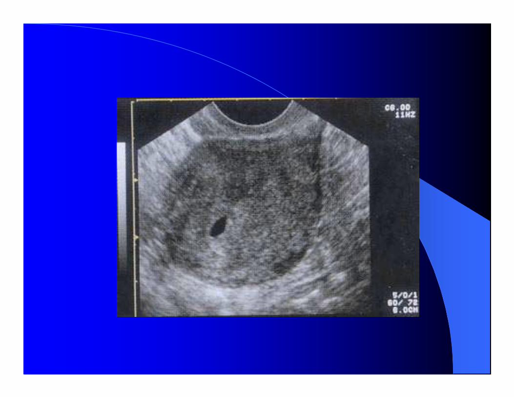
Gestation SacGestation Sac
� What?� The pregnancy or gestation sac: the chorionic cavity� Seen as a round echo- free/ hypoechoic/black area
surrounded by a thick white echoic/echogenic ring = surrounded by a thick white echoic/echogenic ring = (decidual reaction) in the uterus
� When?� Before the embrional pole/embrio becomes visible for
CRL� Usually from 5 weeks TAS/ 4w TVS

SonoembryologySonoembryology
� 4 weeks LMP� (4+0 – 4+6)� TVS 4+3 LMP� TVS 4+3 LMP� BHCG 500 – 1000 mIU/ml IRP� Chorionic sac visible in the uterus on TVS� Visble in the endometrium lateral of the
midline ( ectopic pseudo sac in the midline)

Gestation SacGestation Sac
� Where and How?� Measure the black area from outer to outer –
biggest measurement (do not include the white ring)white ring)
� Diameter: L x B ÷2� Volume: Lx B x H x 0.523 (volume of a
sphere)

YSYS

� Content = gestation� GS + YS ~ 5w� GS + Embryo ~ 6w� GS + Embryo ~ 6w� Viability:
– GS + Embryo +Heart flicker ~ 6w

SonoembryologySonoembryology
� 5weeks � Gestation sac� +� +� Yolc sac

SonoembryologySonoembryology
� 5 weeks LMP� (5+0 – 5+6)� Gestation sac 5-7mm (5+1)-(5+2)� Gestation sac 5-7mm (5+1)-(5+2)� YS (5+1)-(5+2)� Colour doppler arcuate artery, radial and
spiral aa visible� Embryo 5+6� FH (5+5)-(5+6)
SonoembryologySonoembryology
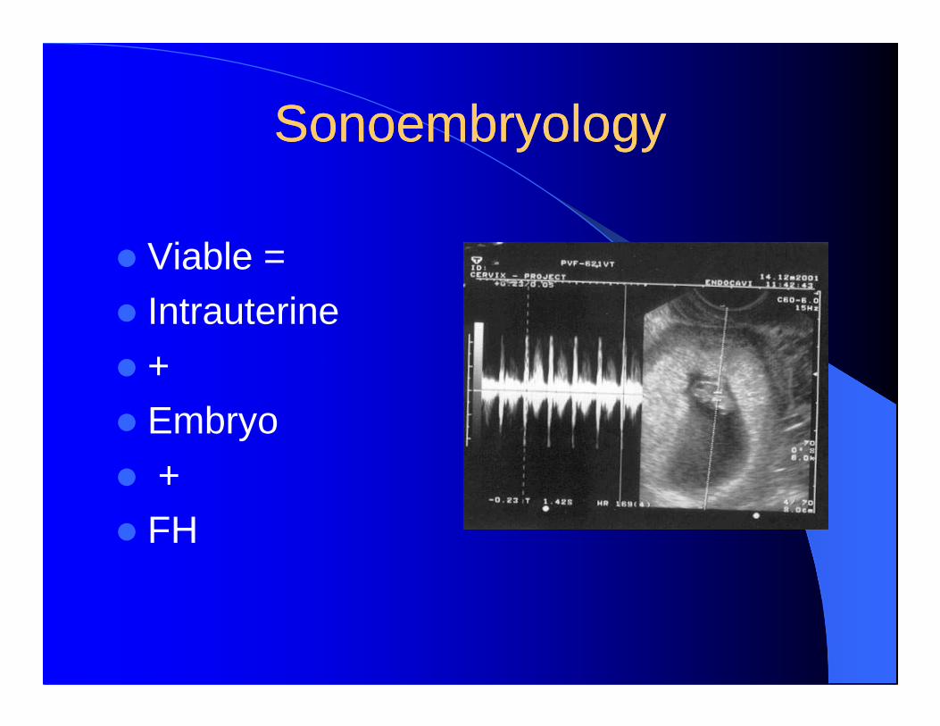
� Viable =� Intrauterine� +� +� Embryo� +� FH

FHRFHR
� Fetal heart rate should be done using M- Mode as far as possible particularly in the First trimesterin the First trimester

SonoembryologySonoembryology
�6 weeks� Embryo� Heartflicker� Heartflicker� YS

SonoembryologySonoembryology

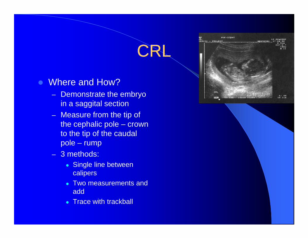
CRLCRL
� What?� Measuring the embryo or embryonic
polepole� When?� TAS 6 – 14 weeks� TVS 5 – 14weeks
CRLCRL
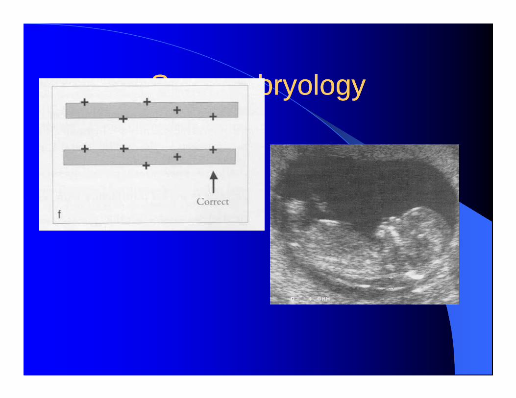
� Where and How?– Demonstrate the embryo
in a saggital section– Measure from the tip of
the cephalic pole – crown to the tip of the caudal pole – rump
– 3 methods:� Single line between
calipers� Two measurements and
add� Trace with trackball

CRLCRL
� CRL is the most accurate measurement for determining gestation if measured correctly.correctly.
� Where gestation from CRL differs from > 14 days to gestation by LMP use CRL

CRLCRL
� Pitfalls:� Faulty measurements due to:� Fetal Position� Excessive:� Excessive:� Flexion or Hyperextension = underestimation� Poor measurement plane� Improper section oblique� Inclusion of YS in CRL measurement in early pregnancy� Faulty measurements can make a gestation error of up to
10days!!!!

Sonoembryology Sonoembryology
� CRL

CRL 9.1 –15.9mm Week 7Day43 -49
Illustrated EmbryologyVolume 2 OrganogenesisH. Tuchman-Duplessis,P.Haegel

SonoembryologySonoembryology
Week 9CRL 22 – 31mm
SonoembryologySonoembryology
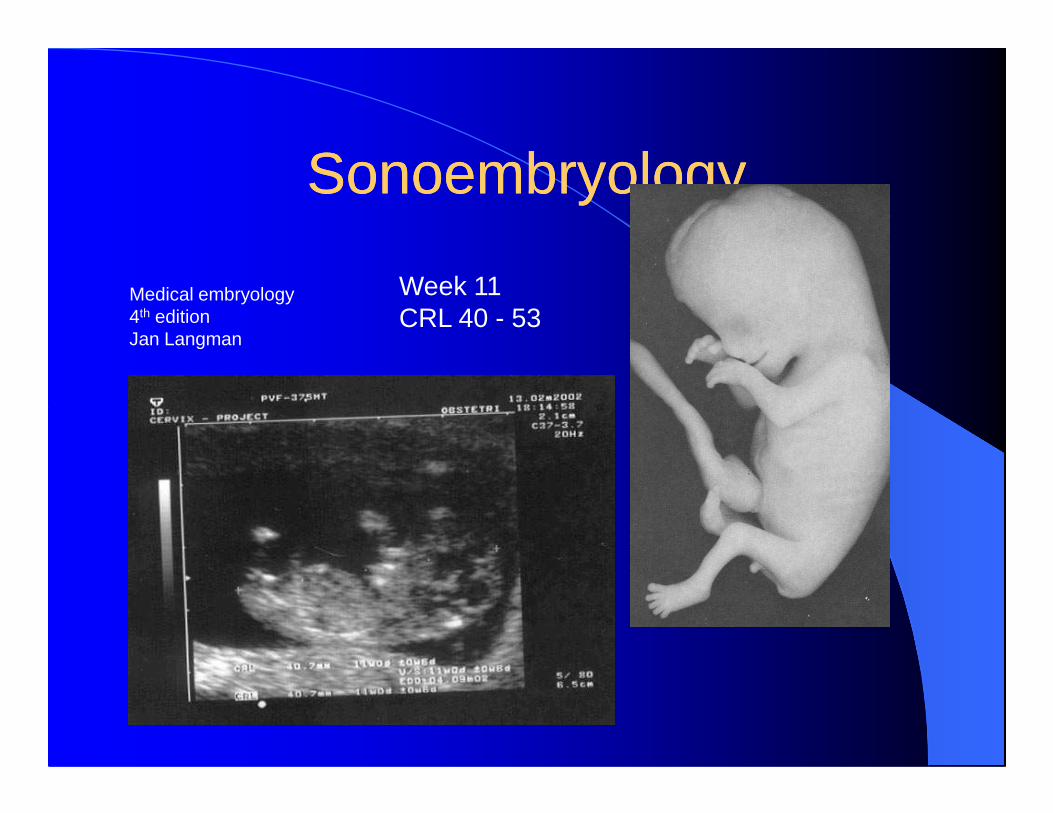
Medical embryology4th editionJan Langman
Week 11CRL 40 - 53

SonoembryologySonoembryology
Tachicardia T13,Turners,TriploidyBradicardia T18
SonoembryologySonoembryology
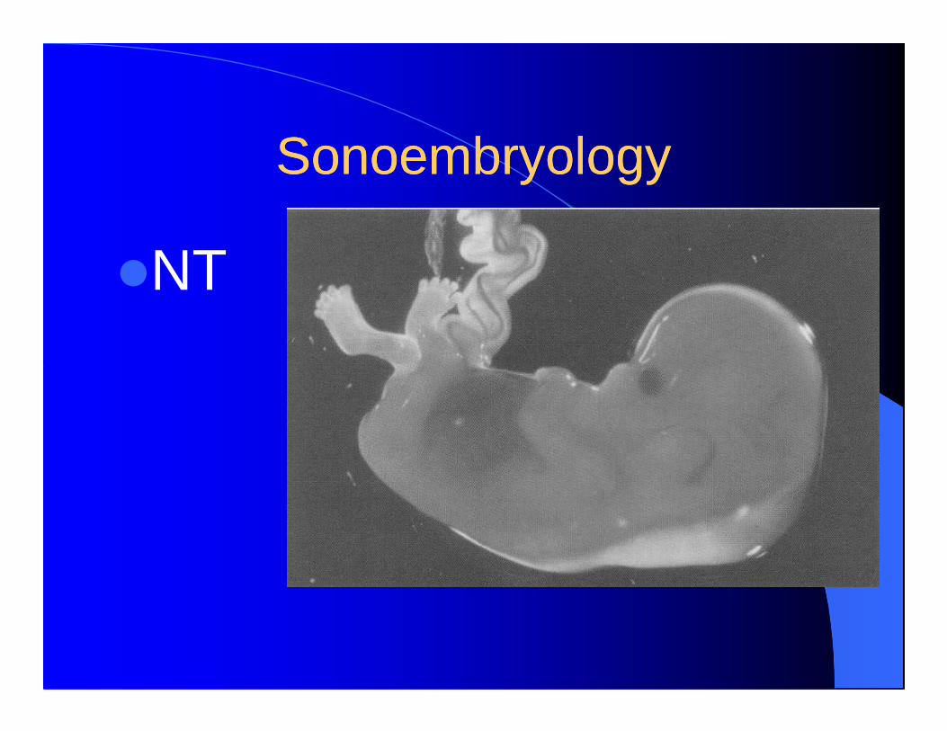
�NT
SonoembryologySonoembryology

SonoembryologySonoembryology
Nuchal cord5-10%smaller NT
Saggital75%“on to on”amnion
smaller NT
NT CRL 45 – 84mm

Normal anatomyNormal anatomy

Normal anatomyNormal anatomy

Normal anatomyNormal anatomy

Normal anatomyNormal anatomy

Normal anatomyNormal anatomy

BPDBPD
� What?� The bi-parietal measurement� When?� When?� From (14)15 – 28 weeks

BPDBPD
� Where?� Demonstrate the fetal skull in a transverse
section – occipito transverse� Correct section should demonstrate the � Correct section should demonstrate the
following:� Midline� Thalami� Basal cisterns� Cavum septum pelucidum� As suggested by the ACOG

BPDBPD
� How?� Leading edge to leading edge*
� 25 tables to date� 25 tables to date

BPDBPD
� Start by placing the transducer suprapubic in transverse on the maternal skin
� Chances are that you will see the fetal skull� If you see fetal spine then you are in a � If you see fetal spine then you are in a
longitudinal section and need to rotate the transducer through 90 degrees
� If you are lucky and get a transverse section then by small sliding, rocking and rotation movements you will be able to demonstrate the correct level/ section

BPDBPD
� Pitfalls:� Fetal position:Bx,DOP,Tx� Incorrect measurment level: too low, too � Incorrect measurment level: too low, too
high,rotated, angled� Fetal head shape:
dolicocephaly,brachicephaly (NB anomalies)
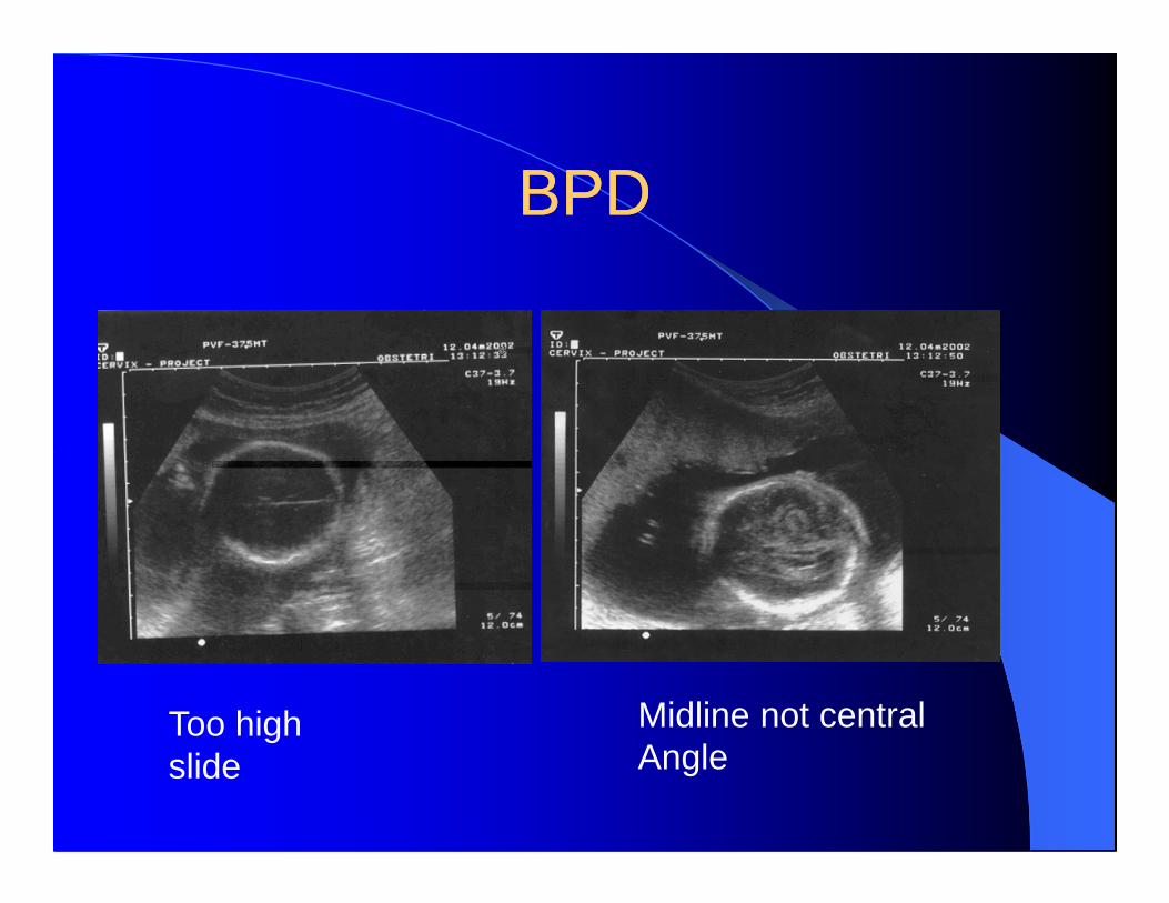
BPDBPD
Too highslide
Midline not centralAngle

BPDBPD
Low anteriorrotate Low Posterior
Rotate

BPD and OFDBPD and OFD
� Machine calibration incorrect 1540m/s @ 1600 m/s 4% measurement error!!
� OFD and CI� Occipito frontal diameter� Occipito frontal diameter� Where abnormal head shape� Dolicocephalic – long flat
– (oligohydramnios,PPROM,– microcephalic)
� Brachicephalic - short round – breech, anomalies
�

BPD and OFDBPD and OFD
� Where?� Same level as BPD� How?� How?� Occiput to front� CI = BPD ÷ OFD x 100 = 78.3%� CI ≤ 75% = dolicocephalic� CI ≥ 85% = brachicephalic

HCHC
� What?� Circumference of the skull� Indirect estimate of the size of the fetal � Indirect estimate of the size of the fetal
brain (skull sutures have not yet closed)� When? � From 15 weeks
HCHC
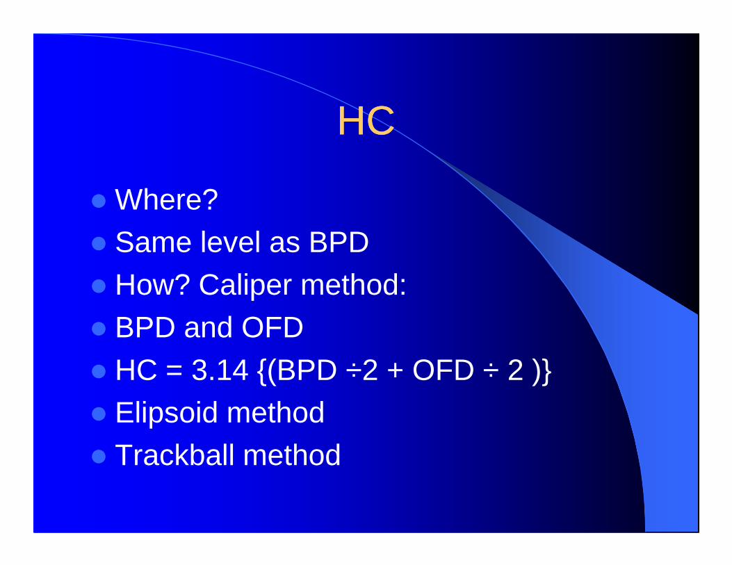
� Where?� Same level as BPD� How? Caliper method:� How? Caliper method:� BPD and OFD� HC = 3.14 {(BPD ÷2 + OFD ÷ 2 )}� Elipsoid method� Trackball method

ACAC
� What?
� The circumference of the abdomen� The circumference of the abdomen– Indirect estimate of fetal liver size and
glycogen storage/ nutritional status
� When? � 15 –23 weeks

ACAC
� Where? � Demonstrate a cross section through the fetal
abdomen� Correct section should demonstrate the � Correct section should demonstrate the
following:� Stomach� Fetal liver & middle 1/3 of umbilical vein� Spine L/R ( 3 dots of vertebral body)� Campbell and Wilken 1975

ACAC

ACAC
� How to get there� From the BPD slide the transducer upwards
in a transverse position – if you see the fetal stomach you are there – you now need to get stomach you are there – you now need to get the perfect section by a range of sliding, rocking and angling and rotation movements
� if you see the fetal heart you are to high� If you see the fetal bladder or kidneys you are
too low

ACAC
� How?� Caliper method:� TAD and APAD� TAD and APAD� AC = 3.14 {(APAD ÷2 + TAD ÷ 2 )}� Elipsoid method� Trackball method
FLFL
� What?� The femur shaft
� When? � From 15 – 23 weeks

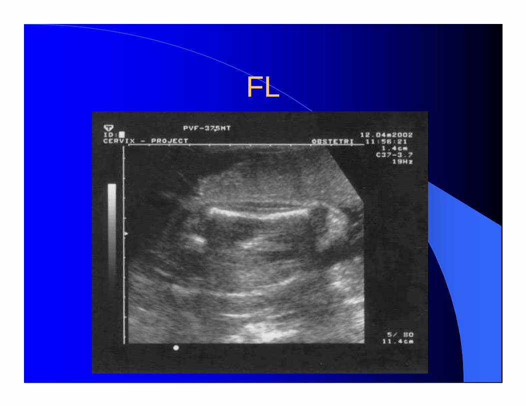
FLFL
� Where and How?� Transverse section through the fetal
femurfemur� Measure the “white” = the metaphisis of
the bone from the greater tubercle of the femur to the distal end of the femur
� “U to U”

FLFL
� How to get there?� If you see the fetal head slide the transducer
away from the head in a transverse position� If you see the bladder you are close� If you see the bladder you are close� The fetal femur can be seen as a white dot in
the thigh on cross section/ transverse rotate through 90 degrees here and you should start seeing the femur in a longitudinal section by a range of rocking,sliding,angling and rotational movements you should get the perfect section.

FLFL
� If you are too close to the BPD you may erroneously measure the humerus.
� If you see two dots in transverse section � If you see two dots in transverse section you are too low – tibia , fibula
FLFL
� Pitfalls:� Poor section: ie obligue also remember
the physics – poor lateral resolutionthe physics – poor lateral resolution� Wrong bone!!!
FLFL

AFIAFI
� Amniotic fluid index� 4 quadrants summate� Tables� Tables� Single pocket > 8cm = polyhydramnios� Single pocket < 2cm = oligohydramnios�

CervixCervix
� It is important to identify the relationship of the bladder: cervix
� It will help you orientate yourself� It will help you determine if the pregnancy is
intrauterine� It will help you exclude a placental praevia� It will identify pregnancies at risk for PTD

CXCX
� Remember the bladder should not be over extended
� The ideal mode of examination is a TVS with empty bladder this can be done at The ideal mode of examination is a TVS with empty bladder this can be done at the end of the exam
� How? � Internal to external os� < 20 mm is considered short

CXCX

CXCX
Measurements and helpMeasurements and help
� Remember� YOU are not alone� If you struggle or are unsure� If you struggle or are unsure� WE can help and would actually be glad
to do so
Measurements and helpMeasurements and help
� Your sources of help:� The hospital nearest to you will have an
radiology/ultrasound department:� Radiologists� Radiologists� Medical sonographers� BTech sonographers (have 4 years of sonar
training)� There are a handful of Fetal Medicine
Specialists/Units

Measurements and helpMeasurements and help
�SASUOG��
�www. sasuog.org.za

Measurements and helpMeasurements and help
� Some useful contact numbers:� Gauteng: State Hospitals� Kalafong 012 318 6675 Fetal Medicine Unit Dr Bridget Jeffery� Pretoria Academic:� Pretoria West: Dr Swanepoel� Pretoria West: Dr Swanepoel� Tembisa Hospital Dr L Meiring� Coronation Hospital: � 011 470 9054 Mrs Lucille Israel � Johannesburg General: � 011 488 3152 Mrs Z Holland� Baragwanath: � 011 933 8147 Dr E Nicolaou� Klerksdorp: Dr Alma Piek

Measurements and helpMeasurements and help
� Private Practice:– Fetal Medicine Specialists in SA
� Dr Ermos Nicolaou� Dr Bridgit Jeffery� Dr Lut Geerts� Dr Logi Govender� Dr Lizette De Koning
– Referral Doctors Gauteng� Dr Mark Van der Griendt Johannesburg � Dr Emmerich Frohlich� Dr’s Fouchs and Hattingh Femina� Dr Marieta Fourie Unitas� Dr Pierre Davis LCM� Dr Isabel Hough Eugene Marais� Dr Alma Piek Klerksdorp
�

Measurements and helpMeasurements and help
� National:� Western Cape:� Tygerberg Hospital� Fetal Medicine Unit� Dr Karen Norman� Dr Shannon Morris� Dr Linnie Miller - Private� Free State� Dr Lizette de Koning Bloemfonteint Private� Natal� Dr Logi Govender King Edward – State

THANK YOU FOR YOUR THANK YOU FOR YOUR ATTENTIONATTENTIONATTENTIONATTENTION