bcf uk foetal sexing guide
DESCRIPTION
BCF UK Foetal Sexing GuideTRANSCRIPT
Foetal Sexing Quick Guide
+44 (0)1506 460 023 | [email protected] | www.bcftechnology.com
Your ultrasound and X-ray people
Position the foetus
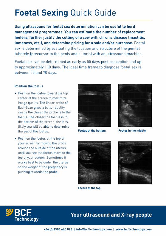
• Position the foetus toward the top center of the screen to maximize image quality. The linear probe of Easi-Scan gives a better quality image the closer the probe is to the foetus. The closer the foetus is to the bottom of the screen, the less likely you will be able to determine the sex of the foetus.
• Position the foetus at the top of your screen by moving the probe around the outside of the uterus until you see the foetus move to the top of your screen. Sometimes it works best to be under the uterus so the weight of the pregnancy is pushing towards the probe.
Using ultrasound for foetal sex determination can be useful to herd management programmes. You can estimate the number of replacement heifers, further justify the culling of a cow with chronic disease (mastitis, lameness, etc.), and determine pricing for a sale and/or purchase. Foetal sex is determined by evaluating the location and structure of the genital tubercle (precursor to the penis and clitoris) with an ultrasound machine.
Foetal sex can be determined as early as 55 days post conception and up to approximately 110 days. The ideal time frame to diagnose foetal sex is between 55 and 70 days.
Foetus at the bottom
Foetus at the top
Foetus in the middle

Foetal Sexing Quick GuideMale foetus determination
• It is easier to identify males so you should always start by checking for a male genital tubercle (GT). If you do not see a distinct male feature, you can then look to confirm it is female.
• The male and female GT have a similar appearance. The location is key to determining between male and female.
• Start by identifying the umbilical cord, and follow it into the abdomen. Look closely at the area where the umbilical cord connects to the foetus.
• You should be looking for:
° The male GT, it appears as two bright white parallel lines (bi-lobed structure), it can appear tri-lobed in older foetuses
° It is located at the base of the umbilical cord
° The scrotum is located between the hind limbs. It appears as a tri-lobed structure.
Umbilical cord looking unconnected
Umbilical cord connected
Male GT
Scrotum
Male GT
Male GT Abdomen
Umbilical cord Umbilical cord
Scrotum
Scrotum
Hind limbs Front limbs
Umbilical cord
Tail
Male GT
Front limbs
Umbilical cord
Genital tubercle
Hindlimbs

+44 (0)1506 460 023 | [email protected] | www.bcftechnology.com
Female foetus determination
• To identify a female foetus, start by searching the tail region.
• You should be looking for:
° The female GT, it appears as two bright white parallel lines (bi-lobed structure),
° It is located behind the hind limbs and under the tail
• Once tail is located, try to see both the tail and female GT at the same time. This ensures you are not incorrectly identifying the tail as the female GT.
• Once you have identified the tail and female GT you should locate the hind limbs. This ensures you are not confusing a leg bone with a female GT.
Female GT
Tail and GT
Tail, GT and hind limbs
Female GTFemale GT
Hind limbsHind limbs
Female GT
Female GT
TailTail
Umbilical cord
Hind limbsFemale GT
Tail
Tail
Tail
Tail
Genital tubercle
Hindlimbs
Your ultrasound and X-ray people

BCF Technology Ltd
UK +44 (0)1506 460 023 [email protected]
IRE +353 (0)42 932 0070 [email protected]
North America 800-210-9665 [email protected]
www.bcftechnology.com
To find your nearest BCF account manager, please visit our website.
Visit our learning zone at www.bcftechnology.com for a wide range of clinical guides and materials.
This quick guide is intended to help you start the learning process of identifying foetal sex. Training courses that cover this topic are available to help you advance your skills.
© BCF Technology, May 2013