bd cytometric bead array (cba) human th1/th2/th17 … · (bsa)* and proclin®-150 as a preservative...
TRANSCRIPT

BD OptEIA™
Mouse TNF ELISA KitInstruction Manual
Catalog No. 560478

BD flow cytometers are Class 1 Laser Products.For Research Use Only. Not for use in diagnostic or therapeutic procedures.© 2014 Becton, Dickinson and Company. All rights reserved. No part of this publication may bereproduced, transmitted, transcribed, stored in retrieval systems, or translated into any language orcomputer language, in any form or by any means: electronic, mechanical, magnetic, optical, chemical,manual, or otherwise, without prior written permission from BD Biosciences.Purchase does not include or carry any right to resell or transfer this product either as a stand-alone product or as a component of another product. Any use of this product other than the permitted use without the express written authorization of Becton, Dickinson and Company is strictly prohibited.BD, BD Logo and all other trademarks are property of Becton, Dickinson and Company. © 2014 BD
Table of Contents
Introduction................................................................................................... 4
Principle of the Test....................................................................................... 4
Reagents Provided.......................................................................................... 5
Materials Required but not Provided............................................................. 6
Storage Information....................................................................................... 6
Warnings and Precautions.............................................................................. 6
Specimen Collection and Handling................................................................. 8
Reagent Preparation....................................................................................... 8
Assay Procedure............................................................................................. 9
Assay Procedure Summary........................................................................... 11
Calculation of Results.................................................................................. 12
Typical Data................................................................................................ 12
Limitations of the Procedure........................................................................ 13
Performance Characteristics......................................................................... 13
Limit of Detection................................................................................ 13
Recovery.............................................................................................. 13
Linearity.............................................................................................. 13
Specificity............................................................................................. 14
Precision.............................................................................................. 14
Intra-assay.................................................................................... 14
Inter-assay.................................................................................... 14
Standardization.................................................................................... 14
Experimental Results............................................................................ 15
Serum........................................................................................... 15
Cell Culture Supernatants............................................................. 15
Troubleshooting.......................................................................................... 15
References.................................................................................................... 16
Plate Templates............................................................................................ 17

For Research Use Only. Not for use in diagnostic or therapeutic procedures.
www.bdbiosciences.com4
IntroductionTumor necrosis factor (TNF, formerly known as TNF-α) is a potent mediator of immune and inflammatory responses.TNF is produced by many activated cell types including monocytes, macrophages, astrocytes, granulocytes, T and B lymphocytes, NK cells, keratinocytes, fibroblasts, and certain tumor cells. TNF exerts many regulatory influences on the activation, growth, and differentiation of leukocytes and other cells. For example, TNF can costimulate the proliferation of activated T and B lymphocytes, upregulate the expressed levels of MHC class I and class II molecules by various cell types, as well as induce the expression of adhesion molecules by endothelial cells. TNF is selectively cytotoxic for some transformed cell lines and can exert cytotoxic effects against certain solid tumors. In vivo, TNF serves as a primary mediator in protective immune responses against microbial and viral pathogens. However, TNF has also been implicated as a central mediator in a number of pathologic responses including septic shock, cachexia, and autoimmune diseases.1-2
Activated cells initially express TNF as transmembrane proteins that associate to form homotrimeric complexes. After proteolytic cleavage, the extracellular region of membrane TNF sheds as a soluble homotrimer. Membrane and soluble TNF homotrimers are biologically active, whereas monomeric TNF is not. The mature mouse TNF monomer contains 156 amino acid residues, has a predicted size of ~17 kDa, and contains one potential N-glycosylation site. TNF exerts its biological activities by binding and signaling through membrane Type I and II TNF Receptors (TNFRI and TNFRII). Interestingly, TNF binds to soluble forms of TNFR (sTNFRI and sTNFRII) which are naturally shed by activated cells and can modulate biological activities of TNF.
The BD OptEIA™ Mouse TNF ELISA Kit is for the quantitative determination of mouse TNF in serum, plasma, and cell culture supernatant.
Principle of the TestThe BD OptEIA test is a solid phase sandwich ELISA (Enzyme-Linked Immunosorbent Assay). It utilizes a monoclonal antibody specific for mouse TNF coated on a 96-well plate. Standards and samples are added to the wells, and any TNF present binds to the immobilized antibody. The wells are washed, and biotinylated polyclonal anti-mouse TNF antibody is added, producing an antibody-antigen-antibody "sandwich." After a second wash, streptavidin-horseradish peroxidase conjugate is added. The wells are again washed and TMB substrate solution is added, which produces a blue color in direct proportion to the amount of TNF present in the initial sample. The Stop Solution changes the color from blue to yellow, and the wells are read at 450 nm.

For Research Use Only. Not for use in diagnostic or therapeutic procedures.
www.bdbiosciences.com 5
Reagents ProvidedAntibody Coated Wells: 2 plates of 96 breakable wells
(12 strips × 8 wells) coated with anti-mouse TNF monoclonal antibody
Detection Antibody: 30 mL of biotinylated anti-mouse TNF phage Fab antibody with fetal bovine serum (FBS) and with 0.1% ProClin®-150 as a preservative
Standards: 4 vials of lyophilized recombinant mouse TNF
Enzyme Concentrate (250×): 150 μL of 250× concentrated Streptavidin-horseradish peroxidase conjugate with bovine serum albumin (BSA)* and ProClin®-150 as a preservative
Standard/Sample Diluent: 30 mL of animal serum* with 0.09% sodium azide as preservative
Enzyme Diluent: 30 mL of animal serum* base with 0.15% ProClin®-150 as a preservative
ELISA Diluent: 12 mL of a buffered protein base with 0.09% sodium azide as preservative
Wash Concentrate (20×): 100 mL of 20× concentrated detergent solution with ProClin®-150 as a preservative
TMB One-Step Substrate Reagent:
30 mL of 3,3',5,5'-tetramethylbenzidine (TMB) in buffered solution
Stop Solution: 13 mL of 1 M phosphoric acid
Plate Sealers: 4 sheets with adhesive backing
*Source of all serum proteins is from USDA inspected abattoirs located in the United States

For Research Use Only. Not for use in diagnostic or therapeutic procedures.
www.bdbiosciences.com6
Materials Required but not Provided Microplate reader capable of measuring absorbance at 450 nm
Precision pipettes to deliver 50 μL and 100 μL volumes
Adjustable 1 mL, 5 mL, 10 mL, 25 mL pipettes for reagent preparation
Deionized or distilled water
Wash bottle or automated microplate washer
Log-log graph paper or computer and software for ELISA data analysis
Tubes to prepare standard dilutions
Laboratory timer
Absorbent paper
Storage Information1. Store the unopened kit at 2 - 8°C. Do not use the kit after the
expiration date.
2. Before use, bring all reagents to room temperature (18 - 25°C). Immediately after use, return to proper storage conditions.
3. Lyophilized standards are stable until the kit expiration date. After reconstitution, use freshly reconstituted standard within 12 hours (stored at 2 - 8°C).
Warnings and Precautions1. Reagents that contain preservatives may be toxic if ingested, inhaled,
or brought in contact with skin.
2. Avoid contact of skin, eyes, or clothing with Stop Solution or Substrate Reagents.
3. Handle all serum and plasma specimens in accordance with CLSI guidelines for preventing transmission of blood-borne infections.
4. Standard/Sample Diluent and ELISA Diluent contain less than 0.1% sodium azide. Sodium azide yields highly toxic hydrazoic acid under acidic conditions. Dilute azide compounds in running water before discarding to avoid accumulation of potentially explosive deposits in plumbing.

For Research Use Only. Not for use in diagnostic or therapeutic procedures.
www.bdbiosciences.com 7
5. Warning
Wash Concentrate (20X) (component 51-9003738) contains 0.002% (w/w), Mouse TNF Lyophilized Standard (component 51-26676E) contains 0.03% (w/w), Enzyme Diluent (component 51-2718KD) contains 0.003% (w/w) and Detection Antibody Biotin Anti-Mouse TNF (component 51-9006279) contains 0.003% (w/w) of a CMIT/MIT mixture (3:1), which is a mixture of: 5-chloro-2-methyl-4-isothiazolin-3-one [EC No 247-500-7] and 2-methyl-4-isothiazolin-3-one [EC No 220-239-6] (3:1).
Hazard statements
May cause an allergic skin reaction.
Precautionary statements
Wear protective gloves / eye protection.
Wear protective clothing.
Avoid breathing mist/vapours/spray.
If skin irritation or rash occurs: Get medical advice/attention.
IF ON SKIN: Wash with plenty of water.
Dispose of contents/container in accordance with local/regional/national/international regulations.
6. Danger
Stop Solution (component 51-2608KC) contains 15.23% phosphoric acid (w/w).
Hazard statements
Causes severe skin burns and eye damage.
Precautionary statements
Wear protective gloves / eye protection.
Wear protective clothing.
IF ON SKIN (or hair): Remove/Take off immediately all contaminated clothing. Rinse skin with water/shower.
IF IN EYES: Rinse cautiously with water for several minutes. Remove contact lenses, if present and easy to do.
Continue rinsing.
IF INHALED: Remove victim to fresh air and keep at rest in a position comfortable for breathing.
Dispose of contents/container in accordance with local/regional/national/international regulations.

For Research Use Only. Not for use in diagnostic or therapeutic procedures.
www.bdbiosciences.com8
Specimen Collection and HandlingSpecimens should be clear, non-hemolyzed, and non-lipemic. Samples with expected values higher than the top standard, 2000 pg/mL, should be diluted with Standard/Sample Diluent prior to running the assay.
Cell culture supernatants: Remove any particulate material by centrifugation and assay immediately or store samples at -20°C. Avoid repeated freeze-thaw cycles.
Serum: Use a serum tube (eg, BD Vacutainer® Cat. No. 366430) and allow samples to clot for 30 minutes, then centrifuge for 10 minutes at 1000 × g. Remove serum and assay immediately or store samples at -20°C. Avoid repeated freeze-thaw cycles.
Plasma: Collect plasma using citrate, EDTA, or heparin as anticoagulant. Centrifuge for 10 minutes at 1000 × g within 30 minutes of collection. Assay immediately or store samples at -20°C. Avoid repeated freeze-thaw cycles.
Reagent Preparation1. Bring all reagents to room temperature (18 - 25°C) before use.
2. Standards
a. Reconstitute 1 vial lyophilized Standard with the required volume (noted on vial label) of Standard/Sample Diluent to prepare a 2000 pg/mL stock standard. Allow the standard to equilibrate for at least 15 minutes before making dilutions. Vortex to mix.
b. Add 300 μL Standard/Sample Diluent to 6 tubes. Label the tubes as 1000 pg/mL, 500 pg/mL, 250 pg/mL, 125 pg/mL, 62.5 pg/mL, and 31.3 pg/mL.
c. Perform serial dilutions by adding 300 μL of each standard to the next tube and vortexing between each transfer. The undiluted standard serves as the high standard (2000 pg/mL). The Standard/Sample Diluent serves as the zero standard (0 pg/mL).

For Research Use Only. Not for use in diagnostic or therapeutic procedures.
www.bdbiosciences.com 9
3. Wash Buffer
Note: If the Wash Concentrate contains visible crystals, warm to room temperature and mix gently until dissolved. Dilute the required quantity of 20× Wash Concentrate with deionized or distilled water and mix. (To prepare 2,000 mL, add 100 mL Wash Concentrate to 1,900 mL water. At least 500 mL solution should be prepared for a full 96-well plate).
4. TMB One-Step Substrate Reagent
No more than 15 minutes prior to use, add required volume of TMB One-Step Substrate Reagent to a clean tube or reservoir. To prevent contamination, pipette out from the tube/ reservoir instead of directly from bottle. Avoid prolonged exposure to light or contact with metal, air, or extreme temperature as color may develop.
Assay Procedure1. Bring all reagents and samples to room temperature (18 - 25°C) prior
to use. It is recommended that all standards and samples be run in duplicate.
2. Remove the required quantity of test strips/wells and place in well holder.
Note: Wells are provided in breakable 8-well strips. Strips may be “broken” into individual wells, replaced in a well holder, and assayed. Return any unused wells to sealed pouch for 2 - 8°C storage.
3. Pipette 50 μL of ELISA Diluent into each well.
4. Pipette 50 μL of each standard (see Reagent Preparation, step 2) and sample into appropriate wells. Cover wells with Plate Sealer and incubate for 2 hours at room temperature.
5. Decant or aspirate contents of wells. Wash wells by filling with at least 300 μL/well prepared Wash Buffer (see Reagent Preparation, step 3) followed by decanting/aspirating. Repeat the wash 4 times for a total of 5 washes. After the last wash, blot the plate on absorbent paper to remove any residual buffer. Complete removal of liquid is required for proper performance.

For Research Use Only. Not for use in diagnostic or therapeutic procedures.
www.bdbiosciences.com10
6. Add 100 μL of Detection Antibody to each well. Cover wells with Plate Sealer and incubate for 1 hour at room temperature.
7. a. Prepare Enzyme Working Reagent.
b. Pipette the required volume of Enzyme Diluent into a clean tube or flask. Add in the required quantity of Enzyme Concentrate (250×) and vortex or mix well. For a full 96-well plate, add 48 μL of Enzyme Concentrate into 12 mL of Enzyme Diluent.
8. Wash wells as in Step 5.
9. Add 100 μL of Enzyme Working Reagent (see step 7 above) to each well. Cover wells with Plate Sealer and incubate for 30 minutes at room temperature.
10. Wash wells as in Step 5, but a total of 7 times.
Note: In this final wash step, soak wells in wash buffer for 30 seconds to 1 minute for each wash. Thorough washing at this step is very important.
11. Add 100 μL of TMB One-Step Substrate Reagent to each well. Incubate plate (without Plate Sealer) for 30 minutes at room temperature in the dark.
12. Add 50 μL of Stop Solution to each well.
13. Read absorbance at 450 nm within 30 minutes of stopping the reaction. If wavelength correction is available, subtract the optical density readings at 570 nm from readings at 450 nm.
A standard curve is required in each assay run.

For Research Use Only. Not for use in diagnostic or therapeutic procedures.
www.bdbiosciences.com 11
Assay Procedure Summary1. Add 50 μL ELISA Diluent to each well.
2. Add 100 μL standard or sample to each well.
Incubate 2 hours at room temperature.
3. Aspirate and wash 5 times.
4. Add 100 μL Detection Antibody to each well.
Incubate 1 hour at room temperature.
5. Aspirate and wash 5 times.
6. Add 100 μL Enzyme Working Reagent to each well.
Incubate 30 minutes at room temperature.
7. Aspirate and wash/soak 7 times.
8. Add 100 μL TMB One-Step Substrate Reagent to each well.
Incubate 30 minutes at room temperature.
9. Add 50 μL Stop Solution to each well.
Read at 450 nm within 30 minutes.
correction 570 nm.

For Research Use Only. Not for use in diagnostic or therapeutic procedures.
www.bdbiosciences.com12
Calculation of ResultsCalculate the mean absorbance for each set of duplicate standards, controls, and samples. Subtract the mean zero standard absorbance (ie, plate background) from each.
Plot the standard curve on log-log graph paper, with TNF concentration on the x-axis and absorbance on the y-axis. Draw the best fit straight line through the standard points.
To determine the TNF concentration of the unknowns, find the unknowns’ mean absorbance value on the y-axis and draw a horizontal line to the standard curve. At the point of intersection, draw a vertical line to the x-axis and read the TNF concentration. If samples were diluted, multiply the interpolated TNF concentration by the dilution factor.
Computer-based curve-fitting statistical software may also be employed.
Typical DataThis standard curve is for demonstration only. A standard curve must be run with each assay.
Concentration(pg/mL) OD1 OD2 Mean
Zero StandardSubtracted
0 0.027 0.027 0.027 0.000
31.3 0.103 0.104 0.104 0.077
62.5 0.171 0.162 0.167 0.140
125 0.317 0.308 0.312 0.286
250 0.596 0.619 0.607 0.580
500 1.042 1.089 1.066 1.039
1000 1.932 1.934 1.933 1.906
2000 3.318 3.811 3.565 3.538
For Research Use Only. Not for use in diagnostic or therapeutic procedures.
www.bdbiosciences.com 13
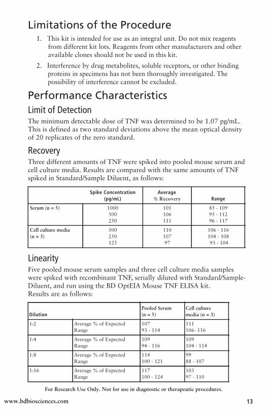
Limitations of the Procedure1. This kit is intended for use as an integral unit. Do not mix reagents
from different kit lots. Reagents from other manufacturers and other available clones should not be used in this kit.
2. Interference by drug metabolites, soluble receptors, or other binding proteins in specimens has not been thoroughly investigated. The possibility of interference cannot be excluded.
Performance CharacteristicsLimit of DetectionThe minimum detectable dose of TNF was determined to be 1.07 pg/mL. This is defined as two standard deviations above the mean optical density of 20 replicates of the zero standard.
RecoveryThree different amounts of TNF were spiked into pooled mouse serum and cell culture media. Results are compared with the same amounts of TNF spiked in Standard/Sample Diluent, as follows:
Spike Concentration(pg/mL)
Average% Recovery Range
Serum (n = 5) 1000500250
101106111
85 - 10995 - 11296 - 117
Cell culture media (n = 3)
500250125
11010797
106 - 116104 - 108 93 - 104
LinearityFive pooled mouse serum samples and three cell culture media samples were spiked with recombinant TNF, serially diluted with Standard/Sample-Diluent, and run using the BD OptEIA Mouse TNF ELISA kit. Results are as follows:
DilutionPooled Serum (n = 5)
Cell culture media (n = 3)
1:2 Average % of Expected Range
107 93 - 114
111 106- 116
1:4 Average % of Expected Range
109 94 - 116
109 104 - 114
1:8 Average % of Expected Range
114 100 - 121
99 88 - 107
1:16 Average % of Expected Range
117 100 - 124
103 97 - 110

For Research Use Only. Not for use in diagnostic or therapeutic procedures.
www.bdbiosciences.com14
SpecificityCross Reactivity: The following factors were tested using the BD OptEIA assay at 10 ng/mL, and no cross-reactivity (value 15 pg/mL) was identified.
Recombinant Human
TNF
Recombinant Mouse
IL-1, IL-1, IL-2, IL-3, IL-4, IL-5, IL-6, IL-7, IL-9, IL-10, IL-12 (p70), IL-13, IL-15, IL-17, IL-18, GM-CSF, IFN-LT-, MCP-1, M-CSF, MIG, MIP-1, MCIP-1, sTNFRI, sTNFRII, RANTES
Recombinant Rat
IL-1IL-2, IL-4, IL-6, IL-10, IL-18, GM-CSF, IFN-
PrecisionIntra-assay
Twenty-four replicates each of three different levels of TNF were tested in one plate. The following results were observed:
Number of Replicates 24 24 24
Mean Concentration 585.5 pg/mL 287.4 pg/mL 135.8 pg/mL
SD 34.2 13.9 14.8
%CV 5.8 4.8 10.9
Inter-assay
Three different levels of TNF were tested in four different plates. The following results were observed:
Number of Replicates 32 32 32
Mean Concentration 555.3 pg/mL 277.2 pg/mL 136.1 pg/mL
SD 46.7 24.2 16.8
%CV 8.4 8.7 12.4
StandardizationThe immunoassay was calibrated against recombinant mouse TNF.
During the development of this product, the NIBSC/WHO mouse TNF reference Standard 88/532 (recombinant mouse TNF) was evaluated in this kit. The conversion factor for NIBSC material is as follows:
1 mg NIBSC (88/532) TNF = 0.3 BD OptEIA recombinant mouse TNF

For Research Use Only. Not for use in diagnostic or therapeutic procedures.
www.bdbiosciences.com 15
Experimental ResultsSerum
Seventeen apparently healthy normal mouse serum samples were tested in this assay. Fourteen samples measured less than 31.3 pg/mL (lowest standard level). Three samples measured above the lowest standard at 36.5, 37.3, and 73.5 pg/mL.
Cell Culture Supernatants
Mouse splenocytes or peritoneal exudate cells (PECs) from apparently healthy, normal mice were cultured in RPMI 1640 complete medium with 7.5% FBS at 1 × 10⁶ cells/mL, and stimulated with Con A (5 μg/mL) for 24 hours (1), or IFN- (10 ng/mL)/LPS (1 μg/mL) for 7 days (2), or PMA/ionomycin for 24 hours (3) or 48 hours (4). PECs were differentiated into MiCK-2 cells (5). Culture supernatants were collected and quantified for TNF using the BD OptEIA Mouse TNF ELISA Kit. The results are as follows:Mouse No. TNF (pg/mL)
1 182 Balb/c splenocytes, 24 hr Con A (5 μg/mL) stimulation
2 2,247 PEC, 7 day Ms IFN (10 ng/mL) + LPS (1μg/mL) stimulation
3 1,215 C57BL/6 splenocytes, 24 hr PMA/ionomycin stimulation
4 2,251 C57BL/6 splenocytes, 48 hr PMA/ionomycin stimulation
5 21,458 MiCK-2 cells, (6 hr PMA (5 ng/mL) + Ionomycin (500 μg/mL) stimulation
TroubleshootingProblem Possible Source Corrective Action
Poor Precision
• Inadequate washing / aspiration of wells
• Inadequate mixing of reagents • Imprecise / inaccurate pipetting • Imprecise sealing of plate
• Check function of washing system • Ensure adequate mixing • Check / calibrate pipettes • Ensure complete sealing of plate
Poor Standard Curve
• Improper standard handling / dilution
• Incomplete washing / aspiration of wells
• Imprecise / inaccurate pipetting
• Ensure correct preparation of standards
• Check function of washing system • Check / calibrate pipettes
Low Signal • Inadequate reagent volumes added to wells
• Incorrect incubation times / temperature
• Overly high wash / aspiration pressure from automated plate-washer.
• Check / calibrate pipettes
• Ensure sufficient incubation times / reagents warmed to room temperature • Utilize manual washing

For Research Use Only. Not for use in diagnostic or therapeutic procedures.
www.bdbiosciences.com16
References1. Beyaert R, Fiers W. Tumor necrosis factor and Iymphotoxin. In: Mire-Sluis AR,
Thorpe R, eds. Cytokines. San Diego, CA: Academic Press; 1998:335-360.
2. Ware CF, Santee S, Glass A. Tumor necrosis factor-related ligands and receptors.In: Thompson A, ed. The Cytokine Handbook. 3rd ed. San Diego, CA: AcademicPress; 1998:549-592.

For Research Use Only. Not for use in diagnostic or therapeutic procedures.
www.bdbiosciences.com 17
Plate Templates
1 2 3 4 5 6 7 8 9 10 11 12
A
B
C
D
E
F
G
H
1 2 3 4 5 6 7 8 9 10 11 12
A
B
C
D
E
F
G
H
For Research Use Only. Not for use in diagnostic or therapeutic procedures.
www.bdbiosciences.com18
Notes


Catalog No. 560478Rev# 2
United States877.232.8995
Canada866.979.9408
Europe32.2.400.98.95
Japan0120.8555.90
Asia/Pacific65.6861.0633
Latin America/Caribbean55.11.5185.9995
Becton, Dickinson and CompanyBD Biosciences2350 Qume Dr.San Jose, CA 95131 USA(US) Ordering 855.236.2772Technical Service 877.232.8995Fax [email protected]