benjamin j. fregly , jeffrey a. reinbolt, kelly l. rooney ...fregly/pdfs/tbme2007a.pdf · the...
TRANSCRIPT

IEEE TRANSACTIONS ON BIOMEDICAL ENGINEERING, VOL. 54, NO. 9, SEPTEMBER 2007 1687
Design of Patient-Specific Gait Modifications forKnee Osteoarthritis Rehabilitation
Benjamin J. Fregly�, Jeffrey A. Reinbolt, Kelly L. Rooney, Kim H. Mitchell, and Terese L. Chmielewski
Abstract—Gait modification is a nonsurgical approach forreducing the external knee adduction torque in patients withknee osteoarthritis (OA). The magnitude of the first adductiontorque peak in particular is strongly associated with knee OAprogression. While toeing out has been shown to reduce the secondpeak, no clinically realistic gait modifications have been identifiedthat effectively reduce both peaks simultaneously. This studypredicts novel patient-specific gait modifications that achieve thisgoal without changing the foot path. The modified gait motionwas designed for a single patient with knee OA using dynamicoptimization of a patient-specific, full-body gait model. The costfunction minimized the knee adduction torque subject to con-straints limiting how much the new gait motion could deviate fromthe patient’s normal gait motion. The optimizations predicteda “medial-thrust” gait pattern that reduced the first adductiontorque peak between 32% and 54% and the second peak be-tween 34% and 56%. The new motion involved three synergistickinematic changes: slightly decreased pelvis obliquity, slightlyincreased leg flexion, and slightly increased pelvis axial rotation.After gait retraining, the patient achieved adduction torque re-ductions of 39% to 50% in the first peak and 37% to 55% in thesecond one. These reductions are comparable to those reportedafter high tibial osteotomy surgery. The associated kinematicchanges were consistent with the predictions except for pelvisobliquity, which showed little change. This study demonstratesthat it is feasible to design novel patient-specific gait modificationswith potential clinical benefit using dynamic optimization ofpatient-specific, full-body gait models. Further investigation isneeded to assess the extent to which similar gait modifications maybe effective for other patients with knee OA.
Index Terms—Dynamic optimization, knee adduction moment,movement prediction, osteoarthritis (OA).
Manuscript received May 5, 2006; revised December 15, 2006. This work wassupported in part by the Whitaker Foundation and in part by the National Insti-tutes of Health (NIH) National Library of Medicine under Grant R03-LM07332.Asterisk indicates corresponding author.
B. J. Fregly is with the Departments of Mechanical and Aerospace Engi-neering, Biomedical Engineering, and Orthopaedics and Rehabilitation, Uni-versity of Florida, Gainesville, FL 32611 USA (e-mail: [email protected]).
J. A. Reinbolt was with the the Department of Mechanical and AerospaceEngineering, University of Florida, Gainesville, FL 32611 USA. He is now withthe Department of Bioengineering, Stanford University, Stanford, CA 94305USA (e-mail: [email protected])..
K. L. Rooney is with the Department of Biomedical Engineering, Universityof Florida, Gainesville, FL 32611 USA.
K. H. Mitchell is with the Biomotion Foundation, Palm Beach, FL 33480USA.
T. L. Chmielewski is with the Department of Physical Therapy, University ofFlorida, Gainesville, FL 32611 USA (e-mail: [email protected]).
This paper contains supplementary multimedia material available at http://ieeexplore.ieee.org, provided by the author. The material consists of videos inavi format.
Digital Object Identifier 10.1109/TBME.2007.891934
I. INTRODUCTION
DESPITE the need for early treatment, few clinical inter-ventions slow the progression of knee osteoarthritis (OA)
and minimize future functional limitations. One of the most con-servative surgical interventions is high tibial osteotomy (HTO).During this procedure, a wedge of bone is typically added to orremoved from the proximal tibia, changing the alignment of theleg from bow legged to slightly knock kneed. The change in limbalignment is intended to shift some of the contact load from thediseased medial to the healthy lateral compartment of the knee.Since articular cartilage is responsive to the magnitude of jointloading, reducing compressive loads in the diseased compart-ment may slow the rate of cartilage breakdown and delay theneed for joint replacement.
Since medial compartment load cannot be measured nonin-vasively in vivo, researchers have sought an external measure toquantify the load reduction achieved by HTO surgery. The bestcandidate found thus far is the external knee adduction torqueduring gait [1], [2]. This torque exhibits two peaks during thegait cycle—one during early stance and the other during latestance. Only the first peak during early stance has been shownto be higher in patients with knee OA compared to healthy con-trols [3]–[5], making the first peak the most critical one to lower.Following HTO surgery, patients with the lowest first peak tendto have the best long-term outcome [6], [7], particularly thosewhose peak is below approximately 2.5% body weight timesheight (%BW HT) [8]. Such patients typically obtain adduc-tion torque reductions on the order of 30% to 50% from HTOsurgery [3], [6], [9]. A high-peak knee adduction torque has alsobeen correlated with higher proximal tibial bone density on themedial side [10], increased OA disease severity [11], and an in-creased rate of OA disease progression [12].
A noninvasive early treatment option that has received limitedattention is gait modification to decrease the peak knee adduc-tion torque. Ideally, if simple gait modifications could reduce thepeak adduction torque by as much as HTO surgery, then the ben-efits of the surgery could be made available to a broad clinicalpopulation without the risks and costs of an invasive procedure.To date, at least five basic gait modifications have been shownto reduce the adduction torque in patients with knee OA: toeingout, walking more slowly, walking with decreased stride length,walking with increased medial-lateral trunk sway, or using lat-eral heel wedges. Walking with the toes pointed outward can re-duce the second peak of the adduction torque curve by as muchas 40% but has little influence on the first peak [2], [4], [13],[14]. Walking slower or with decreased stride length can re-duce both peaks significantly in some patients but not others[15], but the speed or stride length decrease that is required to
0018-9294/$25.00 © 2007 IEEE

1688 IEEE TRANSACTIONS ON BIOMEDICAL ENGINEERING, VOL. 54, NO. 9, SEPTEMBER 2007
achieve a significant reduction may be larger than many patientswould tolerate. While increasing medial-lateral trunk sway canreduce the first peak by up to 66% [16], the resulting gait mo-tion does not look normal, which could deter many patients fromadopting it. Recent gait studies using lateral heel wedges havereported only modest reductions in the peak adduction torque[17], [18]. Thus, no clinically realistic gait modifications havebeen identified that will reduce the first peak or both peaks ofthe knee adduction torque curve to an extent comparable to HTOsurgery.
This feasibility study seeks to design a novel yet “normallooking” gait motion that reduces both adduction torque peaksto the same extent as HTO surgery but without changing thefoot path or trunk orientation. The design process was tailoredto an individual patient, utilized dynamic optimization of a pa-tient-specific full-body gait model, and required the patient’spretreatment gait data as a starting point. The optimization re-sults were used to teach the patient how to walk differently toreduce both adduction torque peaks simultaneously. Gait datacollected from the patient following gait retraining were used toevaluate the extent to which the predicted modifications can beachieved and sustained in clinical practice.
II. MATERIALS AND METHODS
A. Experimental Data Collection
Gait data were collected from a single highly functional kneeOA patient (male, age 37 years, height 170 cm, mass 69 kg,alignment 5 varus with Kellgren and Lawrence grade 2 medialOA in both knees based on radiographic assessment). The sub-ject gave informed consent and performed all experimental trialswhile wearing New Balance 608 sneakers. Three-dimensional(3-D) surface marker data using the Cleveland Clinic markerset with additional markers on the feet were collected at 120 Hzusing a six-camera video-based motion analysis system (Mo-tion Analysis Corporation, Santa Rosa, CA). The 3-D resid-uals from the camera calibration trial averaged 1.1 mm with astandard deviation of 0.5 mm. Individual markers were locatedon both wrists, elbows, and shoulders, with three markers persegment placed on the pelvis and each thigh, shank, and hind-foot [19]. A static trial with additional markers over anatomicallandmarks was performed to define segment coordinate systemsand marker locations within those coordinate systems. Dynamicjoint motion trials were performed for the hip, knee, and ankle toexercise their primary functional axes for determination of jointpositions and orientations in the segment coordinate systems[19]. For the gait trials, the subject walked along a 15-m runwayat a self-selected speed of 1.4 m/s, which was measured usingthe progression of the pelvis markers over the time betweensuccessive left heel strikes. Ground reaction forces and torquesunder each foot were measured at 1000 Hz around the electricalcenters of two force plates (Advanced Mechanical Technology,Inc., Watertown, MA). The raw marker data were filtered usinga fourth-order, zero phase-shift, low-pass Butterworth filter witha cutoff frequency of 6 Hz. The data collection and subsequentcomputer simulations were approved by the institutional reviewboard.
One complete gait cycle (left heel strike to left heel strike)without surface marker dropout was selected as the nominal dataset for use in the optimization study. Due to the limitation ofonly two force plates, no ground reaction data were availablefor the right leg at the start of the cycle (i.e., the right leg wasin contact with the ground, but not on a force plate, at the initialleft heel strike). For the initial time frames when no right groundreaction data were available, the patient-specific full-body gaitmodel developed for the optimization studies was used to esti-mate the right ground reactions from the left ones. Since onlythe left leg had experimental ground reaction data for the entirecycle, results from the subsequent optimization studies are pre-sented only for that leg.
After optimization predictions were developed using thepatient’s nominal gait data, the patient attempted to trainhimself to produce the predicted gait motion on a qualitativebasis. Training consisted of studying plots of the optimizedkinematics, kinetics, center of pressure, and ground reactions,as well as animations comparing the nominal and optimalgait motions. The patient implemented the general kinematicalterations in a manner that was as natural and easy to achieveas possible compared to his original gait motion. Over anine-month self-training period, the patient sought to incorpo-rate these gait modifications into his normal walking patternby constantly reminding himself to walk the new way, thegoal being for the new gait motion to become second nature.After this period, the patient gave informed consent and wasretested wearing the same sneakers. Testing was performedat a self-selected walking speed, which again was 1.4 m/s,under two conditions. First, the patient was asked to try to walkusing his old gait motion prior to retraining, and second, hewas asked to walk using an exaggerated version of the newgait motion predicted by the optimizations. These two walkingpatterns were selected to bound the experimental adductiontorque changes that the patient could achieve as a result of thegait retraining process.
B. Dynamic Model Development
A parametric, 3-D, dynamic gait model was developed usingtwo separate software packages. The first was Autolev (OnlineDynamics, Inc., Sunnyvale, CA), a symbolic manipulator forderiving dynamical equations, and the second was Softwarefor Interactive Musculoskeletal Modeling (SIMM) with theDynamics Pipeline (Motion Analysis Corporation, Santa Rosa,CA). The Autolev model provided direct access to the symbolicform of the equations of motion, making it well suited forcalibrating joint and inertial parameters to the patient’s nominalgait data. In contrast, the SIMM/Pipeline model provided awell-structured dynamic simulation environment along with theability to visualize the predicted motions and ground reactionforce vectors using a skeletal model scaled to the patient’sdimensions. Having two versions of the same dynamic modelalso permitted validation of the inverse dynamic analyses usedin the optimization studies.
The gait model possessed 27 degrees of freedom (DOFs)composed of gimbal (3 DOFs), universal (2 DOFs), and pin(1 DOF) joints (Fig. 1). Similar to the model structure in [20],

FREGLY et al.: DESIGN OF PATIENT-SPECIFIC GAIT MODIFICATIONS FOR KNEE OSTEOARTHRITIS REHABILITATION 1689
Fig. 1. Schematic of the 27 degree-of-freedom (DOF) full-body gait modelused to predict novel gait motions that reduce the peak knee adduction torque.All joints are traditional engineering joints (gimbal, universal, pin) with theexception of the ground to pelvis joint which possesses 6 DOFs. Parametersdefining the positions and orientations of joints in the body segments and themasses, mass centers, and moments of inertia of the body segments were cali-brated to gait data collected from a single patient with knee osteoarthritis.
three translational and three rotational DOFs defined the po-sition and orientation of the pelvis in the laboratory referenceframe. For the lower body joints, each hip was modeled asa gimbal joint, each knee as a pin joint, and each ankle astwo nonintersecting and nonorthogonal pin joints [21]. Forthe upper body, the back was modeled as a gimbal joint, eachshoulder as a universal joint, and each elbow as a pin joint.The axes of each segment coordinate system were assumed tocoincide with the central principal axes of the segment [22].In lieu of a deformable ground contact model utilizing springsand dampers [22], [23], the ground reaction forces and torquescalculated using the force plate electrical centers were treatedas external loads applied to the feet during periods when thefoot was known to be in contact with the ground [24]. Thisapproach also eliminated the need for a toes segment in eachfoot model.
Inverse dynamic analyses were performed using thestate-space form of the equations of motion from both full-bodygait models. Consequently, 27 control forces and torques werecalculated from the 27 equations of motion and the experi-mentally determined joint kinematics and ground reactions.The forces and torques calculated by the two models wereidentical to within roundoff error, providing a check on theequations of motion. The external knee adduction torque foreach leg was calculated as the negative of the internal reaction
torque about an axis directed anteriorly through the origin ofthe tibial coordinate system (i.e., midpoint between the femoralepicondyles). External (i.e., residual) forces and torques actingon the pelvis were calculated from the 6 DOF joint betweenthe ground and pelvis. Since no external loads act on the pelvisin real life, nonzero external force or torque components atany time frame represent error in the model structure, modelparameter values, and/or experimental data.
C. Model Parameter Calibration
Joint and inertial parameters in the model were calibrated tomatch the nominal gait data as close as possible. Calibrationwas performed using the Levenberg–Marquardt nonlinearleast-squares optimization algorithm in Matlab (The Math-works, Natick, MA) along with an Autolev version of theequations of motion that included the locations of the surfacemarkers in the segment coordinate systems. The cost functionsimultaneously minimized errors between model and exper-imental marker locations in the laboratory reference frame,external pelvis forces and torques, and changes in inertialparameter values away from their initial values. The designvariables were joint parameters (i.e., joint positions and ori-entations in the segment coordinate systems) for joints thatunderwent large 25 rotations [25], all inertial parameters(i.e., masses, mass centers, and moments of inertia), and poly-nomial and Fourier coefficients defining the trajectory of eachjoint translation and rotation [26]. Initial guesses for all jointparameters (hip, knee, and ankle on each side) were calculatedfrom optimization of the isolated joint trial data [19], whileinitial guesses for all inertial parameters were calculated from[27].
Each optimizer function evaluation involved an inversedynamics analysis performed with the Autolev version of themodel over all time frames for which experimentally measuredground reaction forces and torques were available (or knownto be zero) for both feet. To reach the optimal solution, jointand inertial parameters required relatively small changes fromtheir initial guesses, at most 0.005 m for joint positions, 3.6for joint orientations, 0.57 kg for segment masses, 0.019 m forsegment mass centers, and 0.079 kg m for segment inertias.The resulting root-mean-square (rms) errors were 0.013 m insurface marker positions and 24.5 N and 3.5 Nm in externalpelvis forces and torques, respectively.
D. Dynamic Optimization Predictions
We used an inverse dynamic optimization approach to de-sign “normal looking” gait motions capable of reducing bothadduction torque peaks simultaneously. In this context, “normallooking” means a gait motion whose trunk orientations, arm mo-tion, foot paths, and pelvis translations are similar to the pa-tient’s normal gait motion. Rather than varying control torqueinputs and predicting motion outputs using forward dynamics,our optimizations varied motion (and ground reaction) inputsand predicted control torque outputs using inverse dynamics[26]. In this way, new gait motions could be predicted usingstable inverse rather than unstable forward dynamic simulationsfor each function evaluation.

1690 IEEE TRANSACTIONS ON BIOMEDICAL ENGINEERING, VOL. 54, NO. 9, SEPTEMBER 2007
The design variables for the one-cycle gait optimizationswere coefficients defining the shape of each motion and groundreaction input curve. To develop initial guesses, we fitted eachnominal input curve as a function of time using a combinationof a cubic polynomial and eight Fourier harmonics (i.e., 20coefficients per curve). RMS errors between experimental andfitted curves were 0.26 mm for joint translations, 0.17 for jointrotations, 4.1 N for ground reaction forces, and 0.75 Nm forground reaction torques. Differentiating the polynomial-Fouriermotion curves twice with respect to time produced the joint ve-locities and accelerations needed for inverse dynamic analyses.During each optimization, the following motion and ground re-action curves were varied by changing their polynomial-Fouriercoefficients: pelvis superior/inferior translation, all back, pelvis,hip, knee, and ankle rotations, and all ground reaction forcesand torques (i.e., 29 curves for a total of 580 design variables).Ground reactions were set to zero for time frames when thefoot was known to be off the floor. Coefficients for shoulderand elbow rotations and pelvis horizontal translations were notvaried so as to match the nominal gait motion. Walking speedwas not varied by the dynamic optimizations.
We incorporated these design variables into an optimizationcost function (1) that minimized the left and right knee adduc-tion torques subject to several “reality” constraints implementedvia a penalty method and consistent with our “normal looking”gait requirement
(1)
where
vector of 580 design variables(polynomial-Fourier coefficients);
time frame (1 through 101);
joint translational or rotational axis (1 through2, 3, or 6);
side (1 through 2);
through are cost function weights;
knee adduction torque;
change in a hip, knee, or ankle control torqueaway from its nominal value;
change in a center pressure location away fromits nominal value measured with respect to thefoot frame;
change in a foot translation or rotation awayfrom its nominal value measured with respectto the lab frame (these quantities are not DOFsin the model);
change in a trunk rotation away from its nominalvalue measured with respect to the lab frame(these quantities are not DOFs in the model);
change in a residual pelvis force from itsnominal value (close to zero) expressed in thelab frame;
change in a residual pelvis torque away from itsnominal value (close to zero) expressed in thelab frame.
Units for the various cost function terms were mm for transla-tions, deg for rotations, for forces, and Nm for torques. Thepenalty terms (i.e., the terms with weight factors through
) forced the optimization to use muscle controls similar tothe nominal case, keep the center of pressure under each foot,follow the nominal foot paths and trunk orientation, and elim-inate external forces and torques acting on the pelvis. Sincethe cost function minimized a weighted sum of squares of er-rors, we again chose to use the Levenberg–Marquardt nonlinearleast-squares algorithm in Matlab.
We performed two gait optimizations with this cost function,each with a different set of cost function weights. For both sets,one of the goals was for the nominal foot paths , trunkorientation , and residual pelvis loads to be trackedclosely, similar to a constraint. We developed the first set of costfunction weights by setting all weights to 1 and performing atrial optimization. We then systematically increased weightsthrough by a factor of 10 until rms errors in the foot path,trunk orientation, and pelvis residual loads were all less than 1.The resulting weights were 1 for through and 10 forthrough . The second set of cost function weights started fromthe first set but added 50% more weight to the adduction torqueterms and 50% less weight to the loosely tracked controltorque and center of pressure terms ( and ), thereby givingthe optimizer greater freedom to reduce the adduction torque.The resulting weights were 1.5 for , 0.5 for and , and10 for through . The predictive gait optimizations wereperformed with the SIMM/Pipeline version of the model. De-spite the use of 580 design variables, each optimization requiredonly 45 min of CPU time on a 1.7-GHz Pentium M laptop.
III. RESULTS
Both optimizations predicted “normal looking” gait motionsthat significantly reduced both adduction torque peaks duringleft leg stance (Table I). SIMM animations of the predictedmotions revealed a “medial-thrust” gait pattern that drove theleft knee inward, causing the ground reaction force vector

FREGLY et al.: DESIGN OF PATIENT-SPECIFIC GAIT MODIFICATIONS FOR KNEE OSTEOARTHRITIS REHABILITATION 1691
TABLE ISUMMARY OF OPTIMIZED AND POSTTRAINING KNEE ADDUCTION TORQUE REDUCTIONS RELATIVE TO PRETRAINING PEAK VALUES
Fig. 2. Visualization of the moment arm of the ground reaction force vector about the knee center for experimental and optimized gait motions. (a) Nominal exper-imental gait motion. (b) Gait motion predicted using tighter tracking of leg control torques and center of pressure locations. (c) Gait motion measured posttrainingwhen the patient tried to walk using his old gait pattern. (d) Gait motion predicted using looser tracking of leg control torques and center of pressure locations.(e) Gait motion measured posttraining when the patient tried to walk using an exaggerated version of the predicted gait motions. The selected time frame is thelocation of the first peak in the knee adduction torque curve.
to pass more laterally to the knee center than in the nominalexperimental situation (Fig. 2). For the first set of cost func-tion weights, the predicted adduction torque reductions were1.2 %BW HT (32%) and 1.6 %BW HT (34%) in the first andsecond peaks, respectively, while for the second set, they were2.1 %BW HT (54%) and 2.6 %BW HT (56%) (Fig. 3). Theprimary difference between the two sets of optimization resultswas that the second set predicted larger kinematic and kineticchanges to achieve larger adduction torque reductions.
The predicted medial-thrust gait motion involved three subtlekinematic changes: slightly decreased pelvis obliquity, slightlyincreased pelvis axial rotation, and slightly increased leg (i.e.,simultaneous hip, knee, and ankle) flexion. During the stancephase, less pelvis obliquity created a more level pelvis whilemore pelvis axial rotation caused the stance leg hip to move an-teriorly (Fig. 4). Coupled with a more flexed leg, these changescaused the stance leg knee to shift medially under the center ofmass of the body. Most of the leg flexion increase came fromthe hip and knee, with a small amount coming from the ankle aswell (Fig. 5). While the increases in hip, knee, and ankle flexionwere proportional to the decrease in the knee adduction torque,
Fig. 3. Left knee adduction torque curves measured experimentally for thenominal gait motion (solid lines), predicted by the optimizations (dashed lines),and measured experimentally after gait retraining (dotted lines). (a) Results cor-responding to tighter tracking of nominal experimental data. (b) Results corre-sponding to looser tracking of nominal experimental data.
the changes in pelvis rotations were approximately the same forboth optimizations.
These kinematic changes were produced by three primary ki-netic changes: decreased hip adduction torque, increased kneeextension torque, and increased ankle inversion torque (Fig. 6).In all three cases, the magnitude of the change was proportional

1692 IEEE TRANSACTIONS ON BIOMEDICAL ENGINEERING, VOL. 54, NO. 9, SEPTEMBER 2007
Fig. 4. Pelvis obliquity and axial rotation curves measured experimentally forthe nominal gait motion (solid lines), predicted by the optimizations (dashedlines), and measured experimentally after gait retraining (dotted lines). (a) Re-sults corresponding to tighter tracking of nominal experimental data. (b) Resultscorresponding to looser tracking of nominal experimental data.
Fig. 5. Left hip, knee, and ankle flexion curves measured experimentally forthe nominal gait motion (solid lines), predicted by the optimizations (dashedlines), and measured experimentally after gait retraining (dotted lines). (a) Re-sults corresponding to tighter tracking of nominal experimental data. (b) Resultscorresponding to looser tracking of nominal experimental data.
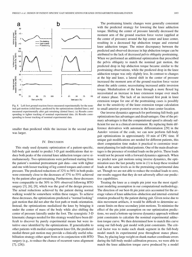
to the magnitude of the adduction torque reduction. These jointtorque changes resulted in a lateral shift of the center of pres-sure under the foot (Fig. 7) with little corresponding change inthe ground reaction force (Fig. 8). The magnitude of the centerof pressure change was also proportional to the magnitude ofthe adduction torque reduction. Kinetic changes predicted at theother joints were less prominent.
After gait retraining, the patient was able to achieve ex-ternal knee adduction torque reductions comparable to theoptimization predictions (Table 1; Figs. 2 and 3). When the
Fig. 6. Left hip abduction, knee extension, and ankle inversion torque curvesmeasured experimentally for the nominal gait motion (solid lines), predictedby the optimizations (dashed lines), and measured experimentally after gait re-training (dotted lines). (a) Results corresponding to tighter tracking of nominalexperimental data. (b) Results corresponding to looser tracking of nominal ex-perimental data.
Fig. 7. Left foot center of pressure locations on the bottom of the foot mea-sured experimentally for the nominal gait motion (solid lines), predicted by theoptimizations (dashed lines), and measured experimentally after gait retraining(dotted lines). (a) Results corresponding to tighter tracking of nominal exper-imental data. (b) Results corresponding to looser tracking of nominal experi-mental data.
patient attempted to walk using his old gait pattern, he pro-duced adduction torque reductions of 1.5 %BW HT (39%) and1.7 %BW HT (37%) in the first and second peak, respectively.When he walked using an exaggerated version of the predictedmedial-thrust gait motion, the reductions were 1.9 %BW HT(50%) and 2.5 %BW HT (55%).
These decreases were achieved by kinematic and kineticchanges that were generally consistent with the optimizationpredictions (Figs. 4–7). However, several exceptions werenoted. No decrease in pelvis obliquity and a larger than pre-dicted increase in pelvis axial rotation were observed (Fig. 4).The increase in ankle flexion was also larger than predicted(Fig. 5). For kinetic quantities, the experimental decrease inhip adduction torque was generally less than predicted (Fig. 6).The increase in the first peak of the knee extension torque was
FREGLY et al.: DESIGN OF PATIENT-SPECIFIC GAIT MODIFICATIONS FOR KNEE OSTEOARTHRITIS REHABILITATION 1693
Fig. 8. Left foot ground reaction forces measured experimentally for the nom-inal gait motion (solid lines), predicted by the optimizations (dashed lines), andmeasured experimentally after gait retraining (dotted lines). (a) Results corre-sponding to tighter tracking of nominal experimental data. (b) Results corre-sponding to looser tracking of nominal experimental data.
smaller than predicted while the increase in the second peakwas larger.
IV. DISCUSSION
This study used dynamic optimization of a patient-specific,full-body gait model to predict 3-D gait modifications that re-duce both peaks of the external knee adduction torque curve si-multaneously. Two optimizations were performed starting fromthe patient’s nominal pretreatment gait data—one with tighterand one with looser tracking of leg control torques and center ofpressure. The predicted reductions of 32% to 56% in both peakswere extremely close to the decreases of 37% to 55% achievedby the patient after gait retraining. Furthermore, these decreaseswere comparable to the 30% to 50% observed following HTOsurgery [3], [6], [9], which was the goal of the design process.The actual reductions achieved by the patient during normalwalking would be somewhere within these ranges. To achievethese decreases, the optimizations predicted a “normal looking”gait motion that did not alter the foot path or trunk orientation.Instead, the optimizations medialized the knee by bringing itunder the center of mass of the body while also shifting thecenter of pressure laterally under the foot. The synergistic 3-Dkinematic changes needed for this strategy would have been dif-ficult to discover by purely experimental means but were in-tuitive once identified by the optimizations. If generalizable toother patients with medial compartment knee OA, the predictedmedial-thrust gait motion may provide a clinically useful reha-bilitation strategy either apart from or in conjunction with HTOsurgery (e.g., to reduce the chance of recurrent varus alignment[7], [28]).
The posttraining kinetic changes were generally consistentwith the predicted strategy for lowering the knee adductiontorque. Shifting the center of pressure laterally decreased themoment arm of the ground reaction force vector (applied atthe center of pressure) around the hip center and knee center,resulting in a decreased hip abduction torque and externalknee adduction torque. The minor discrepancy between thepredicted and observed decrease in hip abduction torque can beattributed to the lack of decreased pelvis obliquity posttraining.When we performed an additional optimization that prescribedthe pelvis obliquity to match the nominal gait motion, thepredicted drop in hip abduction torque became similar to theposttraining observations, while the predicted drop in the kneeadduction torque was only slightly less. In contrast to changesat the hip and knee, a lateral shift in the center of pressureincreased the moment arm of the ground reaction force vectorabout the ankle center, necessitating increased ankle inversiontorque. Medialization of the knee through a more flexed legnecessitated an increase in knee extension torque over muchof stance phase. The lack of an increased first peak in kneeextension torque for one of the posttraining cases is possiblydue to the sensitivity of the knee extension torque calculationto small anterior–posterior errors in the knee center location.
Our inverse dynamics approach for performing full-body gaitoptimizations has advantages and disadvantages. One of the pri-mary advantages is that the computational speed is already suf-ficient for use in a clinical environment. By replacing finite dif-ference derivatives with automatic differentiation [29] in theAutolev version of the code, we can now perform full-bodygait optimizations in approximately 10 min of CPU time. Ifunique gait modifications are needed for different patients, thisshort computation time makes it practical to customize treat-ment planning for individual patients. One of the main disadvan-tages is the presence of residual loads on the pelvis. These loadswould not be present with a forward dynamics approach. Whenwe predict new gait motions using inverse dynamics, the opti-mization uses the last penalty term in (1) to keep these residualloads at the same levels as in the pretraining experimental dataset. Though we are not able to reduce the residual loads to zero,our results suggest that they do not adversely affect our predic-tive capabilities.
Treating the knee as a simple pin joint was the most signif-icant modeling assumption in our computational methodology.The direction of our best-fit pin joint axis accounted for the av-erage values of knee adduction–abduction and internal–externalrotation produced by the patient during gait. Given noise due toskin movement artifacts, it would be difficult to determine ac-curate limits on these secondary joint motions. To minimize theeffect of the pin joint assumption on our optimization predic-tions, we used a bottom–up inverse dynamics approach withoutjoint constraints to calculate the nominal experimental adduc-tion torque curve. We then determined how to match this curveusing our full-body gait model with joint constraints. The crit-ical factor was to make each shank segment in the full-bodymodel match its experimental pose throughout stance phase.Thus, by placing large weight on shank marker coordinate errorsduring the full-body model calibration process, we were able tomatch the knee adduction torque curve produced by a model

1694 IEEE TRANSACTIONS ON BIOMEDICAL ENGINEERING, VOL. 54, NO. 9, SEPTEMBER 2007
without joint constraints. Since the predicted adduction torquereductions were comparable to those observed experimentally,we do not believe that our pin joint knee assumption adverselyaffected our prediction process.
Another assumption in our computational methodology wasthat the selected cost function weights were representative of thepatient’s control strategy. Though we followed a systematic ap-proach for selecting two sets of cost function weights, differentweights will produce different optimization results. As in anyengineering design study, the main goal is to achieve a final de-sign that is better than the nominal one, whether or not the finaldesign is the best one possible. Since the predicted gait modifi-cations did, in fact, result in significant adduction torque reduc-tions when implemented, this limitation is not a serious one.
The biggest limitation of our study was the evaluation ofonly a single patient. Since the goal of the study was to assessthe feasibility of using dynamic optimization of a patient-spe-cific gait model to design a patient-specific treatment, we be-lieve that use of a single patient was reasonable. If we were un-able to demonstrate that the methodology works for at least onepatient, there would be little motivation for studying a largernumber of patients. Our next step will be to evaluate whetherthe same methodology will work for other patients with kneeOA. These patients will have different nominal gait patterns, ad-duction torque peaks, varus malalignment, and lateral collateralligament laxity, all of which may affect the effectiveness of ourmodel-based treatment approach.
Neural adaptation may explain why the subject achieved re-duced knee adduction torque peaks posttraining when tryingto walk with his pretraining gait pattern. The goal of gait re-training was for the subject to make the new gait pattern secondnature. When the subject tried to walk posttraining with his pre-training gait pattern, he attempted to give his knees a slight lat-eral thrust. However, the review of video data revealed that hestill exhibited increased medialization of the knees compared topretraining. Consequently, the subject’s adduction torque peaksdid not reach pretraining levels, possibly because he had in-grained the new gait pattern through the retraining process. Atleast two previous studies have shown that gait retraining canproduce sustained changes in gait mechanics [30], [31]. Sus-tained changes are also common in athletes, who make a con-scious effort to modify their mechanics during training so thatthey will perform better during competition.
A related issue is how to perform gait retraining for future pa-tients. The patient used in our study was highly knowledgeableon gait mechanics and so was able to understand and imple-ment the necessary gait modifications by studying plots and an-imations of the optimization results. The nine-month retrainingperiod was a function of the amount of time required to obtainaccess to a gait lab for retesting rather than the amount of timeneeded to learn and assimilate the new gait pattern. For other pa-tients to learn the medial-thrust gait pattern and make it secondnature, some type of real-time feedback coupled with a system-atic retraining process, as recently performed successfully fortibial shock reduction [31], will likely be necessary.
A final clinical implementation issue is whether the proposedmedial-thrust gait motion will result in detrimental loadingchanges at other joints. After a year and a half of walking with
the medial-thrust gait pattern, the patient involved in our studyhas experienced reduced knee pain without any problems atother joints. Apart from the knee adduction torque, the largestload changes were in the internal ankle inversion and kneeextension torque, both of which increased. For the nonexagger-ated posttraining gait motion, the 25% increase in peak ankleinversion torque is comparable to that caused by 6 lateral heelwedges [18]. Thus, the medial-thrust gait pattern is likely to beno worse for the ankle than it is for the introduction of lateralheel wedges. Furthermore, ankle OA is rare in clinical practiceand is usually the result of previous traumatic injury [32], [33].
Though both knee extension torque peaks also increased post-training, these increases do not necessarily imply an increase inmedial contact force. First, little correlation exists between themagnitude of the knee extension torque and the magnitude ofin vivo medial contact force measured by an instrumented kneereplacement during gait [34]. Second, the largest quadricepsforces do not necessarily correspond to periods of the largestknee extension torque [35]. Third, patients with knee OA ex-hibit increased quadriceps-hamstrings co-contraction comparedto normal subjects [36], suggesting that increased knee exten-sion torque could be created by decreased hamstrings co-con-traction, leading to a reduced medial contact force. Fourth, kneeextensor strength training has been reported to decrease ratherthan increase pain in patients with knee OA [37]. Finally, ourpatient reduced both knee adduction torque peaks by 1.5 to2.5 %BW HT, which is highly significant as the risk of OA pro-gression increases by a factor of 6.5 for each 1 %BW HT in-crease in peak knee adduction torque [12]. Given these observa-tions for the ankle and knee, we believe that clinical evaluationof the generalizability, efficacy, and safety of the medial-thrustgait pattern in a small patient population is a logical next step.
REFERENCES
[1] O. D. Schipplein and T. P. Andriacchi, “Interaction between active andpassive knee stabilizers during level walking,” J. Orthop. Res., vol. 9,pp. 113–119, 1991.
[2] T. P. Andriacchi, “Dynamics of knee malalignment,” Orthop. Clin.North Am., vol. 25, pp. 395–403, 1994.
[3] M. Wada, S. Imura, K. Nagatani, H. Baba, S. Shimada, and S. Sasaki,“Relationship between gait and clinical results after high tibial os-teotomy,” Clin. Orthop. Relat. Res., vol. 354, pp. 180–188, 1998.
[4] D. E. Hurwitz, A. B. Ryals, J. P. Case, J. A. Block, and T. P. Andri-acchi, “The knee adduction moment during gait in subjects with kneeosteoarthritis is more closely correlated with static alignment than ra-diographic disease severity, toe out angle and pain,” J. Orthop. Res.,vol. 20, pp. 101–107, 2002.
[5] A. Mündermann, C. O. Dyrby, and T. P. Andriacchi, “Secondary gaitchanges in patients with medial compartment knee osteoarthritis,”Arthritis Rheum., vol. 52, pp. 2835–2844, 2005.
[6] C. C. Prodromos, T. P. Andriacchi, and J. O. Galante, “The relationshipbetween gait and clinical changes following high tibial osteotomy,” J.Bone Joint Surg., vol. 67A, pp. 1188–1194, 1985.
[7] J. W. Wang, K. N. Kuo, T. P. Andriacchi, and J. O. Galante, “Theinfluence of walking mechanics and time on the results of proximaltibial osteotomy,” J. Bone Joint Surg., vol. 72A, pp. 905–913, 1990.
[8] J. M. Bryan, D. E. Hurwitz, B. R. Bach, T. Bittar, and T. P. Andriacchi,“A predictive model of outcome in high tibial osteotomy,” in Proc. 43rdAnnu. Meeting Orthop. Res. Soc., 1997, p. 718.
[9] L. Weidenhielm, O. K. Svensson, and L.-Å. Broström, “Surgicalcorrection of leg alignment in unilateral knee osteoarthritis reducesthe load on the hip and knee bilaterally,” Clin. Biomech., vol. 10, pp.217–221, 1995.
[10] D. E. Hurwitz, D. R. Sumner, T. P. Andriacchi, and D. A. Sugar, “Dy-namic knee loads during gait predict proximal tibial bone distribution,”J. Biomech., vol. 31, pp. 423–430, 1998.

FREGLY et al.: DESIGN OF PATIENT-SPECIFIC GAIT MODIFICATIONS FOR KNEE OSTEOARTHRITIS REHABILITATION 1695
[11] L. Sharma, D. E. Hurwitz, E. J.-M. A. Thonar, J. A. Sum, M. E. Lenz,D. D. Dunlop, T. J. Schnitzer, G. Kirwan-Mellis, and T. P. Andriacchi,“Knee adduction moment, serum hyaluronan level, and disease severityin medial tibiofemoral osteoarthritis,” Arthritis Rheum., vol. 41, pp.1233–1240, 1998.
[12] T. Miyazaki, M. Wada, H. Kawahara, M. Sato, H. Baba, and S. Shi-mada, “Dynamic load at baseline can predict radiographic disease pro-gression in medial compartment knee osteoarthritis,” Ann. Rheum. Dis.,vol. 61, pp. 617–622, 2002.
[13] M. Andrews, F. R. Noyes, T. E. Hewett, and T. P. Andriacchi, “Lowerlimb alignment and foot angle are related to stance phase knee adduc-tion in normal subjects: A critical analysis of the reliability of gait anal-ysis data,” J. Orthop. Res., vol. 14, pp. 289–295, 1996.
[14] M. Guo, M. J. Axe, and K. Manal, “The Influence of foot progres-sion angle on the knee adduction moment during walking and stairclimbing in pain free individuals with knee osteoarthritis,” Gait Pos-ture (in press).
[15] A. Mündermann, C. O. Dyrby, D. E. Hurwitz, L. Sharma, and T. P. An-driacchi, “Potential strategies to reduce medial compartment loading inpatients with knee osteoarthritis of varying severity,” Arthritis Rheum.,vol. 50, pp. 1172–1178, 2004.
[16] J. L. DeMarre, L. Mündermann, L., T. P. Andriacchi, and A. Mün-dermann, “A mechanism to lower the knee adduction moment duringwalking: Gait retraining as intervention for knee OA,” presented at the30th Annu. Meeting Am. Soc. Biomech., 2006, paper #173.
[17] W. Kakihana, M. Akai, N. Yamasaki, T. Takashima, and K. Nakazawa,“Changes in joint moments in the gait of normal subjects wearing later-ally wedged insoles,” Am. J. Phys. Med. Rehab., vol. 83, pp. 273–278,2004.
[18] W. Kakihana, M. Akai, K. Nakazawa, T. Takashima, K. Naito, and S.Torii, “Effects of laterally wedged insoles on knee and subtalar jointmoments,” Arch. Phys. Med. Rehab., vol. 86, pp. 1465–1471, 2005.
[19] J. A. Reinbolt, J. F. Schutte, B. J. Fregly, B. I. Koh, R. T. Haftka, A. D.George, and K. H. Mitchell, “Determination of patient-specific multi-joint kinematic models through two-level optimization,” J. Biomech.,vol. 38, pp. 621–626, 2005.
[20] F. C. Anderson and M. G. Pandy, “Dynamic optimization of humanwalking,” J. Biomech. Eng., vol. 123, pp. 381–390, 2001.
[21] A. J. Van den Bogert, G. D. Smith, and B. M. Nigg, “In vivo determina-tion of the anatomical axes of the ankle joint complex: An optimizationapproach,” J. Biomech., vol. 27, pp. 1477–1488, 1994.
[22] F. C. Anderson and M. G. Pandy, “A dynamic optimization solutionfor vertical jumping in three dimensions,” Comput. Meth. Biomech.Biomed. Eng., vol. 2, pp. 201–231, 1999.
[23] R. R. Neptune, I. C. Wright, and A. J. Van den Bogert, “A method fornumerical simulation of single limb ground contact events: Applicationto heel-toe running,” Comput. Meth. Biomech. Biomed. Eng., vol. 3, pp.321–334, 2001.
[24] Z. Popovic, “Motion transformation by physically based spacetime op-timization,” Ph.D. dissertation, Dept. Comput. Sci., Carnegie MellonUniv., Pittsburgh, PA, 1999.
[25] L. Chéze, B. J. Fregly, and J. Dimnet, “Determination of joint func-tional axes from noisy marker data using the finite helical axis,” Hum.Mov. Sci., vol. 17, pp. 1–15, 1998.
[26] M. L. Nagurka and V. Yen, “Fourier-based optimal control of nonlineardynamic systems,” J. Dyn. Syst. Meas. Cont., vol. 112, pp. 17–26, 1990.
[27] P. D. Leva, “Adjustments to Zatsiorsky-Seluyanov’s segment inertiaparameters,” J. Biomech., vol. 29, pp. 1223–1230, 1996.
[28] G. Magyar, S. Toksvig-Larsen, and A. Lindstrand, “Changes in osseouscorrection after proximal tibial osteotomy,” Acta. Orthop. Scand., vol.70, pp. 473–477, 1999.
[29] A. Griewank, D. Juedes, and J. Utke, “ADOL-C: A package for theautomatic differentiation of algorithms written in C/C++,” Assoc.Comput. Mach. Trans. Math. Software, vol. 22, pp. 131–167, 1996.
[30] J. Schröter, V. Güth, M. Overbeck, D. Rosenbaum, and W. Winkel-mann, “The ‘Entlastungsgang’. A hip unloading gait as a new con-servative therapy for hip pain in the adult,” Gait Posture, vol. 9, pp.151–157, 1999.
[31] H. P. Crowell and I. S. Davis, I.S., “Reducing lower extremity loadsthrough gait retraining using real-time feedback methods,” in Proc.30th Annu. Meeting Am. Soc. Biomech., 2006, paper #318.
[32] J. A. Buckwalter, C. Saltzman, and T. Brown, “The impact of os-teoarthritis: Implications for research,” Clin. Orthop. Rel. Res., vol.427, pp. S6–S15, 2004.
[33] C. L. Saltzman, M. L. Salamon, G. M. Blanchard, T. Huff, A. Hayes,J. A. Buckwalter, and A. Amendola, “Epidemiology of ankle arthritis:Report of a consecutive series of 639 patients from a tertiary or-thopaedic center,” Iowa Orthop. J., vol. 24, pp. 44–46, 2004.
[34] D. Zhao, S. A. Banks, K. H. Mitchell, D. D. D’Lima, C. W. Colwell,and B. J. Fregly, “Correlation between the knee adduction torque andmedial contact force for a variety of gait patterns,” J. Orthop. Res., vol.25, pp. 789–797, 2007.
[35] K. Manal, J. Gardinier, and N. Chimera, “What are we missing whenusing inverse dynamics,” presented at the 30th Annu. Meeting Am. Soc.Biomech., 2006, paper #323.
[36] T. Hortobagyi, L. Westerkamp, S. Beam, J. Moody, J. Garry, D. Hol-bert, and P. DeVita, “Altered hamstring-quadriceps muscle balance inpatients with knee osteoarthritis,” Clin. Biomech., vol. 20, pp. 97–104,2005.
[37] L. A. Talbot, J. M. Gaines, S. M. Ling, and E. J. Metter, “A home-based protocol of electrical muscle stimulation for quadriceps musclestrength in older adults with osteoarthritis of the knee,” J. Rheum., vol.30, pp. 1571–1578, 2003.
Benjamin J. Fregly, photograph and biography not available at the time ofpublication.
Jeffrey A. Reinbolt, photograph and biography not available at the time ofpublication.
Kelly L. Rooney, photograph and biography not available at the time ofpublication.
Kim H. Mitchell, photograph and biography not available at the time ofpublication.
Terese L. Chmielewski, photograph and biography not available at the time ofpublication.