bid mediates selective killing of apc-deficient cells in ... · bid mediates selective killing of...
TRANSCRIPT

BID mediates selective killing of APC-deficient cells inintestinal tumor suppression by nonsteroidalantiinflammatory drugsBrian Leibowitza,b, Wei Qiua,b, Monica E. Buchanana,c, Fangdong Zoua,c,d, Philip Vernona, Mary P. Moyere,Xiao-Ming Yinb,f, Robert E. Schoena,g, Jian Yua,b,1, and Lin Zhanga,c,1
aUniversity of Pittsburgh Cancer Institute, Departments of bPathology, cPharmacology & Chemical Biology, and gMedicine, University of Pittsburgh School ofMedicine, Pittsburgh, PA 15213; dCollege of Life Sciences, Sichuan University, Chengdu, Sichuan, 610064, P.R. China; eINCELL Corporation, San Antonio, TX78249; and fDepartment of Pathology and Laboratory Medicine, Indiana University School of Medicine, Indianapolis, IN 46202
Edited by Bert Vogelstein, Johns Hopkins University, Baltimore, MD, and approved October 8, 2014 (received for review August 7, 2014)
Colorectal tumorigenesis is driven by genetic alterations in theadenomatous polyposis coli (APC) tumor suppressor pathway andeffectively inhibited by nonsteroidal antiinflammatory drugs(NSAIDs). However, how NSAIDs prevent colorectal tumorigenesishas remained obscure. We found that the extrinsic apoptotic path-way and the BH3 interacting-domain death agonist (BID) are acti-vated in adenomas from NSAID-treated patients. Loss of BIDabolishes NSAID-mediated tumor suppression, survival benefit,and apoptosis in tumor-initiating stem cells in APCMin/+ mice.BID-mediated cross-talk between the extrinsic and intrinsic apo-ptotic pathways is responsible for selective killing of neoplasticcells by NSAIDs. We further demonstrate that NSAIDs induce deathreceptor signaling in both cancer and normal cells, but only acti-vate BID in cells with APC deficiency and ensuing c-Myc activation.Our results suggest that NSAIDs suppress intestinal tumorigenesisthrough BID-mediated synthetic lethality triggered by death re-ceptor signaling and gatekeeper mutations, and provide a ratio-nale for developing more effective cancer prevention strategiesand agents.
colorectal cancer | APC | chemoprevention | BID | apoptosis
Colorectal cancer (CRC) is the third leading cause of cancer-related death in the United States (1). However, treatment
options for advanced colorectal cancer, including radiation, cy-totoxic, and targeted therapies, are of limited effectiveness.Colorectal tumorigenesis is driven by genetic alterations in theAPC tumor suppressor pathway through aberrant Wnt signaling,leading to β-catenin accumulation and activation of oncogenessuch as c-Myc and CCND1 (2). One of the most promisingstrategies for reducing the morbidity and mortality of CRC is toinhibit tumorigenesis by using natural products or pharmacologicagents, such as nonsteroidal antiinflammatory drugs (NSAIDs)(3). The chemopreventive activities of NSAIDs, such as aspirinand sulindac, have been demonstrated in epidemiological studies(4), clinical trials (5, 6), and animal models (7). However, thecritical cellular activities and molecular targets of NSAIDs inchemoprevention have remained elusive.It is suggested that the antitumor effects of NSAIDs require
selective killing of neoplastic cells through apoptosis (8), a majorturnover mechanism of intestinal epithelial cells (9). Apoptoticdeath is regulated by the death receptor (DR or extrinsic) andmitochondrial (intrinsic) pathways (10). The DR pathwayinvolves activation of death receptors such as DR4 and DR5,recruitment of FADD and procaspase 8, and depletion of pro-survival proteins such as c-FLIP, resulting in activation of therecruited caspase 8 and other caspases (11). The mitochondrialpathway is regulated by the Bcl-2 family proteins (12) andcharacterized by mitochondrial dysfunction, release of cyto-chrome c, and activation of caspase 9 (10). These two pathwayscross talk under certain conditions through caspase-8–mediatedcleavage of BID, a BH3-only Bcl-2 family member (13). The
activated and truncated BID (tBID) then engages the intrinsicpathway for efficient apoptosis induction (13). NSAIDs rely onthe intrinsic pathway to kill CRC cells (14, 15). Nonetheless, it isstill unclear whether apoptosis induction is essential for the an-titumor activities of NSAIDs.In this study, we found that NSAID treatment activates BID
and the extrinsic apoptotic pathway in human intestinal adeno-mas. Loss of BID almost completely abolishes NSAID-mediatedtumor suppression and killing of oncogenic intestinal stem cellsin APC-deficient mice. BID is activated by a synthetic lethalinteraction and mediates the effects of NSAIDs through cross-talk between the extrinsic and intrinsic pathways. Our resultsindicate that BID-dependent killing of tumor-initiating stemcells is critical for cancer prevention by NSAIDs.
ResultsNSAIDs Activate Caspase 8 and BID in Human Colonic Adenomas. Todetermine the role of the extrinsic apoptotic pathway in NSAID-mediated tumor suppression, we used terminal deoxynucleotidyltransferase dUTP nick end labeling (TUNEL) and active caspase8 staining to analyze advanced colonic adenomas from patientstaking aspirin or other NSAIDs and control samples frompatients without NSAID use (16). The number of TUNEL-pos-itive cells in the adenomas of the NSAID-treated patients was5.0-fold higher compared with those in the control group (Fig.1A). Active caspase 8 positive cells were markedly increased in
Significance
Colorectal cancer is the third leading cause of cancer-relateddeath in the United States, but treatment options for this dis-ease are of limited effectiveness. Most human colorectaltumors begin with an inactivating mutation in the adenoma-tous polyposis coli (APC) gene. We demonstrate a mechanismby which nonsteroidal antiinflammatory drugs (NSAIDs) pro-tect against colon cancer development by killing intestinal stemcells that have lost functional APC. NSAID treatment com-bines with APC loss-induced gene expression changes to se-lectively activate BID and induce apoptosis in these cells, whileleaving normal cells unharmed. These results provide a ratio-nale for developing more effective cancer prevention strate-gies and agents.
Author contributions: B.L., J.Y., and L.Z. designed research; B.L., W.Q., M.E.B., F.Z., andP.V. performed research; B.L., M.P.M., X.-M.Y., and R.E.S. contributed new reagents/ana-lytic tools; B.L., J.Y., and L.Z. analyzed data; and B.L., J.Y., and L.Z. wrote the paper.
The authors declare no conflict of interest.
This article is a PNAS Direct Submission.1To whom correspondence may be addressed. Email: [email protected] or [email protected].
This article contains supporting information online at www.pnas.org/lookup/suppl/doi:10.1073/pnas.1415178111/-/DCSupplemental.
16520–16525 | PNAS | November 18, 2014 | vol. 111 | no. 46 www.pnas.org/cgi/doi/10.1073/pnas.1415178111
Dow
nloa
ded
by g
uest
on
May
17,
202
0

the patients taking NSAIDs (Fig. 1B), and detected in 8 of 9adenomas from NSAID users, but only in 1 of 7 from nonusers.Upon analyzing olfactomedin 4 (OLFM4) (Fig. 1C, Left), whichmarks human crypt base columnar cells (CBCs), representingactive intestinal stem cells (ISCs) (17), we found active caspase8/OLFM4 double-positive cells were significantly increased bymore than 12-fold in the adenomas from NSAID takers com-pared with nonusers (Fig. 1C, Right). Most notably, BID cleavagewas detected in several adenomas with high BID expression fromNSAID users, but not in any adenomas from the patients nottaking NSAIDs (Fig. 1D). These results suggest that NSAIDs killoncogenic stem cells in human adenomas, and activation of theDR pathway and BID may contribute to this effect of NSAIDs.
BID Is Required for Tumor Suppression by NSAIDs in APCMin/+ Mice.To determine whether BID is required for the chemopreventiveeffects of NSAIDs, we crossed APCMin/+ mice, a genetic model ofintestinal adenomas (18), with BID KO mice (19) and generatedage- and sex-matched cohorts of C57BL/6J APCMin/+ mice withdifferent BID genotypes. These mice were then fed an AIN-93Gdiet containing 0 (control) or 200 ppm sulindac, and analyzedfor intestinal polyp formation. Sulindac treatment suppressedsmall intestinal adenoma formation in BID+/+/APCMin/+ miceby more than 80% compared with the untreated mice (Fig. 2A–C). Remarkably, this effect of sulindac was almost com-pletely lost in BID−/−/APCMin/+ mice, in which the number of
small intestinal polyps was only slightly reduced (Fig. 2C).Sulindac was also less effective in the small intestine of BIDheterozygous APCMin/+ mice (Fig. 2C). Similar observationswere made in the colon of APCMin/+ mice (Fig. 2 A–C).Indomethacin (10 ppm), also an NSAID with antitumor activity(15), was similarly BID-dependent in reducing small intestinaladenoma formation (Fig. S1). In untreated animals, loss of BIDdid not significantly affect the size and number of small in-testinal polyps, but led to an increase in polyp number in thecolon of APCMin/+ mice (Fig. 2C).We then compared the effects of prolonged sulindac treat-
ment on the survival of BID+/+ and BID−/− APCMin/+ mice. Instriking contrast to BID+/+/APCMin/+ mice (n = 9), which werealive after 70 wk of treatment, all of the BID−/−/APCMin/+ mice(n = 11) died before 35 wk, surviving only slightly longer thanthose on the control diet (∼30 wk) (Fig. 2D). The differencebetween the BID+/+ and BID−/− groups is highly significant (70vs. 33 wk, P = 0.0005) (Fig. 2D). Together, these results dem-onstrate that BID plays an essential role in NSAID-mediatedchemoprevention in APCMin/+ mice.
BID Is Required for NSAID-Induced Killing of Intestinal Stem Cells inAPCMin/+ Mice. We investigated how BID mediates the chemo-preventive effects of NSAIDs in APCMin/+ mice. We found thatcrypt cell apoptosis detected by TUNEL and active caspase 3staining was significantly reduced in the small intestine andcolon of sulindac- or indomethacin-treated BID−/−/APCMin/+
mice relative to BID+/+/APCMin/+ mice (Fig. 3 A and B and Fig.S2 A and B). In contrast, activation of caspase 8 by sulindac orindomethacin was not affected by the absence of BID (Fig.S2C). Sulindac was shown to preferentially kill ISCs in the cryptbottom of APCMin/+ mice (16), which can be detected bymarkers such as Lgr5 and OLFM4 (20). Analysis of Lgr5-EGFP–marked mice revealed that the killing effect of sulindacand indomethacin on ISCs indicated by TUNEL/EGFP double-positive staining was reduced in the small intestine and colon ofBID−/−/APCMin/+ mice relative to BID+/+/APCMin/+ mice (Fig.3C and Fig. S2D). Using OLFM4 RNA in situ hybridization todetect CBCs, we found the killing effect of sulindac and in-domethacin on ISCs indicated by TUNEL/OLFM4 double-positive staining was also reduced in the small intestine ofBID−/−/APCMin/+ mice (Fig. S2E). Inhibition of cyclooxygenase2 (COX 2) by NSAIDs has been implicated as a mechanismof NSAID-mediated chemoprevention (21). Sulindac andindomethacin treatment reduced plasma prostaglandin E2metabolites (PGEM) by ∼50% in BID+/+/APCMin/+, but theseeffects were intact in BID−/−/APCMin/+ mice (Fig. S2F), sug-gesting that BID functions downstream of COX 2 inhibition insuppressing tumorigenesis. Together, these results indicate thatBID mediates the chemopreventive effects of NSAIDs throughselective killing of APC-deficient ISCs.
BID Mediates NSAID-Induced Apoptosis Through the MitochondrialPathway. To further delineate the functional role of BID inNSAID-mediated tumor suppression, we analyzed HCT116 co-lon cancer cells, which contain a β-catenin–activating mutation andcan recapitulate the anticancer and apoptotic effects of NSAIDsin mice (14–16). We generated BID knockout (BID KO) HCT116cells by using homologous recombination (Fig. S3 and Fig. 4A)and compared WT and BID KO cells for their responses toNSAIDs and other anticancer agents. Strikingly, apoptosis in-duced by sulindac and indomethacin, as determined by nuclearfragmentation and annexin V staining, was almost completelyblocked in BID KO cells (Fig. 4B and Fig. S4A). BID deficiencyabrogated NSAID-induced Bax multimerization (Fig. 4C), mito-chondrial membrane depolarization (Fig. 4D), cytosolic release ofcytochrome c and SMAC (Fig. 4E), and activation of caspases 9, 7,and 3 (Fig. 4F). Reconstituting BID expression in BID KO cells
Fig. 1. NSAID treatment activates caspase 8 and BID in human advancedadenomas. (A–C) Advanced adenomas from nine patients taking NSAIDs andseven patients not taking NSAIDs were analyzed by TUNEL staining (green)(A), active caspase 8 (Casp 8) staining (red) (B), and OLFM4 (green) and activecaspase 8 (red) double staining (C ). (Left) Representative staining pic-tures. (Right) Mean numbers + SD of positive cells per field. (Scale bars:25 μm.) ***P < 0.001. Arrows indicate example positive cells. (D) BIDexpression in the adenomas was analyzed by Western blotting usinghomogenized tissue lysates. Each lane represents an individual patient.
Leibowitz et al. PNAS | November 18, 2014 | vol. 111 | no. 46 | 16521
MED
ICALSC
IENCE
S
Dow
nloa
ded
by g
uest
on
May
17,
202
0

restored sulindac-induced apoptosis (Fig. S4B). In contrast towild-type (WT) cells, the long-term survival of BID KO cells, asanalyzed by colony formation assay, did not decrease followingNSAID treatment (Fig. S4C).NSAIDs also required BID to induce apoptosis in other CRC
cell lines, including APC-mutant DLD1 and HT29 cells, andAPC-WT RKO cells (Fig. S4D). Furthermore, BID-dependentapoptosis was induced by other NSAIDs, including sulindacsulfone, a sulindac derivative (22), and SC236, a COX 2-specificinhibitor structurally unrelated to sulindac and indomethacin(23) (Fig. S5 A–C). In contrast, BID deficiency did not affectapoptosis induced by staurosporine, camptothecin, or over-expression of the BH3-only protein PUMA, but attenuated ap-optosis induced by tumor necrosis factor-related apoptosis-inducing ligand (TRAIL) (Fig. S5D). These results demonstrate
a prominent and specific role of BID in mediating the anticancerand apoptotic effects of NSAIDs.
BID Is Activated by NSAIDs Through the Extrinsic Apoptotic Pathway.We determined how NSAIDs activate BID to engage the mito-chondrial pathway to kill colon cancer cells. tBID was observedin HCT116 cells treated with different NSAIDs (Fig. 5A and Fig.S5 A and B) and could be detected as early as 18 h after treat-ment (Fig. 5A). The short form of DR5, but not DR4, was in-duced by NSAIDs in WT and BID KO HCT116 cells (Fig. 5Band Fig. S6 A and B). The induction of DR5 by NSAIDs wasindependent of p53 (Fig. S6C), which can activate DR5 followingDNA damage (24), and observed in different CRC cells (Fig.S6D). The short form of c-FLIP (FLIPs), a negative regulator ofcaspase 8 activation, was markedly down-regulated before BID
Fig. 2. BID is required for tumor suppression by NSAIDs in APCMin/+ mice. Age- and sex-matched APCMin/+ mice with different BID genotypes were fed controlor NSAID-containing AIN93G diet. (A) Representative images of small intestine and colon from BID+/+ and BID−/−APCMin/+mice treated with sulindac (200 ppm) for 4mo, with arrows indicating visible macroadenomas. (B) Representative images of small intestine and colon of control mice and mice treated with sulindac (200 ppm)for 2 mo, with arrows indicating microscopic lesions. (C) Mean numbers + SD of small intestinal and colonic adenomas (≥0.5 mm in diameter) in BID+/+, BID+/−, andBID−/− APCMin/+ mice treated ± sulindac as in B. *P < 0.05; **P < 0.01; ***P < 0.001 (n = 4 per group). (D) Kaplan–Meier survival curves of BID+/+ and BID−/− APCMin/+
mice treated ± sulindac (200 ppm) for 70 wk. BID+/+ vs. BID−/− APCMin/+ mice, P = 0.0005 (log-rank test). (Scale bars: A, 1 cm; B, 2 mm.)
Fig. 3. BID deficiency abolishes NSAID-induced apoptosis in intestinal stem cells in APCMin/+ mice. (A and B) BID+/+ and BID−/− APC Min/+ mice were fed control(CT) or experimental AIN93G diet containing 200 ppm sulindac (SUL) or 10 ppm indomethacin (INDO) for 1 wk. Small intestinal and colonic sections wereanalyzed for apoptosis by TUNEL staining (green) (A) and active caspase 3 staining (brown) (B). (Upper) Representative staining pictures of small intestinalsections, with arrows indicating example cells with positive staining. (Lower) Mean + SD of positive signals in the small intestinal and colonic crypts. (C) Lgr5-EGFP–marked BID+/+ and BID−/− APCMin/+ mice were treated as in A and analyzed for intestinal stem cell apoptosis by TUNEL (green) and EGFP (Lgr5; red)double staining. (Upper) Representative staining pictures of small intestinal sections, with arrows indicating example double-positive cells. (Lower) Mean + SDof TUNEL/Lgr5 double-positive signals in the small intestinal and colonic crypts. (Scale bars: 25 μm.) *P < 0.05; **P < 0.01; ***P < 0.001 (n = 3 per group).
16522 | www.pnas.org/cgi/doi/10.1073/pnas.1415178111 Leibowitz et al.
Dow
nloa
ded
by g
uest
on
May
17,
202
0
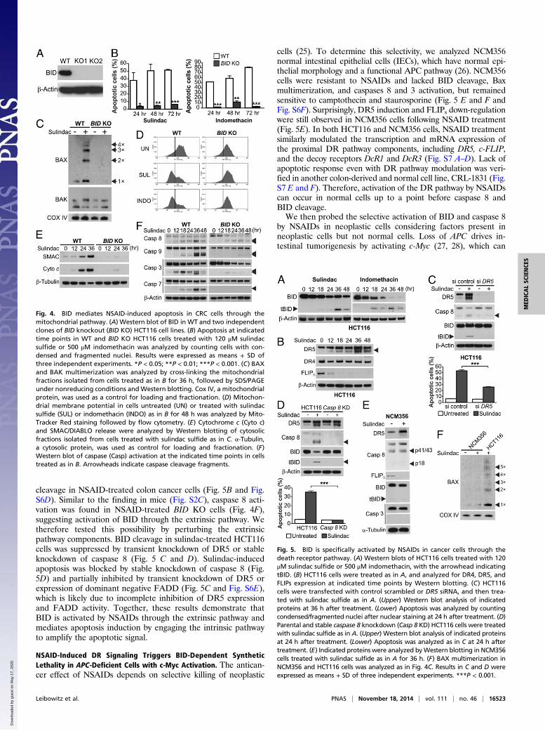
cleavage in NSAID-treated colon cancer cells (Fig. 5B and Fig.S6D). Similar to the finding in mice (Fig. S2C), caspase 8 acti-vation was found in NSAID-treated BID KO cells (Fig. 4F),suggesting activation of BID through the extrinsic pathway. Wetherefore tested this possibility by perturbing the extrinsicpathway components. BID cleavage in sulindac-treated HCT116cells was suppressed by transient knockdown of DR5 or stableknockdown of caspase 8 (Fig. 5 C and D). Sulindac-inducedapoptosis was blocked by stable knockdown of caspase 8 (Fig.5D) and partially inhibited by transient knockdown of DR5 orexpression of dominant negative FADD (Fig. 5C and Fig. S6E),which is likely due to incomplete inhibition of DR5 expressionand FADD activity. Together, these results demonstrate thatBID is activated by NSAIDs through the extrinsic pathway andmediates apoptosis induction by engaging the intrinsic pathwayto amplify the apoptotic signal.
NSAID-Induced DR Signaling Triggers BID-Dependent SyntheticLethality in APC-Deficient Cells with c-Myc Activation. The antican-cer effect of NSAIDs depends on selective killing of neoplastic
cells (25). To determine this selectivity, we analyzed NCM356normal intestinal epithelial cells (IECs), which have normal epi-thelial morphology and a functional APC pathway (26). NCM356cells were resistant to NSAIDs and lacked BID cleavage, Baxmultimerization, and caspases 8 and 3 activation, but remainedsensitive to camptothecin and staurosporine (Fig. 5 E and F andFig. S6F). Surprisingly, DR5 induction and FLIPs down-regulationwere still observed in NCM356 cells following NSAID treatment(Fig. 5E). In both HCT116 and NCM356 cells, NSAID treatmentsimilarly modulated the transcription and mRNA expression ofthe proximal DR pathway components, including DR5, c-FLIP,and the decoy receptors DcR1 and DcR3 (Fig. S7 A–D). Lack ofapoptotic response even with DR pathway modulation was veri-fied in another colon-derived and normal cell line, CRL-1831 (Fig.S7 E and F). Therefore, activation of the DR pathway by NSAIDscan occur in normal cells up to a point before caspase 8 andBID cleavage.We then probed the selective activation of BID and caspase 8
by NSAIDs in neoplastic cells considering factors present inneoplastic cells but not normal cells. Loss of APC drives in-testinal tumorigenesis by activating c-Myc (27, 28), which can
Fig. 4. BID mediates NSAID-induced apoptosis in CRC cells through themitochondrial pathway. (A) Western blot of BID in WT and two independentclones of BID knockout (BID KO) HCT116 cell lines. (B) Apoptosis at indicatedtime points in WT and BID KO HCT116 cells treated with 120 μM sulindacsulfide or 500 μM indomethacin was analyzed by counting cells with con-densed and fragmented nuclei. Results were expressed as means + SD ofthree independent experiments. *P < 0.05; **P < 0.01; ***P < 0.001. (C) BAXand BAK multimerization was analyzed by cross-linking the mitochondrialfractions isolated from cells treated as in B for 36 h, followed by SDS/PAGEunder nonreducing conditions and Western blotting. Cox IV, a mitochondrialprotein, was used as a control for loading and fractionation. (D) Mitochon-drial membrane potential in cells untreated (UN) or treated with sulindacsulfide (SUL) or indomethacin (INDO) as in B for 48 h was analyzed by Mito-Tracker Red staining followed by flow cytometry. (E) Cytochrome c (Cyto c)and SMAC/DIABLO release were analyzed by Western blotting of cytosolicfractions isolated from cells treated with sulindac sulfide as in C. α-Tubulin,a cytosolic protein, was used as control for loading and fractionation. (F)Western blot of caspase (Casp) activation at the indicated time points in cellstreated as in B. Arrowheads indicate caspase cleavage fragments.
Fig. 5. BID is specifically activated by NSAIDs in cancer cells through thedeath receptor pathway. (A) Western blots of HCT116 cells treated with 120μM sulindac sulfide or 500 μM indomethacin, with the arrowhead indicatingtBID. (B) HCT116 cells were treated as in A, and analyzed for DR4, DR5, andFLIPs expression at indicated time points by Western blotting. (C) HCT116cells were transfected with control scrambled or DR5 siRNA, and then trea-ted with sulindac sulfide as in A. (Upper) Western blot analysis of indicatedproteins at 36 h after treatment. (Lower) Apoptosis was analyzed by countingcondensed/fragmented nuclei after nuclear staining at 24 h after treatment. (D)Parental and stable caspase 8 knockdown (Casp 8 KD) HCT116 cells were treatedwith sulindac sulfide as in A. (Upper) Western blot analysis of indicated proteinsat 24 h after treatment. (Lower) Apoptosis was analyzed as in C at 24 h aftertreatment. (E) Indicated proteins were analyzed byWestern blotting in NCM356cells treated with sulindac sulfide as in A for 36 h. (F) BAX multimerization inNCM356 and HCT116 cells was analyzed as in Fig. 4C. Results in C and D wereexpressed as means + SD of three independent experiments. ***P < 0.001.
Leibowitz et al. PNAS | November 18, 2014 | vol. 111 | no. 46 | 16523
MED
ICALSC
IENCE
S
Dow
nloa
ded
by g
uest
on
May
17,
202
0

trigger a synthetic lethal interaction in normal and tumor cellswith high levels of DR5 (29). We therefore tested whether eitherAPC loss or c-Myc activation sensitizes IECs to NSAIDs in anotherwise normal background. Knockdown of APC by siRNA, inconjunction with sulindac treatment, resulted in caspase 8 andBID cleavage, apoptosis induction, caspase 3 activation, loss ofviability, and enhanced DR5 induction and FLIPs down-regula-tion in NCM356 cells (Fig. 6 A and B and Fig. S8 A and E). Thekilling effects of APC depletion require DR5, BID, and FLIPsdepletion, as knockdown of DR5 or BID, or transfection ofFLIPS suppressed apoptosis and restored cell viability in APC-depleted and NSAID-treated NCM356 cells (Fig. S8 B–E and
Fig. 6B). Conversely, c-FLIP knockdown or DR5 transfectionphenocopied APC depletion in inducing apoptosis, loss of cellviability, and caspase 8 and BID cleavage in NSAID-treatedNCM356 cells (Fig. 6 C and D and Fig. S8E).As expected, APC depletion led to c-Myc induction in
NCM356 cells (Fig. 6A), and knockdown of c-Myc substantiallydecreased caspase 8 activation, and abolished BID cleavage,apoptosis, and loss of cell viability induced by APC deletion andsulindac treatment (Fig. 6 A and B and Fig. S8E). Transfection ofc-Myc, but not a control vector, induced apoptosis in NSAID-treated NCM356 cells (Fig. 6E and Fig. S8 F and G). Consis-tent with the notion that c-Myc modulates the DR pathway(29), c-Myc transfection further down-regulated FLIPS protein,mRNA, and promoter activity and enhanced the expression ofDR5, DR4, and DcR2 in sulindac-treated NCM356 cells (Figs.S8G and S9). The effects of c-Myc on apoptosis and cell viabilityalso depended on BID and DR5 and were inhibited by FLIPS(Fig. 6E and Fig. S8F). These results demonstrate that APCdeficiency and subsequent c-Myc activation interact with NSAID-induced DR signaling, leading to caspase 8 and BID cleavage andselective killing of APC-deficient IECs.
DiscussionDespite the well-defined precursor lesions and genetic changesin colorectal tumorigenesis (2), how NSAIDs inhibit this processhas remained obscure. Our results demonstrate that activation ofBID through the extrinsic pathway plays a vital role in NSAID-mediated chemoprevention. It is possible that the adenomas withhigh levels of BID and/or caspase 8 activity are less likely torecur, compared with those with relatively low levels. Long-termNSAID use is associated with a variety of side effects, such asgastrointestinal, renal, and cardiovascular toxicities (25). It isconceivable that the side effects of NSAIDs can be mitigated byselectively enhancing NSAID-induced apoptotic signaling, or bycombining other agents with NSAIDs at lower and well-tolerateddoses to achieve safer and more effective cancer prevention (30).BID largely accounts for the antitumor and apoptotic effects
of NSAIDs in APCMin/+ mice, a classical model that recapitulatesthe chemopreventive effects of NSAIDs in humans (7). Thegeneral role of BID and the extrinsic pathway in chemo-prevention needs to be verified by using carcinogen tumormodels and other genetically engineered mice. The increasedpolyp number in the colon, but not in the small intestine ofBID-deficient APCMin/+ mice, suggests that a BID-mediatedeffect is more engaged in colonic epithelial cells and con-tributes to relatively lower tumor incidence in the colon ofAPCMin/+ mice. It may reflect differences in apoptosis andexpression of other Bcl-2 family members in normal epithelialcells of small intestine and colon (31). ISCs with regenerativecapability can sustain driver mutations and are likely to be themost relevant cell target of NSAIDs in chemoprevention.Tumor-initiating ISCs are likely to be more vulnerable toapoptosis relative to normal ISCs because of c-Myc activationby APC loss (32, 33), and compared with differentiated cellson account of intrinsic high levels of c-Myc in ISCs (34). Inaddition to their effects on colorectal tumorigenesis, NSAIDshave been shown to be useful in treating precancerous lesionsof other tissues, and in adjuvant therapy for treating a varietyof cancers (25). It is possible that these effects of NSAIDs arealso mediated by BID through the DR pathway. Comparedwith other anticancer drugs, NSAIDs seem to be more relianton BID to kill tumor cells, which is likely because BID is themajor, if not the only, BH3-only Bcl-2 family protein activatedby NSAIDs. In contrast, other drugs such as DNA-damagingagents modulate multiple BH3-only proteins, including BID,PUMA, Noxa, and BIM.Suppression of intestinal tumorigenesis by NSAIDs seems to
be mediated by the BID-dependent synthetic lethal interaction
Fig. 6. APC depletion or c-Myc induction triggers BID activation and BID-dependent apoptosis in NSAID-treated normal colonic epithelial cells. (A)NCM356 normal colonic epithelial cells were transfected with the indicatedsiRNA and then treated with 120 μM sulindac sulfide for 48 h. Indicatedproteins were analyzed by Western blotting. C Casp 3, cleaved caspase 3. (B)NCM356 cells were transfected with indicated siRNA or siRNA/plasmid combi-nations, and then treated with 120 μM sulindac sulfide for 48 h. Apoptosis wasmeasured by counting apoptotic nuclei after nuclear staining. (C and D) Westernblotting of the indicated proteins in NCM356 cells transfected as indicated,and then treated with 120 μM sulindac sulfide for 36 h. (E) Apoptosis inNCM356 cells transfected as indicated and treated with 120 μM sulindac sulfidefor 24 h was measured as in B. (F) A model of NSAID-mediated apoptosis inAPC-deficient cells. Results in B and E were expressed as means + SD of threeindependent experiments. **P < 0.01; ***P < 0.001; N.S., not significant.
16524 | www.pnas.org/cgi/doi/10.1073/pnas.1415178111 Leibowitz et al.
Dow
nloa
ded
by g
uest
on
May
17,
202
0

of DR signaling and aberrant Wnt signaling, which can be causedby inherited or sporadic APC mutations, activating β-cateninmutations, or other alterations (2). NSAIDs do not discriminatebetween cancer and normal cells in modulating the expression ofDR5, c-FLIP, and other DR family members, which is in-sufficient for cell death. However, these changes in conjunctionwith activation of Wnt signaling and ensuing c-Myc induction areboth sufficient and required to trigger caspase 8 and BIDcleavage, leading to apoptosis induction in an otherwise normalbackground. Therefore, premalignant cells with APC loss andaberrant Wnt signaling are primed for apoptosis due to c-Myc–induced DR signaling, and the enhanced DR signaling by NSAIDshelps reach a critical threshold for caspase 8 activation (Fig. 6F).NSAIDs induce a number of apical signaling events (21), some ofwhich, such as endoplasmic reticulum (ER) stress, can trigger DRsignaling (35). Studying these signaling events may help identifythe direct target of NSAIDs in chemoprevention, which shouldbe an important area of future investigation. Exploiting syntheticlethal interactions has been recognized as a powerful targetingstrategy (36) and may hold the key for developing effective pre-ventive agents for other cancer types with defined gatekeeper ordriver mutations.
Materials and MethodsAnalysis of Human Colonic Polyps. Frozen specimens from patients withsporadic colonic adenomas, including nine patients treated with NSAIDs andseven untreated patients, were acquired from theHealth Sciences Tissue Bankof the University of Pittsburgh. Acquisition of the tissue samples was ap-proved by the Institutional Review Board. Informed consent was receivedfrom all participating patients. TUNEL and the intestinal stem cell markerOLFM4 were analyzed by immunostaining of paraffin-embedded tissue asdescribed (16). Active caspase 8 staining was performed as described in SIMaterials and Methods.
Mice and Treatment. The procedures for all animal experiments were ap-proved by the Institutional Animal Care and Use Committee of the Uni-versity of Pittsburgh. C57BL/6J BID−/− (BID KO) mice, which were described(19), were crossed with APCMin/+ mice (Jackson Laboratory) to generateAPCMin/+ mice with different BID genotypes. Lgr5-EGFP–marked BID+/+ andBID−/− APCMin/+ mice were generated as described in SI Materials andMethods. For NSAID treatment, 4-wk-old mice were fed AIN-93G diets(Dyets) with or without 200 ppm sulindac (Sigma) or 10 ppm indomethacin(Sigma) for 1 wk for apoptosis analysis, or for 2 or 4 mo for tumor phe-notype analysis. Following treatment and killing of mice, analysis of ade-noma numbers and apoptosis in adenoma tissues was done as described inSI Materials and Methods. For survival analysis, BID+/+ and BID−/− APCMin/+
mice were fed sulindac or control diet for up to 70 wk when all micewere killed.
Analysis of NSAID-Induced and BID-Mediated Apoptosis in Human CRC CellLines and NCM356 Cells. Human CRC cell lines, including HCT116, HT29,RKO, DLD1 and their derivatives, and NCM356 cells (Incell) (26), were treatedby NSAIDs and analyzed for apoptosis by nuclear staining, annexin V/pro-pidium iodide staining, colony formation, mitochondrial membrane in-tegrity, cytochrome c release, and Bax conformational change as describedin SI Materials and Methods.
Statistical Analysis. Statistical analyses were performed by using GraphPadPrism IV software. Differences were considered significant if the probabilityof the difference occurring by chance was less than 5 in 100 (P < 0.05).
ACKNOWLEDGMENTS. We thank laboratory members, Dr. Wendie Cohick,and Dr. Thomas Kensler for critical reading. This work was supported byNational Institute of Health Grants CA106348, CA121105, CA172136 (to L.Z.),CA129829, and U01DK085570 (to J.Y.) and American Cancer Society GrantsRSG-07-156-01-CNE (to L.Z.) and RGS-10-124-01-CCE (to J.Y.). This projectused the University of Pittsburgh Cancer Institute shared facilities that weresupported in part by Award P30CA047904.
1. Siegel R, Naishadham D, Jemal A (2012) Cancer statistics, 2012. CA Cancer J Clin 62(1):10–29.
2. Vogelstein B, Kinzler KW (2004) Cancer genes and the pathways they control. NatMed 10(8):789–799.
3. William WN, Jr, Heymach JV, Kim ES, Lippman SM (2009) Molecular targets for cancerchemoprevention. Nat Rev Drug Discov 8(3):213–225.
4. Bosetti C, Rosato V, Gallus S, Cuzick J, La Vecchia C (2012) Aspirin and cancer risk: Aquantitative review to 2011. Ann Oncol 23(6):1403–1415.
5. Rothwell PM, et al. (2010) Long-term effect of aspirin on colorectal cancer in-cidence and mortality: 20-year follow-up of five randomised trials. Lancet 376(9754):1741–1750.
6. Giardiello FM, et al. (2002) Primary chemoprevention of familial adenomatous poly-posis with sulindac. N Engl J Med 346(14):1054–1059.
7. Corpet DE, Pierre F (2005) How good are rodent models of carcinogenesis in pre-dicting efficacy in humans? A systematic review and meta-analysis of colon chemo-prevention in rats, mice and men. Eur J Cancer 41(13):1911–1922.
8. Sun SY, Hail N, Jr, Lotan R (2004) Apoptosis as a novel target for cancer chemo-prevention. J Natl Cancer Inst 96(9):662–672.
9. Hall PA, Coates PJ, Ansari B, Hopwood D (1994) Regulation of cell number in themammalian gastrointestinal tract: The importance of apoptosis. J Cell Sci 107(Pt 12):3569–3577.
10. Danial NN, Korsmeyer SJ (2004) Cell death: Critical control points. Cell 116(2):205–219.11. Ashkenazi A (2008) Directing cancer cells to self-destruct with pro-apoptotic receptor
agonists. Nat Rev Drug Discov 7(12):1001–1012.12. Cory S, Adams JM (2002) The Bcl2 family: Regulators of the cellular life-or-death
switch. Nat Rev Cancer 2(9):647–656.13. Yin XM (2006) Bid, a BH3-only multi-functional molecule, is at the cross road of life
and death. Gene 369:7–19.14. Kohli M, et al. (2004) SMAC/Diablo-dependent apoptosis induced by nonsteroidal
antiinflammatory drugs (NSAIDs) in colon cancer cells. Proc Natl Acad Sci USA 101(48):16897–16902.
15. Zhang L, Yu J, Park BH, Kinzler KW, Vogelstein B (2000) Role of BAX in the apoptoticresponse to anticancer agents. Science 290(5493):989–992.
16. Qiu W, et al. (2010) Chemoprevention by nonsteroidal anti-inflammatory drugseliminates oncogenic intestinal stem cells via SMAC-dependent apoptosis. Proc NatlAcad Sci USA 107(46):20027–20032.
17. van der Flier LG, Haegebarth A, Stange DE, van de Wetering M, Clevers H (2009)OLFM4 is a robust marker for stem cells in human intestine and marks a subset ofcolorectal cancer cells. Gastroenterology 137(1):15–17.
18. Su LK, et al. (1992) Multiple intestinal neoplasia caused by a mutation in the murinehomolog of the APC gene. Science 256(5057):668–670.
19. Yin XM, et al. (1999) Bid-deficient mice are resistant to Fas-induced hepatocellularapoptosis. Nature 400(6747):886–891.
20. Li L, Clevers H (2010) Coexistence of quiescent and active adult stem cells in mammals.Science 327(5965):542–545.
21. Gupta RA, Dubois RN (2001) Colorectal cancer prevention and treatment by inhibitionof cyclooxygenase-2. Nat Rev Cancer 1(1):11–21.
22. Piazza GA, et al. (1997) Sulindac sulfone inhibits azoxymethane-induced colon carci-nogenesis in rats without reducing prostaglandin levels. Cancer Res 57(14):2909–2915.
23. Penning TD, et al. (1997) Synthesis and biological evaluation of the 1,5-diarylpyrazoleclass of cyclooxygenase-2 inhibitors: Identification of 4-[5-(4-methylphenyl)-3-(tri-fluoromethyl)-1H-pyrazol-1-yl]benze nesulfonamide (SC-58635, celecoxib). J Med Chem40(9):1347–1365.
24. Wu GS, et al. (1997) KILLER/DR5 is a DNA damage-inducible p53-regulated deathreceptor gene. Nat Genet 17(2):141–143.
25. Thun MJ, Henley SJ, Patrono C (2002) Nonsteroidal anti-inflammatory drugs as anti-cancer agents: Mechanistic, pharmacologic, and clinical issues. J Natl Cancer Inst 94(4):252–266.
26. Zhang L, et al. (2010) Chemoprevention of colorectal cancer by targeting APC-deficient cells for apoptosis. Nature 464(7291):1058–1061.
27. He TC, et al. (1998) Identification of c-MYC as a target of the APC pathway. Science281(5382):1509–1512.
28. Sansom OJ, et al. (2007) Myc deletion rescues Apc deficiency in the small intestine.Nature 446(7136):676–679.
29. Wang Y, et al. (2004) Synthetic lethal targeting of MYC by activation of the DR5death receptor pathway. Cancer Cell 5(5):501–512.
30. Meyskens FL, Jr, et al. (2008) Difluoromethylornithine plus sulindac for the preventionof sporadic colorectal adenomas: A randomized placebo-controlled, double-blindtrial. Cancer Prev Res (Phila) 1(1):32–38.
31. Merritt AJ, et al. (1995) Differential expression of bcl-2 in intestinal epithelia. Cor-relation with attenuation of apoptosis in colonic crypts and the incidence of colonicneoplasia. J Cell Sci 108(Pt 6):2261–2271.
32. Barker N, et al. (2009) Crypt stem cells as the cells-of-origin of intestinal cancer. Na-ture 457(7229):608–611.
33. Powell AE, et al. (2012) The pan-ErbB negative regulator Lrig1 is an intestinal stem cellmarker that functions as a tumor suppressor. Cell 149(1):146–158.
34. Takahashi K, Yamanaka S (2006) Induction of pluripotent stem cells from mouseembryonic and adult fibroblast cultures by defined factors. Cell 126(4):663–676.
35. Lu M, et al. (2014) Cell death. Opposing unfolded-protein-response signals convergeon death receptor 5 to control apoptosis. Science 345(6192):98–101.
36. Kaelin WG, Jr (2009) Synthetic lethality: A framework for the development of wisercancer therapeutics. Genome Med 1(10):99.
Leibowitz et al. PNAS | November 18, 2014 | vol. 111 | no. 46 | 16525
MED
ICALSC
IENCE
S
Dow
nloa
ded
by g
uest
on
May
17,
202
0