bio 211 anatomy & physiology i · describe the major functions of each organ system....
TRANSCRIPT
NORTHWESTERN CONNECTICUT COMMUNITY COLLEGE COURSE SYLLABUS
Course Title: Anatomy and Physiology I Lecture Course #: BIO* 211 Course Description: This course is the first semester of a two-semester sequence designed to provide a
comprehensive study of the structure and function of human body systems and their relationships to other systems. Anatomy and Physiology I covers the following topics: anatomical terminology, review of prerequisite biological and chemical principles, histology, integumentary, skeletal, muscular, and nervous systems. Pathology examples are covered within body each system. Four semester hours. (3 hours of lecture and 3 hours of lab)
Prerequisite: Prerequisite: BIO* 121 or BIO* 127 Prerequisite/Co-requisite: CHE 111or equivalent Goals: To provide the student with a comprehensive understanding of the
structure and function of mammalian systems. To apply knowledge of cell biology to tissue and organ structure and function. To investigate and integrate the structure and function of the integumentary, skeletal, muscular, and nervous system, especially as it pertains to movement. To consider disorders and clinical syndromes associated with specific systems as they pertain to both human and veterinary medicine.
Outcomes: At the end of this course students should be able to: BODY PLAN & ORGANIZATION
1. Anatomical position
1. Describe a person in anatomical position. 2. Describe how to use the terms right and left in anatomical
reference.
2. Body planes & sections
1. Identify the various planes in which a body might be dissected. 2. Describe the appearance of a body presented along various
planes.
3. Body cavities & regions
1. Describe the location of the body cavities and identify the major organs found in each cavity.
2. Identify the location of the major anatomical regions of the
body. 3. Describe the location of the four abdominopelvic quadrants
and the nine abdominopelvic regions and list the major organs located in each.
4. Directional terms
1. Identify the major directional terms used in anatomy.

2. Describe the location of body structures, using appropriate directional terminology.
5. Basic terminology
1. Define the terms anatomy and physiology. 2. Give specific examples to show the interrelationship between
anatomy and physiology. 3. Describe the location of structures of the body, using basic
regional and systemic terminology.
6. Levels of organizations
1. Describe, in order from simplest to most complex, the major levels of organization in the human organism.
2. Give an example of each level of organization.
7. Survey of body systems
1. Identify the organ systems of the human body and their major components.
2. Describe the major functions of each organ system.
HOMEOSTASIS
1. Definition
Define homeostasis
2. General types of homeostatic
mechanisms
1. Identify the components of a feedback loop and explain the function of each. 2. Compare and contrast positive and negative feedback in terms of the relationship between stimulus and response. 3. Explain why negative feedback is the most commonly used mechanism to maintain homeostasis in the body.
3. Examples of homeostatic
mechanisms
1. Provide an example of a negative feedback loop that utilizes the nervous system to relay information. Describe the
specific organs, structures, cells or molecules (receptors, neurons, CNS structures, effectors, neurotransmitters) included in the feedback loop.
2. 2. Provide an example of a negative feedback loop that utilizes the endocrine system to relay information.
Describe the specific cells or molecules (production cells, hormones, target cells) included in the feedback loop. 3. Provide an example of a positive feedback loop in the body. Describe the specific structures (organs, cells or molecules) included in the feedback loop.
The following four outcomes are also included with most of the other modules in Anatomy & Physiology I and II. They will be difficult to answer at this early point in the course, but are listed here to emphasize the importance of homeostasis in the study of anatomy and physiology.
4. Application of homeostatic
mechanisms
1. Provide specific examples to demonstrate how organ systems respond to maintain homeostasis.
2. Explain how different organ systems relate to one another to
maintain homeostasis.
5. Predictions related to
homeostatic imbalance, including disease states & disorders
1. Predict factors or situations affecting various organ systems that could disrupt homeostasis.
2. Predict the types of problems that would occur in the body if
various organ systems could not maintain homeostasis and allowed regulated variables (body conditions) to move away from normal.
6. Diagnostic imaging
Identify specific diagnostic tests and describe their uses including x-ray, MRI, CT scan, ultrasound, PET scan, and diagnostic tests using radio-labeled isotopes.
CHEMISTRY & CELL BIOLOGY
1. Atoms & molecules
1. With respect to the structure of an atom: a. Describe the charge, mass, and relative location of electrons, protons and neutrons. b. Relate the number of electrons in an electron shell to an
atom’s chemical stability and its ability to form chemical bonds.
c. Identify how ions and isotopes are produced by changing the relative number of specific subatomic particles.
d. Distinguish among the terms atomic number, mass number, and atomic weight.
2. Compare and contrast the terms ions, electrolytes, free
radicals, isotopes, and radioisotopes. 3. Compare and contrast the terms atoms, molecules, elements,
and compounds.
2. Chemical bonding
1. With respect to non-polar covalent, polar covalent, ionic, and hydrogen bonds:
a. Identify each type of bond in order by relative strength. b. Analyze specific compounds to determine the specific type
of bond.
3. Inorganic compounds &
solutions
1. Identify the physiologically important properties of water. 2. Distinguish among the terms solution, solute, solvent, colloid
suspension, and emulsion.
3. Define the term salt and give examples of physiological significance.
4. Define the terms pH, acid, base, and buffer and give examples
of physiological significance. 5. Predict whether a solution is acidic, basic, or neutral when
given the pH or concentration of H+ ions. 6. Predict the effect on pH if an acid or base is added to a
buffered solution.
4. Organic compounds
1. Define the term organic molecule. 2. Explain the relationship between monomers and polymers. 3. Define and give examples of dehydration synthesis and
hydrolysis reactions. 4. With respect to carbohydrates, proteins, lipids, and nucleic
acids: a. Identify the monomers and polymers. b. Compare and contrast general molecular structure. c. Provide specific examples. d. Identify dietary sources. e. Discuss physiological and structural roles in the human
body. 5. Describe the four levels of protein structure and discuss the
importance of protein shape for protein function. 6. Demonstrate factors that affect enzyme activity, including
denaturation, and interpret graphs showing the effects of various factors on the rate of enzyme-catalyzed reactions.
7. Identify specific functional groups and give examples where
they are found, including amino, hydroxyl, carboxyl, phosphate, sulfhydryl, methyl
5. Energy transfer using ATP
Describe the generalized reversible reaction for release of energy from ATP and explain the role of ATP in the cell.
6. Intracellular organization of
nucleus & cytoplasm
1. Identify the three main parts of a cell, and list the general functions of each.
2. Explain how cytoplasm and cytosol are different.
7. Membrane structure & function
1. Describe how lipids are distributed in a cell membrane, and explain their functions.
2. Describe how carbohydrates are distributed in a cell membrane, and explain their functions.
3. Describe how proteins are distributed in a cell membrane, and
explain their functions.
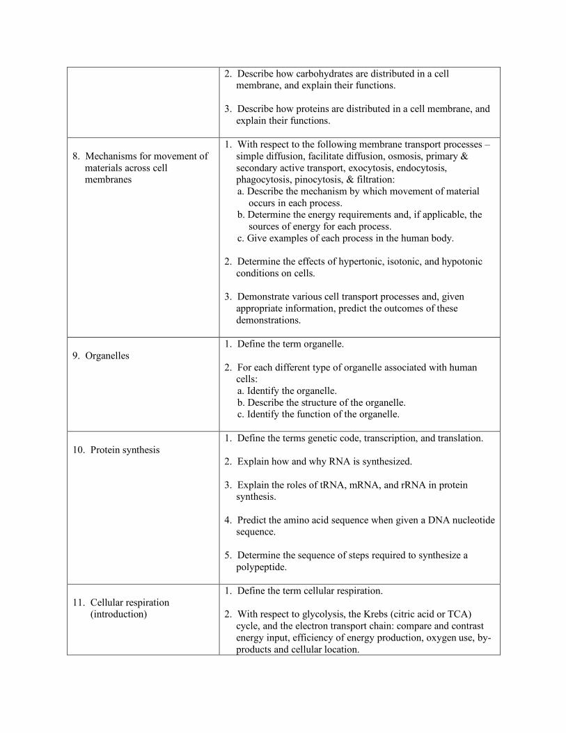
8. Mechanisms for movement of
materials across cell membranes
1. With respect to the following membrane transport processes – simple diffusion, facilitate diffusion, osmosis, primary & secondary active transport, exocytosis, endocytosis, phagocytosis, pinocytosis, & filtration:
a. Describe the mechanism by which movement of material occurs in each process.
b. Determine the energy requirements and, if applicable, the sources of energy for each process.
c. Give examples of each process in the human body. 2. Determine the effects of hypertonic, isotonic, and hypotonic
conditions on cells. 3. Demonstrate various cell transport processes and, given
appropriate information, predict the outcomes of these demonstrations.
9. Organelles
1. Define the term organelle. 2. For each different type of organelle associated with human
cells: a. Identify the organelle. b. Describe the structure of the organelle. c. Identify the function of the organelle.
10. Protein synthesis
1. Define the terms genetic code, transcription, and translation. 2. Explain how and why RNA is synthesized. 3. Explain the roles of tRNA, mRNA, and rRNA in protein
synthesis. 4. Predict the amino acid sequence when given a DNA nucleotide
sequence. 5. Determine the sequence of steps required to synthesize a
polypeptide.
11. Cellular respiration (introduction)
1. Define the term cellular respiration. 2. With respect to glycolysis, the Krebs (citric acid or TCA)
cycle, and the electron transport chain: compare and contrast energy input, efficiency of energy production, oxygen use, by-products and cellular location.
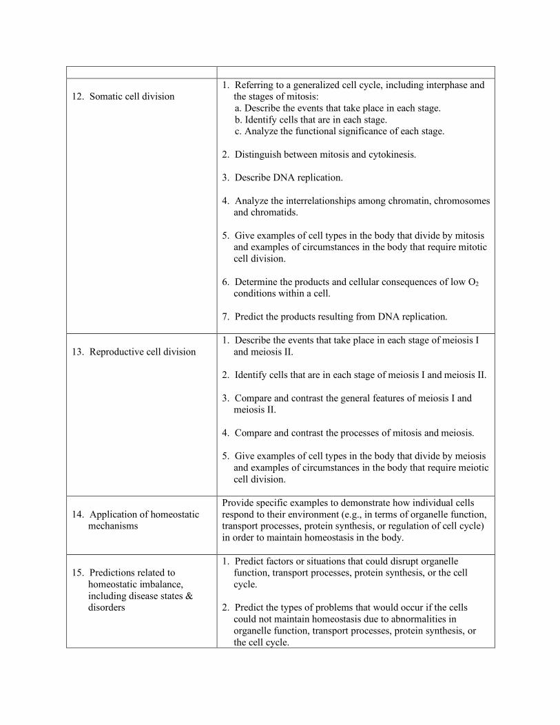
12. Somatic cell division
1. Referring to a generalized cell cycle, including interphase and the stages of mitosis:
a. Describe the events that take place in each stage. b. Identify cells that are in each stage. c. Analyze the functional significance of each stage. 2. Distinguish between mitosis and cytokinesis. 3. Describe DNA replication. 4. Analyze the interrelationships among chromatin, chromosomes
and chromatids. 5. Give examples of cell types in the body that divide by mitosis
and examples of circumstances in the body that require mitotic cell division.
6. Determine the products and cellular consequences of low O2
conditions within a cell. 7. Predict the products resulting from DNA replication.
13. Reproductive cell division
1. Describe the events that take place in each stage of meiosis I and meiosis II.
2. Identify cells that are in each stage of meiosis I and meiosis II. 3. Compare and contrast the general features of meiosis I and
meiosis II. 4. Compare and contrast the processes of mitosis and meiosis. 5. Give examples of cell types in the body that divide by meiosis
and examples of circumstances in the body that require meiotic cell division.
14. Application of homeostatic
mechanisms
Provide specific examples to demonstrate how individual cells respond to their environment (e.g., in terms of organelle function, transport processes, protein synthesis, or regulation of cell cycle) in order to maintain homeostasis in the body.
15. Predictions related to
homeostatic imbalance, including disease states & disorders
1. Predict factors or situations that could disrupt organelle function, transport processes, protein synthesis, or the cell cycle.
2. Predict the types of problems that would occur if the cells
could not maintain homeostasis due to abnormalities in organelle function, transport processes, protein synthesis, or the cell cycle.
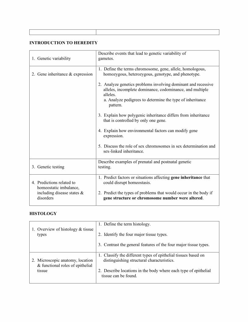
INTRODUCTION TO HEREDITY
1. Genetic variability
Describe events that lead to genetic variability of gametes.
2. Gene inheritance & expression
1. Define the terms chromosome, gene, allele, homologous, homozygous, heterozygous, genotype, and phenotype.
2. Analyze genetics problems involving dominant and recessive
alleles, incomplete dominance, codominance, and multiple alleles.
a. Analyze pedigrees to determine the type of inheritance pattern.
3. Explain how polygenic inheritance differs from inheritance
that is controlled by only one gene. 4. Explain how environmental factors can modify gene
expression. 5. Discuss the role of sex chromosomes in sex determination and
sex-linked inheritance.
3. Genetic testing
Describe examples of prenatal and postnatal genetic testing.
4. Predictions related to
homeostatic imbalance, including disease states & disorders
1. Predict factors or situations affecting gene inheritance that could disrupt homeostasis.
2. Predict the types of problems that would occur in the body if
gene structure or chromosome number were altered.
HISTOLOGY
1. Overview of histology & tissue
types
1. Define the term histology. 2. Identify the four major tissue types. 3. Contrast the general features of the four major tissue types.
2. Microscopic anatomy, location
& functional roles of epithelial tissue
1. Classify the different types of epithelial tissues based on distinguishing structural characteristics.
2. Describe locations in the body where each type of epithelial
tissue can be found.
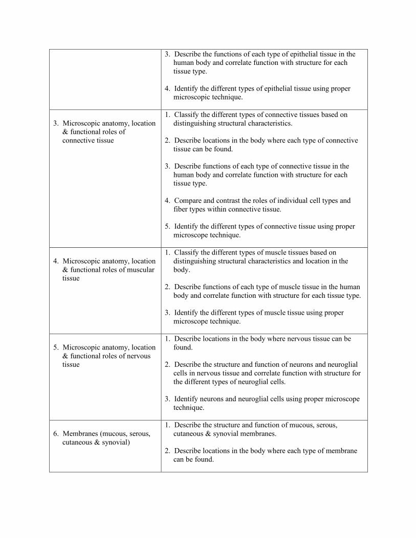
3. Describe the functions of each type of epithelial tissue in the human body and correlate function with structure for each tissue type.
4. Identify the different types of epithelial tissue using proper
microscopic technique.
3. Microscopic anatomy, location
& functional roles of connective tissue
1. Classify the different types of connective tissues based on distinguishing structural characteristics.
2. Describe locations in the body where each type of connective
tissue can be found. 3. Describe functions of each type of connective tissue in the
human body and correlate function with structure for each tissue type.
4. Compare and contrast the roles of individual cell types and
fiber types within connective tissue. 5. Identify the different types of connective tissue using proper
microscope technique.
4. Microscopic anatomy, location
& functional roles of muscular tissue
1. Classify the different types of muscle tissues based on distinguishing structural characteristics and location in the body.
2. Describe functions of each type of muscle tissue in the human
body and correlate function with structure for each tissue type. 3. Identify the different types of muscle tissue using proper
microscope technique.
5. Microscopic anatomy, location
& functional roles of nervous tissue
1. Describe locations in the body where nervous tissue can be found.
2. Describe the structure and function of neurons and neuroglial
cells in nervous tissue and correlate function with structure for the different types of neuroglial cells.
3. Identify neurons and neuroglial cells using proper microscope
technique.
6. Membranes (mucous, serous,
cutaneous & synovial)
1. Describe the structure and function of mucous, serous, cutaneous & synovial membranes.
2. Describe locations in the body where each type of membrane
can be found.
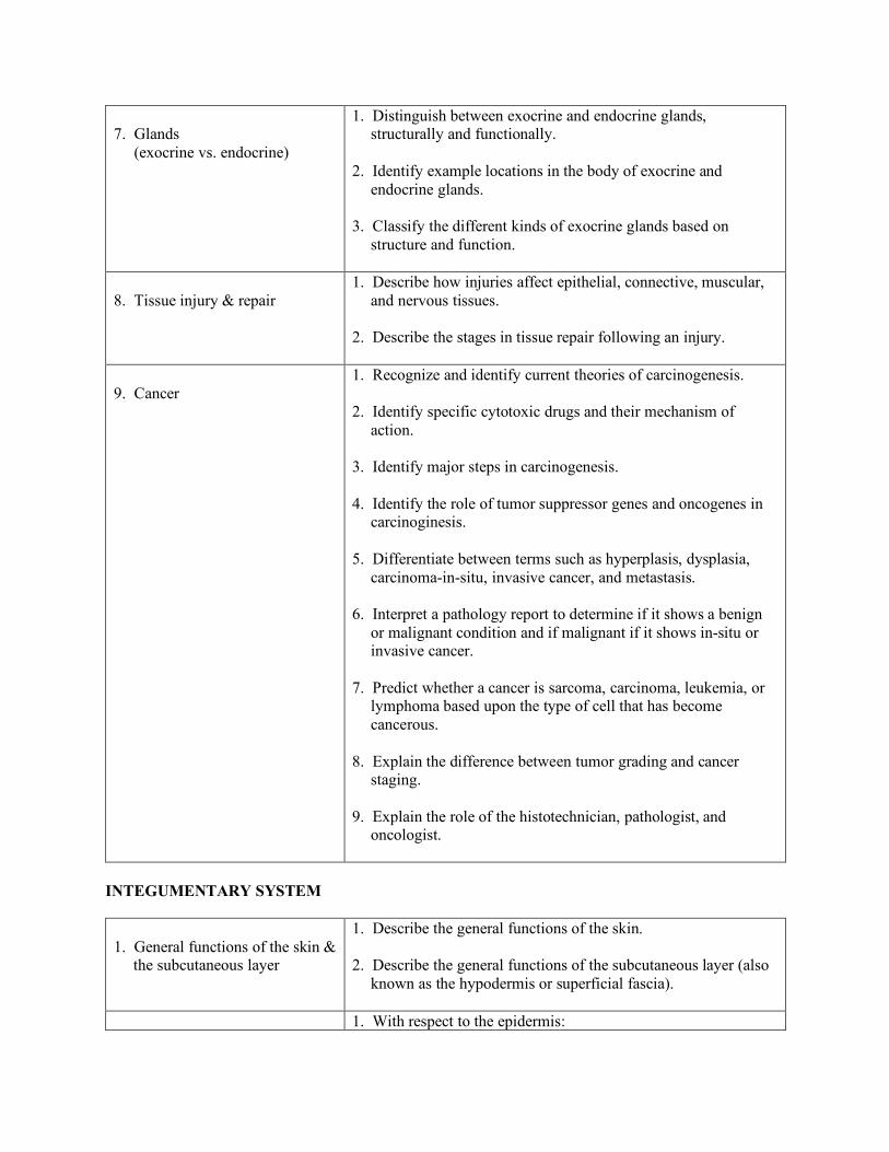
7. Glands (exocrine vs. endocrine)
1. Distinguish between exocrine and endocrine glands, structurally and functionally.
2. Identify example locations in the body of exocrine and
endocrine glands. 3. Classify the different kinds of exocrine glands based on
structure and function.
8. Tissue injury & repair
1. Describe how injuries affect epithelial, connective, muscular, and nervous tissues.
2. Describe the stages in tissue repair following an injury.
9. Cancer
1. Recognize and identify current theories of carcinogenesis. 2. Identify specific cytotoxic drugs and their mechanism of
action. 3. Identify major steps in carcinogenesis. 4. Identify the role of tumor suppressor genes and oncogenes in
carcinoginesis. 5. Differentiate between terms such as hyperplasis, dysplasia,
carcinoma-in-situ, invasive cancer, and metastasis. 6. Interpret a pathology report to determine if it shows a benign
or malignant condition and if malignant if it shows in-situ or invasive cancer.
7. Predict whether a cancer is sarcoma, carcinoma, leukemia, or
lymphoma based upon the type of cell that has become cancerous.
8. Explain the difference between tumor grading and cancer
staging. 9. Explain the role of the histotechnician, pathologist, and
oncologist.
INTEGUMENTARY SYSTEM
1. General functions of the skin &
the subcutaneous layer
1. Describe the general functions of the skin. 2. Describe the general functions of the subcutaneous layer (also
known as the hypodermis or superficial fascia).
1. With respect to the epidermis:
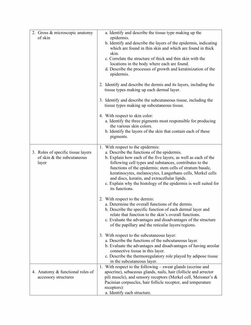
2. Gross & microscopic anatomy of skin
a. Identify and describe the tissue type making up the epidermis.
b. Identify and describe the layers of the epidermis, indicating which are found in thin skin and which are found in thick skin.
c. Correlate the structure of thick and thin skin with the locations in the body where each are found.
d. Describe the processes of growth and keratinization of the epidermis.
2. Identify and describe the dermis and its layers, including the
tissue types making up each dermal layer. 3. Identify and describe the subcutaneous tissue, including the
tissue types making up subcutaneous tissue. 4. With respect to skin color: a. Identify the three pigments most responsible for producing
the various skin colors. b. Identify the layers of the skin that contain each of these
pigments.
3. Roles of specific tissue layers
of skin & the subcutaneous layer
1. With respect to the epidermis: a. Describe the functions of the epidermis. b. Explain how each of the five layers, as well as each of the
following cell types and substances, contributes to the functions of the epidermis: stem cells of stratum basale, keratinocytes, melanocytes, Langerhans cells, Merkel cells and discs, keratin, and extracellular lipids.
c. Explain why the histology of the epidermis is well suited for its functions.
2. With respect to the dermis: a. Determine the overall functions of the dermis. b. Describe the specific function of each dermal layer and
relate that function to the skin’s overall functions. c. Evaluate the advantages and disadvantages of the structure
of the papillary and the reticular layers/regions. 3. With respect to the subcutaneous layer: a. Describe the functions of the subcutaneous layer. b. Evaluate the advantages and disadvantages of having areolar
connective tissue in this layer. c. Describe the thermoregulatory role played by adipose tissue
in the subcutaneous layer. 4. Anatomy & functional roles of
accessory structures
1. With respect to the following – sweat glands (eccrine and apocrine), sebaceous glands, nails, hair (follicle and arrector pili muscle), and sensory receptors (Merkel cell, Meissner’s & Pacinian corpuscles, hair follicle receptor, and temperature receptors):
a. Identify each structure.
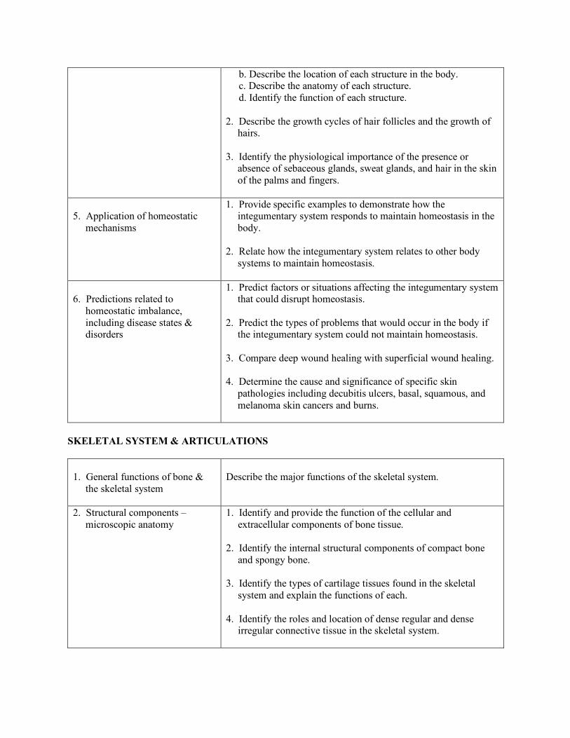
b. Describe the location of each structure in the body. c. Describe the anatomy of each structure. d. Identify the function of each structure. 2. Describe the growth cycles of hair follicles and the growth of
hairs. 3. Identify the physiological importance of the presence or
absence of sebaceous glands, sweat glands, and hair in the skin of the palms and fingers.
5. Application of homeostatic
mechanisms
1. Provide specific examples to demonstrate how the integumentary system responds to maintain homeostasis in the body.
2. Relate how the integumentary system relates to other body
systems to maintain homeostasis.
6. Predictions related to
homeostatic imbalance, including disease states & disorders
1. Predict factors or situations affecting the integumentary system that could disrupt homeostasis.
2. Predict the types of problems that would occur in the body if
the integumentary system could not maintain homeostasis. 3. Compare deep wound healing with superficial wound healing. 4. Determine the cause and significance of specific skin
pathologies including decubitis ulcers, basal, squamous, and melanoma skin cancers and burns.
SKELETAL SYSTEM & ARTICULATIONS
1. General functions of bone &
the skeletal system
Describe the major functions of the skeletal system.
2. Structural components – microscopic anatomy
1. Identify and provide the function of the cellular and extracellular components of bone tissue.
2. Identify the internal structural components of compact bone
and spongy bone. 3. Identify the types of cartilage tissues found in the skeletal
system and explain the functions of each. 4. Identify the roles and location of dense regular and dense
irregular connective tissue in the skeletal system.

3. Structural components – gross
anatomy
1. Identify the structural components of a long bone, with emphasis on region of longitudinal growth.
2. Explain the functions of those structural components in the
context of a whole bone.
4. Physiology of embryonic bone
formation (ossification, osteogenesis)
1. Identify the roles osteogenic cells play in the formation of bone tissue.
2. Identify the steps required for appositional and interstitional
bone growth. 3. Identify the necessary factors required for bone growth. 4. Describe the principles of bone repair and healing. 5. Identify specific types of bone pathology, including
osteoporosis, ostheomalacia, chondromalcia, fracture types and osteosarcoma.
6. Interpret the significance of nuclear medicine bone scans in
identifying abnormal pathologies. 7. Interpret an MRI report to explain a pathological condition
such as spondylolisthesis, disk herniation, and foraminal stenosis, including information based on information provided from T1 and T2-weighted images.
8. Compare and contrast intramembranous and endochondral
(intracartilaginous) bone formation.
5. Physiology of bone growth,
repair & remodeling
1. Compare and contrast the function of osteoblasts and osteoclasts during bone growth, repair, and remodeling.
2. Identify the hormonal regulation of skeleton growth. 3. Identify the roles of calcitonin, parathyroid hormone and
calcitriol in bone remodeling and blood calcium regulation. 4. Identify the principles of bone repair & healing. 5. Identify different types of fractures including simple &
compound comminuted, Saltar-Harris, greenstick, impatic spiral.
6. Organization of the skeletal
system
Define the two major divisions of the skeletal system (axial and appendicular) and list the general bone structures contained within each.
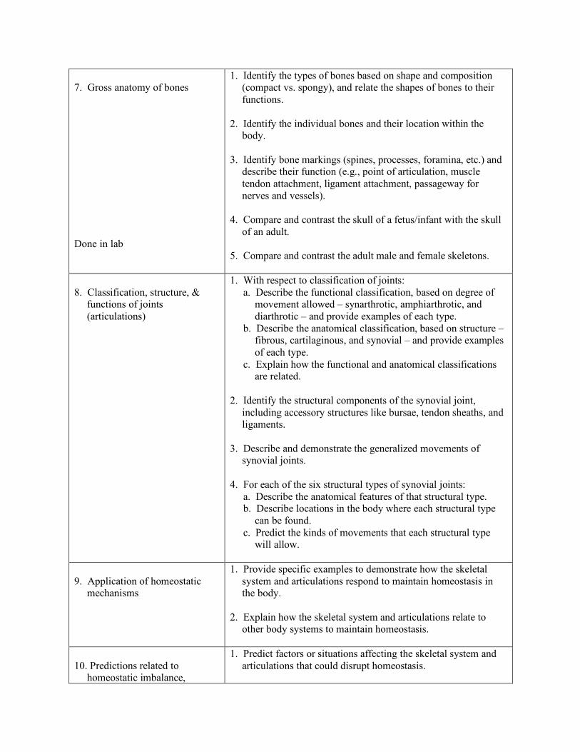
7. Gross anatomy of bones Done in lab
1. Identify the types of bones based on shape and composition (compact vs. spongy), and relate the shapes of bones to their functions.
2. Identify the individual bones and their location within the
body. 3. Identify bone markings (spines, processes, foramina, etc.) and
describe their function (e.g., point of articulation, muscle tendon attachment, ligament attachment, passageway for nerves and vessels).
4. Compare and contrast the skull of a fetus/infant with the skull
of an adult. 5. Compare and contrast the adult male and female skeletons.
8. Classification, structure, &
functions of joints (articulations)
1. With respect to classification of joints: a. Describe the functional classification, based on degree of
movement allowed – synarthrotic, amphiarthrotic, and diarthrotic – and provide examples of each type.
b. Describe the anatomical classification, based on structure – fibrous, cartilaginous, and synovial – and provide examples of each type.
c. Explain how the functional and anatomical classifications are related.
2. Identify the structural components of the synovial joint,
including accessory structures like bursae, tendon sheaths, and ligaments.
3. Describe and demonstrate the generalized movements of
synovial joints. 4. For each of the six structural types of synovial joints: a. Describe the anatomical features of that structural type. b. Describe locations in the body where each structural type
can be found. c. Predict the kinds of movements that each structural type
will allow.
9. Application of homeostatic
mechanisms
1. Provide specific examples to demonstrate how the skeletal system and articulations respond to maintain homeostasis in the body.
2. Explain how the skeletal system and articulations relate to
other body systems to maintain homeostasis.
10. Predictions related to
homeostatic imbalance,
1. Predict factors or situations affecting the skeletal system and articulations that could disrupt homeostasis.
including disease states & disorders
2. Predict the types of problems that would occur in the body if the skeletal system and articulations could not maintain homeostasis.
3. Interpret an MRI report to explain a pathological condition
such as spondylolisthesis, oseophytes, herniated nucleus pulposis.
4. Identify the advantage of the use of a contrast agent in MRI.
MUSCULAR SYSTEM
1. General functions of muscle
tissue
Identify the major functions of muscle tissue.
2. Identification, general location,
& comparative characteristics of skeletal, smooth, & cardiac muscle tissue
1. Identify skeletal, cardiac, and smooth muscle tissue types. 2. Describe the structure, location in the body and function of
skeletal, cardiac and smooth muscle. 3. Compare and contrast the characteristics of skeletal, cardiac
and smooth muscle.
3. Detailed gross & microscopic
anatomy of skeletal muscle
1. Describe the organization of muscle tissue from cell to whole muscle to groups of muscles.
2. Identify the connective tissue layers that surround each cell,
fascicle, muscle, and group of muscles and indicate the specific type of connective tissue that composes all of these layers.
3. Describe a skeletal muscle fiber including the transverse (T)
tubules, sarcoplasmic reticulum, and myofibrils. 4. Explain the organization of a myofibril. 5. Identify and describe the function of each of the contractile,
regulatory, and structural protein components of a sarcomere. 6. Describe the anatomy of the neuromuscular junction. 7. Identify the anatomical and metabolic characteristics of fast,
slow, and intermediate muscle fibers.
4. Physiology of skeletal muscle
contraction
1. Explain the sliding filament theory of muscle contraction. 2. Describe the sequence of events involved in the contraction
cycle of skeletal muscle.
3. Explain how an electrical signal from the nervous system arrives at the neuromuscular junction.
4. Describe, in order, the events that occur at the neuromuscular
junction that elicit an action potential in the muscle fiber. 5. Explain what is meant by the expression “excitation-
contraction coupling”.
5. Skeletal muscle metabolism
1. Identify the sources of energy stored in a typical muscle fiber. 2. Describe the mechanisms that muscle fibers use to obtain ATP
for muscle contraction. 3. Identify factors that contribute to muscle fatigue. 4. Summarize the events that occur during the recovery period of
muscle contraction. 5. Compare and contrast the metabolism of skeletal, cardiac and
smooth muscle.
6. Principles & types of whole
muscle contraction
1. Interpret a myogram of a twitch contraction with respect to the duration of the latent, contraction and relaxation periods and describe the events that occur in each period.
2. Define the terms tension and contraction, with respect to
muscles. 3. Define the term motor unit. 4. With respect to the mechanisms by which muscles generate
variable amounts of tension: a. Interpret a myogram or graph of tension vs. stimulus
frequency and explain the physiological basis for the phenomena of treppe, summation and tetanus.
b. Interpret a myogram or graph of tension vs. stimulus intensity and explain the physiological basis for the phenomenon of recruitment.
c. Interpret a graph of the length-tension relationship and discuss the anatomical basis for that relationship.
5. Demonstrate isotonic and isometric contraction and interpret
graphs of tension vs. time and muscle length vs. time for each type of contraction.
6. Distinguish between concentric and eccentric contraction and
contrast the relative tension and resistance that exists, as well as the change in muscle length that occurs, in each type of contraction.
7. Nomenclature of skeletal
muscles
Recognize how the name of a muscle can help identify its action, appearance, or location.
8. Location & function of the
major skeletal muscles
Identify the origin, insertion, and action of the major skeletal muscles and demonstrate these muscle actions.
9. Group actions of skeletal
muscles
1. Define the terms prime mover (or agonist), antagonist, synergist or fixator.
2. For a given movement, differentiate specific muscles that
function as prime mover, antagonist, synergist or fixator.
10. Lever systems
1. Differentiate among the three classes of levers in terms of the relative position of fulcrum, effort and load, as well as in terms of the relative power and range of motion.
2. Give examples in the human body of muscles and their
associated joints to illustrate each type of lever system.
11. Application of homeostatic
mechanisms
1. Provide specific examples to demonstrate how the muscular system responds to maintain homeostasis in the body.
2. Explain how the muscular system relates to other body
systems to maintain homeostasis. 3. Identify specific types of muscle pathologies, such as, strain,
rhahdomyalysis.
12. Predictions related to
homeostatic imbalance, including disease states & disorders
1. Predict factors or situations affecting the muscular system that could disrupt homeostasis.
2. Predict the types of problems that would occur in the body, if
the muscular system could not maintain homeostasis.
NERVOUS SYSTEM
1. General functions of the
nervous system
Describe the major functions of the nervous system
2. Organization of the nervous
system from both anatomical & functional perspectives
1. Describe the nervous system as a control system identifying nervous system elements that are sensory receptors, the afferent pathway, control centers, the efferent pathway, and effector organs.
2. Differentiate between the somatic and autonomic divisions of
the nervous system.
3. Gross & microscopic anatomy
of nervous tissue
1. Identify the parts of the nervous system that constitute the central nervous system (CNS) and those that constitute the peripheral nervous system (PNS).
2. With respect to the three structure types of neurons (unipolar,
bipolar & mulipolar): a. Identify each type of neuron. b. Identify soma (cell body), axon, and dendrites. c. State which parts of each type of neuron receive
information, which parts integrate information, and which parts conduct the output signal of the neuron.
d. Identify the location of the cell bodies of each type of neuron within the nervous system.
e. Identify a function of each type of neuron. f. Describe how the anatomy of each type of neuron supports
its function. 3. With respect to glial cells found in the CNS: a. Identify four types of CNS glial cells. b. Describe functions for each of those cells. c. Explain how the anatomy of each CNS glial cell supports
its function. 4. With respect to glial cells found in the PNS: a. List two types of PNS glial cells. b. Describe functions for each of those cells. c. Explain how the anatomy of each PNS glial cell supports its
function. 5. Define the term nerve. 6. Differentiate between a nerve and a CNS tract.
4. Neurophysiology, including
mechanism of resting membrane potential, production of action potentials, & impulse transmission
1. Define permeability. 2. Explain how ion channels affect neuron selective permeability. 3. Contrast the relative concentrations of sodium, potassium, and
chloride ions inside and outside of a cell. 4. Differentiate between a concentration gradient and an
electrical potential. 5. Define electrochemical gradient. 6. With respect to ion channels: a. Differentiate between passive and active ion channels. b. Explain how passive ion channels cause development of the
resting membrane potential in neurons.
c. Differentiate between voltage-gated and chemically-gated ion channels.
d. Describe the voltage-gated ion channels that are essential for development of the action potential.
7. Discuss the sequence of events that must occur for an action
potential to be generated. 8. Describe the role of the sodium-potassium exchange pump in
maintaining the resting membrane potential and making continued action potentials possible.
9. Define threshold. 10. Discuss the role of positive feedback in generation of the
action potential. 11. Interpret a graph showing the voltage vs. time relationship of
an action potential, and relate the terms depolarize, repolarize, and hyperpolarize to the events of an action potential.
12. With respect to the refractory periods: a. Define absolute and relative refractory periods. b. Explain the physiological basis of the absolute and relative
refractory periods. c. Discuss the consequence of a neuron having an absolute
refractory period. 13. With respect to impulse conduction: a. Describe how local circuit currents cause impulse
conduction in an unmyelinated axon. b. Explain how axon diameter and myelination affect
conduction velocity. c. Describe salutatory conduction.
5. Neurotransmitters & their roles
in synaptic transmission
1. Identify the presynaptic and postsynaptic cells at a synapse. 2. Identify the structures that comprise a chemical synapse. 3. Describe the synaptic (axon) terminal. 4. Restate the steps that lead from the action potential arriving in
the synaptic terminal to the release of neurotransmitter from synaptic vesicles.
5. Discuss the relationship between a neurotransmitter and its
receptor. 6. Explain how the receptors for neurotransmitters are related to
chemically-gated ion channels.
7. Describe the events of synaptic transmission in proper chronological order.
8. Define excitatory postsynaptic potential (EPSP) and inhibitory
postsynaptic potential (IPSP) and interpret graphs showing the voltage vs. time relationship of an EPSP and an IPSP.
9. Explain termporaland spatial summation of synaptic potentials. 10. Explain how movement of sodium ions alone, or movement of
both sodium and potassium ions, across the postsynaptic cell membrane can excite a neuron.
11. Explain how movement of potassium or chloride ions across
the postsynaptic cell membrane can inhibit a neuron. 12. Compare and contrast synaptic potentials with action
potentials. 13. Explain how a single neurotransmitter may be excitatory at
one synapse and inhibitory at another. 14. Describe the mechanism by which neurotransmitters may
have indirect (metabotropic) effects on postsynaptic cells. 15. List the most common excitatory neurotransmitter(s) in the
CNS and the most common inhibitory neurotransmitter(s) in the CNS.
16. Propose a possible CNS function for each biogenic amine
neurotransmitter. 17. Compare and contrast chemical and electrical synapses.
6. Sensory receptors & their roles
1. Describe exteroceptors, interoceptors and proprioceptors in terms of the general location of each in the body and the origin of the stimuli that each receives.
2. Describe each of the following types of receptors, indicating
what sensation it detects and giving an example of where it can be found in the body: pain receptors (nociceptors), temperature receptors, mechanoreceptors (including proprioceptors and barorceptors/pressorecptors), chemoreceptors, and photoreceptors.
3. Explain the generator potential that occurs when receptors for
general senses are stimulated. 4. Describe the relationship between unipolar neurons and
receptors for general senses.
5. Differentiate between the site of action potential generation in a unipolar neuron and a multipolar neuron.
6. Explain the phenomenon of adaptation. 7. Compare and contrast receptors for the special senses with
receptors for general sensation.
7. Division, origin, & function of
component parts of the brain
1. Identify the five developmental regions of the brain and identify the major areas of the adult brain that arise from each region.
2. Correlate functions with each major area of the adult brain. 3. Describe the orientation of the brain relative to bones of the
skull. 4. Identify the five lobes of the cerebral cortex and describe how
the motor and sensory functions of the cerebrum are distributed among the lobes.
5. Explain why the sensory and motor homunculi are relevant
clinically. 6. Discuss the concept of cerebral hemispheric specialization and
the role of the corpus callosum in connecting the two halves of the cerebrum.
7. Describe the location and functions of the limbic system. 8. Describe the parts of the brain involved in storage of long term
memory and discuss possible mechanisms of memory consolidation.
9. Describe the location and functions of the reticular activating
system.
8. Protective roles of the cranial
bones, meninges, & cerebrospinal fluid
1. Describe how the bones of the skull protect the brain. 2. Identify the meninges and describe their functional
relationship to the brain and cranial bones. 3. Describe the functions of cerebrospinal fluid, as well as the
details of its production, its circulation within the central nervous system, and its ultimate reabsorption into the bloodstream.
4. Describe the structural basis for, and the importance of the
blood brain barrier.
1. Identify the cranial nerves by name and number.

9. Structure & function of cranial nerves
2. Describe the specific functions of each of the cranial nerves
and classify each as sensory, motor or mixed. 3. Describe the location of the cranial nerve nuclei and the
ganglia associated with the cranial nerves. 4. Propose how knowledge of the anatomy of cranial nerve nuclei
can be used to help pinpoint damage to particular regions of the brain stem.
10. Anatomy of the spinal cord,
spinal and cranial nerves, and brain
1. Describe the gross anatomy of the spinal cord and spinal nerves and specify their location relative to the anatomy of the skeletal system.
2. Identify the anatomical features seen in a cross sectional view
of the spinal cord. 3. Contrast the relative position of gray matter and white matter
in the spinal cord with the corresponding arrangement of gray and white matter in the brain.
4. Identify the dorsal root ganglia, dorsal and ventral roots, and
spinal nerves. 5. Discuss how the structures root, nerve, ramus, plexus, tract and
ganglion relate to one another. 6. Identify the four spinal nerve plexuses and give examples of
nerves that emerge from each. 7. Distinguish between ascending and descending tracts in the
spinal cord. 8. Describe the concept of dermatomes and explain why they are
clinically significant. 9. Locate and determine the functions of important brain regions
11. Reflexes & their roles in
nervous system function
1. Define the term reflex. 2. Describe reflex responses in terms of the major structural and
functional components of a reflex arc. 3. Distinguish between each of the following pairs of reflexes:
intrinsic (inborn) reflexes vs. learned reflexes, somatic vs. visceral reflexes, monosynaptic vs. polysynaptic reflexes, and ipsilateral vs. contralateral reflexes, cranial and spinal reflexes.
4. Explain the terms spinal reflex and intersegmental spinal
reflex.
5. Describe a stretch reflex, a flexor (withdrawal) reflex, and a
crossed-extensor reflex, and name all components of each reflex arc.
6. Demonstrate a stretch reflex (e.g., patellar or plantar). 7. Propose how specific reflexes would be used in clinical
assessment of nervous system function. 8. Give examples of autonomic reflexes