biofunctionalized magnetic-vortex microdiscs for targeted ... · pdf filearticles nature...
TRANSCRIPT

ARTICLESPUBLISHED ONLINE: 29 NOVEMBER 2009 | DOI: 10.1038/NMAT2591
Biofunctionalized magnetic-vortex microdiscs fortargeted cancer-cell destructionDong-Hyun Kim1, Elena A. Rozhkova2*, Ilya V. Ulasov3, Samuel D. Bader1,2, Tijana Rajh2,Maciej S. Lesniak3 and Valentyn Novosad1*
Nanomagnetic materials offer exciting avenues for probing cell mechanics and activating mechanosensitive ion channels, aswell as for advancing cancer therapies. Most experimental works so far have used superparamagnetic materials. This reportdescribes a first approach based on interfacing cells with lithographically defined microdiscs that possess a spin-vortex groundstate. When an alternating magnetic field is applied the microdisc vortices shift, creating an oscillation, which transmits amechanical force to the cell. Because reduced sensitivity of cancer cells toward apoptosis leads to inappropriate cell survivaland malignant progression, selective induction of apoptosis is of great importance for the anticancer therapeutic strategies. Weshow that the spin-vortex-mediated stimulus creates two dramatic effects: compromised integrity of the cellular membrane,and initiation of programmed cell death. A low-frequency field of a few tens of hertz applied for only ten minutes was sufficientto achieve∼90% cancer-cell destruction in vitro.
Since the discovery of the compass, magnetic phenomenahave become an inseparable feature of everyday life, mostnoticeably in magnetic recording and advanced information
technologies1. Nanobiomagnetic applications, including magneticresonance imaging contrast-enhancement agents, targeted drugdelivery, bioseparation and magnetically induced hyperthermia2–5are also becoming prominent. Furthermore, magnetic actuationhas been explored for ‘distant’ control and manipulation ofcell adhesion, receptor clustering and intercellular signalling6–8.Integration of magnetic materials with biological molecules andtherapeutics creates hybrid materials with advanced properties9,10.Most experimental work so far has used superparamagneticparticles (that is, those with zero magnetic moment at remanence)and their bioconjugates. Because of their superparamagnetic natureand typically small value of the magnetization of saturation, highmagnetic fields are required to detect or manipulate these particles.Conversely, if highly magnetic particles are fabricated (that is,particles with larger single domains), agglomeration may occurowing to magnetic fringe fields. Other challenges related to thetraditional approach include difficulty in controlling the growthprocess, broad size and shape distributions, and variable structuraland magnetic properties. Magnetic structures confined to twodimensions, including the arrangement known as a spin vortex, donot suffer from these concerns.
Herein, we report on the interfacing of whole cells withbiocompatible lithographically defined ferromagnetic microdiscs(MDs) with a spin-vortex ground state. A magnetic vortex ischaracterized by an in-plane flux-closure spin distribution withnet zero magnetization in the absence of a magnetic field11–15.Although the dipole–dipole interaction is known to decrease thevortex state stability for small interdisc separation distances16,the vanishing magnetization in remanence is significant becausethis reduces the long-range magnetostatic forces responsible forparticle agglomeration. Candidate materials include 3d transitionmetals with high values of the saturationmagnetizationMs, becauseof their ability to induce a high magnetomotive force. When
1Materials Sciences Division; 2Center for Nanoscale Materials, Argonne National Laboratory, Argonne, Illinois 60439, USA, 3The Brain Tumor Center, TheUniversity of Chicago Pritzker School of Medicine, Chicago, Illinois 60637, USA. *e-mail: [email protected]; [email protected].
the biofunctionalized MDs selectively bind to cancer cells, thismechanical force is efficiently transduced to the membrane andfurther on to subcellular components (Fig. 1).We demonstrate thata spatially uniform and time-varying magnetic field as small astens of oersteds, with a frequency as low as a few tens of hertzand applied for only 10min, is sufficient to achieve cancer-celldestruction in vitro. This antineoplastic activity is the combinedresult of the compromised integrity of the cellular membrane andmagnetic-vortex-mediated initiation of programmed cell death.
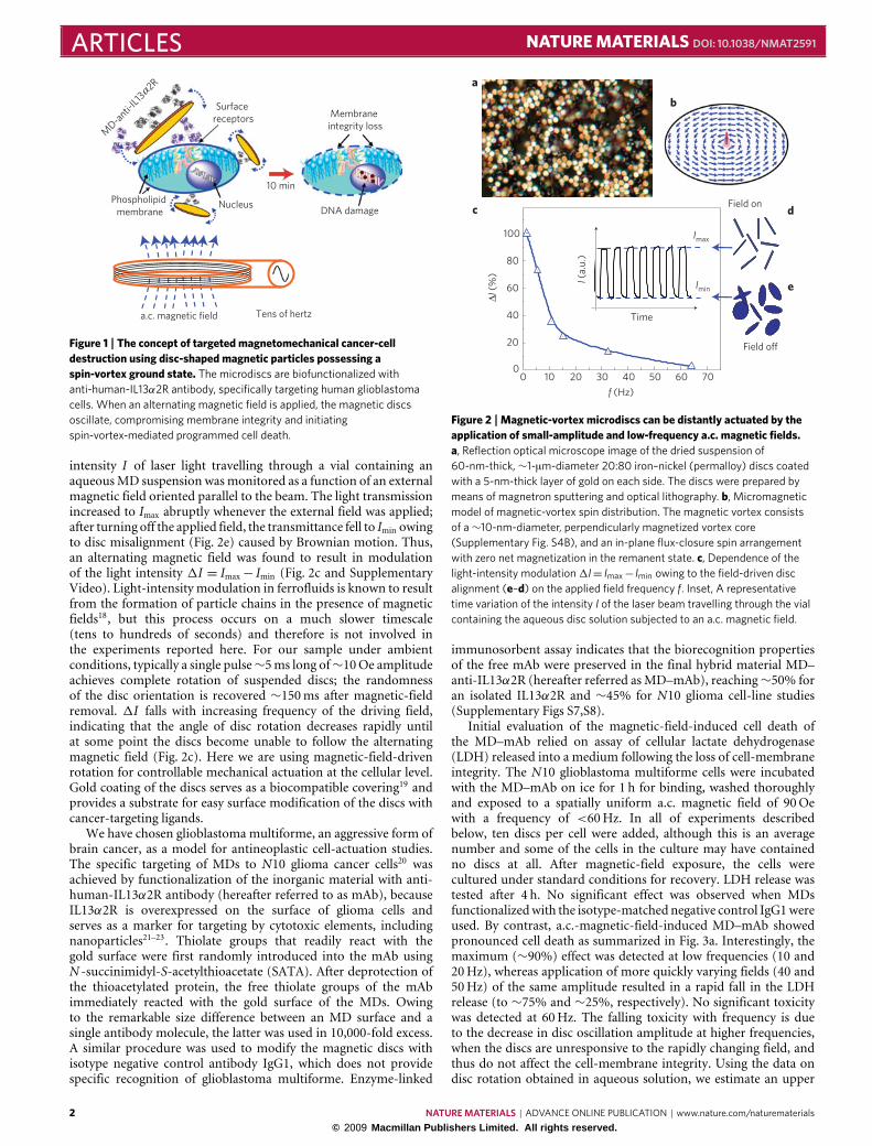
The 60-nm-thick, ∼1-µm-diameter 20:80% iron–nickel(permalloy) discs, coated with a 5-nm-thick layer of gold oneach side, were prepared by magnetron sputtering and opticallithography (ref. 17 and Supplementary Fig. S1) before release fromthe wafer into aqueous solution, ready for surface modification.Figure 2a shows a representative optical microscope image of theas-fabricated microdiscs dried on a glass slide. The geometry ofthe sample, combined with intrinsic properties of the permalloymaterial, results in a magnetic-vortex spin state (Fig. 2b) whenthe system is in remanence. The magnetic vortex nucleateswhen the demagnetizing fields within each particle are no longercounterbalanced by the external applied field. The existence ofmagnetic vortices in our samples was experimentally confirmedby hysteresis-loop measurements (Supplementary Fig. S2) andmagnetic-force microscopy imaging of the reference sample(Supplementary Fig. S3). The MDs show zero in-plane magneticmoment in remanence, a linear M (H ) dependence in small fieldsand nucleation and saturation fields of ∼250Oe and ∼600Oe,respectively, in agreement with the model. When a magnetic field isapplied, the vortices in each disc shift, developing a magnetizationcomponent parallel to the field direction. MDs subjected toa magnetic field H will therefore experience a driving torqueτm =m×H, where m is the disc magnetic moment, proportionalto bothMs and the magnetic susceptibility. This torque rotates thedisc, aligning the plane of the disc parallel to the direction of themagnetic field (Fig. 2d). The dynamics of MDs in aqueous solutionwere separately investigated before cell-culture experiments. The
NATUREMATERIALS | ADVANCE ONLINE PUBLICATION | www.nature.com/naturematerials 1© 2009 Macmillan Publishers Limited. All rights reserved.
ARTICLES NATURE MATERIALS DOI: 10.1038/NMAT2591
Phospholipid membrane
MD-a
nti-IL1
3 2R
αSurface
receptors
Nucleus
a.c. magnetic field Tens of hertz
Membrane integrity loss
DNA damage
10 min
Figure 1 | The concept of targeted magnetomechanical cancer-celldestruction using disc-shaped magnetic particles possessing aspin-vortex ground state. The microdiscs are biofunctionalized withanti-human-IL13α2R antibody, specifically targeting human glioblastomacells. When an alternating magnetic field is applied, the magnetic discsoscillate, compromising membrane integrity and initiatingspin-vortex-mediated programmed cell death.
intensity I of laser light travelling through a vial containing anaqueousMD suspension wasmonitored as a function of an externalmagnetic field oriented parallel to the beam. The light transmissionincreased to Imax abruptly whenever the external field was applied;after turning off the applied field, the transmittance fell to Imin owingto disc misalignment (Fig. 2e) caused by Brownian motion. Thus,an alternating magnetic field was found to result in modulationof the light intensity 1I = Imax− Imin (Fig. 2c and SupplementaryVideo). Light-intensity modulation in ferrofluids is known to resultfrom the formation of particle chains in the presence of magneticfields18, but this process occurs on a much slower timescale(tens to hundreds of seconds) and therefore is not involved inthe experiments reported here. For our sample under ambientconditions, typically a single pulse∼5ms long of∼10Oe amplitudeachieves complete rotation of suspended discs; the randomnessof the disc orientation is recovered ∼150ms after magnetic-fieldremoval. 1I falls with increasing frequency of the driving field,indicating that the angle of disc rotation decreases rapidly untilat some point the discs become unable to follow the alternatingmagnetic field (Fig. 2c). Here we are using magnetic-field-drivenrotation for controllable mechanical actuation at the cellular level.Gold coating of the discs serves as a biocompatible covering19 andprovides a substrate for easy surface modification of the discs withcancer-targeting ligands.
We have chosen glioblastoma multiforme, an aggressive form ofbrain cancer, as a model for antineoplastic cell-actuation studies.The specific targeting of MDs to N10 glioma cancer cells20 wasachieved by functionalization of the inorganic material with anti-human-IL13α2R antibody (hereafter referred to as mAb), becauseIL13α2R is overexpressed on the surface of glioma cells andserves as a marker for targeting by cytotoxic elements, includingnanoparticles21–23. Thiolate groups that readily react with thegold surface were first randomly introduced into the mAb usingN -succinimidyl-S-acetylthioacetate (SATA). After deprotection ofthe thioacetylated protein, the free thiolate groups of the mAbimmediately reacted with the gold surface of the MDs. Owingto the remarkable size difference between an MD surface and asingle antibody molecule, the latter was used in 10,000-fold excess.A similar procedure was used to modify the magnetic discs withisotype negative control antibody IgG1, which does not providespecific recognition of glioblastoma multiforme. Enzyme-linked
Δl (
%)
100
80
60
40
20
0
f (Hz)
0 10 20 30 40 50 60 70
Field off
d
e
Field on
l (a
.u.)
Time
Imax
Imin
a
b
c
Figure 2 |Magnetic-vortex microdiscs can be distantly actuated by theapplication of small-amplitude and low-frequency a.c. magnetic fields.a, Reflection optical microscope image of the dried suspension of60-nm-thick,∼1-µm-diameter 20:80 iron–nickel (permalloy) discs coatedwith a 5-nm-thick layer of gold on each side. The discs were prepared bymeans of magnetron sputtering and optical lithography. b, Micromagneticmodel of magnetic-vortex spin distribution. The magnetic vortex consistsof a∼10-nm-diameter, perpendicularly magnetized vortex core(Supplementary Fig. S4B), and an in-plane flux-closure spin arrangementwith zero net magnetization in the remanent state. c, Dependence of thelight-intensity modulation1I= Imax− Imin owing to the field-driven discalignment (e–d) on the applied field frequency f. Inset, A representativetime variation of the intensity I of the laser beam travelling through the vialcontaining the aqueous disc solution subjected to an a.c. magnetic field.
immunosorbent assay indicates that the biorecognition propertiesof the free mAb were preserved in the final hybrid material MD–anti-IL13α2R (hereafter referred as MD–mAb), reaching∼50% foran isolated IL13α2R and ∼45% for N10 glioma cell-line studies(Supplementary Figs S7,S8).
Initial evaluation of the magnetic-field-induced cell death ofthe MD–mAb relied on assay of cellular lactate dehydrogenase(LDH) released into a medium following the loss of cell-membraneintegrity. The N10 glioblastoma multiforme cells were incubatedwith the MD–mAb on ice for 1 h for binding, washed thoroughlyand exposed to a spatially uniform a.c. magnetic field of 90Oewith a frequency of <60Hz. In all of experiments describedbelow, ten discs per cell were added, although this is an averagenumber and some of the cells in the culture may have containedno discs at all. After magnetic-field exposure, the cells werecultured under standard conditions for recovery. LDH release wastested after 4 h. No significant effect was observed when MDsfunctionalizedwith the isotype-matched negative control IgG1wereused. By contrast, a.c.-magnetic-field-induced MD–mAb showedpronounced cell death as summarized in Fig. 3a. Interestingly, themaximum (∼90%) effect was detected at low frequencies (10 and20Hz), whereas application of more quickly varying fields (40 and50Hz) of the same amplitude resulted in a rapid fall in the LDHrelease (to ∼75% and ∼25%, respectively). No significant toxicitywas detected at 60Hz. The falling toxicity with frequency is dueto the decrease in disc oscillation amplitude at higher frequencies,when the discs are unresponsive to the rapidly changing field, andthus do not affect the cell-membrane integrity. Using the data ondisc rotation obtained in aqueous solution, we estimate an upper
2 NATUREMATERIALS | ADVANCE ONLINE PUBLICATION | www.nature.com/naturematerials
© 2009 Macmillan Publishers Limited. All rights reserved.

NATURE MATERIALS DOI: 10.1038/NMAT2591 ARTICLES
b
d e
a
Frequency (Hz)
LDH
leak
age
(%)
No field 10 20 40 50 60
IL13-coated MDsIgG-coated MDs
100
80
60
40
20
0
20 µm
20 µm
c
100 µm
100 µm
Figure 3 |A low-frequency spatially uniform magnetic field applied to theMDs–mAb–cell complex results in compromised integrity of the cellularmembrane and cell death. a, Magnetic-field-induced cell death of theMD–mAb towards N10 cells and MD-IgG loss of cell-membrane integrityusing a cellular LDH test for different field frequencies. No remarkable LDHrelease was observed when MDs functionalized with the isotype-matchednegative control IgG1 were applied. Error bars denote the standarddeviation for experiments across six wells. b–e, Optical images of control(b,d) and treated (c,e) N10 glioma cells. A 90 Oe–20 Hz magnetic field wasapplied for 10 min. The treated cells are rounded off with membraneshrinkage and a loss of membrane integrity.
limit on the disc oscillation angle to be in the range from 20◦ downto 5◦ for driving frequencies from 10 to 50Hz. The temperatureof the cell–MD solution, remotely monitored with an infraredcamera, always remained below ∼22 ◦C during our experiments,and varied not more than a few degrees (owing to a small amountof convective heat exchange from the coil) during magnetic-fieldexposure. This excludes hyperthermia as a possible mechanismof cell damage. The observed LDH leakage might be caused bytargeted membrane rupture or ‘magnetomechanically induced celldestruction’.Magnetic field exposure times of 10 and 30min did notaffect LDH levels, so 10minwas chosen as theworking condition forall further experiments.
Dramatic cell-morphology changes were observed aftermagnetic-field application. Figure 3 compares representative op-tical images of the control N10 cells pre-incubated with MD–mAb(Fig. 3b,d) with cells after 90Oe–20Hz a.c. magnetic-field exposurefollowing 1 h culture under standard conditions (Fig. 3c,e). Thetreated cells are rounded off, with membrane shrinkage and loss ofmembrane integrity. Furthermore, atomic forcemicroscope imagesof the fixed cells show that the control N10 cells (Fig. 4a) appearas ∼500-nm-thick, rough and elongated shapes with well-defined∼1-µm-high nuclei. The treated cells (Fig. 4b) are characterized
by an apparent fractioning and redistribution of nucleus material,shape rounding and flattening, surface smoothing and ratherabrupt edges. It is apparent that the force produced by slowlyoscillating MDs on the cell surface could not alone cause suchprofound effects. On the basis of a simple magnetic-torque model,we estimate that each disc can deliver a force of the order of tens ofpiconewtons (see Supplementary Section SIV), whereas the forcesneeded to rupture a cell membrane are reported to be in the rangeof hundreds of piconewtons24–26. Therefore, it is feasible that theobserved effects have not resulted from membrane rupture alone,but instead are due to the triggering of intracellular pathwaysactivating programmed cell death through application of magneticfield to the MD–mAb–cell complex.
Since the observed nucleus fractioning and cell shrinkage areprincipal morphological characteristics of apoptosis, we assayedone of the hallmarks of late apoptotic cascades, namely genomicDNA fragmentation. To identify nuclear DNA damage, a terminaldeoxynucleotidyl transferase dUTP nick end labelling (TUNEL)assay was used27. The optical images show that untreated controlcells (Fig. 5b,d) with well-organized chromatin structures havea blue fluorescence, whereas treated cells (Fig. 5c,e) are stainedwith a dark orange–brown dye owing to chromatin fragmentation.Quantified data from TUNEL-positive fluorescent cells are sum-marized for different a.c.-magnetic-field amplitudes in Fig. 5a. Thelargest number of TUNEL-positive cells (∼60%) was observed fora 90Oe–20Hz field. The number of apoptotic cells decreased withfurther increase in the field strength. We attribute this observationto increasing numbers of necrotic cells at higher field strengthsowing to more severe magnetomechanically induced membranedamage. Application of lower fields (30Oe) still results in a no-ticeable (∼20%) population of cells undergoing apoptosis. Theindication of apoptosis was observed both 4 h (Fig. 5) and 24 h (notshown) after magnetic-field exposure. As expected, the numberof TUNEL-stained cells was observed to fall remarkably (∼50%)after the addition of 20 µM Z-VAD-FMK (carbobenzoxy-valyl-alanyl-aspartyl-[O-methyl]-fluoromethylketone), an inhibitor ofcaspase proteases, the key players in apoptosis. DNA denatura-tion at the terminal stage of apoptosis is known to be catalysedby calcium-dependent endonucleases28, and DNA fragmentationcould only barely be directly achieved by the applied mechanicalforces. In other words, the observed late apoptotic changes are farremoved from the primary stimulus, and therefore are the result ofintercellular signal transduction cascades29.
Calcium signalling is involved in a number of key mecha-nisms implicated in cell death29–32. Stimulus-induced calcium influxthrough membrane ionic channels, or calcium release from theinternal cell sources (for example, the endoplasmic reticulum),can result in the total perturbation of cellular calcium home-ostasis and triggering of apoptosis29,32. In the present case, someimportant apoptosis-related supramolecular membrane structures,including surface ‘death’ receptors (for example, Fas or TNF-R), ormechanosensitive or other cation channels, may be non-specificallyactivated by magnetic-vortex-mediated mechanical forces6–8. Fur-thermore, it is well established that membrane disturbances, evenif transient and gentle, lead to the induction of stretch-activatedchannels33,34, and consequently to increasing levels of intracellu-lar calcium ions35,36. Therefore, we investigated whether calciumsignalling is involved in the observed magnetic-vortex-driven celldeath. For intracellular calcium imaging, N10 cells were preloadedwith Fluo-4AM, a calcium indicator dye (excitation wavelengthλex = 488 nm, emission wavelength λem = 520 nm), whereas cellnuclei were stained with Hoechst 33342 (λex = 350 nm). A dual-wavelength laser confocal microscope was used to qualitativelydetermine changes in calcium intracellular concentration and spa-tial distribution during magnetic-field application. A magneticcoil was deployed on the microscope sample stage. All assays
NATUREMATERIALS | ADVANCE ONLINE PUBLICATION | www.nature.com/naturematerials 3© 2009 Macmillan Publishers Limited. All rights reserved.

ARTICLES NATURE MATERIALS DOI: 10.1038/NMAT2591
a
b
0.2
0.4
0.6
0.8
1.0
1.2
0
0
10 20 30 40 50
0 10 20 30 40 500
0.2
0.4
0.6
0.8
1.0
1.2
90 Oe/20Hz a.c. magnetic field
Cel
l hei
ght (
µm)
Distance (µm)
Distance (µm)
Cel
l hei
ght (
µm)
Figure 4 | Comparison of representative atomic force microscope amplitude error, height and cross-section scans for the control and treated cells.a, The control N10 cells appear as∼500-nm-thick, rough and elongated shapes with well-defined∼1-µm-high nuclei at their centres. b, The treated cellsare characterized by a smoother surface, thickening, shape rounding, rather abrupt edges and an apparent flattening and fractioning of the genetic materialstoring organelle—the nucleus. The blue and red curves show the cross-section profiles across the centre of the cell along horizontal and verticaldirections, respectively. The scan size is 50×50 µm2.
b
e
Apo
ptot
ic c
ells
(%
)
*
*
*
*
IL13-coated MDsNegative control
100
80
60
40
20
0130 90 60 30
Magnetic-field amplitude H (Oe)
50 µm
d 20 µm
c 100 µm
100 µm
a
Figure 5 |Magnetic-vortex-mediated mechanical stimuli trigger intracellular biochemical pathways activating programmed cell death. a, Apoptosis ofthe N10 cells induced by an a.c. magnetic field. b–e, Images of negative control (b,d) and MD–mAb-functionalized cells subjected to 20 Hz–90 Oe a.c.fields for 10 min and TUNEL stained 4 h after the magnetic-field exposure (c,e). The control cells with well-organized chromatin structures have a bluefluorescence, whereas the treated cells are stained with a dark orange–brown dye owing to chromatin fragmentation—an indication of apoptosis. Theproportion of apoptotic populations was calculated by counting a total of 1,000 nuclei in each group using optical microscope images. Error bars denotethe standard deviation for experiments across six wells. Asterisks indicate that differences between experimental groups and negative controls wereconsidered significant at p<0.05.
were performed in nominally calcium-free Hank’s buffer solution.Figure 6a shows an optical micrograph of the imaged area. The ini-tial calcium concentration of cells 1 and 2 seemed to be much lowerthan that observed in cell 3. This could be true, because the cells
were at different stages of their life cycles. To ensure that baselinecalcium equilibrium was reached, the dye-loaded cells were imagedfor 2min before magnetic-field application. Next, an alternatingmagnetic fieldwith a frequency of 10Hz and amplitude of 90Oewas
4 NATUREMATERIALS | ADVANCE ONLINE PUBLICATION | www.nature.com/naturematerials
© 2009 Macmillan Publishers Limited. All rights reserved.

NATURE MATERIALS DOI: 10.1038/NMAT2591 ARTICLES
3
2
1
0 min
a b
10 min
d
7 min
c
Figure 6 |Optical imaging of intracellular calcium. The N10 cells were preloaded with calcium indicator Fluo-4-AM dye (λex=488 nm, λem= 520 nm),green, whereas cell nuclei were stained with Hoechst 33342 (λex= 350 nm), blue. a, Cell optical images. b–d, Fluorescent cell images: snapshots at 0, 7and 10 min application of alternating magnetic field of 10 Hz–90 Oe. Yellow arrows show representative calcium flashes or spikes.
applied in situ for 10min to stimulate the biofunctionalized MDs.Cell 3 showed well-pronounced characteristic apoptotic membraneblebbing and shrank significantly during the next few minutes ofthe experiment. Although its total fluorescence faded by the end ofthe magnetic-field exposure, owing to cell death or pumping outof calcium and stain, calcium delocalization was clearly observed(for example, compare images Fig. 6b,c). Cells 1 and 2 also showedmembrane shrinkage, although to a less significant extent. Signif-icantly, the calcium level in these cells increased, and spectaculardynamic oscillations, spikes and relocalization of calcium wereobserved (Fig. 6b–d). The most likely explanation is that calciummobilization arose from intracellular calcium depots, but externalcalcium pumping cannot be excluded simply because nominallycalcium-free buffer was used. Although wide horizons continueto exist, with exciting possibilities for further mechanistic studies,on the basis of our observations we propose that magnetic-vortex-mediated mechanical stimuli are converted and amplified into achemical ionic signal able to initiate apoptosis and cancer-cell death.
The present study used rather large discs for these proof-of-concept experiments. Because it is well established that themicromagnetic properties of an object fabricated from a givenmagnetically soft material are determined solely by its geometricalaspect ratio (in the case of MDs, the thickness-to-diameter ratio),the biofunctionalized MD system can be scaled down to smallerlateral dimensions (∼100 nm) while preserving its closed-fluxspin structure and magnetomechanical properties (ref. 11, andSupplementary Fig. S4).
It is instructive to compare the proposed strategy withthe well-explored use of magnetic nanoparticles to achievetumour regression through hyperthermia effects. The principal
remaining problems with magnetic hyperthermia are invasiveness,targeting (restricting the hyperthermia effect to the specific areaof interest) and achieving homogenous heat distributions withinthe target organ4,5,37–39. Failure to solve these problems may leadto either insufficient treatment effects or, worse, lethal exposureof neighbouring cells. In contrast to hyperthermia treatment,magnetomechanic stimulus induced byMDs is transmitted directlyto the targeted cell with high specificity and high efficiency. Owingto the use of a magnetically soft material with a unique spinarrangement, namely a spin-vortex state, the biologically relevanteffect was achieved through application of unprecedentedly weakmagnetic fields<100Oe of a frequency of a few tens of Hz, appliedfor a duration of only 10min. This is in stark contrast to themuch stronger fields, running at high frequencies of hundreds ofkilohertz, required to achieve heating-induced cell toxicity withsuperparamagnetic particles37. In other words, in our experiments,the external power (proportional to f ×H 2) supplied to cell cultureswas on average at least 100,000 times smaller than that used atpresent in hyperthermia treatments with magnetic nanoparticles.The lowoperating field strengthmay create treatment opportunitieswith low cost, large working volume, and minimal invasiveness.Because the operating paradigm is not directly mechanical orthermal—instead, the mechanical oscillation of MDs attached tothe cancer-cell membrane is transformed into an ionic electricalsignal, which triggers the programmed cell-death pathway—thetotal energy necessary to accomplish cell death is minute. It shouldbe noted, however, that MDs subjected to a.c. field at typicalhyperthermia frequencies are likely to demonstrate inductiveheating owing to the combined effects of eddy currents and dynamichysteresis losses. Aside from applications to magnetomechanically
NATUREMATERIALS | ADVANCE ONLINE PUBLICATION | www.nature.com/naturematerials 5© 2009 Macmillan Publishers Limited. All rights reserved.

ARTICLES NATURE MATERIALS DOI: 10.1038/NMAT2591
induced cancer-cell destruction and hyperthermia, which werediscussed, the magnetic microdiscs with a spin-vortex ground statemay find further application in probes of mechanotransduction incells, distant cell manipulation and separation, controllable forceapplication during regenerative tissue growth and studies of cellelasticity in a diagnostic capacity.
MethodsBiofunctionalization of MDs. 27.8 µl of 55mM SATA solution in dimethylsulphoxide and 2.8 µl of 60 µM IL13 solution were mixed and incubated atroom temperature with continuous gentle shaking in the dark for 30min. Todeprotect thiolate groups the SATA-modified antibody solution was combinedwith 4 µl of 0.5M hydroxylamine·HCl and 25mM ethylenediamine tetra-aceticacid solution in 0.1M phosphate-buffered saline, pH 7.4, and incubated for 2 h atroom temperature. 500 µl of MD suspension (5×10⁸ eaml−1) were degassed bynitrogen gas bubbling for 15min, and then the deacetylated antibody solution wasimmediately mixed with the MD suspension with continuous gentle shaking in thedark at room temperature for ∼2 h. The final MD–mAb was spin-washed threetimes with 500 µl 0.1M phosphate-buffered saline, pH 7.4, to remove unboundprotein, and then redispersed in 500 µl of 0.1M phosphate-buffered saline,pH 7.4, and stored at 4 ◦C. Fourier transform infrared spectroscopy, wavelength,cm−1: 1650 (amide I) and 1550 (amide II).
Magnetomechanical cell destruction. A magnetic-field station plate was madeby inserting a 1/4 inch soft-iron rod into the under-well in a 96-well plate. Thestation circumference was wound by Cu wire as a coil. The power supply–amplifier(KEPCO, Flushing, NY, USA), function generator (Agilent 33220A, USA)and oscilloscope (Tektronix, Beverton, OR, USA) were connected with themagnetic-field station for generating a.c. magnetic field. For the experiment the96-well plate with samples was positioned on top of the magnetic-field stationplate. Magnetic-field strength was calibrated with a Hall probe. After the treatment,the necrosis and apoptosis of treated cells was assessed with a standard LDHtest and TUNEL assay.
Morphological analysis of cell viability by optical and atomic force microscopy.Human glioma cells from the N10 cell line were cultivated on poly-l-lysine-coatedcover-slips. The human glioma tumour cell line No. 10 cells (N10) were grownon the cover-slips in 24-well plates for 24 h to reach 104 cells per cover-slip.The cells on the cover-slips were exposed to a.c. magnetic fields as described inthe magnetomechanical cell-destruction set-up. The treated cells were fixed in4% paraformaldehyde and analysed by optical microscopy or recovery culturedin standard conditions for 4 h and then fixed for atomic force microscopy.Cover-slips were thoroughly rinsed with MilliQ water before imaging. Bright- ordark-field images of the cells were taken using ×10, ×20, ×50 and ×100 objectivelenses. Atomic force microscope surface images of the cells were acquired innon-contact (tapping) mode.
Laser confocal intracellular calcium imaging. For the calcium assays the N10cells were grown in 35mm Fluorodish cell-culture dishes (World PrecisionInstruments, Florida, USA) to reach ∼10,000 cells per dish and then incubatedwith MD–mAb at standard conditions. Cells were loaded with 1 µM Fluo-4-AMcontaining 0.02% Pluronic F-127 (Molecular Probes) and 1 µgml−1 Hoechst33342 (Molecular Probes) at 37 ◦C for 20 and 5min respectively, washed and thenassayed in Hank’s buffer. A magnetic coil was placed on top of a sample stageof a dual-wavelength Leica SP5 tandem scanner spectral two-photon confocalmicroscope. After 2min of dye-loaded cell initial fluorescence imaging, analternating magnetic field of 10Hz (90Oe) was applied to stimulate MDs on thecell surface for 10min. Images were digitized, and fluorescent intensities wereanalysed using Image J software.
Statistical analysis. All data are expressed as mean± standard deviation, and wereanalysed by one-way analysis of variance. Statistical significance was determined,with P values less than 0.05 considered statistically significant.
Received 8 July 2009; accepted 2 November 2009;published online 29 November 2009
References1. Chappert, C., Fert, A. & Van Dau, F. N. The emergence of spin electronics in
data storage. Nature Mater. 6, 813–823 (2007).2. Bruns, O. T. et al. Real-time magnetic resonance imaging and quantification
of lipoprotein metabolism in vivo using nanocrystals. Nature Nanotech. 4,193–201 (2009).
3. Ferrari, M. Cancer nanotechnology: Opportunities and challenges.Nature Rev. Cancer 5, 161–171 (2005).
4. Pankhurst, Q. A., Connolly, J., Jones, S. K. & Dobson, J. Applications ofmagnetic nanoparticles in biomedicine. J. Phys. D 36, R167–R175 (2003).
5. Hergt, R., Dutz, S., Muller, R. & Zeisberger, M.Magnetic particle hyperthermia:Nanoparticle magnetism andmaterials development for cancer therapy. J. Phys.Condens. Matter. 18, S2919–S2923 (2006).
6. Dobson, J. Remote control of cellular behaviour with magnetic nanoparticles.Nature Nanotech. 3, 139–148 (2008).
7. Mannix, R. J. et al. Nanomagnetic actuation of receptor-mediated signaltransduction. Nature Nanotech. 3, 36–40 (2007).
8. Wang, N., Butler, J. P. & Ingber, D. E. Mechanotransduction across the cellsurface and through the cytoskeleton. Science 260, 1124–1127 (1993).
9. Jun, Y.-W., Seo, J.-W. & Cheon, J. Nanoscaling laws of magneticnanoparticles and their applicability in biomedical science. Acc. Chem. Res. 41,179–186 (2008).
10. Cheon, J. & Lee, J.-H. Synergistically integrated nanoparticles as multimodalprobes for nanobiotechnology. Acc. Chem. Res. 41, 1630–1635 (2008).
11. Cowburn, R. P., Koltsov, D. K., Adeyeye, A. O. &Welland,M. E. Single-domaincircular nanomagnets. Phys. Rev. Lett. 83, 1042–1045 (1999).
12. Shinjo, T., Okuno, T., Hassdorf, R., Shigeto, K. & Ono, T. Magnetic vortex coreobservation in circular dots of permalloy. Science 289, 930–933 (2000).
13. Wachowiak, A. et al. Direct observation of internal spin structure of magneticvortex cores. Science 298, 577–580 (2002).
14. Buchanan, K. S. et al. Soliton-pair dynamics in patterned ferromagnetic ellipses.Nature Phys. 1, 172–176 (2005).
15. Ade, H. & Stoll, H. Near-edge X-ray absorption fine-structure microscopy oforganic and magnetic materials. Nature Mater. 8, 281–290 (2009).
16. Novosad, V. et al. Effect of interdot magnetostatic interaction onmagnetizationreversal in circular dot arrays. Phys. Rev. B 65, 060402 (2002).
17. Rozhkova, E. A. et al. Ferromagnetic microdisks as carriers for biomedicalapplications. J. Appl. Phys. 105, 07B306 (2009).
18. Martin, J. E., Hill, K. M. & Tigges, C. P. Magnetic-field-induced opticaltransmittance in colloidal suspensions. Phys. Rev. E 59, 5676–5692 (1999).
19. Nel, A. E. et al. Understanding biophysicochemical interactions at the nano-biointerface. Nature Mater. 8, 543–557 (2009).
20. Da, K., Shiyama, K., Naka, R., Hiyama, A. & Anishi, T. GFAP-positive humanglioma cell lines: No. 10, no. 11. Hum. Cell 3, 251–256 (1990).
21. Debinski, W., Gibo, D., Hulet, S., Connor, J. & Gillespie, G. Receptor forinterleukin 13 is amarker and therapeutic target for human high-grade gliomas.Cancer Res. 5, 985–990 (1999).
22. Kawakami, K., Kawakami, M., Snoy, P. J., Husain, S. R. & Puri, R. K. In vivooverexpression of IL-13 receptor {alpha}2 chain inhibits tumorigenicity ofhuman breast and pancreatic tumors in immunodeficient mice. J. Exp. Med.194, 1743–1754 (2001).
23. Rozhkova, E. A. et al. A high-performance nanobio photocatalyst for targetedbrain cancer therapy. Nano Lett. 9, 3337–3342 (2009).
24. Sen, S., Subramanian, S. & Discher, D. E. Indentation and adhesive probing ofa cell membrane with AFM: Theoretical model and experiments. Biophys. J. 89,3203–3213 (2005).
25. Afrin, R., Yamada, T. & Ikai, A. Analysis of force curves obtained on the livecell membrane using chemically modified AFM probes. Ultramicroscopy 100,187–195 (2004).
26. Muller, D., Helenius, J., Alsteens, D. & Dufrêne, Y. Force probing surfaces ofliving cells to molecular resolution. Nature Chem. Biol. 5, 383–391 (2009).
27. Mpoke, S. S. & Wolfe, J. Differential staining of apoptotic nuclei inliving cells: Application to macronuclear elimination in tetrahymena.J. Histochem. Cytochem. 45, 675–684 (1997).
28. Yakovlev, A. G. et al. Role of DNAS1L3 in Ca2+- and Mg2+-dependentcleavage of DNA into oligonucleosomal and high molecular mass fragments.Nucl. Acids Res. 27, 1999–2005 (1999).
29. Mattson, M. P. & Chan, S. L. Calcium orchestrates apoptosis. Nature Cell Biol.5, 1041–1043 (2003).
30. Clapham, D. E. Calcium signaling. Cell 80, 259–268 (1995).31. Duchen, M. R. Mitochondria and calcium: From cell signalling to cell death.
J. Phys. 529, 57–68 (2000).32. Boehning, D. et al. Cytochrome c binds to inositol (1,4,5) trisphosphate
receptors, amplifying calcium-dependent apoptosis. Nature Cell Biol. 5,1051–1061 (2003).
33. Martinac, B. Mechanosensitive ion channels: Molecules ofmechanotransduction. J. Cell Sci. 117, 2449–2460 (2004).
34. Guharay, F. & Sachs, F. J. Stretch-activated single ion channel currents intissue-cultured embryonic chick skeletal muscle. Physiol. (London) 352,685–701 (1984).
35. Diamond, S. L., Sachs, F. & Sigurdson,W. J.Mechanically inducedmobilizationin cultured endothelial cells is dependent on actin and phospholipase.Arterioscler. Thromb. Vasc. Biol. 14, 2000–2006 (1994).
36. Adachi, T., Sato, K. & Tomita, Y. Directional dependence of osteoblasticcalcium response to mechanical stimuli. Biomech. Model Mechanobiol. 2,73–82 (2003).
37. Hergt, R. & Dutz, S. Magnetic particle hyperthermia—biophysical limitationsof a visionary tumour therapy. J. Magn. Magn. Mater. 311, 187–192 (2007).
38. Goya, G. F., Grazu, V. & Ibarra, M. R. Magnetic nanoparticles for cancertherapy. Current Nanosci. 4, 1–16 (2008).
6 NATUREMATERIALS | ADVANCE ONLINE PUBLICATION | www.nature.com/naturematerials
© 2009 Macmillan Publishers Limited. All rights reserved.

NATURE MATERIALS DOI: 10.1038/NMAT2591 ARTICLES39. Gupta, A. K. & Gupta, M. Synthesis and surface engineering of iron oxide
nanoparticles for biomedical applications. Biomaterials 26, 3995–4021 (2005).
AcknowledgementsWe thank D. Clapham, R. Hergt, J. Dobson and A. Datesman for valuable suggestions andcritical reading of the manuscript. We also thank J. Pearson for help with developing themagnetic-field induction set-up, R. Divan for discussing the microfabrication strategies,and V. Bindokas for technical assistance in Ca imaging at the UC Biological SciencesDivision Light Microscopy Core Facility. Work at Argonne and its Center for NanoscaleMaterials and Electron Microscopy Center is supported by the US Department of EnergyOffice of Science, Basic Energy Sciences, under contract No DE-AC02-06CH11357.Work at the University of Chicago is supported by the National Cancer Institute(R01-CA122930), the National Institute of Neurological Disorders and Stroke(K08-NS046430), the Alliance for Cancer Gene Therapy Young Investigator Award andthe American Cancer Society (RSG-07-276-01-MGO).
Author contributionsV.N. and E.A.R. conceived the experimental idea. M.S.L. advanced the conceptualdesign for the glioma cell targeting. I.V.U. performed in vitro and cell cytotoxicitystudies. D.-H.K. and V.N. fabricated the magnetic microdiscs and carried out themagnetic characterization and micromagnetic modelling. D.-H.K. and E.A.R. ranthe biofunctionalization experiments. D.-H.K. carried out atomic force and opticalmicroscopy characterizations. E.A.R. and I.V.U. designed and analysed the intracellularCa imaging experiments. D.-H.K., E.A.R., I.V.U., T.R., S.D.B., M.S.L. and V.N. analysedthe data and wrote the manuscript.
Additional informationThe authors declare no competing financial interests. Supplementary informationaccompanies this paper on www.nature.com/naturematerials. Reprints and permissionsinformation is available online at http://npg.nature.com/reprintsandpermissions.Correspondence and requests formaterials should be addressed to E.A.R. or V.N.
NATUREMATERIALS | ADVANCE ONLINE PUBLICATION | www.nature.com/naturematerials 7© 2009 Macmillan Publishers Limited. All rights reserved.