biological psychiatry sponsored document from a, suzanne
TRANSCRIPT
Systemic Inflammation Induces Acute Behavioral and CognitiveChanges and Accelerates Neurodegenerative Disease
Colm Cunninghama,⁎, Suzanne Campionb, Katie Lunnonb, Carol L. Murraya, Jack F.C.Woodsa, Robert M.J. Deaconc, J. Nicholas P. Rawlinsc, and V. Hugh Perryb
aDepartment of Biochemistry, Trinity College, Institute of Neuroscience, Trinity College Dublin, Republic ofIreland bCNS Inflammation Group, Southampton Neurosciences Group, School of Biological Sciences,University of Southampton, Bassett Crescent East cDepartment of Experimental Psychology, University ofOxford, South Parks Road, Oxford, United Kingdom
AbstractBackground—Chronic neurodegeneration results in microglial activation, but the contribution ofinflammation to the progress of neurodegeneration remains unclear. We have shown that microgliaexpress low levels of proinflammatory cytokines during chronic neurodegeneration but are “primed”to produce a more proinflammatory profile after systemic challenge with bacterial endotoxin(lipopolysaccharide [LPS]).
Methods—Here, we investigated whether intraperitoneal (IP) challenge with LPS, to mimicsystemic infection, in the early stages of prion disease can 1) produce exaggerated acute behavioral(n = 9) and central nervous system (CNS) inflammatory (n = 4) responses in diseased animalscompared with control animals, and 2) whether a single LPS challenge can accelerate diseaseprogression (n = 34–35).
Results—Injection of LPS (100 μg/kg), at 12 weeks postinoculation (PI), resulted in heightenedCNS interleukin-1 beta (IL-1β), tumor necrosis factor-alpha (TNF-α), and interferon-beta (IFN-β)transcription and microglial IL-1β translation in prion-diseased animals relative to control animals.This inflammation caused exaggerated impairments in burrowing and locomotor activity, andinduced hypothermia and cognitive changes in prion-diseased animals that were absent in LPS-treated control animals. At 15 weeks PI, LPS (500 μg/kg) acutely impaired motor coordination andmuscle strength in prion-diseased but not in control animals. After recovery, these animals alsoshowed earlier onset of disease-associated impairments on these parameters.
Conclusions—These data demonstrate that transient systemic inflammation superimposed onneurodegenerative disease acutely exacerbates cognitive and motor symptoms of disease andaccelerates disease progression. These deleterious effects of systemic inflammation haveimplications for the treatment of chronic neurodegeneration and associated delirium.
© 2009 Elsevier Inc..⁎Address reprint requests to Colm Cunningham, Ph.D., Trinity College Dublin, Trinity College Institute of Neuroscience, Departmentof Biochemistry, Trinity College, Dublin, Ireland [email protected] document was posted here by permission of the publisher. At the time of deposit, it included all changes made during peer review,copyediting, and publishing. The U.S. National Library of Medicine is responsible for all links within the document and for incorporatingany publisher-supplied amendments or retractions issued subsequently. The published journal article, guaranteed to be such by Elsevier,is available for free, on ScienceDirect.
Sponsored document fromBiological Psychiatry
Published as: Biol Psychiatry. 2009 February 15; 65(4): 304–312.
Sponsored Docum
ent Sponsored D
ocument
Sponsored Docum
ent
Key WordsAlzheimer's disease; cytokines; delirium; inflammation; microglial priming; neurodegeneration;systemic
Chronic neurodegeneration is accompanied by an inflammatory response characterized by aselective activation of the microglial cells of the central nervous system (CNS) (1). Thisinflammation is assumed to contribute to disease progression through the production ofinflammatory mediators. Although long-term nonsteroidal anti-inflammatory drug (NSAID)use modestly protects against the development of Alzheimer's disease (AD) (2), prospectivestudies with these drugs in AD patients have been disappointing (3–5). This emphasizes theconsiderable gaps in our knowledge about how microglial activation influences chronicneurodegeneration.
Systemic infections have more severe consequences in the elderly and demented population.Systemic infection is a common trigger for episodes of delirium (6–8), and episodes of deliriumare predictive of increased morbidity and mortality (9–11). Surprisingly, direct investigationsas to whether systemic inflammation accelerates the progression of dementia andneurodegenerative disease are lacking. Thus, whether and how systemic inflammation andCNS disease interact to accelerate neurodegeneration and to induce delirium is poorlyunderstood.
It is well known that systemic inflammation, induced by endotoxin or proinflammatorycytokines, can induce CNS-mediated behavioral responses known collectively as sicknessbehavior (12). These include the fever response and decreased locomotor and reward-seekingactivities and are driven by the CNS synthesis of inflammatory mediators. We have previouslyshown that sickness behavior responses to peripheral challenge with bacterial endotoxin(lipopolysaccharide [LPS]) are exaggerated in late-stage prion-diseased mice with chronicneurodegeneration and associated microglial “priming” (13). This priming consists ofmorphological and cell surface marker evidence of activation (14) but minimalproinflammatory cytokine synthesis (15–17). However, these primed hippocampal microgliaundergo a phenotypic switch and produce a pronounced proinflammatory profile, characterizedby microglial interleukin-1 beta (IL-1β), upon systemic challenge with LPS (13,18).
In the current study, we hypothesized that a systemic inflammatory episode induced bybacterial endotoxin (100 μg/kg) in animals with prion disease would induce exaggeratedinflammatory and sickness behavioral responses and acute hippocampal-dependent cognitiveimpairment, in a novel shallow water Y-maze, that is absent upon similar challenge in normalanimals or in saline-treated prion animals. We also hypothesized that a single systemic LPSchallenge at 14 to 15 weeks postinoculation would induce acute impairments in motorcoordination and muscle strength only in prion-diseased animals and would accelerate orexacerbate progression of the disease.
Methods and MaterialsAnimals and Stereotaxic Surgery
Female C57BL/6 mice, n = 233 (Harlan Olac, United Kingdom), were housed in cages of fiveat 21°C with a 12:12 hour light-dark cycle (lights on 0700 to 1900) with free access to foodand water. They were anesthetized intraperitoneally (IP) with Avertin (Sigma, Poole, UnitedKingdom) and positioned in a stereotaxic frame. One microliter of a 10% wt/vol scrapie-infected (ME7 strain) C57BL/6 brain homogenate (or 10% wt/vol normal brain homogenate[NBH]) was injected into both dorsal hippocampi (from bregma: anterior-posterior −2.5 mm,
Cunningham et al. Page 2
Published as: Biol Psychiatry. 2009 February 15; 65(4): 304–312.
Sponsored Docum
ent Sponsored D
ocument
Sponsored Docum
ent

lateral −1.7 mm, depth −1.6 mm) using a microsyringe (Hamilton, Reno, Nevada). Furtheranimals (n = 9) were injected intracerebrally (IC) with N-methyl-D-aspartate (NMDA) (10 mg/mL) to ablate the hippocampus. This surgery was performed under isoflurane anesthesia(approximately 2%) with perisurgical analgesia (carprophen, 5 mg/kg). Chlordiazepoxide(CDZP; 10 mg/kg) and atropine (.075 mg/kg) were given to minimize seizure activity andbronchial secretions, respectively. In all other respects, the hippocampal lesions wereperformed as previously described (19). Sham-operated animals (n = 12) had eight holes drilledin the skull but no IC injections made. Female animals were used for all studies to minimizethe occurrence of fighting, since this has significant effects on behavior.
Intraperitoneal ChallengesExperimental groups at 12 weeks postinoculation with ME7 or NBH were injected with 100μg/kg of LPS IP (Equine abortus [L5886], Sigma) in a volume of 200 μL saline. Prior studiesdemonstrated exacerbated neuronal death and floor effects on sickness behavior with LPS at500 μg/kg (13). We thus injected 500 μg/kg to examine the longitudinal effect of LPS on diseaseprogression at 14 to 15 weeks (n = 34–35) but injected 100 μg/kg to elucidate exaggeratedsickness effects at 12 weeks (n = 9). ME7 animals were given lower doses of LPS than NBHanimals in Y-maze experiments (100 μg/kg vs. 500 μg/kg, n = 23–24) as a conservativemeasure, such that ME7 + LPS animals were not more sick than the NBH + LPS animals (toverify that any cognitive change observed was not attributable to some nonspecific aspect ofexaggerated sickness). This was verified by open field activity assessment (data not shown).Control animals were administered 200 μL nonpyrogenic saline.
BurrowingMice were placed in individual cages with a food pellet-filled, opaque plastic burrow tube aspreviously described (20). The weight of pellets displaced in a 2-hour period was measuredfor several baseline sessions prior to LPS administration. On the day of LPS challenge, micewere placed in the cages to measure burrowing 2 hours after LPS and also at 24 and 48 hourslater.
Open Field ActivityOpen field activity was assessed using activity monitor software (Med Associates Inc.,Georgia, Vermont) as previously described (21). The open field consisted of an aluminiumbase (27 × 27cm) enclosed on four sides with a .7-cm-thick clear acrylic sheet, surrounded byan opaque screen. Animals were tested at weekly intervals from 6 weeks postinoculation, thusensuring baseline stability. Locomotor activity was measured immediately before LPSchallenge and then 5 hours and 24 hours following IP challenges.
Body TemperatureA thermocouple rectal probe (Thermalert TH5, Physitemp, Clifton, New Jersey) was used tomeasure core body temperature. The mice were preadapted to measurements of rectaltemperature for 2 days prior to the IP challenges to minimize stress effects. Temperatures weretaken at baseline and then at 5 hours and 24 hours postchallenge (approximately 5:00 PM and11:00 AM).
Spatial LearningWe have developed a novel “paddling” Y-maze task to investigate cognitive status in sickanimals since reduced appetitive, motivational, and locomotor drives confound data from manycommon tests of hippocampal function. This Y-maze task has recently been published (22)and combines elements of two hippocampal-dependent tasks: the paddling pool spatialcognition test and the appetitive Y-maze (23,24). Briefly, a clear perspex Y-maze mounted on
Cunningham et al. Page 3
Published as: Biol Psychiatry. 2009 February 15; 65(4): 304–312.
Sponsored Docum
ent Sponsored D
ocument
Sponsored Docum
ent

a white plastic base (constructed in the School of Biomedical Sciences, University ofSouthampton) was filled with 2 cm of water at 20°C to 22°C, sufficient to motivate mice toleave the maze by paddling to an exit tube at the distal end of one arm, 2 cm above the floor.The mouse exits to a burrowing tube in which it is returned to its home cage. Burrowing tubeswere also placed over the false exit tubes so that, from the center, all arms looked identical.The maze was placed in the middle of a small room surrounded by prominent visual cues. Thelocation of the exit was fixed for each animal. Mice were placed in one of the two possiblestart arms in a pseudorandomized sequence for 10 trials and the groups were counterbalancedwith respect to the location of the exit. ME7 and NBH mice were treated with saline or LPSand assessed, starting 3 hours postchallenge, on incorrect trials (failure to choose the correctarm first) and total number of arms entered. An arm entry was defined as entry of the wholebody, excluding the tail. Mice with NMDA lesions of the hippocampus and sham-operatedanimals were also tested to verify hippocampal dependence of the task.
Motor Tests: Horizontal Bar and Inverted ScreenThe horizontal bar assessed forelimb muscular strength and coordination. It consisted of a 26cm long metal bar, .2 cm diameter, supported by a 19.5 cm high column at each end. Eachmouse was held by the tail, placed with its front paws at the central point of the bar, and rapidlyreleased. A time was assigned depending on whether and when the mouse fell, held on for 60seconds, or reached a supporting column (the latter two results scoring the maximum of 60seconds). The inverted screen (25) assessed muscular strength for all four limbs. It consistedof a wooden frame, 43 cm square, covered with wire mesh (12 mm squares of 1 mm diameterwire). The mouse was placed on the screen, which was then slowly (2 seconds) inverted. Thetime it took for the mouse to fall was measured, up to a criterion time of 60 seconds. Paddingwas provided to cushion mice falling off either apparatus.
Quantitative PCRAnimals were terminally anesthetized and transcardially perfused with heparinized saline 2hours postadministration of LPS or saline. Brains were rapidly removed and the hippocampiwere dissected out, snap frozen in liquid nitrogen, and stored at −80°C. Total RNA wasextracted using Qiagen RNeasy mini columns (Qiagen, Crawley, United Kingdom) accordingto the manufacturer's instructions. Contaminating genomic DNA was degraded duringextraction with Qiagen DNase1 (Qiagen). Two hundred nanograms of total RNA were reverse-transcribed in a 10 μL reaction volume and 1 μL of the reverse transcription (RT) reaction wasused for polymerase chain reaction (PCR). Equipment and reagents for quantitative PCR weresupplied by Applied Biosystems (Warrington, United Kingdom). Assays for IL-1β, tumornecrosis factor-alpha (TNF-α), and interferon-beta (IFN-β) were designed using the publishedsequences for these genes and Primer Express software (Applied Biosystems, Warrington,United Kingdom) and quantified using a relative standard curve. These methods and primersequences have been described elsewhere (26) and are included in full in Supplement 1. AllPCR data were normalized to the expression of the housekeeping gene glyceraldehyde-3-phosphate dehydrogenase (GAPDH).
ImmunohistochemistryImmunohistochemistry for cyclooxygenase-2 (COX-2) and IL-1β was carried out on formalin-fixed, paraffin-embedded sections. Sections were dewaxed, rehydrated, quenched with 1%hydrogen peroxide (H2O2) in absolute methanol, and washed briefly in phosphate bufferedsaline (PBS) before antigen retrieval by microwaving in citrate buffer (pH 6) for 2 × 5 minutes.Sections were blocked with 10% horse serum (for COX-2) or 20% normal goat serum (forIL-1β) before overnight incubation with the primary antibody at 1/1000 (goat polyclonal anti-COX-2, Santa Cruz Biotechnology Inc., Santa Cruz, California) or 1/50 (rabbit polyclonal anti-
Cunningham et al. Page 4
Published as: Biol Psychiatry. 2009 February 15; 65(4): 304–312.
Sponsored Docum
ent Sponsored D
ocument
Sponsored Docum
ent

IL-1β, Peprotech, London, United Kingdom) prepared in 20% serum to block nonspecificinteractions. The sections were washed and then incubated with the appropriate biotinylatedsecondary antibody. Labeling was visualized using the avidin-biotin-peroxidase complex(ABC method, using .015% vol/vol hydrogen peroxide as substrate and diaminobenzidine aschromagen (Sigma) as previously described (26).
Statistical AnalysesAcute biochemical experiments were analyzed by one-way analysis of variance (ANOVA)with Bonferroni post hoc comparisons. Interaction between disease and acute treatment wasanalyzed using two-way ANOVA. All acute behavioral experiments were analyzed using aplanned comparison by one-way ANOVA, with Bonferroni post hoc tests, at the time predictedto produce clearest differences in LPS-induced effects. Parametricity was tested byKolmogorov-Smirnov test. Horizontal bar and inverted screen data were nonparametric andwere analyzed by Kruskal-Wallis at 6 hours post-LPS challenge and across the post-LPS timecourse by repeated measures two-way ANOVA. The numbers reaching a criterion of 60seconds were also compared by one-tailed Fisher exact test, with Tocher's modification.Median numbers of errors made on Y-maze acquisition were compared by the Kruskal-Wallisone-way ANOVA with Dunn post hoc tests and also analyzed by repeated measures ANOVAto assess acquisition across two blocks of trials. Longitudinal effects of LPS on diseaseprogression were assessed by repeated measures ANOVA.
ResultsAcute LPS-Induced Changes at 12 Weeks
At 12 weeks postinoculation, ME7 and NBH mice showed equivalent burrowing and openfield performance prior to LPS challenge. All treatment groups burrowed, on average, between150 g and 170 g of pellets in baseline 2-hour sessions. Average baseline distance travelled wasbetween 240 cm and 330 cm for all groups.
BurrowingBetween 2 and 4 hours postsystemic challenge with LPS, all LPS-treated animals showedcomplete inhibition of burrowing (Figure 1A). At 24 hours, there was good recovery ofburrowing in NBH + LPS animals (not significantly different from that at 0 time [baselineburrowing taken 24 hours before LPS challenge, p > .05) but ME7 + LPS animals were stillimpaired to approximately 50% (one-way ANOVA, p < .05). All animals returned to normalby 48 hours. Thus, systemic inflammatory challenge induced a greater inhibition of burrowingin prion-diseased than in control animals.
Open Field ActivityLipopolysaccharide (100 μg/kg) reduced locomotor activity in both normal NBH and prion-diseased mice (Figure 1B) but this inhibition was greater (approximately 80% vs. 50%) in ME7animals than NBH control animals (p < .05, one-way ANOVA with Bonferroni post hoc testat 5 hours post-LPS). Normal open field activity had recovered in all groups by 24 hours.
Body TemperatureIt is known that LPS causes hypothermia rather than hyperthermia in the C57BL/6 mouse atambient temperatures (27). However, small changes in body temperature are difficult to detectusing rectal probe measurements. Accordingly, LPS did not induce a measurable hypothermiaat 5 hours in NBH control animals (Figure 1C). In contrast, a clear and statistically significanthypothermic response was observed in ME7 animals given the same dose of LPS (p < .05, one-
Cunningham et al. Page 5
Published as: Biol Psychiatry. 2009 February 15; 65(4): 304–312.
Sponsored Docum
ent Sponsored D
ocument
Sponsored Docum
ent
way ANOVA with Bonferroni post hoc test at 5 hours post-LPS). Thus, LPS at 100 μg/kginduces a measurable hypothermia in ME7 but not in NBH animals.
Inflammatory Changes at 12 WeeksExpression of messenger RNA (mRNA) for the genes IL-1β, TNFα, and IFN-β was observed2 hours after challenge of NBH animals with LPS (100 μg/kg). Bonferroni post hoc tests aftera significant one-way ANOVA showed that the magnitude of this increase, for all three genes,was significantly higher in ME7 animals given the same LPS challenge (Figure 2A–C): NBH+ LPS versus ME7 + LPS, p < .001 (IL-1β), p < .01 (TNFα), and p < .05 (IFN-β). A two-wayANOVA analysis revealed that there were, as expected, clear main effects of LPS on all threegenes. However, there was also a significant interaction of disease and acute treatment onexpression of IL-1β [F(3,13) = 70.49, p < .0001] and TNFα [F(3,13) = 16.76, p = .0013],whereas that for IFN-β was not quite significant [F(3,13) = 3.54, p = .0825]. Microglialactivation was apparent in all ME7 animals as evidenced by increased numbers of microglialcells positively stained for COX-2 in the hippocampus compared with that in NBH animals(Figure 2D–F). As previously observed (18), COX-2 staining was also observed constitutivelyin perivascular macrophages and inducibly in endothelial cells after LPS challenge. Phenotypicswitching of microglia to the more proinflammatory state is manifest as increased IL-1βstaining in ME7 + LPS animals compared with NBH + LPS animals, which do not showdetectable microglial IL-1β staining (Figure 2G-I). Interleukin-1 beta positive microglia werefound proximal to hippocampal blood vessels, but we cannot rule out the possibility of IL-1βthat is below the limit of detection in NBH + LPS animals or deeper into the parenchyma ofME7 + LPS animals.
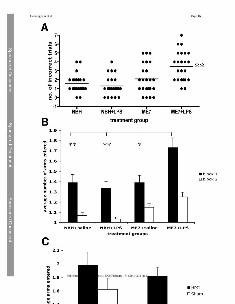
Spatial Reference Memory in the Y-MazeNormal brain homogenate and ME7 animals were treated with saline or LPS (n = 23 for NBH,n = 24 for ME7) and tested in the visuospatial reference memory paddling Y-maze, between3 and 7 hours, to test for LPS-induced learning impairments. The ME7 animals challengedwith LPS made more incorrect arm entries across 10 trials than any of the other groups (Figure3A; p < .0001 Kruskal-Wallis test). Dunn's multiple comparison post hoc tests revealed ME7+ LPS animals to be significantly different from all other groups (p < .05). Plotted as the averagenumber of arms entered, in two blocks of five trials (Figure 3B), ME7 + LPS animals madeconsiderably more errors in block 1 and this persisted to a lesser degree in block 2. Theseanimals were hypoactive at this time, so increased arm entries cannot be explained by increasedactivity. Despite the sickness of the NBH + LPS animals, their learning was not significantlyimpaired compared with NBH + saline animals (Figure 3B). This simple Y-maze has beendesigned to allow learning to be achieved in a single session, while LPS effects persist, toeliminate state-dependent effects. Thus, comparison of block 2 with block 1 (paired t tests p< .0013) is suggestive of some learning in all groups. Repeated measures ANOVA comparisonof arm entries by ME7 + LPS and ME7 animals shows main effects of treatment (F = 9.66, p= .0032, df = 1,46) and of trial (F = 9.9, p < .0001, df = 9,414) but that for NBH + saline andNBH + LPS shows an effect of trial (F = 24.88, p < .0001, df = 9,396) but no effect of treatment(F = 1.62, p = .2091, df = 1,44). Thus, LPS has no impact on the accuracy of NBH animals inthis maze task but does impair that of ME7 animals.
Acquisition of correct responding in this maze was hippocampal-dependent (Figure 3C). Sham-operated animals quickly learned the location of the exit while hippocampal-lesioned animalscontinued responding at chance levels (50%). A repeated measures ANOVA analysis showsa significant effect of treatment (F = 10.78, p = .0039, df = 1,19).
Cunningham et al. Page 6
Published as: Biol Psychiatry. 2009 February 15; 65(4): 304–312.
Sponsored Docum
ent Sponsored D
ocument
Sponsored Docum
ent
Acute LPS-Induced Changes at 15 WeeksHorizontal Bar and Inverted Screen—All animal groups performed the horizontal bartask well at 14 weeks (Figure 4A). Upon IP challenge with LPS, ME7 animals showed adecrease in performance relative to the NBH + LPS group. Repeated measures ANOVA revealsan interaction of time and treatment (F = 3.05, p = .0085, df = 6,114) and Bonferroni post hoctest at 6 hours reveals significant impairment of ME7 + LPS (p < .05). ME7 + saline animalsperformed well throughout the experiment. Since the data were nonparametric, a scatter dotplot is also shown (Figure 4B). Nine out of 12 NBH + LPS animals reached a criterion of 60(median = 60), while 9 out of 14 ME7 + LPS animals failed to reach this criterion (median =38). The proportions of each group that reached criterion are significantly different by one-tailed Fisher exact test with Tocher's modification. Furthermore, when the times are comparedby Kruskal-Wallis, with a Dunn post hoc test, ME7 + LPS and NBH + LPS are significantlydifferent (p < .05).
All animal groups showed good performance on the inverted screen at 14 weeks (Figure 4C).Lipopolysaccharide induced a clear decrease in times in ME7 animals compared with the samechallenge in NBH animals and to saline challenges in ME7 animals (Bonferroni post hoc testat 6 hours, p < .001). This decrease is most obvious at 6 hours but is still apparent at 24 hoursand repeated measures ANOVA reveals a main effect of treatment (F = 5.35, p = .0088, df =2,39) and an interaction of time and treatment (F = 4.49, p = .0004, df = 6,117). We also providescatter dot plots of the 6-hour data (Figure 4D). When these data are compared by Kruskal-Wallis, with Dunn post hoc tests, there is a statistically significant difference between ME7 +LPS and NBH + LPS (p < .05).
All groups showed good recovery on both tasks by 15 weeks. These data demonstrate a greateracute effect of LPS on motor coordination and muscle strength in ME7 animals than in NBHcontrol animals. When administered at 12 weeks postinoculation with prion disease, LPS didnot have any acute effects on motor coordination or muscle strength (data not shown).
Longitudinal Effects of Systemic Infection on Disease ProgressionAs previously shown, there is a clear impairment in performance on the horizontal bar and theinverted screen in the ME7 groups between 16 and 20 weeks (Figure 5). ME7 animalschallenged with 500 μg/kg LPS between 14 and 15 weeks subsequently showed moreimpairment on the horizontal bar than ME7 + saline animals, and their performance on thistask remained worse throughout the experiment (Figure 5A). A repeated measures ANOVAanalysis of the two ME7 groups revealed main effects of treatment (F = 6.04, p < .05, df =1,65) and of time (F = 43.77, p < .0001, df = 4,260) and an interaction of treatment and time(F = 3.06, p < .05, df = 4,260).
ME7 animals challenged with 500 μg/kg LPS between 14 and 15 weeks also subsequentlyshowed an earlier onset of impairment on the inverted screen than ME7 + saline animals, andthis performance remained worse than that for ME7 + saline animals throughout the experiment(Figure 5B). A repeated measures ANOVA analysis of the two ME7 groups revealed maineffects of treatment (F = 6.41, p < .05, df = 1,47) and of time (F = 53.86, p < .0001, df = 4,188)and an interaction of treatment and time (F = 2.76, p < .05, df = 4,188).
ME7 animals challenged with LPS 500 μg/kg between 14 and 15 weeks also subsequentlyshowed an earlier decrease in open field locomotor hyperactivity. Hyperactivity of both ME7groups relative to NBH control animals is clear in Figure 5C, but this is less marked in theME7 + LPS group, suggesting the beginning of the late-stage decline. Repeated measuresANOVA analysis of the two ME7 groups shows a main effect of time (F = 12.86, p < .0001,df = 6,228) and a main effect of treatment (F = 9.15, p = .0044, df = 1,38).
Cunningham et al. Page 7
Published as: Biol Psychiatry. 2009 February 15; 65(4): 304–312.
Sponsored Docum
ent Sponsored D
ocument
Sponsored Docum
ent

All of these LPS-induced decrements are temporally separated from the acute LPS effects onthese tasks shown in Figure 4 (A,B) and together these data demonstrate that a single, transient,peripheral inflammatory event can hasten the disease process, accelerating the appearance ofthree different neurological signs of disease.
DiscussionIn the current study, we have shown that systemic LPS during presymptomatic chronicneurodegenerative disease can induce exaggerated CNS inflammation and sickness behaviorresponses compared with those in normal animals. Acute symptoms also now extend beyondthe typical spectrum of sickness behavior responses to include exacerbation of symptoms morecommonly associated with the underlying disease: acute cognitive impairments (LPS at 12weeks) and acute impairments of motor coordination and muscle strength (LPS at 15 weeks).In addition, a single LPS challenge accelerates and exacerbates the progression of theunderlying disease. Thus, on multiple levels, systemic inflammation impacts negatively onchronic neurodegeneration.
Exaggerated Sickness Behavior and Microglial PrimingWe had previously shown that at 18 weeks postinoculation with the ME7 strain of prion disease,these animals were susceptible to exaggerated sickness behavior responses to LPS (13). Suchresponses were absent at 8 weeks postinoculation, before large numbers of microglia becomeactivated. Thus, microglia that are activated by the process of neurodegeneration (18,28) oraging (29,30) but which do not synthesize robust levels of proinflammatory cytokines aresusceptible to phenotypic switching by systemic inflammatory insults. We demonstratedIL-1β synthesis specifically in ME7 hippocampal microglial cells after systemic LPS challengebut not in NBH + LPS animals or in ME7 animals injected with saline, despite markedneurodegeneration and extracellular prion protein (PrPSc) deposits in the latter (18). In thecurrent study, we have shown that this phenotypic switching, heightened cytokine production,and exaggerated sickness behavior are also induced at 12 weeks postinoculation with priondisease in the absence of neuronal loss but at the onset of hippocampal CA1 synaptic loss andhippocampal-dependent impairments (31,32). This, therefore, places subjects at risk of diseaseexacerbation from an early or even presymptomatic stage of disease. Whether the switch fromthe primed state to that expressing IL-1β is key to this disease exacerbation remains to beestablished, but given the high sensitivity of IL-1 type I receptors (33), neuroanatomicallydefined, microglial IL-1β protein expression constitutes a biologically significant event.
Acute Cognitive ImpairmentsLipopolysaccharide induced acute cognitive impairments in ME7 animals but not in NBHanimals. Primed microglial cells are limited to the hippocampus and limbic system at this stageof disease (18,31), and therefore we hypothesized that hippocampal-dependent tasks would besensitive to these insults. We have shown here that correct responding in the Y-maze spatialreference memory task is hippocampal-dependent. Thus, we have induced an acute memorydeficit in LPS-treated ME7 animals that is hippocampal-dependent and is absent in LPS-treatedNBH animals, despite obvious sickness behavior. Lipopolysaccharide is reported to affectcognitive function in young and aged mice (34–37), but there are potential confounding factorsin these studies, directly referred to by the authors. The novel shallow water Y-maze taskdeveloped here assesses incorrect responses rather than time taken to solve the maze, thusminimizing such confounding factors. The cognitive impairment that we describe upon acuteinflammatory insult during chronic neurodegeneration and similar impairments reported inaged animals challenged with LPS (38,39) have parallels with the acute cognitive impairmentsseen in episodes of delirium during dementia. Delirium is an acute and transient confusionalstate that commonly occurs after infection, surgery, or injury (40). Delirium is remarkably
Cunningham et al. Page 8
Published as: Biol Psychiatry. 2009 February 15; 65(4): 304–312.
Sponsored Docum
ent Sponsored D
ocument
Sponsored Docum
ent

prevalent in the aged and demented population: 10% of over 65s admitted to the emergencyroom (41) and 22% to 89% in demented patients (42). The etiology of delirium is unknownand given its very high frequency in aged and demented populations, it is a major researchquestion. The novel Y-maze easily distinguishes between sham and hippocampal-lesionedanimals. Larger group sizes are required to distinguish between ME7 and ME7 + LPS animals,reflecting interanimal variability in the onset of cognitive changes in ME7 animals. This mirrorsthe difficulties in distinguishing chronic and acute impairments in dementia and delirium. Wepropose that further refinement of the model system used here will be useful in the study ofhow systemic inflammation and underlying dementia interact to produce these episodes.
Acute Exacerbation Leads to Acceleration of Chronic DiseaseDementia is the single biggest risk factor for episodes of delirium and systemic inflammationis a major trigger in these patients. It is also now clear that a single episode of delirium ispredictive of an accelerated rate of clinical decline in demented patients (9–11). This impliesthat the insults that induce episodes of delirium are also responsible for an acceleration of thedisease process. We previously showed that a single challenge with LPS was sufficient toexacerbate neuronal death during prion disease (18). Repeated challenges with LPS have beenshown to exacerbate axonal damage and disease progression in a transgenic model ofamyotrophic lateral sclerosis (43). Similarly, there is evidence for exacerbation of features ofpathology in a triple transgenic model of Alzheimer's disease (44) and for exacerbation ofinflammation (45) and amyloid deposition (46) in AD models. In the current study, we haveshown that a single challenge with LPS is sufficient to accelerate and exacerbate permanentloss of neurological function. We observed acute impairments of motor function during thesystemic inflammatory episode, recovery of normal function postinflammation, andaccelerated onset of progressive and permanent neurological changes. This mirrors theaccelerated cognitive decline of demented patients after recovery from acute symptoms ofdelirium (47) and indicates that systemic inflammatory insults accelerate disease progression.It is of note that LPS administered at 12 weeks postinoculation with prion disease did not affectmotor coordination, consistent with the idea that as pathology becomes more global, later indisease, the range of functions that may be affected by systemic inflammation becomes broader.It is of interest to examine whether pathology and behavioral symptoms in Tg2576 and CRND8mice are similarly affected by systemic inflammation. However, that our hypothesis and thecurrent model have validity in human neurodegenerative disease has been demonstrated inrecent and ongoing clinical studies: carer-reported systemic infection and elevatedconcentrations of IL-1β in sera were independently associated with accelerated cognitivedecline in Alzheimer's disease patients (48).
ConclusionWe have shown a clear systemic inflammation-induced exacerbation of neurodegenerativedisease. The mechanism of this exacerbation remains unclear. It is well known that peripheralinflammation can activate CNS centers by a number of routes, including the circumventricularorgans, vagal afferents, and the brain endothelium (see 12 for review). Bacterial endotoxin candirectly activate the brain endothelium (49,50), but it is well known that behavioral and CNSinflammatory changes can be induced by systemic administration of proinflammatorycytokines (12). Furthermore, it has recently been shown that systemic IL-1β exacerbatesneurodegeneration and motor symptoms in a rat model of Parkinson's disease (51). Due to thelarge numbers of animals required to overcome interindividual variability in the current study,mechanistic analysis was impractical. However, preliminary investigations in normal animalsshow that indomethacin does not inhibit CNS proinflammatory cytokine transcription despiteclear effects on LPS-induced behavioral changes and on prostaglandins (52). There remainmany unanswered questions about the nature of the interactions between systemic and CNSinflammatory compartments in this model and in human disease. Studies addressing these
Cunningham et al. Page 9
Published as: Biol Psychiatry. 2009 February 15; 65(4): 304–312.
Sponsored Docum
ent Sponsored D
ocument
Sponsored Docum
ent

interactions and the relative roles of endotoxin, cytokines, and prostaglandins will haveimplications for our understanding of the role of inflammation in neurodegenerative diseaseand in episodes of delirium.
Supplementary dataRefer to Web version on PubMed Central for supplementary material.
Supplementary dataRefer to Web version on PubMed Central for supplementary material.
AcknowledgementsWe gratefully acknowledge the financial support of the Wellcome Trust.
The authors report no biomedical financial interests or potential conflicts of interest.
References1. Akiyama H. Barger S. Barnum S. Bradt B. Bauer J. Cole G.M. Inflammation and Alzheimer's disease.
Neurobiol Aging 2000;21:383–421. [PubMed: 10858586]2. Etminan M. Gill S. Samii A. Effect of non-steroidal anti-inflammatory drugs on risk of Alzheimer's
disease: Systematic review and meta-analysis of observational studies. BMJ 2003;327:128. [PubMed:12869452]
3. Aisen P.S. The potential of anti-inflammatory drugs for the treatment of Alzheimer's disease. LancetNeurol 2002;1:279–284. [PubMed: 12849425]
4. Firuzi O. Pratico D. Coxibs and Alzheimer's disease: Should they stay or should they go? Ann Neurol2006;59:219–228. [PubMed: 16402383]
5. McGeer P.L. McGeer E.G. NSAIDs and Alzheimer disease: Epidemiological, animal model andclinical studies. Neurobiol Aging 2007;28:639–647. [PubMed: 16697488]
6. Lerner A.J. Hedera P. Koss E. Stuckey J. Friedland R.P. Delirium in Alzheimer disease. AlzheimerDis Assoc Disord 1997;11:16–20. [PubMed: 9071440]
7. George J. Bleasdale S. Singleton S.J. Causes and prognosis of delirium in elderly patients admitted toa district general hospital. Age Ageing 1997;26:423–427. [PubMed: 9466291]
8. Rahkonen T. Makela H. Paanila S. Halonen P. Sivenius J. Sulkava R. Delirium in elderly people withoutsevere predisposing disorders: Etiology and 1-year prognosis after discharge. Int Psychogeriatr2000;12:473–481. [PubMed: 11263714]
9. Murray A.M. Levkoff S.E. Wetle T.T. Beckett L. Cleary P.D. Schor J.D. Acute delirium and functionaldecline in the hospitalized elderly patient. J Gerontol 1993;48:M181–M186. [PubMed: 8366260]
10. Rockwood K. Cosway S. Carver D. Jarrett P. Stadnyk K. Fisk J. The risk of dementia and death afterdelirium. Age Ageing 1999;28:551–556. [PubMed: 10604507]
11. Pitkala K.H. Laurila J.V. Strandberg T.E. Tilvis R.S. Prognostic significance of delirium in frail olderpeople. Dement Geriatr Cogn Disord 2005;19:158–163. [PubMed: 15627764]
12. Dantzer R. Cytokine-induced sickness behaviour: A neuroimmune response to activation of innateimmunity. Eur J Pharmacol 2004;500:399–411. [PubMed: 15464048]
13. Combrinck M.I. Perry V.H. Cunningham C. Peripheral infection evokes exaggerated sicknessbehaviour in pre-clinical murine prion disease. Neuroscience 2002;112:7–11. [PubMed: 12044467]
14. Betmouni S. Perry V.H. Gordon J.L. Evidence for an early inflammatory response in the centralnervous system of mice with scrapie. Neuroscience 1996;74:1–5. [PubMed: 8843071]
15. Cunningham C. Boche D. Perry V.H. Transforming growth factor beta1, the dominant cytokine inmurine prion disease: Influence on inflammatory cytokine synthesis and alteration of vascularextracellular matrix. Neuropathol Appl Neurobiol 2002;28:107–119. [PubMed: 11972797]
Cunningham et al. Page 10
Published as: Biol Psychiatry. 2009 February 15; 65(4): 304–312.
Sponsored Docum
ent Sponsored D
ocument
Sponsored Docum
ent

16. Cunningham C. Wilcockson D.C. Boche D. Perry V.H. Comparison of inflammatory and acute-phaseresponses in the brain and peripheral organs of the ME7 model of prion disease. J Virol2005;79:5174–5184. [PubMed: 15795301]
17. Walsh D.T. Betmouni S. Perry V.H. Absence of detectable IL-1beta production in murine priondisease: A model of chronic neurodegeneration. J Neuropathol Exp Neurol 2001;60:173–182.[PubMed: 11273005]
18. Cunningham C. Wilcockson D.C. Campion S. Lunnon K. Perry V.H. Central and systemic endotoxinchallenges exacerbate the local inflammatory response and increase neuronal death during chronicneurodegeneration. J Neurosci 2005;25:9275–9284. [PubMed: 16207887]
19. Deacon R.M. Bannerman D.M. Kirby B.P. Croucher A. Rawlins J.N. Effects of cytotoxichippocampal lesions in mice on a cognitive test battery. Behav Brain Res 2002;133:57–68. [PubMed:12048174]
20. Deacon R.M. Burrowing in rodents: A sensitive method for detecting behavioral dysfunction. NatProtoc 2006;1:118–121. [PubMed: 17406222]
21. Felton L.M. Cunningham C. Rankine E.L. Waters S. Boche D. Perry V.H. MCP-1 and murine priondisease: Separation of early behavioural dysfunction from overt clinical disease. Neurobiol Dis2005;20:283–295. [PubMed: 15886005]
22. Deacon R.M. Cholerton L.L. Talbot K. Nair-Roberts R.G. Sanderson D.J. Romberg C. Age-dependentand -independent behavioral deficits in Tg2576 mice. Behav Brain Res 2008;189:126–138. [PubMed:18261809]
23. Deacon R.M. Rawlins J.N. Learning impairments of hippocampal-lesioned mice in a paddling pool.Behav Neurosci 2002;116:472–478. [PubMed: 12049328]
24. Reisel D. Bannerman D.M. Schmitt W.B. Deacon R.M. Flint J. Borchardt T. Spatial memorydissociations in mice lacking GluR1. Nat Neurosci 2002;5:868–873. [PubMed: 12195431]
25. Kondziela W. A new method for the measurement of muscle relaxation in white mice [in German].Arch Int Pharmacodyn Ther 1964;152:277–284. [PubMed: 14265648]
26. Cunningham C. Campion S. Teeling J. Felton L. Perry V.H. The sickness behaviour and CNSinflammatory mediator profile induced by systemic challenge of mice with synthetic double-strandedRNA (poly I:C). Brain Behav Immun 2007;21:490–502. [PubMed: 17321719]
27. Greer G.G. Rietschel E.T. Lipid A-induced tolerance and hyperreactivity to hypothermia in mice.Infect Immun 1978;19:357–368. [PubMed: 631875]
28. Perry V.H. Newman T.A. Cunningham C. The impact of systemic infection on the progression ofneurodegenerative disease. Nat Rev Neurosci 2003;4:103–112. [PubMed: 12563281]
29. Frank M.G. Barrientos R.M. Biedenkapp J.C. Rudy J.W. Watkins L.R. Maier S.F. mRNA up-regulation of MHC II and pivotal pro-inflammatory genes in normal brain aging. Neurobiol Aging2006;27:717–722. [PubMed: 15890435]
30. Perry V.H. Matyszak M.K. Fearn S. Altered antigen expression of microglia in the aged rodent CNS.Glia 1993;7:60–67. [PubMed: 8423063]
31. Guenther K. Deacon R.M. Perry V.H. Rawlins J.N. Early behavioural changes in scrapie-affectedmice and the influence of dapsone. Eur J Neurosci 2001;14:401–409. [PubMed: 11553290]
32. Cunningham C. Deacon R. Wells H. Boche D. Waters S. Diniz C.P. Synaptic changes characterizeearly behavioural signs in the ME7 model of murine prion disease. Eur J Neurosci 2003;17:2147–2155. [PubMed: 12786981]
33. Sims J.E. Gayle M.A. Slack J.L. Alderson M.R. Bird T.A. Giri J.G. Interleukin 1 signaling occursexclusively via the type I receptor. Proc Natl Acad Sci U S A 1993;90:6155–6159. [PubMed:8327496]
34. Sparkman N.L. Kohman R.A. Garcia A.K. Boehm G.W. Peripheral lipopolysaccharide administrationimpairs two-way active avoidance conditioning in C57BL/6J mice. Physiol Behav 2005;85:278–288.[PubMed: 15936787]
35. Sparkman N.L. Kohman R.A. Scott V.J. Boehm G.W. Bacterial endotoxin-induced behavioralalterations in two variations of the Morris water maze. Physiol Behav 2005;86:244–251. [PubMed:16115658]
Cunningham et al. Page 11
Published as: Biol Psychiatry. 2009 February 15; 65(4): 304–312.
Sponsored Docum
ent Sponsored D
ocument
Sponsored Docum
ent

36. Sparkman N.L. Martin L.A. Calvert W.S. Boehm G.W. Effects of intraperitoneal lipopolysaccharideon Morris maze performance in year-old and 2-month-old female C57BL/6J mice. Behav Brain Res2005;159:145–151. [PubMed: 15795008]
37. Kohman R.A. Tarr A.J. Byler S.L. Boehm G.W. Age increases vulnerability to bacterial endotoxin-induced behavioral decrements. Physiol Behav 2007;91:561–565. [PubMed: 17499821]
38. Barrientos R.M. Higgins E.A. Biedenkapp J.C. Sprunger D.B. Wright-Hardesty K.J. Watkins L.R.Peripheral infection and aging interact to impair hippocampal memory consolidation. NeurobiolAging 2006;27:723–732. [PubMed: 15893410]
39. Chen J. Buchanan J.B. Sparkman N.L. Godbout J.P. Freund G.G. Johnson R.W. Neuroinflammationand disruption in working memory in aged mice after acute stimulation of the peripheral innateimmune system. Brain Behav Immun 2008;22:301–311. [PubMed: 17951027]
40. Brown T.M. Boyle M.F. Delirium. BMJ 2002;325:644–647. [PubMed: 12242179]41. Elie M. Rousseau F. Cole M. Primeau F. McCusker J. Bellavance F. Prevalence and detection of
delirium in elderly emergency department patients. CMAJ 2000;163:977–981. [PubMed: 11068569]42. Fick D.M. Agostini J.V. Inouye S.K. Delirium superimposed on dementia: A systematic review. J
Am Geriatr Soc 2002;50:1723–1732. [PubMed: 12366629]43. Nguyen M.D. D'Aigle T. Gowing G. Julien J.P. Rivest S. Exacerbation of motor neuron disease by
chronic stimulation of innate immunity in a mouse model of amyotrophic lateral sclerosis. J Neurosci2004;24:1340–1349. [PubMed: 14960605]
44. Kitazawa M. Oddo S. Yamasaki T.R. Green K.N. LaFerla F.M. Lipopolysaccharide-inducedinflammation exacerbates tau pathology by a cyclin-dependent kinase 5-mediated pathway in atransgenic model of Alzheimer's disease. J Neurosci 2005;25:8843–8853. [PubMed: 16192374]
45. Sly L.M. Krzesicki R.F. Brashler J.R. Buhl A.E. McKinley D.D. Carter D.B. Chin J.E. Endogenousbrain cytokine mRNA and inflammatory responses to lipopolysaccharide are elevated in the Tg2576transgenic mouse model of Alzheimer's disease. Brain Res Bull 2001;56:581–588. [PubMed:11786245]
46. Brugg B. Dubreuil Y.L. Huber G. Wollman E.E. Delhaye-Bouchaud N. Mariani J. Inflammatoryprocesses induce beta-amyloid precursor protein changes in mouse brain. Proc Natl Acad Sci U S A1995;92:3032–3035. [PubMed: 7708769]
47. Jackson J.C. Gordon S.M. Hart R.P. Hopkins R.O. Ely E.W. The association between delirium andcognitive decline: A review of the empirical literature. Neuropsychol Rev 2004;14:87–98. [PubMed:15264710]
48. Holmes C. El-Okl M. Williams A.L. Cunningham C. Wilcockson D. Perry V.H. Systemic infection,interleukin 1beta, and cognitive decline in Alzheimer's disease. J Neurol Neurosurg Psychiatry2003;74:788–789. [PubMed: 12754353]
49. Chakravarty S. Herkenham M. Toll-like receptor 4 on nonhematopoietic cells sustains CNSinflammation during endotoxemia, independent of systemic cytokines. J Neurosci 2005;25:1788–1796. [PubMed: 15716415]
50. Singh A.K. Jiang Y. How does peripheral lipopolysaccharide induce gene expression in the brain ofrats? Toxicology 2004;201:197–207. [PubMed: 15297033]
51. Godoy M.C. Tarelli R. Ferrari C.C. Sarchi M.I. Pitossi F.J. Central and systemic IL-1 exacerbatesneurodegeneration and motor symptoms in a model of Parkinson's disease. Brain 2008;131(Pt 7):1880–1894. [PubMed: 18504291]
52. Teeling J.L. Felton L.M. Deacon R.M. Cunningham C. Rawlins J.N. Perry V.H. Sub-pyrogenicsystemic inflammation impacts on brain and behavior, independent of cytokines. Brain Behav Immun2007;21:836–850. [PubMed: 17367989]
Cunningham et al. Page 12
Published as: Biol Psychiatry. 2009 February 15; 65(4): 304–312.
Sponsored Docum
ent Sponsored D
ocument
Sponsored Docum
ent

Cunningham et al. Page 13
Published as: Biol Psychiatry. 2009 February 15; 65(4): 304–312.
Sponsored Docum
ent Sponsored D
ocument
Sponsored Docum
ent

Figure 1.Acute sickness behavioral responses to intraperitoneal LPS. (A) Weight burrowed over 2 hoursby C57BL/6 mice at baseline, 2 hours, 24 hours, and 48 hours post-IP challenge with 100 μg/kg LPS or sterile saline. ME7 and NBH animals were 12 weeks postinoculation with the ME7murine scrapie strain or normal brain homogenate, respectively. Statistical significance wasdetermined by planned comparisons by one-way ANOVA with Bonferroni post hoc test at 24hours. (B) Distance traveled in an open field by the same animals at baseline, 5 hours, 24 hours,and 48 hours postchallenge with LPS. Statistical significance was determined by plannedcomparisons by one-way ANOVA with Bonferroni post hoc test at 5 hours. (C) Core bodytemperature of the same animals at −24, 0, 5, and 24 hours postchallenge with LPS or saline.Planned comparison by one-way ANOVA at 5 hours was performed. All data are plotted asmean ± SEM and n = 9 in all groups. *p < .05. ANOVA, analysis of variance; IP, intraperitoneal;LPS, lipopolysaccharide; NBH, normal brain homogenate.
Cunningham et al. Page 14
Published as: Biol Psychiatry. 2009 February 15; 65(4): 304–312.
Sponsored Docum
ent Sponsored D
ocument
Sponsored Docum
ent
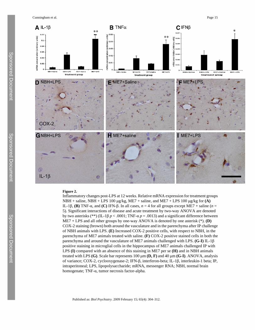
Figure 2.Inflammatory changes post-LPS at 12 weeks. Relative mRNA expression for treatment groupsNBH + saline, NBH + LPS 100 μg/kg, ME7 + saline, and ME7 + LPS 100 μg/kg for (A)IL-1β, (B) TNF-α, and (C) IFN-β. In all cases, n = 4 for all groups except ME7 + saline (n =5). Significant interactions of disease and acute treatment by two-way ANOVA are denotedby two asterisks (**) (IL-1β p < .0001; TNF-α p = .0013) and a significant difference betweenME7 + LPS and all other groups by one-way ANOVA is denoted by one asterisk (*). (D)COX-2 staining (brown) both around the vasculature and in the parenchyma after IP challengeof NBH animals with LPS. (E) Increased COX-2 positive cells, with respect to NBH, in theparenchyma of ME7 animals treated with saline. (F) COX-2 positive stained cells in both theparenchyma and around the vasculature of ME7 animals challenged with LPS. (G-I) IL-1βpositive staining in microglial cells in the hippocampus of ME7 animals challenged IP withLPS (I) compared with an absence of this staining in ME7 per se (H) and in NBH animalstreated with LPS (G). Scale bar represents 100 μm (D, F) and 40 μm (G-I). ANOVA, analysisof variance; COX-2, cyclooxygenase-2; IFN-β, interferon-beta; IL-1β, interleukin-1 beta; IP,intraperitoneal; LPS, lipopolysaccharide; mRNA, messenger RNA; NBH, normal brainhomogenate; TNF-α, tumor necrosis factor-alpha.
Cunningham et al. Page 15
Published as: Biol Psychiatry. 2009 February 15; 65(4): 304–312.
Sponsored Docum
ent Sponsored D
ocument
Sponsored Docum
ent
Cunningham et al. Page 16
Published as: Biol Psychiatry. 2009 February 15; 65(4): 304–312.
Sponsored Docum
ent Sponsored D
ocument
Sponsored Docum
ent

Figure 3.Spatial learning in the paddling Y-maze: normal and prion-diseased animals and hippocampal-lesioned and sham-operated control animals. (A) Cognitive performance of animals in thepaddling Y-maze at 12 weeks postinoculation with ME7 (100 μg/kg LPS or saline) and NBH(500 μg/kg LPS or saline) 3 to 7 hours after IP challenge. This plot represents the total numberof incorrect trials (out of 10) by each treatment group. Each animal is shown as an individualpoint and the median is illustrated by the bar. Kruskal-Wallis ANOVA revealed a differencein the medians (p < .0001) and pairwise comparisons showed ME7 + LPS to be significantlydifferent to all other groups (**), n = 23 in the case of both NBH groups and n = 24 for bothME7 groups. (B) The same data are plotted as two successive blocks of five trials, showingthe average number of arms entered including the final correct one. Block 2 is compared withblock 1 by Student t tests in all cases (*p < .05). The average number of arms entered in block1 by each group was also compared by ANOVA with Bonferroni post hoc test (*p < .05, **p< .01). (C) Hippocampal-lesioned animals compared with sham-operated control animalstested across two blocks of five trials on the same paddling Y-maze. A repeated measuresANOVA analysis shows a significant effect of treatment (F = 10.78, p = .0039, df = 1,19) andof trial (F = 3.54, p = .0005, df = 9,171). ANOVA, analysis of variance; IP, intraperitoneal;LPS, lipopolysaccharide; NBH, normal brain homogenate.
Cunningham et al. Page 17
Published as: Biol Psychiatry. 2009 February 15; 65(4): 304–312.
Sponsored Docum
ent Sponsored D
ocument
Sponsored Docum
ent

Figure 4.Acute effects on motor tasks induced by LPS in normal and prion-diseased animals. (A)Performance on a horizontal bar at 14 weeks postinoculation with ME7 or NBH, followed byperformance at 6 hours, 24 hours, and 1 week after IP LPS (500 μg/kg) or saline challenges inthese animals. Data were plotted as mean ± SEM and analyzed by repeated measures ANOVABonferroni post hoc tests (p < .05 between ME7 + LPS and NBH + LPS, n = 13–15 for allgroups). (B) Replot of the 6-hour post-LPS time point showing the median and full range of
Cunningham et al. Page 18
Published as: Biol Psychiatry. 2009 February 15; 65(4): 304–312.
Sponsored Docum
ent Sponsored D
ocument
Sponsored Docum
ent
values. These data were compared by Kruskal-Wallis test with Dunn's post hoc test (*p < .05).(C) Performance on an inverted screen at the same time points as described above. Data areshown as mean ± SEM and were analyzed by repeated measures ANOVA with Bonferronipost hoc test (p < .001 between ME7 + LPS and NBH + LPS, n = 13–15 for all groups). (D)Replot of the inverted screen 6-hour post-LPS time point showing the median and full rangeof values. These data were compared by Kruskal-Wallis test with Dunn's post hoc test (*p < .05). ANOVA, analysis of variance; IP, intraperitoneal; LPS, lipopolysaccharide; NBH, normalbrain homogenate.
Cunningham et al. Page 19
Published as: Biol Psychiatry. 2009 February 15; 65(4): 304–312.
Sponsored Docum
ent Sponsored D
ocument
Sponsored Docum
ent

Cunningham et al. Page 20
Published as: Biol Psychiatry. 2009 February 15; 65(4): 304–312.
Sponsored Docum
ent Sponsored D
ocument
Sponsored Docum
ent

Figure 5.Longitudinal effects of LPS at 14 to 15 weeks on the progression of prion disease. (A)Performance on the horizontal bar for 6 successive weeks post-IP challenge with LPS (500μg/kg) or saline at 15 weeks postinoculation with ME7 or NBH. Data are shown as mean ±SEM (n = 25 for NBH + LPS, n = 34 for ME7 + Saline, and n = 35 for ME7 + LPS). ME7 +LPS and ME7 + saline data were compared by repeated measures ANOVA across the 6 weeksshown and showed both main effects of treatment and time and an interaction of treatment andtime (* denotes p = .0166 for the main effect of treatment). (B) Performance of the same animalson the inverted screen for 5 successive weeks post-IP challenge with LPS (500 μg/kg) or saline.Data are shown as mean ± SEM (n = 25 for NBH + LPS, n = 34 for ME7 + Saline, and n = 35for ME7 + LPS). ME7 + Saline and ME7 + LPS data were compared by repeated measuresANOVA across the 5 weeks shown and showed both main effects of treatment and time andan interaction of treatment and time (** denotes p = .0147 for the main effect of treatment).(C) Open field activity for 7 successive weeks post-IP challenge with LPS 500 μg/kg. Data areshown as mean ± SEM (n = 19, n = 20). Repeated measures ANOVA of ME7 groups showedmain effects of time and treatment (* denotes p = .0044 for the main effect of treatment).ANOVA, analysis of variance; IP, intraperitoneal; LPS, lipopolysaccharide; NBH, normalbrain homogenate.
Cunningham et al. Page 21
Published as: Biol Psychiatry. 2009 February 15; 65(4): 304–312.
Sponsored Docum
ent Sponsored D
ocument
Sponsored Docum
ent