biomechanics of locomotion in sharks, rays, and chimaeras
TRANSCRIPT

5Biomechanics of Locomotion in Sharks, Rays, and Chimaeras
Anabela M.R. Maia, Cheryl A.D. Wilga, and George V.Lauder
CONTENTS5.1 Introduction 125
5.1.1 Approaches to Studying Locomotion in Chondrichthyans 1255.1.2 Diversity of Locomotory Modes in Chondrichthyans 1275.1.3 Body Form and Fin Shapes 127
5.2 Locomotion in Sharks 1285.2.1 Function of the Body during Steady Locomotion and Vertical Maneuvering 1285.2.2 Function of the Caudal Fin during Steady Locomotion and Vertical Maneuvering 1305.2.3 Function of the Pectoral Fins during Locomotion 134
5.2.3.1 Anatomy of the Pectoral Fins 1345.2.3.2 Role of the Pectoral Fins during Steady Swimming 1365.2.3.3 Role of the Pectoral Fins during Vertical Maneuvering 1385.2.3.4 Function of the Pectoral Fins during Benthic Station-Holding 1395.2.3.5 Motor Activity in the Pectoral Fins 139
5.2.4 Routine Maneuvers and Escape Responses 1405.2.5 Synthesis 141
5.3 Locomotion in Skates and Rays 1425.4 Locomotion in Holocephalans 1455.5 Material Properties of Chondrichthyan Locomotor Structures 1465.6 Future Directions 147Acknowledgments 148References 148
5.1 Introduction
The body form of sharks is notable for the distinctiveheterocercal tail with external morphological asymme-try present in most taxa and the ventrolateral winglikepectoral fins extending laterally from the body (Figure5.1)that give the appearance of powerful yet effortlesslocomotion. In contrast, expansion of the pectoral finscoupled with a dorsoventrally flattened body in rays andskates resulted in modification of locomotor mode fromtrunk based to pectoral based, while the chimaera bodyshape is similar to that of actinopterygian fishes in termsof lateral compression. These features are distinct fromthe variety of body forms present in actinopterygianfishes (Lauder, 2000) and have long been of interest toresearchers wishing to understand the functional designof sharks (Aleev,1969;Garman, 1913;Grove and Newell.1936;Harris, 1936;Magnan, 1929;Thomson, 1971).
-
5.1.1 Approaches to StudyingLocomotion in Chondrichthyans
Historically, many attempts have been made to under-stand the function of the median and paired fins insharks and rays, and these studies have included workwith models (Affleck. 1950;Harris, 1936;Simons, 1970),experiments on fins removed from the body (Aleev,1969;Alexander. 1965;Daniel, 1922;Harris, 1936),andquantification of body form and basic physical model-ing (Thomson, 1976;Thomson and Simanek, 1977).Morerecently, direct quantification of fin movement usingvideography has allowed a better understanding of finconformation and movement (Ferry and Lauder, 1996;Fish and Shannahan, 2000;Flammang, 2010;Wilga andLauder, 2000),although such studies have to date beenlimited to relatively few species. Obtaining high-reso-lution, three-dimensional (3D)data on patterns of sharkfin motion is a difficult task, and these studies have
125

126 Biology of Sharks and Their Relatives
Thunniform
Axial Undulatory propulsors
1
Subcaranglform
H
Subcarangiform
3
~':;~4
Appendage Propulsors
Undulatory
0,Oscillatory
5
6
FIGURE 5.1Propulsion mechanisms in chondrichthyans. Numbers indicate body groups (see text). E, epicaudallobe; H, hypochordal lobe: S, subterminallobe. (Based on Webb, 1984; Webb and Blake, 1985.)
been confined to a highly controlled laboratory environ-ment where sharks swim in a recirculating flow tank.Although locomotion of sharks and rays under these con-ditions does not allow the range of behaviors seen in thewild, the ability to obtain data from precisely controlledhorizontal swimming as well as specific maneuveringbehaviors has been vital to both testing classical hypoth-eses of fin function and to the discovery of new aspectsof locomotory mechanics. A key general lesson learnedfrom recent experimental kinematic and hydrodynamicanalyses of shark locomotion is the value of understand-ing the 3D pattern of fin movement and the requirementfor experimental laboratory studies that permit detailedanalyses of fin kinematics and hydrodynamics.Two new laboratory-based approaches in recent
years have been particularly fruitful in clarifying thebiomechanics of shark locomotion. Chief among thesehas been the use of two- and three-camera high-speedvideo systems to quantify patterns of fin motion in 3D(e.g.,Ferry and Lauder, 1996;Standen and Lauder, 2005;Wilga and Lauder, 2000).Two-dimensional (2D) analy-ses are subject to very large errors when motion occursin 3D,and the orientation of a planar surface element in3D can be opposite to the angle appearing in a single 2Dview; an example of this phenomenon relevant to thestudy of shark tails is given in Lauder (2000).The use
of two or more simultaneous high-speed video cameraspermits determination of the x, y, and z locations of indi-vidual points and hence the 3D orientation of fin andbody surface elements and distortion to be extractedfrom the images (Lauder and Madden, 2008). Three-dimensional kinematic analysis has been identified asthe new challenge in fish locomotion (Tytell et al., 2008).The second new approach to studying shark loco-
motor biomechanics has been the application of flowvisualization techniques from the field of fluid mechan-ics. Briefly, the technique of particle image velocim-etry (PlV) (Krothapalli and Lourenco, 1997;Willert andGharib, 1991)allows direct visualization of water flowaround the fins of swimming sharks and quantifica-tion of the resulting body and fin wake (e.g.,Lauder andDrucker, 2002; Lauder et al., 2003; Wilga and Lauder,2002).We now have the ability to understand the hydro-dynamic significance of different fin and body shapesand to measure forces exerted on the water as a result offin motion (Lauder and Drucker, 2002).This represents areal advance over more qualitative previous approaches,such as injection of dye to gain an impression of how thefins of fishes function.Additional techniques that have provided new avenues
for research in fish locomotion and are being appliedto chondrichthyan locomotion are computational fluid

Biomechanics of Locomotion in Sharks, Rays, and Chimaeras
dynamics (CFD) (Tytell et al., 2010)and material prop-erty testing on cartilaginous locomotor structures(Porter and Long, 2010;Porter et al., 2006,2007;Schaeferand Summers, 2005). Finally, more traditional experi-mental techniques such as electromyography to quan-tify the timing ofmuscle activation, in combination withnewer techniques such as sonomicrometry (Donley andShadwick, 2003;Donley et al., 2005),are revealing newaspects of shark muscle function during locomotion.
5.1.2 Diversity of LocomotoryModes in Chondrichthyans
Sharks, rays, and chimaeras have had a long evolution-ary history leading to the locomotor modes observedin extant forms (Carroll, 1988).Chondrichthyans havea remarkable diversity of body forms and locomotormodes for a group containing so few species (Figure5.1).All sharks swim using continuous lateral undula-tions of the axial skeleton; however, angel sharks, whichare dorsoventrally depressed, may supplement axialpropulsion with undulations of their enlarged pectoralfins. Four modes of axial undulatory propulsion havebeen described, based on decreasing proportion of thebody that is undulated during locomotion, which forma continuum from anguilliform to thunniform (Donleyand Shadwick, 2003;Webb and Blake, 1985;Webb andKeyes, 1982). In anguilliform swimmers, the entiretrunk and tail participate in lateral undulations wheremore than one wave is present. This mode is character-istic of many elongate sharks such as orectolobiforms,Chlamydoselachus, and more benthic carcharhiniformsharks such as scyliorhinids. More pelagic sharks,such as squaliforms, most carcharhiniforms, and somelarnniforms, are carangiform swimmers (Breder, 1926;Donley and Shadwick, 2003;Gray, 1968;Lindsey, 1978),whose undulations are mostly confined to the posteriorhalf of the body with less than one wave present. Theamplitude of body motion increases markedly over theposterior half of the body (Donley and Shadwick, 2003;Webb and Keyes, 1982).Only the tail and caudal pedun-cle undulate in thunniform swimmers, which is a dis-tinguishing feature of lamniform sharks, most of whichare high-speed cruisers (Donley et al., 2005).Most batoids (skates and rays) have short, stiff head
and trunk regions with slender tails and reduced dor-sal fins; therefore, they must swim by moving the pec-toral fins. Two modes of appendage propulsion areexhibited by batoids: undulatory and oscillatory (Figure5.1) (Webb, 1984).Similar to axial swimmers, undula-tory appendage propulsors swim by passing undu-latory waves down the pectoral fin from anterior toposterior (Daniel, 1922).Most batoids are undulatoryappendage propulsors; however, some myliobatiforms,such as eagle and manta rays, swim by flapping their
127
pectoral fins up and down in a mode known as oscil-latory appendage propulsion (Rosenberger, 2001). Inaddition, batoids can augment thrust by punting off thesubstrate with the pelvic fins (Koester and Spirito, 1999;Macesic and Kajiura, 2010).Holocephalans are append-age propulsors and utilize a combination of flappingand undulation of the pectoral fins for propulsion andmaneuvering, much like many teleost fishes (Combesand Daniel, 2001;Foster and Higham, 2010).
5.1.3 Body Form and Fin Shapes
Most species of sharks have a fusiform-shaped bodythat varies from elongate in species such as bamboosharks to the more familiar torpedo shape of whitesharks; however, angel sharks and wobbegong sharksare dorsoventrally depressed. There is great variabilityin the morphology of the paired and unpaired fins. Fourgeneral body forms have been described for sharks thatencompass this variation (Thomson and Simanek, 1977),with two additional body forms that include batoidsand holocephalans.Sharks with body type 1 (Figure 5.1)have a conical
head; a large, deep body; large pectoral fins; a narrowcaudal peduncle with lateral keels; and a high-aspect-ratio tail (high heterocercal angle) that is externallysymmetricaL These are typically fast-swimming pelagicsharks such as Carcharodon, lsurus, and Lamna. As istypical of most high-speed cruisers, these sharks havereduced pelvic, second dorsal, and anal fins, which actto increase streamlining and reduce drag; however,Cetorhinus and Rhincodon, which are slow-moving filterfeeders, also fit into this category. In these sharks, theexternally symmetrical tail presumably results in moreefficient slow cruising speeds in large-bodied pelagicsharks, aligns the mouth with the center of mass andthe center of thrust from the tail, and probably increasesfeeding efficiency.Sharks with body type 2 (Figure 5.1) have a more
flattened ventral head and body surface, a less deepbody, large pectoral fins, and a lower heterocercal tailangle, and they lack keels. These are more general-ized, continental swimmers such as Alopias, Carcharias,Carcharhinus, Galeocerdo, Negaprion, Prionace, Sphyrna,Mustdus, and Triakis. Alopias is similar to these sharksdespite the elongate pectoral and caudal fins. Similarly,hammerheads, with the exception of the cephalofoil,also fit into this category. These sharks probably havethe greatest range of swimming speeds. They alsoretain moderately sized pelvic, second dorsal, and analfins and therefore remain highly maneuverable overtheir swimming range.Sharks with body type 3 (Figure 5.1)have relatively
large heads, blunt snouts, more anterior pelvic fins,more posterior first dorsal fins, and a low heterocercal

128
tail angle with a small to absent hypochordal lobeand a large subterminal lobe. These sharks are slow-swimming epibenthic, benthic, and demersal sharkssuch as Scyliorhinus, Ginglymostoma, Chiloscyllium,Galeus, Apristurus, Pseudotriakis, and Hexanchiformes.Pristiophoriforms and pristiforms may fit best into thiscategory. Although the body morphology of hexanchi-form sharks is most similar to these, they have only onedorsal fin that is positioned more posterior on the bodythan the pelvic fins.Body type 4 (Figure 5.1)is united by only a few char-
acteristics and encompasses a variety of body shapes.These sharks lack an anal fin and have a large epicaudallobe.Only squalean or dogfish sharks are represented inthis category. Most of these species are deep-sea sharksand have slightly higher pectoral fin insertions (i.e.,Soualus, Isisiius, Centroscymnus, Centroscyllium, Dalaiius,Echinorhinus, Eimopierus, and Somniosus). Squalus alsofrequent continental waters and have higher aspect tailssimilar to those in type 2.A fifth body type (Figure 5.1)can be described based
on dorsoventral flattening of the body, enlarged pecto-ral fins, and a reduction in the caudal half of the body.This type would include batoids, except for pristiformsand guitarfishes. These chondrichthyans are largelybenthic but also include the pelagic myliobatiform rays.Rajiforms and myliobatiforms locomote by undulatingthe pectoral fins, whereas torpediniforms undulate thetail and rhinobatiforms undulate both the pectoral finsand tail.Holocephalans or chimaeras represent the sixth body
type. They resemble teleosts in that they are laterallycompressed and undulate the pectoral fins rather thanthe axial body in steady horizontal swimming. Tailmorphology ranges from long and tapering (Ieptocer-cal) to distinctly heterocercal.
5.2 Locomotion in Sharks
5.2.1 Function of the Body during SteadyLocomotion and Vertical Maneuvering
The anatomy of the various components of shark finand body musculature and skeleton has previouslybeen reviewed (Bone, 1999; Compagno, 1999; Kemp,1999;Liem and Summers, 1999)and is not covered againhere, where our focus is the biomechanics of fin andbody locomotion. It is worth noting, however, that thereare very few detailed studies of the musculature andconnective tissue within fins and little knowledge ofhow myotomal musculature is modified at the caudalpeduncle (Cemballa et al., 2006;Reif and Weishampel,
Biology of Sharks and Their Relatives
1986;Wilga and Lauder, 2001).Such studies will be par-ticularly valuable for understanding how muscularforces are transmitted to paired and median fins.One of the most important factors in shark locomotion
is the orientation of the body, because this is the primarymeans by which the overall force balance (considered indetail below) is achieved during swimming and maneu-vering. When sharks are induced to swim horizontallyso that the path of any point on the body is at all timesparallel to the x (horizontal) axis with effectively no ver-tical (y) motion, the body is tilted up at a positive angleof attack to oncoming flow (Figure 5.2). This positivebody angle occurs even though sharks are swimmingsteadily and not maneuvering and are maintainingtheir vertical position in the water. This positive bodyangle ranges from 11°to 4° in Triakis and Chiloscullium,respectively, at slow swimming speeds of 0.5 lis. Theangle of body attack varies with speed, decreasing tonear zero at 2 lis swimming speed (Figure 5.2).Duringvertical maneuvering in the water column, the angle ofthe body is altered as well (Figure 5.3).When leopardsharks rise so that all body points show increasing val-ues along the y-axis, the body is tilted to a mean angle of22° into the flow. During sinking in the water, the bodyis oriented at a negative angle of attack averaging _11°in Triakis (Figure 5.3). These changes in body orienta-tion undoubtedly reflect changes in lift forces neces-sary either to maintain body position given the negativebuoyancy ofmost sharks or to effect vertical maneuvers.The locomotor kinematics of the body in sharks at a
variety of speeds has been studied by Webb and Keyes(1982).Recent studies have presented electromyographicrecordings of body musculature to correlate activationpatterns of red myotomal fibers with muscle strain pat-terns and body movement (Donley and Shadwick, 2003;Donley et al., 2005).Red muscle fibers in the body myo-tomes of Triakis are activated to produce the body wave ata consistent relative time all along the length of the body(Donley and Shadwick, 2003).The onset ofmuscle activa-tion always occurred as the red fibers were lengthening,and these fibers were deactivated consistently duringmuscle shortening. The authors concluded that the redmuscle fibers along the entire length of the body producepositive power and hence contribute to locomotor thrustgeneration, in contrast to some previous hypothesessuggesting that locomotion in fishes is powered by ante-rior body muscles alone. Strain in the white axial mus-culature, which is indicative of force transmission, wasmeasured in mako sharks, lsurus oxyrinchus (thunniformswimmers), and showed that there is a decoupling of redmuscle activity and local axial bending (Donley et al.,2005).The presence of well-developed hypaxial lateraltendons that differ markedly from those in teleost fisheslends support to this hypothesis (Donley et al., 2005).Recent studies on musculotendinous anatomy revealed

Biomechanics of Locomotion in Sharks, Rays, and Chimaeras
20 -,-------------,
15
88 13
0 0
00 8
0.5 1.0 1.5 2.0
Flow Speed (1s-1)
o
o
129
FIGURE 5.2Plot of body angle VS. flow speed to show the decreasing angle of the body with increasing speed. Each symbol represents the mean of fivebody angle measurements (equally spaced in time) for five tail beats for four individuals. Tmages show body position at the corresponding flowspeeds in lis, where I is total body length (flow direction is left to right), At all speeds, sharks are holding both horizontal and vertical positionin the flow and not rising or sinking in the water column. Body angle was calculated using a line drawn along the ventral body surface fromthe pectoral fin base to the pelvic fin base and the horizontal (parallel to the flow). A linear regression (y = 15.1-7.4x, adjusted r2= 0.43, P <0.001) was significant and gives the best fit to the data. (From Wilga, CD. and Lauder, C'v. J. Exp. Biol., 203, 2261-2278, 2000. With permission.)
40.,.---------------,
significant implications for force transmission in thunni-form sharks (Gemballa et al.,2006).This study comparedred muscle and tendon changes in subcarangiform tothunniform swimmers. The subcarangiforrn specieshave myosepta with one main anterior-pointing coneand two posterior-pointing ones. Within each myosep-tum the cones are connected by longitudinal tendons,hypaxial and epaxial lateral tendons, and myorhabdoid
30.." 20~"8-" 10 Q~" 0-<~-e 00
"' EJ-10
EJ-20
Hold Rise Sink
tendons, while connection to the skin and vertebral axisis made through epineural and epipleuraltendons witha mediolateral orientation. The lateral tendons do notextend more than 0.075total length (TL)of the shark, andthe red muscles insert in the mid-region of these lateraltendons. The thunniform swimmer (mako), however,has a very different condition, thought to have evolvedas a result of the demands of this locomotor mode. The
FIGURE 5.3Plot of body angle vs. behavior during locomotion at 1.0 lis. Circles indicate holding behavior, triangles show rising behavior, and squaresr~flect sinking behavior. Body angle was calculated as in Figure 5.2. Each point represents the mean of five sequences for each of four indi-vlduals. To the right are representative images showing body position during rising, holding, and sinking behaviors. Body angle is signifi-cantly different among the three behaviors (ANOVA,p;= 0.0001). (From Wilga, CD. and Lauder, CV, J. Exp. BioI., 203, 2261-2278, 2000. Withpermission.)

130 Biology of Sharks and Their Relatives
(B) (C)
FIGURE S.4Muscle and tendon architecture in a thunniform swimmer, lsurus oxyrinchus. (A, B)Transverse sections through main anterior cone and adja-cent hypaxial musculature, lateral to the left. (A) Fresh specimen illustrating the deep position of red muscles within the white muscles. (B)Histological section at 0.54 with 24 hypaxial lateral tendons visible (1 to 12within red muscles, and 13 to 24 within white muscles). Dorso- andventromedially, the fed muscles are separated from the white muscles by a sheath of connective tissue. (C) Three-dimensional reconstructionof a posterior myoseptum. Notice the sections of hypaxial lateral tendons within the red muscle and the correspondence with the sectionsshown in (A) and (B). (From Gemballa, S. et al., J. Morphol., 267,477-493, 2006. With permission.)
red muscle is internalized and surrounded by a lubricat-ing connective tissue sheath, and it inserts onto the ante-rior hypaxial lateral tendon, which increases caudally,spanning as much as 0.19TL. In addition, the medio-lateral fibers are not organized into tendons as in sub-carangiform species (Figure 5.4) (Gemballa et al., 2006).Additional specializations for high-speed swimminghave been found in salmon sharks, Lamna diiropis, whichinhabit cold waters and have internalized red musclethat function at elevated temperatures (20'C and 30'C);thus, this species is closer to mammals in muscle activity(Bernal et al., 2005).Magnetic resonance imaging (MRI)was used to determine the position and volume of inter-nalized red muscle in salmon sharks and confirmed theposition of the hypaxial lateral tendons that transmitforce to the caudal peduncle (Perry et al., 2007).During propulsion and maneuvering in sharks,
skates, and rays, both median fins (caudal, dorsal, andanal) as well as paired fins (pectoral and pelvic) playan important role. In this chapter, however, we focuson the caudal and pectoral fins, as virtually nothingquantitative is known about the function of dorsal, anal,and pelvic fins. Harris (1936)conducted specific experi-ments designed to understand the function of mul-tiple fins using model sharks placed in an unnaturalbody position in a wind tunnel. The first dorsal fin inwhite sharks, Carcharodon carcharias. has been hypoth-esized to function as a dynamic stabilizer during steadyswimming based on dermal fiber arrangement, which
may allow internal hydrostatic pressure to increase(Lingham-Soliar, 2005).The role of the dorsal, anal, andpelvic fins during locomotion in elasmobranchs is a keyarea for future research on locomotor mechanics.
5.2.2 Function of the Caudal Fin during SteadyLocomotion and Vertical Maneuvering
Motion of the tail is an important aspect of shark pro-pulsion, and the heterocercal tail of sharks moves ina complex 3D manner during locomotion. Ferry andLauder (1996)used two synchronized high-speed videocameras to quantify the motion of triangular segmentsof the leopard shark tail during steady horizontal loco-motion. Sample video frames from that study, shownin Figure 5.5, illustrate tail position at six times duringhalf of a tail stroke. One video camera viewed the taillaterally, giving the x and y coordinates of identifiedlocations on the tail, while a second camera aimed ata mirror downstream of the tail provided a posteriorview, giving z and y coordinates for those same loca-tions. Tail marker locations were connected into tri-angular surface elements (Figure 5.6A,B), and theirorientation was tracked through time. This approachis discussed in more detail by Lauder (2000).Analysisof surface element movement through time showedthat for the majority of the tail beat cycle the caudal finsurface was inclined at an angle greater than 90' to thehorizontal (Figure 5.6), suggesting that the downwash

Biomechanics of Locomotion in Sharks, Rays, and Chimaeras 131
FIGURE 5.5Composite video sequence of the tail beating from the leftmost extreme (A), crossing the midline of the beat (B,C, and D), and beating to therightmost extreme or maximum lateral excursion (reached in E and F). In (F), the tail has started its beat back to the left. Times for each imageare shown at the top, with the last three digits indicating elapsed time in milliseconds. Each panel contains images from two separate high-speed video cameras, composited into a split-screen view. (From Ferry, L.A. and Lauder, CY, J. Exp. Bioi., 199,2253-2268, 1996.With permission.)
(C) 10E 8
""" 6.~
" 4~ 2'"N 0
180
'--
""~90<N><
0
180
C
""~ 90-cN><
00
~-----~- - -~Vertical ~
0.05 0.10 0.15 0.20 0.25 0.30 0.35Time (sec)
FIGURE 5.6Images of the tail of a representative leopard shark, Triakis semifasicata, swimming in the flow tank. Landmarks (1-8) are shown in (A) withboth lateral and posterior views and in (B)with the points joined to form the triangles (A-H) for analysis. Points marked "ref" were digitizeda~ reference points. Both views were identically scaled using the grid in the lateral view (1 box = 2 em); the smaller grid visible in the posteriorView is the upstream baffle reflected in the mirror toward which the shark is SWimming. (C) Heterocercal tail kinematics in a representativeleopard shark swimming steadily at 1.2 lis; z-dimension excursions (upper panel) of two points on the tail and the three-dimensional angles~f t~o tail triangles with the xz plane. Note that for most of the tail beat, the orientation of these two triangular elements is greater than 90°,lI~dlCatingthat the tail is moving in accordance with the classical model of heterocercal tail function. (From Ferry, L.A. and Lauder, GY, J. Exp.Biol., 199,2253-2268, 1996; Lauder, G.v., Am. Zool., 40, 101-122, 2000. With permission}

132 Biology of Sharks and Their Relatives
Camera 1 Camera 2
Shark body':&. Laser sheet .........
,5 Wurgon-tonlaser
FIGURE 5.7Schematic diagram of the working section of the flow tank illustrating the defocusing digital particle image velocimetry (DDPIV) system.Sharks swam in the working section of the flow tank with the laser sheet oriented in a vertical (parasagittal, xy) plane. Lenses and mirrorswere used to focus the laser beam into a thin light sheet directed vertically into the flow tank. The shark is shown with the tail cutting throughthe laser sheet. Two high-speed video cameras recorded synchronous images of the body (camera 1)and particles in the wake (camera 2) ofthe freely swimming sharks.
of water from the moving tail would be directed pos-teroventrally. These data provided kinematic corrobo-ration of the classical model of shark heterocercal tailfunction, which hypothesized that the shark caudal finwould generate both thrust and lift by moving waterposteriorly and ventrally (Alexander, 1965; Grove andNewell, 1936; Lauder, 2000).Although kinematic data provide strong evidence in
support of the classical view of heterocercal tail functionin sharks, they do not address what is in fact the primarydirect prediction of that model: the direction of watermovement. To determine if the heterocercal tail of sharksfunctions hydrodynamically as expected under the clas-sical view, a new technique is needed that permits directmeasurement of water flow. Particle image velocimetry(PlV) is such a technique, and a schematic diagram ofthis approach as applied to shark locomotion is illus-trated in Figure 5.7. Sharks swim in a recirculating flowtank, which has been seeded with small (12-f1m meandiameter) reflective hollow glass beads. A 5- to lO-Wlaser is focused into a light sheet 1 to 2 mm thick and 10to 15 em wide, and this beam is aimed into the flow tankusing focusing lenses and mirrors. Sharks are induced toswim with the tail at the upstream edge of the light sheet
so the wake of the shark passes through the light sheetas this wake is carried downstream. Generally, a secondsynchronized high-speed video camera takes imagesof the shark body so orientation and movements in thewater column can be quantified.Analysis of wake flow video images proceeds using
standard PlV processing techniques, and further detailsof PlV as applied to problems in fish locomotion areprovided in a number of recent papers (Drucker andLauder, 1999, 2005; Lauder, 2000; Lauder and Drucker,2002; Lauder et al., 2002, 2003; Nauen and Lauder, 2002;Standen, 2010; Standen and Lauder, 2005, 2007; Wilgaand Lauder, 1999,2000,2001, 2002). Briefly, cross-correla-tion of patterns of pixel intensity between homologousregions of images separated in time is used to gener-ate a matrix of velocity vectors, which reflect the patternof fluid flow through the light sheet. Commercial andfreeware versions of PlV analysis software are availableand used widely (Raffel et al., 2007; Stamhuis, 2006).Sample PlV data are presented in Figure 5.8. From thesematrices of velocity vectors the orientation of fluid accel-erated by the tail can be quantified and any rotationalmovement measured as fluid vorticity. Recent researchon fish caudal fin function has shown that the caudal
...

Biomechanics of Locomotion in Sharks, Rays, and Chimaeras
(A)
Ring axis angle 109"Jet angle _45"
(B)
Ring axis angle 129"Jet angle _42"
15 em 5-1 -+-
133
8]76 .
E 5]u;.. :J
2
1
0
0 1, r, -,r-4
2345678
Xcm
7-
-10
Vorticity (rad/s) 0t A,'!t ,M.-5 0.0 5 10
FIGURE 5.8Defocusing digital particle image velocimetry (DDPIV)analysis of the wake of the tail of representative (A) Triakis semifasciata and (8)Chiloecvllium puncta tum sharks during steady horizontal locomotion at 1.0 lis. On the left is a tracing depicting the position of the tail relativeto the shed vortex ring visible in this vertical section of the wake. The plot to the right shows fluid vorticity with the matrix of black velocityvectors representing the results of DPIV calculations based on particle displacements superimposed on top. A strong jet, indicated by thelarger velocity vectors, passes between two counterrotating vortices representing a slice through the vortex ring shed from the tail at the endof each beat. The black dashed line represents the ring axis angle. Note: Light gray color indicates no fluid rotation, dark gray color reflectsclockwise fluid rotation, and medium gray color indicates counterclockwise fluid rotation. To assist in visualizing jet flow,a mean horizontalflow of 11 = 19and 11 = 24cm/s was subtracted from each vector for T. semifasciata and C. punctatum, respectively. (From Wilga, CD. and Lauder,G.V.,J. Exp. BioI., 205,2365-2374,2002.With permission.)
fin of fishes sheds momentum in the form of vortexloops as the wake rolls up into discrete torus-shapedrings with a central high-velocity jet flow (Drucker andLauder, 1999;Lauder and Drucker, 2002).By quantify-ing the morphology of these wake vortex rings, we candetermine the direction of force application to the waterby the heterocercal tail by measuring the direction ofthe central vortex ring momentum jet. In addition, theabsolute force exerted on the water by the tail can be cal-culated by measuring the strength and shape of the vor-tex rings (Dickinson, 1996;Drucker and Lauder, 1999;Lauder and Drucker, 2002).Using the two-camera arrangement illustrated in
Figure 5.7,Wilga and Lauder (2002)studied the hydro-dynamics of the tail of leopard sharks during bothsteady horizontal locomotion and vertical maneuvering.They measured the orientation of the body relative tothe horizontal, the path of motion of the body through
the water, and the orientation and hydrodynamic char-acteristics of the vortex rings shed by the tail (Figure 5.9).Representative data from that study are shown in Figure5.8,which illustrates the pattern of water velocity andvortex ring orientation resulting from one tail beat intwo species of sharks. Tail vortex rings are inclined sig-nificantly to the vertical and are tilted posterodorsally.The central high-velocity water jet through the center ofeach vortex ring is oriented posteroventrally at an anglebetween 40° and 45° below the horizontal. These dataprovide unequivocal support for the classical modelof heterocercal tail function in sharks by demonstrat-ing that the tail accelerates water posteroventrally andthat there must necessarily be a corresponding reactionforcewith dorsal (lift) and anterior (thrust) components.Analysis of the changing orientation of tail vortex
rings as sharks maneuver vertically in the water dem-onstrates that the relationship between vortex ring

134
- 23B~d=,~g~~:-~ " _~ __ '~'~~_ :-..» .Path of motion angle )J,\' .) Jet angle
90' Y<0'+0' (180' ring "is angl~; ,.
_90'
FIGURE 5.9
Schematic summary illustrating body and wake variables measuredrelative to the horizontal: body angle, from a line drawn along theventral body surface; path of motion of the center of mass; tail anglebetween the caudal peduncle and dorsal tail lobe; ring axis angle,from a line extending between the two centers of vorticity; and meanvortex jet angle. Angle measurements from the variables of inter-est (dotted lines) to the horizontal (dashed line) are indicated bythe curved solid lines. Angles above the horizontal are consideredpositive and below the horizontal negative. Ring axis angle was mea-sured from 00 to 1800 (From Wilga, CD. and Lauder, G. V.,J. Exp. Bioi.,205,2365-2374, 2002. With permission.)
angle and body angle remains constant as body anglechanges during maneuvering (Figure 5.10).These datashow that leopard sharks do not alter the direction offorce application to the water by the tail during verti-cal maneuvering, in contrast to previous data from stur-geon that demonstrated the ability to actively alter tailvortex wake orientation as they maneuver (Liao andLauder, 2000).A newly described intrinsic radialis tail muscle may
function to stiffen the fin to change tail conformation(Flammang, 2010).The radialis muscle extends ventralto the axial myomeres and is composed of red fibersangled dorsoposterior!y. A similar arrangement existsin all sharks examined, with slight changes in angelsharks and rays and absence in skates and chimaeras.Muscle activity in spiny dogfish at slow speed followsan anterior to posterior pattern prior to activation of redaxial muscle in the caudal myomeres (Figure 5.11).Incontrast, at higher speed, only the anterior portion ofthe radialis muscle shows activity (Flammang, 2010).
5,2,3 Function of the Pectoral Finsduring Locomotion
5.2,3,1 Anatomy of the Pectoral Fins
There are two distinct types of pectoral fins in sharksbased on skeletal morphology. In aplesodic fins, thecartilaginous radials are blunt and extend up to 50%into the fin with the distal web supported only by cera-totrichia. In contrast, plesodic fins have radials thatextend more than 50%into the fin to stiffen it and sup-plement the support of the ceratotrichia (Compagno,
Biology of Sharks and Their Relatives
1988)(Figure 5.12).The last row of radials tapers to apoint distally in plesodic fins. Plesodic fins appear inLamniformes, hemigaleids, carcharhinids, sphyrnids,and batoids except for pristids; other groups have aple-sodic fins (Shirai, 1996).The restricted distribution ofplesodic pectoral fins in extant sharks, the differentmorphology in each group, and their occurrence inmore derived members (by other characters) of eachgroup strongly suggest that plesodic pectorals arederived and have evolved independently from aple-sodic pectorals (Bendix-Almgreen, 1975; Compagno,1973, 1988; Zanger!, 1973). The decreased skeletalsupport of aplesodic pectoral fins over plesodic fins
(A) 165
160AQA. ~
o IJOa:t~A
DoIJ08CJo8 c AA.If- A·········d···g··-o-··<:>·········
o [] A
(B)
e
'"lEI Jj 0 A !;;"''''-$0-- ~___ -u." '" ,'1>,,"""
•• 0••••
e
-"b.O 155
~ 150~
145
140
20o
.0. • • • l] Clo ".
[J... • •• "l.J,. -.A.A "-
" 0 -g,Q ", , -o o·4e,,·1.. i,.•••••
ell O~Ac
"~-20::s -40"- -60
-80 ..(C)
160
140-Q,~ 120
~ 100
eo 80 13' ••c 'r!a 60 o
40 -I--_--~--:':""-~ __ :_:_-:~ -10 0 10 W W 40
BodyAngle
c
FIGURE 5.10Plot of body angles. (A) Tail angle, (8) jet angle, and (C) ring axisangle in leopard sharks, Triakis semifasciata, while swimming at1.0 lis. Solid lines indicate a significant linear regression, and the dot-ted line represents the predicted relationship. The lack of significanceof the tail vs. body angle regression (P = 0.731, r2 = 0.003) indicatesthat the sharks are not altering tail angle as body angle changes butinstead are maintaining a constant angular relationship regardless oflocomotor behavior. Jet angle decreases with increasing body angle(P < 0.001, r2 = 0.312, Y= -17 -1.087x) at the same rate as the predictedparallel relationship, indicating that the vortex jet is generated at aconstant angle to the body regardless of body position. Ring axisangle increases with body angle at the same rate as the predicted per-pendicular relationship (P < 0.001, r2 == 0.401, Y = 107 + 1.280x).Circles,triangles, and squares represent holds, rises, and sinks, respectively.(From Wilga, Co. and Lauder, G. V, J. Exp. BioI., 205, 2365-2374, 2002.With permission.)

Biomechanics of Locomotion in Sharks, Rays, and Chimaeras 135
.............. ..•.. .~..... .. , .110 ·m.. v· · ..
I· ··..·..::::":,;·······",,! .. .. ! (r ·····..
L anterior radialis (LAR) 1--11~-!-f---I4-------l-
L posterior radialis (LPR) "~..;. ..
R anterior radialis (RAR) 1-'-11-""'-",,,
R posterior radialis (RPR) I-...f--i--~f--...~-- ....-- ......--L red axial muscle (LR) 1--!-4....!Io-~----.....*.-+-------
L white axial muscle (LW) l:=t:±=Ct:=====::::=====::::o 2 3
Time (5)
FIGURE 5.11Tail kinematics and electromyographic recordings of tail muscles of a spiny dogfish swimming steadily at 0.5 lis. Note the anterior to posterioractivation of the radialis muscle. (From Flammang, S.E., J. Morphol., 271, 340-352, 2010. With permission.)
allows greater freedom of motion in the distal web ofthe fin and may function to increase maneuverability.Chiloscyllium (Orectolobiformes) frequently "walk" onthe substrate using both the pectoral and pelvic fins(Pridmore, 1995)in a manner similar to that of salaman-ders. They can bend the pectoral fins such that an acuteangle is formed ventrally when rising on the substrate,and angles up to 1650 are formed dorsally when station-holding on the substrate. Chiloscyllium are even able towalk backward using both sets of paired fins (AMRMand CDW,pers. obs.), In contrast, the increased skeletalsupport of plesodic fins stiffens and streamlines thedistal web, which reduces drag. Furthermore, the extentof muscle insertion into the pectoral fin appears to cor-relate with the extent of radial support into the fin andthus pectoral fin type. In sharks with aplesodic fins, thepectoral fin muscles insert as far as the third (and last)row of radial pterygiophores, well into the fin. In con-trast, those sharks with plesodic fins have muscles thatinsert only as far as the second row (of three) of radials.Streamlined rigid bodies are characteristic of fishes
that are specialized for cruising and sprinting, whereasflexiblebodies are characteristic of fishes that are special-ized for accelerating or maneuvering (Webb,1985,1988).Applying this analogy to shark pectoral fins, it may be
that plesodic fins are specialized for cruising (fast-swim-ming pelagic sharks) and aplesodic fins are specializedfor accelerating or maneuvering (slow-cruising pelagicand benthic sharks).
-1)["~ ,
>60%
Aplesodic Plesodic
FIGURE 5.12(Left) Skeletal structure of the pectoral fins in aplesodic sharks, suchas leopard, bamboo, and dogfish (Wilga and Lauder, 2001);(right)plesodic sharks, such as lemon, blacktip, and hammerhead (redrawnfrom Campagna, 1988). The left pectoral fin for each species is shownin dorsal view. Dark gray elements are propterygium, mesopteryg-ium, and metapterygium from anterior to posterior; light gray ele-ments are radials. The dotted line delimits the extent of ceratotrichiainto the fin web. Muscle insertion extends to the end of the third rowof radials in aplesodic sharks and to the end of the second row ormiddle of the third row of radials in plesodic sharks.

136
5.2.3.2 Role of the Pectoral Finsduring Steady Swimming
The function of the pectoral fins during steady hori-zontal swimming and vertical maneuvering (risingand sinking) has been tested experimentally in Triakissemifasciata, Chiloseyllium plagiosum, and Squalus aean-thias (Wilga and Lauder, 2000, 2001, 2004). Using 3Dkinematics and fin marking (Figure 5.13), these stud-ies have shown that the pectoral fins of these sharksare held in such a way that negligible lift is producedduring steady horizontal locomotion. The pectoral finsare cambered with an obtuse dorsal angle between theanterior and posterior regions of the fin (mean, 1900 to191°) (Figure 5.14).Thus, the planar surface of the pec-toral fin is held concave downward relative to the flowduring steady swimming (Figure 5.15), as well as con-cave mediolaterally.The posture of the pectoral fins relative to the flow
during steady horizontal swimming in these sharks con-trasts markedly to those of the wings in a cruising pas-senger aircraft. The anterior and posterior planes of thepectoral fins in these sharks during steady horizontalswimming are at negative and positive angles, respec-tively, to the direction of flow (Figure 5.15).When bothplanes are considered together, the chord angle is _4°to _5° to the flow. Conversely, the wings of most cruis-ing passenger aircraft have a positive attack angle to thedirection of oncoming air, which generates positive lift.The planar surface of the pectoral fins of these sharks
is held at a negative dihedral angle (fin angle relative tothe horizontal) from _6° (Chiloscyllium plagiosum) to -23°(Triakis semifaseiata) during steady horizontal swimming(Figure 5.16).The pectoral fins are destabilizing in thisposition (Simons, 1994; Smith, 1992;Wilga and Lauder,2000) and promote rolling motions of the body, such as
.1011
y
Lx121111/ a 17
14 '''"'::::llH~-13
15
16
FIGURE 5.13Schematic diagram of a shark illustrating the digitized points on thebody and pectoral fin. Lateral view of the head and pectoral fin (left)and ventral view of pectoral fin region (right). Note that the refer-ence axes differ for lateral (x,y) and ventral (x,z) views. Data from bothviews were recorded Simultaneously. Points 14 to 16 are the samepoints in lateral and ventral views, and points 17 and 17v representthe same location on the dorsal and ventral fin surfaces. These three-dimensional coordinate data were used to calculate a three-dimen-sional planar angle between the anterior and posterior fin planes (aand ~),as shown in B. (From Wilga, CO. and Lauder, G.v.,]. Exp. Biot.,203,2261-2278,2000. With permission.)
Biology of Sharks and Their Relatives
210.,---- --, .".,---- ...
"o!),:3. 200
-Th~cw: 190"jjEu~CI 180eo
-20 -10 0 10 20 30
Body Angle (degrees)
FIGURE 5.14Graph of three-dimensional pectoral fin angle vs. body angle forrising, holding, and sinking behaviors at 1.0 lis in leopard sharks.Symbols are as in Figure 5.3.Body angle was calculated using the lineconnecting points 12and 13 (see Figure 5.11)and the horizontal (par-allel to the flow). Each point represents the mean of five sequencesfor each of four individuals. Images to the right show sample headand pectoral fin positions during each behavior. Pectoral fin anglesequal to 1800 indicate that the two fin triangles (see Figure 5.11)arecoplanar; angles less than 1800 indicate that the fin surface is concavedorsally; and angles greater than 1800 indicate that the fin surface isconcave ventrally. The three-dimensional internal pectoral fin angleis significantly different among the three behaviors (ANOVA, P =O.OODI). The least-squares regression line is significant (slope, OA1;adjusted r2 = 0.39; P < 0.001). (From Wilga, CD. and Lauder, C'v. ].Exp. Bioi., 203, 2261-2278,2000.With permission.)
those made while maneuvering in the water column.For example, in a roll, the fin with the greatest angleto the horizontal meets the flow at a greater angle ofattack, resulting in a greater force (F,) directed into theroll, while the angle of attack of the more horizontallyoriented fin is reduced by the same amount. This is indirect contrast to previous studies suggesting that thepectoral fins of sharks are oriented to prevent rolling,as in the keel of a ship (Harris, 1936, 1953).Wings thatare tilted at a positive angle with respect to the hori-zontal have a positive dihedral angle, as in passengeraircraft, and are self-stabilizing in that they resist roll-ing motions of the fuselage (Figure 5.16) (Simons, 1994;Smith, 1992).When a passenger aircraft rolls, the morehorizontally oriented wing generates a greater lift forcethan the inclined wing (Simons, 1994; Smith, 1992). Inthis way, a corrective restoring moment arises fromthe more horizontal wing, which opposes the roll, andthe aircraft is returned to the normal cruising position.Interestingly, the negative dihedral wings of fighteraircraft, which are manufactured for maneuverability,function similarly to shark pectoral fins.The flow of water in the wake of the pectoral fins
during locomotion in these three species was quanti-fied using PIV to estimate fluid vorticity and the forcesexerted by the fin on the fluid (see Drucker and Lauder,

Biomechanics of Locomotion in Sharks, Rays, and Chimaeras
M
pb
L
Flow.............-........f-- 4° Chord angle
(A) Lateral
................! .Camber 0.15
(B) Ventrolateral
96" Dihedral
~~~.(C) Anterior
FIGURE 5.15Orientation of the two pectoral fin planes (a and b) in three-dimen-sional space during pelagic holding in bamboo sharks, Chiloscylliumplagiosum (leopard and dogfish sharks show similar conformations).Panels show (A) lateral, (B)ventrolateral, and (C) posterior views ofthe fin planes. Points defining the fin triangles correspond to the fol-lowing digitized locations in Figure 5.1LA, anterior, point 14,blackcircle; L, point 15, black square; P,posterior, point 16;M, medial, point17.Chord angle to the flow is given in the lateral view, camber andinternal fin angles between planes a and b are given in the ventro-lateral view, and the dihedral angle is shown in the posterior view.(Note that in the posterior view the angles are given as acute to the xyplane.) (From Wilga, CD. and Lauder, C'v., J. Morpho/., 249, 195-209,2001. With permission.)
1999;Wilga and Lauder, 2000).These results further cor-roborate the conclusion from the 3Dkinematic data thatthe pectoral fins generate negligible lift during steadyhorizontal swimming. There was virtually no vorticityor downwash detected in the wake of the pectoral finsduring steady horizontal swimming, which shows thatlittle or no lift is being produced by the fins (Figure 5.17).According to Kelvin's law, vortices shed from the pec-toral fin must be equivalent in magnitude but oppositein direction to the theoretical bound circulation aroundthe fin (Dickinson, 1996;Kundu, 1990);therefore, thecirculation of the shed vortex can be used to estimatethe force on the fin. Mean downstream vertical fluidimpulse calculated in the wake of the pectoral fins dur-ing steady horizontal swimming was not significantlydiffergnt from zero. This indicates that the sharks areholding their pectoral fins in such a way that the flowspeed and pressure are equivalent on the dorsal andventral surfaces of the fin. Furthermore, if the pectoral
137
Sink 0_5'__ ..... ...._li...··:)
Shark roll_23" .,- .....
.>..rr-/FF"~J
Airplane roll12'
F;< = a
FIGURE 5.16Schematic diagram of the dihedral orientation of the pectoral fins ina shark during holding, rising, and sinking behaviors. Forces dur-ing a roll are illustrated below for the pectoral fins of a shark and thewings of an airplane. The body and fin are represented as a cross-section at the level of plane a of the pectoral fin (see Figure 5.11).Thin,gray, double-headed arrows represent the dihedral angle between theplane a (dotted line) and pectoral fin. Thick arrows show the direc-tion of movement of the body and fins or wing during a roll. Note thatpositive dihedrals (such as those used in aircraft design) are self-sta-bilizing, while fins oriented at a negative dihedral angle, as in sharks,are destabilizing in roll and tend to amplify roll forces. FXI horizon-tal force; Fy, vertical force; Fu resultant force. (From Wilga, CD. andLauder, cv, J. Exp. BioI., 203, 2261-2278, 2000. With permission.)
fins were generating lift to counteract moments gener~ated by the heterocercal tail, there would necessarily bea downwash behind the wing to satisfy Kelvin's law.The lack of an observable and quantifiable downwashindicates clearly that, during holding behavior, pectoralfins generate negligible lift.These results showing that the pectoral fins of these
sharks do not generate lift during steady forwardswimming stand in stark contrast to previous findingson sharks with bound or amputated fins (Aleev, 1969;

138
"'" Vorticity (rad s-l) 0.....,.1¥jWiill
-10 0.0 10
.,~.. ·."':""'r~'•.•...:......:..•• ''''ii'''''' .. "I..,.·~,' ..'IIII. "0;...--- .... lji!.J" ...~"""-'fl'i, 'tl'I.Hil ..", .4;,'r i/.f'~_/~
j "'-·~/...r:r-~IiJr··_._·.~.I~."' :~~ ...;t...·l_~'~<!~/.'·....,•...""••~,,,u,.,
"~:"'-"':-+M'rC, .~_••• ·I .. ""."'<~! ,,,,,,"'("
,-." .-, >4' ... , .:., .......
:::::::::~~;:~
FIGURE S.17DPIV data from leopard shark pectoral fins during (top) holdingvertical position, (middle) sinking, and (bottom) rising behaviorsat 1.0 lis (patterns for bamboo and dogfish sharks are similar). Thevideo image (on the left) is a single image of a shark with the left pec-toral fin located just anterior to the laser light sheet. Note that the ven-tral body margin is faintly visible through the light sheet. The plot onthe right shows fluid vorticity with velocity vectors with conventionsas in Figure 5.8.Note that the fin in the holding position is held in ahorizontal position, and that the vorticity plot shows effectively nofluid rotation. Hence, the pectoral fins in this position do not gener-ate lift forces. During sinking, note that there is a clockwise vortex(dark gray region of rotating fluid to the right) that resulted from theupward fin flip (curved white arrow) to initiate the sinking event.During rising, note that the fin has flipped ventrally (curved whitearrow) to initiate the rising event and that a counterclockwise vor-tex (medium gray region of rotating fluid to the right) has been shedfrom the fin. To assist in visualizing the flow pattern, a mean hori-zontal flowof U = 33cm/s was subtracted from each vector. (Adaptedfrom Wilga, CD. and Lauder, G.V.,f. Morpllol., 249, 195-209, 2001.)
Biology of Sharks and Their Relatives
Daniel, 1922;Harris, 1936).Although the results of suchradical experiments are difficult to evaluate, it is likelythat the lack of pectoral fin motion prevented the sharksfrom initiating changes in pitch and therefore limitedtheir ability to achieve a horizontal position and adjustto perturbances in oncoming flow. Lift forces measuredon the pectoral fins and body of a plaster model ofMustelus canis in a wind tunnel also suggested that thepectoral fins generated upward lift while the body gen-erated no lift (Harris, 1936).However, the pectoral finswere modeled as rigid flat plates (20) and tilted upward8' to the flow, while the longitudinal axis of the bodywas oriented at 0' to the flow. Although it is possiblethat M. canis locomotes with the body and pectoral finsin this position, the results of current studies on live,freely swimming, and closely related Triakis semifasciaia,which has a very similar body shape, show a radicallydifferent orientation of the body and pectoral fins.Three-dimensional kinematic analyses of swimming
organisms are crucial to deriving accurate hypothesesabout the function of the pectoral fins and body (Wilgaand Lauder, 2000).The 20 angle of the anterior marginof the pectoral fin as a representation of the planar sur-face of the pectoral fin in sharks is extremely mislead-ing. Although the pectoral fin appears to be oriented at apositive angle to the flow in lateral view, 3D kinematicsreveals that the fin is actually concave downward with anegative dihedral. When viewed laterally, this negative-dihedral, concave-downward orientation of the pectoralfin creates a perspective that suggests a positive angle ofattack when the angle is, in fact, negative,
5.2.3.3 Role of the Pectoral Finsduring Vertical Maneuvering
Triakis semijasciata, Chiloscyllium plagiosum, and Squalusacanthias actively adjust the angle of their pectoral finsto maneuver vertically in the water column (Wilga andLauder, 2000,2001,2004).Rising in the water column isinitiated when the posterior plane of the fin is flippeddownward to produce mean obtuse dorsal fin anglesaround 200', while the leading edge of the fin is rotatedupward relative to the flow, This downward flipping ofthe posterior plane of the fin increases the chord angleto +14,and as a result the shark rises in the water. In con-trast, to sink in the water the posterior plane of the pec-toral fin is flipped upward relative to the anterior plane,which produces a mean obtuse dorsal fin angle of 185'.At the same time, the leading edge of the fin is rotateddownward relative to the flow such that the chord angleis decreased to _22', and the shark sinks in the water,The dihedral angle of shark pectoral fins changes sig-
nificantly during vertical maneuvering in the water col-umn (Figure 5.16).The dihedral angle increases to _35'during rising and decreases to _5' during sinking, This

pa
Biomechanics of Locomotion in Sharks, Rays, and Chimaeras
may be due to a need for greater stability during sinkingbehavior because the heterocercal tail generates a liftforce that tends to drive the head ventrally. Holding thepectoral fins at a low dihedral angle results in greaterstability during sinking compared to rising. The greaternegative dihedral angle increases maneuverability andallows rapid changes in body orientation during rising.These angular adjustments of the pectoral fins areused to maneuver vertically in the water column andgenerate negative and positive lift forces, which theninitiate changes in the angle of the body relative to theflow.As the posterior plane of the pectoral fin is flippeddown to ascend, a counterclockwise vortex, indicat-ing upward lift force generation, is produced and shedfrom the trailing edge of the fin and pushes the headand anterior body upward (Figure 5.17). This vortex isreadily visible in the wake as it rolls off the fin and iscarried downstream. The opposite flow pattern occurswhen sharks initiate a sinking maneuver in the watercolumn. A clockwise vortex, indicating downward liftforce generation, is visualized in the wake of the pecto-ral fin as a result of the dorsal fin flip and pulls the headand anterior body of the shark downward (Figure 5.17).Lift forces produced by altering the planar surface ofthe pectoral fin to rise and sink appear to be a mecha-nism to reorient the position of the head and anteriorbody for maneuvering. Changing the orientation ofthe head will alter the force balance on the body as aresult of interaction with the oncoming flow and willinduce a change in vertical forces that will move theshark up or down in the water column. Forces generatedby the pectoral fins are significantly greater in magni-tude during sinking than during rising. This may bedue to the necessity of reorienting the body through agreater angular change to sink from the positive bodytilt adopted during steady swimming. A shark mustreposition the body from a positive body tilt of 8° (meanholding angle) down through the horizontal to a nega-tive body tilt of _11°(mean sinking angle), a change of19°. In contrast, to rise a shark simply increases the posi-tive tilt of the body by 14° (mean rise - hold difference),which should require less force given that the oncomingflow will assist the change from a slightly tilted steadyhorizontal swimming position to a more inclined risingbody position.
5.2.3.4 Function of the Pectoral Finsduring Benthic Station-Holding
Chiloscyllium plagiosum have a benthic lifestyle andspend much of their time resting on the substrate onand ~round coral reefs where current flows can bestrong. To maintain position on the substrate duringsignificant current flow, these sharks shift their bodyposture to reduce drag (Wilga and Lauder, 2001). The
139
sharks reorient the longitudinal axis of the body to theflow with the head pointing upstream during currentflow,but they do not orient when current flow is negli-gible or absent. Body angle steadily decreases from 4°at 0 lis to 0.6° at 1.0 lis as they flatten their body againstthe substrate with increasing flow speed. This reducesdrag in higher current flows, thereby promoting station-holding. This behavior is advantageous in fusiform ben-thic fishes that experience a relatively high flow regime,such as streams where salmon parr are hatched (Arnoldand Webb, 1991) and inshore coral reefs where bamboosharks dwell (Campagna, 1984).Chiloscyllium plagiosum also reorient the pectoral fins
to generate negative lift, increase friction, and opposedownstream drag during station-holding in currentflow (Wilga and Lauder, 2001). They hold the pectoralfins in a concave upward orientation, similar to thatin sinking, which decreases from a mean planar angleof 174° at 0 lis to a mean of 165° at 1.0 lis. At the sametime, the chord angle steadily decreases from a meanof 2.70 at 0 lis to a mean of -3.9° at 1.0 lis. Flattening thebody against the substrate lowers the anterior edge ofthe fin, whereas elevating the posterior edge of the finto decrease the planar angle significantly decreases thechord angle (Figure 5.18). In this orientation, water flowis deflected up and over the fin and produces a clock-wise vortex that is shed from the fin tip. The clockwisevortex produces significant negative lift (mean -0.084 N)directed toward the substrate that is eight times greaterthan that generated during sinking. As the clockwisevortex shed from the fin rotates just behind the fin, flowrecirculates upstream and pushes against the posteriorsurface of the fin, which opposes downstream drag.These movements generate negative lift that is directedtoward the substrate and acts to increase total down-ward force and friction force, thereby promoting station-holding as predicted by previous studies (Arnold andWeb, 1991; Webb and Gerstner, 1996), as well as a novelmechanism leading to vortex shedding that opposesdownstream drag to further aid benthic station-holding(Wilga and Lauder, 2001).
5.2.3.5 Motor Activity in the Pectoral Fins
Movement of the posterior plane of the pectoral fin dur-ing sinking and rising is actively controlled by Triakissemifasciata. At the beginning of a rise, the pectoral findepressors (ventral fin muscles, adductors) are active todepress the posterior portion of the pectoral fin (Figure5.19). Small bursts of activity in the lateral hypaxialis,protractor, and levator muscles are sometimes presentduring rising, probably to stabilize pectoral fin position.In contrast, the pectoral fin levators (dorsal fin muscles,adductors), as well as the cucullaris and ventral hypaxi-alis, are strongly active during elevation of the posterior

-----------------------~ ..~ _ '........_ - _-•• _ _ ,,- <..~ ~ .
~
::::::~;,.;.;.::::::::.. ,, _••....,,' .
............... ~ ........ ·.,,'1·,'
..... '.'.', ,J+.'?', r::::=::~::::::·~:;;~...;.. ' . _ ' .
, "' -- .., ... '. " '. - ' ,
140 Biology of Sharks and Their Relatives
lcm --~ ~ ~................................... ~................................................................................................ ~:::::::::::~:::::::~........_ ~......................................"1............................................... . ... . '. , . , .
~ I I ?:l'i , i!IIlIII""\.,(-45 -225 0 225 4Y
Vorticity (rad 5-1)
FIGURE 5.18
DPIV data from the pectoral fins of a representative bamboo shark, Chiloscyllium plagiosum, while station-holding on the substrate. The videoimage on the left shows a shark with the left pectoral fin located in the anterior end of the laser light sheet; other conventions are as in Figures5.8 and 5.15. Note that the fin is held at a negative chord angle to the flow. A clockwise vortex (negative vorticity) was produced in the wake ofthe pectoral fins, which continued to rotate just behind the fin for several seconds until it was carried downstream by the flow (as seen here),after which a new vortex forms in the wake of the fin. (Adapted from Wilga, CO. and Lauder, C'v., J. Morphol., 249, 195-209, 2001.)
portion of the fin at the beginning of sinking behavior.Virtually no motor activity is present in the pectoral finmuscles while holding position at 0.5 and 1.0 lis, indi-cating that the pectoral fins are not actively held in anyparticular position during steady horizontal locomo-tion. At higher flow speeds (1.5 lis), however, recruit-ment of epaxial and hypaxial muscles occurs with slightactivity in the pectoral fin muscles that may function tomaintain stability.
Epaxial or hypaxial muscles are recruited to elevateor depress the head and anterior body during rising orsinking, respectively. At the initiation of rising behavior,simultaneously with the head pitching upward, a strongburst of activity occurs in the cranial epaxialis, while itis virtually silent during holding and sinking. Similarly,a strong burst of activity occurs in the ventral hypaxialis
Hold 1.0lis Hold 1.511s Sink RiseEpaxialis ., i• i' Itll 1111
Cucullaris 11_
II IAdductor III.
HypaxialLat. I' , Ii '. I I '. IIIProtractor 1lI.11I1~1 " IIAbductor Itr t +
HypaxialVent. I II tI500 ms
FIGURE 5.19Electromyographic data from selected pectoral fin and body musclesduring locomotion in Triakis semifasciata at 1.0 lis for four behaviors:holding position at 1.0 and 1.5 lis and sinking and rising at 1.0 lis.Note the near absence of fin muscle activity while holding position at1.0lis and recruitment of body and fin muscles at 1.5 lis. The hypaxi-alis was implanted in both lateral (mid-lateral dorsal and posteriorto pectoral fin base) and ventral (posterior to coracoid bar) positions.All panels are from the same individual. Scale bar represents SODms.
during the initiation of sinking behavior, again with vir-tually no activity during holding and rising. This showsthat the head is actively elevated or depressed to riseor sink, respectively, and that conformational changesin the anterior body assist the forces generated by thepectoral fins to accomplish vertical maneuvers. Finally,antagonistic pectoral fin muscles become active as ris-ing or sinking slows or during braking (i.e.,the levatorsare active as rising stops and the depressors are activeas sinking stops).
5.2.4 Routine Maneuvers and Escape Responses
Less well studied than steady swimming, routinemaneuvers and escape responses have recently becomethe focus of several shark locomotion studies. Foragingturn kinematics have been analyzed in juveniles of threespecies: Sphyrna tiburo, Sphyrna letoini, and Carcharhinusplumbeus (Kajiura et al., 2003). Scalloped hammerheadsharks, Sphyrna Ieunni, are more maneuverable thansandbar sharks, Carcharhinus plumbeus, based on vari-ables such as turning radius, velocity, and banking(Kajiura et al., 2003).Hammerheads do not roll the bodyduring turns, thus rejecting the hypothesis that thecephalofoil functions as a steering wing. The cephalofoilmight still have hydrodynamic functions by providingstability during maneuvers (Kajiura et al., 2003).Furtherinvestigation with larger individuals and flow visual-ization techniques would clarify cephalofoil function.Compared to sandbar sharks, hammerhead sharks havegreater lateral flexure. This may be due to a smaller sec-ond moment of area in hammerhead sharks, which isrelated to cross-sectional shape of vertebrae, rather thanvertebral count (Kajiura et al., 2003).

Biomechanics of Locomotion in Sharks, Rays, and Chimaeras
Body curvature has been assessed in shark speciesduring routine maneuvers to determine which featuresof axial morphology are good predictors of maneuver-ability (Porter et al., 2009). The species studied wereTriakis semifasciaia, Heterodontus [rancisci, Chiloscylliumplagiosum, Chiloscyllium punctaium, and Hemiscylliumocellatum. The best predictor of body curvature is thesecond moment of area of the vertebral centrum, fol-lowed by length and transverse height of vertebralcentra. Body total length, fineness ratio, and width alsoappear to influence maneuverability (Porter et al., 2009).Another important behavior in terms of selective
pressure is escape behavior, which enables individu-als to elude predators. Escape behaviors in sharks havebeen poorly studied, and only one study has been pub-lished to date (Domenici et al., 2004). Spiny dogfish per-form C-start escape responses, which are characterizedby an initial bend of the body into a "C" shape in stage I(Domenici et al., 2004). This initial conformation allowsthe body to accelerate in stage II when the fish straight-ens by thrusting the tail back to start moving awayfrom the stimulus (Domenici and Blake, 1997). Spinydogfish appear to have two types of escape responseresulting in a bimodal distribution in duration, velocity,and acceleration (Domenici et al., 2004). Fast and slowescape responses have maximum turning rates of 766and 1023 deg. S-1 and 434 and 593 deg. s', respectively(Figure 5.20). It appears that spiny dogfish are capableof modulating the escape response based on some per-ceived stimulus or have two neural circuits for escaperesponses. Compared to bony fishes, escape responsesin spiny dogfish are relatively slow; however, turningrate and turning radius are comparable (Domenici etal.,2004).Traditionally routine and escape maneuvers have
been analyzed using 2D approaches; however, forsharks that can quickly swim in any direction, exceptbackward (Wilga and Lauder, 2000, 2001), 3D analysesand fluid dynamics studies would enable relations ofbody type and maneuverability to be made with escaperesponse behavior. For example, juvenile spiny dogfishperform vertically oriented escape responses, whereashatchling skates move horizontally (AMRM, pers. obs.).
5.2.5 Synthesis
The data presented above on pectoral and caudal finfunction and body orientation in the shark species stud-ied permit construction of a new model of the overallforce balance during swimming (Figure 5.21). It is use-ful to discuss separately the vertical force balance andthe rotational (torque) balance. During steady horizon-tal locomotion, when sharks are holding vertical posi-tion, body weight is balanced by lift forces generated
141
20 em-(A) (B)
Ie) (D)
FIGURE 5.20Midline kinematics of spiny dogfish during escape responses. Centerof mass in represented in gray circles, and the head is indicated byan arrow. Consecutive lines are 40 ms apart after the onset of escape.Traces (A) and (C) are representative of fast responses; (B) and (D)represent slow responses. Note the distance covered by the center ofmass in the same time for fast and slow responses (From Domenici. P.et al., J. Exp. Biol., 207,2339-2349, 2004. With permission.)
by the heterocercal tail and ventral body surface. Theventral surface generates lift both anterior and posteriorto the center of body mass by virtue of its positive angleof attack to the oncoming water. Sharks adjust theirbody angle to modulate the total lift force produced bythe body and can thus compensate for changes in bodyweight over both short and longer time frames.Rotational balance is achieved by balancing the
moments of forces around the center of mass. It has notbeen generally appreciated that the ventral body sur-face generates both positive and negative torques cor-responding to the location of the ventral surface anteriorand posterior to the center of mass. Water impacting theventral body surface posterior to the center of mass willgenerate a counterclockwise torque of the same signas that generated by the heterocercal tail. In contrast,water impacting the ventral body anterior to the cen-ter of mass will generate a clockwise torque, which isopposite in sign to that generated by the ventral bodyand tail posterior to the center of mass. Experimentaldata show that shark pectoral fins do not generate lift ortorque during steady horizontal locomotion (Wilga andLauder, 2000, 2001) as a result of their orientation rela-tive to the flow. This stands in contrast to the textbook

142
Rise• 19"
~r-::> ...6'
Fcaudalventral body surface
Sink_3'
~~;;~--3L Fcaudal
dorsal body surface
FweightFcranial FpectoraJdorsal body surface
FIGURE 5.21Schematic diagram of a force balance on swimming sharks duringholding position, rising, and sinking behaviors (also representativeof bamboo sharks, Chiloscyllium punctatum, and spiny dogfish, Squalusacanthias). The white circle represents the center of mass and vectorsindicate forces F exerted by the fish on the fluid. Lift forces are gener-ated by the ventral body surface, both anterior and posterior to thecenter of mass. The jet produced by the beating of the tail maintains aconstant angle relative to body angle and path angle and results in ananterodorsally directed reaction force oriented dorsal to the center ofmass during all three behaviors, supporting the classical model. Tailvortex jet angles are predicted means. (From Wilga, CD. and Lauder,C.V., j. Exp. Biol., 205, 2365-2374, 2002. With permission.)
depiction of shark locomotion in which the pectoral finsplaya central role in controlling body position duringhorizontal locomotion. In our view, experimental kine-matic and hydrodynamic data obtained over the last 10years on benthic and benthopelagic species demonstratethat control of body orientation is the key to modulatinglift and torques during horizontal swimming, and thepectoral fins are not used for balancing forces duringhorizontal swimming.During maneuvering, however, the pectoral fins do
playa key role in generating both positive and negativelift forces and hence torques about the center of mass(Figure 5.21).To rise in the water, sharks rapidly movethe trailing pectoral fin edge ventrally, and a large vor-tex is shed, generating a corresponding lift force. Thisforce has a clockwise rotational moment about the cen-ter of mass pitching the body up, thus increasing theangle of the body and hence the overal1 lift force. As a
Biology of Sharks and Their Relatives
result, sharks move vertically in the water even whilemaintaining horizontal position via increased thrustproduced by the body and caudal fin.To stop this vertical motion or to maneuver down
(sink) in the water, the trailing pectoral fin edge is rap-idly elevated and sheds a large vortex, which producesa large negative lift force (Figure 5.21).This generates acounterclockwise torque about the center ofmass, pitch-ing the body down, exposing the dorsal surface to inci-dent flow,and producing a net sinking motion. Pectoralfins thus modulate body pitch.Overall, the force balance on swimming sharks is
maintained and adjusted by smal1 alterations in bodyangle and this in turn is achieved by elevation anddepression of the pectoral fins. Pectoral fins thus playa critical role in shark locomotion by controlling bodyposition and facilitating maneuvering, but they do notfunction to balance tail lift forces during steady hori-zontallocomotion.
5.3 Locomotion in Skates and Rays
Most batoids either undulate or oscillate the pectoral finsto move through the water (Figure 5.22).Basal batoids,such as guitarfishes, sawfishes, and electric rays, loco-mote by undulating their relatively thick tails similarto those of lateral1y undulating sharks (Rosenberger,2001).Interestingly, Rhinobatos Ientiginosus, which has asharklike trunk and taillike al1guitar fishes, also adoptsa positive body angle to the flow during steady horizon-tal swimming (Rosenberger, 2001).Sawfishes and mostelectric rays are strict axial undulators and use only thetail for locomotion, whereas guitarfishes and some elec-tric rays may supplement axial locomotion with undula-tions of the pectoral fin (Rosenberger, 2001).Most raysuse strict pectoral fin locomotion; however, some rays,such as Rhinopiera and Gymnura, fly through the waterby oscillating the pectoral fins in broad up and downstrokes in a manner that would provide vertical liftsimilar to that of aerial bird flight (Rosenberger, 2001).Although skates undulate the pectoral fins to swimwhen in the water column, they have enlarged mus-cular appendages on the pelvic fins that are modifiedfor walking or "punting" off the substrate (Koester andSpirito, 1999)in a novel locomotor mechanism. Althoughthey lack the modified pelvic appendages of skates,some rays with similar habitats and prey also use punt-ing locomotion (Macesic and Kajiura, 2010). Puntingkinematics were similar across one skate. and threeray species (Raja eglanteria, Narcine brasiliensis, Urobatis[amaicensis, and Dasyatis sabina), with protraction of the

Biomechanics of Locomotion in Sharks, Rays, and Chimaeras 143
FIGURE 5.22Successive dorsal video images of Atlantic guitarfish (Rhinobatos Lentiginosus, left) and lateral video images of R.lentiginosus (second from left),blue-spotted stingray (Taeniuralymma, second from right), and cownose rays (Rhinoptera bonasus, right) swimming in a flow tank. Like sharks,Rhinobatos tentiginoeus swims primarily with its thick shark-like tail. (From Rosenberger, 1.J. and westneat, M.W.,J. Exp. Bioi., 202, 3523-3539,1999; Rosenberger, 1.J./). Exp. Bio!., 204, 379-394, 2001. With permission.)
anterior edge of the pelvic fins followed by contact withthe substrate and then retraction to push off (Macesicand Kajiura, 2010).Raja eglanteria and N. brasiliensis aretrue punters in which the pelvic fins are used to puntwhile pectorals are held horizontally. In contrast, U.jamaieensis and D. sabina augment punting by undula-tions of the pectoral fins, although this does not increaseperformance as measured by the distance traveled dur-ing a punting cycle. The musculature of true puntersis highly specialized with robust crura, mobile distaljoints, and specialized propterygium levators, depres-sors, and protractors originating from lateral processeson the pelvic girdle (Figure 5.23) (Macesic and Kajiura,2010). In contrast, augmented punters have only onelevator and depressor muscle controlling the protopte-rygium with a reduced pelvic girdle that limits move-ments (Macesic and Kajiura, 2010).Some rays are able to vary the mechanics of the pec-
toral fins during locomotion (Rosenberger, 2001).Thereappears to be a trade-off between the amplitude ofundulatory waves and fin beat frequency: Those thathave higher wave amplitudes have fewer waves and viceversa (Rosenberger, 2001). This phenomenon appearsto be correlated with lifestyle. Fully benthic rays andskates that are mostly sedentary, such as Dasyatis sabinaand D. say, have low-amplitude waves with high finbeat-frequencies, permitting high maneuverability atlow speeds, which is more suited for swimming slowlyalong the substrate to locate food items (Rosenberger,
2001).Fully pelagic rays are able to take advantage ofthe 3D environment of the water column and oscillatethe pectoral fins using high-amplitude waves and lowfin beat frequencies (Rosenberger, 2001).Rays and skatesthat have both benthic and pelagic lifestyles, such as Rajasp. and Dasyatis americana, are typically more active andhave intermediate values of amplitude and frequency(Rosenberger, 2001).Oscillatory appendage propulsors that feed on ben-
thic mollusks and crustaceans, such as cownose andbutterfly rays, do not extend the fins below the ven-tral body axis during swimming, presumably so theycan use the lateral line canals to detect prey and alsoto avoid contact with the substrate (Rosenberger, 2001).In contrast, oscillatory appendage propulsors that feedin the water column (i.e., filter feeders such as mantaand mobulid rays) extend the pectoral fins equallyabove and below the body axis during swimming(Rosenberger, 2001).Some batoids are capable of modi-fying the swimming mechanism dependent on habitat;Gymnura undulates the pectoral fins when swimmingalong a substrate and oscillates them when swimmingin the water column (Rosenberger, 2001).Undulatorymechanisms are efficient at slow speeds, offer reducedbody and fin drag, and are highly maneuverable (Blake,1983a,b; Lighthill and Blake, 1990; Rosenberger, 2001;Walker and Westneat, 2000). In contrast, oscillatorymechanisms are efficient at fast cruising and gener-ate greater lift but are less well suited for maneuvering

144
(A) Raja eglanteria
Biology of Sharks and Their Relatives
IVentral, Dorsal
(E) Narcine brasiliensis
FIGURE 5.23
Schematic representation (top) and photographs (bottom) of pelvic fin skeletal elements and musculature of two benthic true punters: (A)Rajaeglanteria, and (8) Narcine brasiliensis. Skeletal elements are shown in the top and middle illustrations: puboischiac bar (PS), iliac pelvic process(IPL), lateral pelvic process (LPP), metapterygium (Met), and propterygium (Pr). Pelvic musculature is shown at the middle and bottom illus-trations: proximal fin depressor (PDF),distal fin depressor (DFP),distal propterygium depressor (DrD), proximal propterygium depressor(PPO), proximal fin levator (PFL), distal fin levator (DFL), proximal propterygium levator (PPL), and distal propterygium levator (DPL). Thepropterygium retractors (PR) and protractors (PP) were found on both dorsal and ventral sides (PP is occluded from view in the dorsal pho-tograph of N. brasiliensis). Note the specializations in propterygium depressors and levators. (From Macesic, 1.J. and Kajiura, S.M., J. Morphol.,271,1219-1228,2010. With permission.)
(Blake, 1983b;Cheng and Zhaung, 1991;Chopra, 1974;Rosenberger, 2001).A recent study suggested that sen-sory coverage area is inversely related to the propor-tion of the wing used for propulsion in batoids (Jordan,2008).However, Myllobatis califomica, which uses oscil-latory propulsion and thus is expected to have a smaller
sensory area, has extensions of the lateral line and elec-trosensory systems along the anterior edge of the pec-toral fins (Jordan, 2008).Different strategies are employed to increase swim-
ming speed in various batoid species (Rosenberger, 2001).Most Dasyatis species increase fin beat frequency, wave

Biomechanics of Locomotion in Sharks, Rays, and Chimaeras 145
o0.3 0.3.. --:--:--;----------+-'1
Anterior dorsal
-0.30.3
Anterior dorsal
1.---- m2 minimum
Mid-anterior dorsal
'L-- m3 minimum
Mid-posterior dorsal
L..:-- m4 minimum
o-0.3
0.3o
i illf Posterior dorsal
~ 0
o -0.3;:; 0.3
'" o
, I , I , I , I , , I , I , , ,Anterior ventral
1--- m2 maximum
Middle ventral
m3 maximum ---J
Posterior ventral
m4maximum t
-0.30.3
o-0.30.3
o-0.3
o 0.6 0.80.2 0.4
(A)
0-!-1IIjfIIj ..... --- ..... ----0.3 L- m2 minimum0.3
Mid-anterior dorsal
o+---."".o+--- __ ----tt1..--m3 minimum
-0.3 i::;::::==::c=:i=±=:c:=:::==::c=~0.31Mid-posterior dorsal
~m4minimum
0 ... ----11.....----1-8~jPostenor dorsal0"'"_,-----1 ..·---_~j,,1 _
-0.30.3
I I I I , I , I , I , I
Anteriorve~~~\ .JJ. .J 'i!~ m2 maximum fllI"'If- TI
Middle ventral
1.-- m3 maximum
Posterior ventral
m4maximum t
o-0.3
0.3
o-0.30.3
o-0.3
oTime(s)
0.4 0.6 0.80.2
(B)
FIGURE 5.24Electromyographic (EMG)data illustrating the muscle activity for the pectoral fin undulation of blue-spotted stingrays, Taeniura Iymma, at alow speed of 1.2 disk length/a (A)and at a higher speed of 3.0 disk length/s (B). The electrode recordings are taken from the following muscles:anterior dorsal, mid-anterior dorsal, mid-posterior dorsal, posterior dorsal, anterior ventral, middle ventral, and posterior ventral. The arrowsbelow the EMGactivity indicate the point during the fin-beat cycle at which the anterior, middle, and posterior fin markers are at their maxi-mum (peak upstroke) and minimum (peak downstroke) excursion. (From Rosenberger, L.]. and Westneat, M.W.,J. Exp. Biot., 202, 3523~3539,1999; Rosenberger, L.J., J. Exp. Bioi., 204, 379-394, 2001. With permission.)
speed, and stride length to increase swimming speedwhile amplitude is held constant; however, Taeniuralymma and D. americana increase fin beat frequency andwave speed but decrease wave number while holdingamplitude constant to increase speed (Rosenberger, 2001;Rosenberger and Westneat, 1999).Similarly, Raja egian-ieria increases wave speed and decreases wave num-ber to swim faster (Rosenberger, 2001;Rosenberger andWestneat, 1999).Oscillatorypropulsors, such as Rhinopieraand Gymnura, increase wave speed in addition to fin-tipvelocity to increase swimming speed (Rosenberger, 2001;Rosenberger and Westneat, 1999).Interestingly, Gymnurapauses between each fin beat at high flow speeds, similarto the burst and glide flight mechanisms of aerial birds(Rosenberger, 2001;Rosenberger and Westneat, 1999).As expected, the dorsal and ventral fin muscles are
alternately active during undulation of the pectoral finfrom anterior to posterior (Figure 5.24) (Rosenberger andWestneat, 1999). The intensity of muscle contraction is
-
increased to swim faster in Taeniura lymma, and the ven-tral muscles are also active longer than the respective dor-sal muscles, indicating that the downstroke is the majorpower-producing stroke (Rosenberger and Westneat,1999). Chondrichthyans are negatively buoyant; thus,lift must be generated to counter the weight of the fish aswell as for locomotion. Interburst duration is decreased inT. lymma at higher swimming speeds with the finmusclesfiring closer together (Rosenberger and Westneat, 1999).
5.4 Locomotion in Holocephalans
Chimaeras have large flexible pectoral fins that havebeen described as both undulatory and oscillatory. Theleading edge of the pectoral fin is flapped, which thenpasses an undulatory wave down the pectoral fin to the
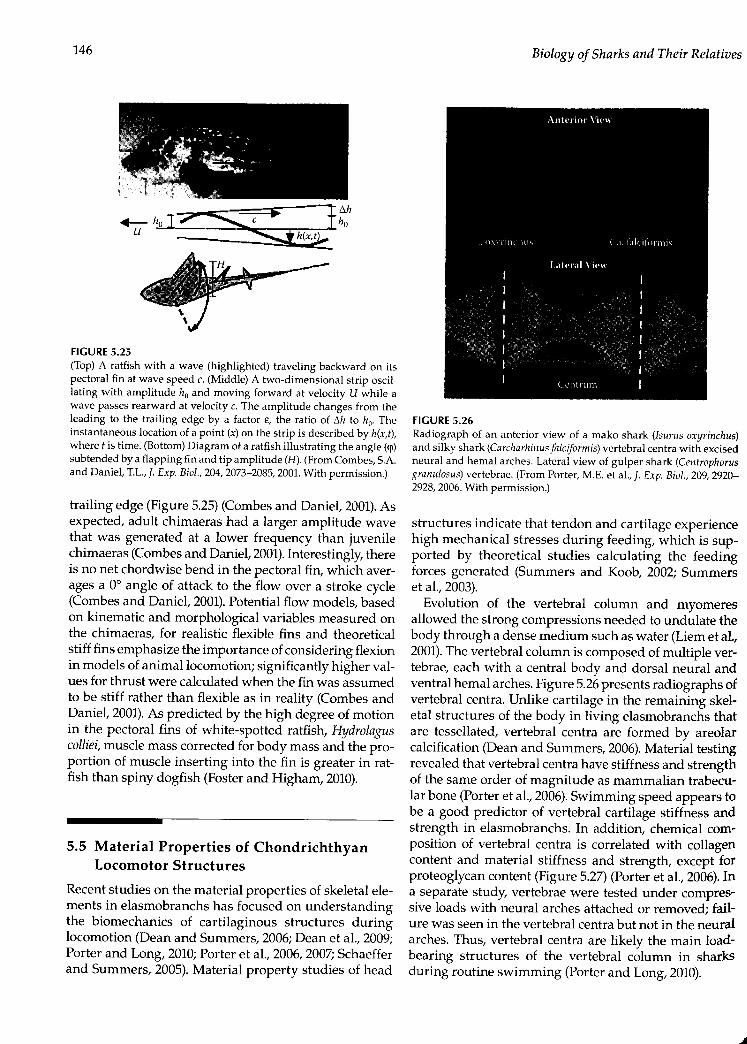
146
+-h.J7'u
FIGURE 5.25(Top) A ratfish with a wave (highlighted) traveling backward on itspectoral fin at wave speed c. (Middle) A two-dimensional strip oscil-lating with amplitude 110and moving forward at velocity U while awave passes rearward at velocity c. The amplitude changes from theleading to the trailing edge by a factor e, the ratio of 6h to ho- Theinstantaneous location of a point (x) on the strip is described by h(x,i),where t is time. (Bottom) Diagram of a ratfish illustrating the angle (q)subtended by a flapping fin and tip amplitude (H). (From Combes, S.A.and Daniel, T.L., f. Exp. Biol., 204, 2073-2085, 2001.With permission.)
trailing edge (Figure 5.25)(Combes and Daniel, 2001).Asexpected, adult chimaeras had a larger amplitude wavethat was generated at a lower frequency than juvenilechimaeras (Combes and Daniel, 2001).Interestingly, thereis no net chordwise bend in the pectoral fin, which aver-ages a 0° angle of attack to the flow over a stroke cycle(Combes and Daniel, 2001).Potential flow models, basedon kinematic and morphological variables measured onthe chimaeras, for realistic flexible fins and theoreticalstiff fins emphasize the importance ofconsidering flexionin models of animal locomotion; significantly higher val-ues for thrust were calculated when the fin was assumedto be stiff rather than flexible as in reality (Combes andDaniel, 2001).As predicted by the high degree of motionin the pectoral fins of white-spotted ratfish, Hydrolaguscolliei, muscle mass corrected for body mass and the pro-portion of muscle inserting into the fin is greater in rat-fish than spiny dogfish (Foster and Higham, 2010).
5.5 Material Properties of ChondrichthyanLocomotor Structures
Recent studies on the material properties of skeletal ele-ments in elasmobranchs has focused on understandingthe biomechanics of cartilaginous structures duringlocomotion (Dean and Summers, 2006;Dean et a!., 2009;Porter and Long, 2010;Porter et a!., 2006,2007;Schaefferand Summers, 2005).Material property studies of head
Biology of Sharks and Their Relatives
FIGURE 5.26Radiograph of an anterior view of a mako shark (Isurus oxyrinchus)and silky shark (CarciJarhinus falciform is) vertebral centra with excisedneural and hemal arches. Lateral view of gulper shark (Centrophorusgranulosus) vertebrae. (From Porter, M.E. et al., J. Exp. BioI., 209, 2920-2928,2006. With permission.)
structures indicate that tendon and cartilage experiencehigh mechanical stresses during feeding, which is sup-ported by theoretical studies calculating the feedingforces generated (Summers and Koob, 2002; Summerset a!., 2003).Evolution of the vertebral column and myomeres
allowed the strong compressions needed to undulate thebody through a dense medium such as water (Liem et al.,2001).The vertebral column is composed ofmultiple ver-tebrae, each with a central body and dorsal neural andventral hemal arches. Figure 5.26presents radiographs ofvertebral centra. Unlike cartilage in the remaining skel-etal structures of the body in living elasmobranchs thatare tessellated, vertebral centra are formed by areolarcalcification (Dean and Summers, 2006).Material testingrevealed that vertebral centra have stiffness and strengthof the same order of magnitude as mammalian trabecu-lar bone (Porter et al., 2006).Swimming speed appears tobe a good predictor of vertebral cartilage stiffness andstrength in elasmobranchs. In addition, chemical com-position of vertebral centra is correlated with collagencontent and material stiffness and strength, except forproteoglycan content (Figure 5.27) (Porter et a!., 2006).Ina separate study, vertebrae were tested under compres-sive loads with neural arches attached or removed; fail-ure was seen in the vertebral centra but not in the neuralarches. Thus, vertebral centra are likely the main load-bearing structures of the vertebral column in sharksduring routine swimming (Porter and Long, 2010).

Biomechanics of Locomotion in Sharks, Rays, and Chimaeras
(A) Box: 95% confidence intervalsB, C Line: species mean
~
Whiskers: species rangeB,C+ ~ 0+ 1 '!' E
-------------------------------------_± --
£' 5064D-"~30"~ 20.0;• 10§5 0
A
B
+
(B) 1200'";?1000
~ 800
" 600
~~
A,E
~400200o ~--=-=--~--=--=--"'"--=--=--=-=--=--=--=--=--=--=-=--=--=--=--""""'"
F
(C) 0.5
FIGURE 5.27Material properties of mineralized cartilage in shark vertebral cen-tra. (A) Ultimate strength (MPa) of vertebral cartilages from sevenelasmobranch species showing significant differences (F6,151 = 182.8,p < 0.001). The broken horizontal line represents the lower limits oftrabecular bone. Letters above the box and whisker plot denote sig-nificant differences between species. (B)Material stiffness was sig-nificantly different among the species (F6,151 = 54.4, P < 0.001).Torpedocalifornica, the only batoid, was less stiff than all shark species (P< 0.001).The horizontal line shows the lower limits of stiffness fortrabecular bone. (C) Yield strain was significantly different amongthe species (F6,147 = 27.6,P < 0.001).T. californica had the greatest yieldstrain of all species (P < 0.007).(From Porter, M.E. et al., J. Exp. Biol.,209,2920-2928,2006.With permission.)
Locomotormode is also a good predictor ofbatoid wingskeletalmorphology, indicating a role of flexural stiffness(Schaeffer and Summers, 2005). Undulatory swimmerssuch as Dasyatis have chain-like catenated calcificationpatterns with staggered joints in the radials; oscillatorySwimmers have radials completely covered with min-eralization in a crustal calcification pattern. Some oscil-latory species also had cross-bracing between adjacentradials that is thought to increase fin stiffness. Theoreticalshffness of fin morphology agreed with observed stiff-ness (Schaeffer and Summers, 2005).These studies pro-VIdefurther evidence of the role of material propertiesin determining the function and evolution of skeletal ele-ments and locomotor modes in Chondrichthyans.
147
5.6 Future Directions
The diversity of shark species for which we haveeven basic functional data on locomotor mechanics isextremely limited. Most papers to date have focused onleopard (Triakis), spiny dogfish (Squalus), and bamboo(Chiloscyllium) sharks swimming under controlled labo-ratory conditions. A high priority for future studies oflocomotion in sharks, skates, and rays is to expand thediversity of taxa studied, especially for analyses of sharkmechanics. The data obtained by Rosenberger (2001)onbatoid locomotion are exemplary for their broadly com-parative character, but studies like this are rare, perhapsnecessarily so when detailed functional data must beobtained for a variety of behaviors.Experimental studies of kinematics and hydrody-
namics would benefit from increased spatial and tem-poral resolution so that a more detailed picture could beobtained of patterns of fin deformation and the result-ing hydrodynamic wake, especially during unsteadymaneuvering behaviors. New high-resolution, high-speed digital video systems will permit a new level ofunderstanding of fin function and its impact on loco-motor performance. Such increased resolution may alsopermit further observations of boundary layer flows inrelation to surface denticle patterns to follow up on theobservation by Anderson et a1.(2001)that the boundarylayer of Mustelus swimming at 0.5 lis did not separateand remained attached along the length of the body.Defocusing digital particle image velocimetry (DDPIV)and stereoscopic PIV can be used to compute flow inthree dimensions and offer a new avenue for compre-hensive studies of fluid dynamics in the wake of swim-ming elasmobranchs (Cordon et al., 2002;Lauder, 2010;Raffel et al., 2007). These studies can be coupled withcomputational fluid dynamics (CFD) (Lauder, 2010) toinvestigate limitations and compare different locomotormodes, similar to what has been done for actinopter-ygian fishes (Tytell et al., 2010).Biorobotics and biomi-metics studies that are emerging use elasmobranchsas models and have focused mainly on batoids (Clarkand Smits, 2006;Cao et al., 2009;Xu et al., 2007;Yang etal., 2009;Zhou and Low, 2010).Biorobotics offers a wayto test scenarios that are not possible with living ani-mals, such as altering Reynolds and Strouhal numbers,aspect ratios, and material properties (Clark and Smits,2006;Lauder, 2010).Biomimetics has great potential toadvance the field through the demand for detailed kine-matics, material properties, and activation patterns ofelasmobranch locomotion for application purposes.More studies on the mechanical properties of elasmo-
branch connective tissue elements and of the role theseplay in transmitting forces to hydrodynamic fin con-trol surfaces are needed. This is a keyarea in which in

148
vitro studies of material properties and in vivo analysesof how elasmobranch connective tissues function cangreatly enhance our understanding of elasmobranchlocomotor mechanics.Finally, new information on locomotor structures
such as fin placement and diversity might come fromadvances in developmental and molecular biology meth-ods. Shark models have been used to study the evolutionof paired and median fins (Cole and Currie, 2007) as wellas skeletogenesis and the early origins of bone (Eameset al., 2007). To the extent that equipment and elasmo-branch behavior permits, it would be extremely valuableto have quantitative 3D field data over the natural loco-motor behavioral repertoire to answer such questionsas what are routine swimming speeds, what are typicalvertical and lateral maneuvering velocities, and whatis the natural range of body angles observed duringdiverse locomotor behaviors? Advances in technology,such as accelerometers, can be used in shark researchand can provide great insight to natural routine locomo-tor behaviors (Sims, 2010). Such data would serve as alink between experimental laboratory studies of sharkbiomechanics and locomotor performance in nature.
AcknowledgmentsSupport for preparation of this paper was provided byMCTESjFCTjSFRHjBDj36852j2007 to AMRM, by NSFgrant DB! 97-07846 to CDW, and grants ONR N00014-09-1-0352, ONR N00014-03-1-0897, and NSF EFRl-0938043 to GVL.
ReferencesAffleck,R]. (1950).Some points in the function, development,
and evolution of the tail in fishes. Froc. Zool. Soc. Lond.120:349-368.
Aleev, y.G. (1969).Function and Gross Morphology in Fish, trans.from the Russian by M. Raveh. Keter Press, Jerusalem.
Alexander, RM. (1965).The lift produced by the heterocercaltails of Selachii. J. Exp. Bioi. 43:131-138.
Anderson, E.j., McGillis, W., and Grosenbaugh, M.A (2001).The boundary layer of swimming fish. J. Exp. Bioi.204:81-102.
Arnold, G.P. and Webb, P.W. (1991).The role of the pectoralfins in station-holding of Atlantic salmon parr (Sa/rnasalar L). J. Exp. BioI. 156:625-629.
Bendix-Almgreen, S.E. (1975). The paired fins and shouldergirdle in Cladoselache, their morphology and phyletic sig-nificance. Colloq. Int. CN.R.S. Paris 218:111-123.
Biology of Sharks and Their Relafives
Bernal, D.,Donley, j.M., Shadwick, RE., and Syme. D.A. (2005).Mammal-like muscles power swimming in a cold-watershark. Nature 437:1349-1352.
Blake, RW (1983a). Fish Locomotion. Cambridge UniversityPress, Cambridge.
Blake, RW (1983b). Median and paired fin propulsion. In:Webb, PW and Weihs, D. (Eds.), Fish Biomechanics.Praeger, New York,pp. 214-247.
Bone, Q. (1999). Muscular system: microscopical anatomy,physiology, and biochemistry of elasmobranch musclefibers. In: Hamlett, We. (Ed.), Sharks, Skates, and Rays:The Biology of Elasmobranch Fishes. The johns HopkinsUniversity Press, Baltimore, MD, pp. 115-143.
Breder, CM. (1926). The locomotion of fishes. Zool. (N.Y.)4:159-256.
Carroll, R1. (1988). Vertebrate Paleontology and Evolution. WHFreeman, New York.
Cheng, j. and Zhaung, 1. (1991).Analysis of swimming three-dimensional waving plates. J. Fluid Mech. 232:341-355.
Chopra, M.G. (1974).Hydrodynamics of lunate-tail swimmingpropulsion. J. Fluid Mech. 64:375-391.
Clark, RP. and Smits, Aj. (2006).Thrust production and wakestructure of a batoid-inspired oscillating fin. J. FluidMech. 562:415-429.
Cole, N.j. and Currie, p.o. (2007).Insights from sharks: evolu-tionary and developmental models of fin development.Dev. Dynam. 236:2421-2431.
Combes, A. and Daniel, T.1. (2001).Shape, flapping and flex-ion: wing and fin design for forward flight. J. Exp. Biol.204:2073-2085.
Campagna, 1.J.v. (1973). Interrelationships of living elasmo-branchs. In: Greenwood, PH., Miles, RS., and Patterson,e. (Eds.), Interrelationships of fishes, Zool. ]. Linn. Soc.53(Suppl. 1):15-61.
Campagna, Lj.V (1984). Sharks of the World. United NationsDevelopment Program, Rome.
Campagna, Lj.V. (1988).Sharks of the Order Curcharhinijormes.Princeton University Press, Princeton, NJ.
Campagna, 1.j.V. (1999). Endoskeleton. In: Hamlett, We.(Ed.), Sharks, Skates, and Rays: The Biology cf ElaemobrancnFishes. The Johns Hopkins University Press, Baltimore,MD, pp. 69-92.
Daniel, j.F. (1922). The Elasmobranch Fishes. University ofCalifornia Press, Berkeley.
Dean, M.N. and Summers, AF (2006). Mineralized carti-lage in the skeleton of chondrichthyan fishes. Zoology109:164-168.
Dean, M.N., Mull, CG., Garb, S.N., and Summers, A.P. (2009).Ontogeny of the tessellated skeleton: insight from theskeletal growth of the round stingray Urobatis halleri. J.Anal. 215:227-239.
Dickinson, M.H. (1996). Unsteady mechanisms of force gen-eration in aquatic and aerial locomotion. Am. Zool.36:537-554.
Domenici, P. and Blake, RW (1997).Fish fast-start kinematicsand performance. J. Exp. Bioi. 200:1165-1178.
Domenici, F, Standen, E.M., and Levine, RP. (2004). Escapemanoeuvres in the spiny dogfish (Squatus acanthias). J.Exp. BioI. 207:2339-2349.

Biomechanics of Locomotion in Sharks, Rays, and Chimaeras
Donley, j. and Shadwick, R (2003).Steady swimming muscledynamics in the leopard shark Yriakis semifasciata. J. Exp.BioI. 206:1117-1126.
Donley, j.M., Shadwick, RE., Sepulveda, e.A., Konstantinidis,P.,and Cemballa, S. (2005).Patterns of red muscle strain/activation and body kinematics during steady swimmingin a lamnid shark, the shortfin mako (Isurus oxyrinchus).J. Exp. Bioi. 208:2377-2387.
Drucker, E.G. and Lauder, CV. (1999).Locomotor forces on aswimming fish: three-dimensional vortex wake dynam-ics quantified using digital particle image velocimetry.].Exp. BioI. 202:2393-2412.
Drucker, E.G. and Lauder, G.V. (2005).Locomotor function ofthe dorsal fin in rainbow trout: kinematic patterns andhydrodynamic forces. J. Exp. BioI. 208:4479-4494.
Eames, B.E, Allen, N., Young, j., Kaplan, A., Helms, j.A., andSchneider, RA. (2007).Skeletogenesis in the swell sharkCephaloscyl/ium ventriosum. J. Anat. 210:542-554.
Ferry, L.A. and Lauder, G.v. (1996).Heterocercal tail functionin leopard sharks: a three-dimensional kinematic analy-sis of two models. J. Exp. BioI. 199:2253-2268.
Fish, EE. and Shannahan, L.D. (2000).The role of the pectoralfins in body trim of sharks. J. Fish Bioi. 56:1062-1073.
Flammang, B.E. (2010).Functional morphology of the radialismuscle in shark tails. J. Morphol. 271:340-352.
Foster, KL. and Higham, T.E. (2010).How to build a pectoralfin: functional morphology and steady swimming kine-matics of the spotted ratfish (Hydrolagus col/iei). Can. J.Zool. 88:774-780.
Gao, j.. Bi, S., u. j., and Liu, e. (2009). Design and experi-ments of robot fish propelled by pectoral fins. ROBIa2009:445-450.
Garman, S. (1913).The Plagiostoma (sharks, skates, and rays).Mem. Mus. Camp. Zool. Harvard Col/. 36.
Gemballa, S., Konstantinidis, P., Donley, j.M., Sepulveda,C; and Shadwick, RE. (2006). Evolution of high-per-formance swimming in sharks: transformations of themusculotendinous system from subcarangiform to thun-niforrn swimmers. J. MorpllOl. 267:477-493.
Gordon, M.S., Hove, [R; and Bartol, I.K (2002). Dynamicsand energetics of animal swimming and flying: introduc-tion. Integr. Camp. Bioi. 42:960-963.
Gray, [. (1968). Animal Locomotion. Weidenfeld & Nicolson,London.
Grove, A.j. and Newell, G.E. (1936).A mechanical investiga-tion into the effectual action of the caudal fin of someaquatic chordates. Ann. Mag. Nat. Hist. 17:280-290.
Harris, j.E. (1936). The role of the fins in the equilibrium ofthe swimming fish. I. Wind tunnel tests on a model ofMustelus canis (Mitchell).]. Exp. Bioi. 13:476-493.
Harris, j.E. (1953). Fin patterns and mode of life in fishes.In: Marshall, S.M. and Orr, A.P. (Eds.), Essays in MarineBiology. Oliver & Boyd, Edinburgh, pp. 17-28.
jordan, L.K (2008).Comparative morphology of stingray lat-eral line canal and electrosensory systems. J. Morphol.269:1325-1339.
Kajiura,SM., Forni,j.B., and Summers,A.P. (2003).Maneuveringin juvenile carcharhinid and sphyrnid sharks: the role ofthe hammerhead shark cephalofoil. Zoology 106:19-28.
149
Kemp, N.E. (1999). Integumentary system and teeth. In:Hamlett, We. (Ed.), Sharks, Skates, and Rays: The Biologyof Elasmobranch Fishes. The johns Hopkins UniversityPress, Baltimore, MD, pp. 43-68.
Koester, D.M. and Spirito, CP. (1999).Pelvic fin locomotion inthe skate, Leucoraja erinacea. Am. Zool. 39:5SA.
Krothapalli, A. and Lourenco, L. (1997). Visualization of veloc-ity and vorticity fields. In: Nakayama, Y. and Tanida,Y. (Eds.), Atlas of Visualization, Vol. 3. CRC Press, BocaRaton, FL, pp. 69-82.
Kundu, P. (1990).Fluid Mechanics. Academic Press, San Diego.Lauder, G.v. (2000).Function of the caudal fin during locomo-
tion in fishes: kinematics, flow visualization, and evolu-tionary patterns. Am. Zool. 40:101-122.
Lauder, C'v. (2010).Swimming hydrodynamics: ten questionsand the technical approaches needed to resolve them. In:Taylor, GK, Triantafyllou, M.S., and Tropea, e. (Eds.),Animal Locomotion. Springer-Verlag, Berlin, pp. 3-15.
Lauder, G.v. and Drucker E. (2002).Forces, fishes, and fluids:hydrodynamic mechanisms of aquatic locomotion. NewsPhysiol. Sci. 17:235-240.
Lauder, G.v. and Madden, P.G.A. (2008).Advances in compar-ative physiology from high-speed imaging of animal andfluid motion. Annu. Rev. Physiol. 70:143-163.
Lauder, G.v., Nauen. j., and Drucker, E.G (2002).Experimentalhydrodynamics and evolution: function of median finsin ray-finned fishes. Integr. Compo Bioi. 42:1009-1017.
Lauder, c.v. Drucker, E.G., Nauen, j., and Wilga, CD. (2003).Experimental hydrodynamics and evolution: caudal finlocomotion in fishes. In: Bels. V, Gasc, J.P., and Casinos,A. (Eds.), Vertebrate Biomechanics and Evolution, BiosScientific Publishers, Oxford, pp. 117-135.
Liao, j. and Lauder, G V. (2000). Function of the heterocer-cal tail in white sturgeon: flow visualization duringsteady swimming and vertical maneuvering. J. Exp. Biol.203:3585-3594.
Liem, K.E and Summers, A.P. (1999). Muscular system. In:Hamlett, We. (Ed.), Sharks, Skates, and Rays: The Biologyof Elasmobranch Fishes, The johns Hopkins UniversityPress, Baltimore, MD, pp. 93-114.
Liem, KE, Bemis,WE., Walker, jr., W.E, and Grande, L. (2001).Functional Anatomy of the Vertebrates: An EvolutionaryPerspective, 3rd ed. Harcourt, New York.
Lighthill, j. and Blake, R (1990). Biofluid dynamics of balis-tiform and gymnotiform locomotion. Part 1. Biologicalbackground and analysis by elongated-body theory. ].Fluid. Mech. 212:183-207.
Lindsey, e.e. (1978). Form, function, and locomotory hab-its in fish. In: Hoar, W.5. and Randall, D.j. (Eds.), FishPhysiology. Vol. 7. Locomotion. Academic Press, New York,pp.1-100.
Lingham-Soliar, T. (2005). Dorsal fin in the white shark,Carcharodon carcharias: a dynamic stabilizer for fast swim-ming.]. Morphol. 263:1-11.
Macesic, L.j. and Kajiura, S.M. (2010). Comparative puntingkinematics and pelvic fin musculature of benthic batoids.]. Morphol. 271:1219-1228.
Magnan, A. (1929).Les characteristiques geometriqes et phy-siques des poisons. Ann. Sci. Nat. Zool. 10:1-~32.

150
Nauen, I.C and Lauder, G.Y. (2002). Quantification of thewake of rainbow trout (Oncorhynchus mykiss) usingthree-dimensional stereoscopic digital particle imagevelocimetry. I. Exp. Bioi. 205:3271-3279.
Perry,CN., Cartamil, D.P.,Bernal, D. et a1.(2007). Quantificationof red myotomal muscle volume and geometry in theshortfin mako shark (Isurus oxyrinchus) and the salmonshark (Lamna ditropis) using Tl-weighted magnetic reso-nance imaging. I. Morphol. 268:284-292.
Porter, M.E. and Long, [r., j.H. (2010). Vertebrae in compres-sion: mechanical behavior of arches and centra in thegray smooth-hound shark (Mustclus califomicus). J.Morphoi. 271:366-375.
Porter, M.E., Beltran, j.L., Koob, rr,and Summers, A.P. (2006).Material properties and biochemical composition ofmineralized vertebral cartilage in seven elasmobranchspecies (Chondrichthyes).]. Exp. BioI. 209:2920-2928.
Porter, M.E., Koob, r.j., and Summers, A.P. (2007). The contri-bution of mineral to the material properties of vertebralcartilage from the smooth-hound shark Mustelus califor-nicus.]. Exp. BioI. 210:3319-3327.
Porter, M.E., Roque, CM., and Long, [r., j.H. (2009). Turningmaneuvers in sharks: predicting body curvature fromaxial morphology. J. Morphol. 270:954-965.
Pridmore, P.A. (1995). Submerged walking in the epauletteshark Hemiscyllium ocellatum (Hemiscyllidae) and itsimplications for locomotion in rhipidistian fishes andearly tetrapods. Zoology 98:278-297.
Raffel, M., Willert, CE., Wereley, S.T.., and Kompenhans. j.(2007). Particle Image Velocimetry: A Practical Guide, 2nded. Springer, Berlin.
Reif W.E. and Weishampel, D.B. (1986). Anatomy and mechan-ics of the lunate tail in Iamnid sharks. Zool. Jahrb. Anat.114:221-234.
Rosenberger, L. (2001). Pectoral fin locomotion in batoid fishes:undulation versus oscillation. J. Exp. Biol. 204:379-394.
Rosenberger, L.j. and Westneat, M.W (1999). Functional mor-phology of undulatory pectoral fin locomotion in thestingray Taeniura lymma (Chondrichthyes: Dasyatidae). J.Exp. BioI. 202:3523-3539.
Schaeffer, j.T and Summers, A.P. (2005). Batoid wing skeletalstructure: novel morphologies, mechanical implications,and phylogenetic patterns. J. Morphoi. 264:298-313.
Shirai, S. (1996). Phylogenetic interrelationships of neosela-chians (Chondrichthyes: Euselachii). In: Stiassny, M.,Parenti, L., and johnson, GD. (Eds.), fnterrelationships ofFishes. Academic Press, San Diego, pp. 9-34.
Simons, j.R (1970). The direction of the thrust produced by theheterocercal tails of two dissimilar elasmobranchs: thePort Jackson shark, Heterodontus portusiacksoni (Meyer),and the piked dogfish, Squalus megalops (Macleay). J. Exp.Bioi. 52:95-107.
Simons, M. (1994). Model Aircraft Aerodynamics. Argus Books,Herts, U.K
Sims, D.W. (2010). Trackingand analysis techniques for under-standing free-ranging shark movements and behavior.In: Carrier, jC.; Musick, j.A., and Heithaus, M.R. (Eds.),Sharks and Their Relatives II. CRC Press, Boca Raton, FL,pp. 351-392.
Biology of Sharks and Their Relatives
Smith, H.C (]992). The filustrated Guide to Aerodynamics. TABBooks, New York.
Stemhuis, E.j. (2006). Basics and principles of particle imagevelocimetry (PN) for mapping biogenic and biologicallyrelevant flows. Aquat. Ecol. 40:463-479.
Standen, E.M. (2010). Muscle activity and hydrodynamic func-tion of pelvic fins in trout (Oncorhynchus mykiss). J. Exp.Bioi. 213:83]-841.
Standen, E.M. and Lauder, G.Y. (2005). Dorsal and anal finfunction in bluegill sunfish Lepomis macrochirus: three-dimensional kinematics during propulsion and maneu-vering. J. Exp. BioI. 208:2753-2763.
Standen, E.M. and Lauder, G.Y. (2007). Hydrodynamic func-tion of dorsal and anal fins in brook trout (Salvelinus fon-tinalis). j. Exp. BioI. 210:340-356.
Summers, A.P. and Koob. Tj. (2002). The evolution of tendon:morphology and material properties. Compo Biochem.Phys. A 133:1159-1170.
Summers, A.P, Koob-Emurtds, M.M., Kajiura,S.M., and Koob,T.J. (2003). A novel fibrocartilaginous tendon from anelasmobranch fish (Rhinoptera bonasus). Cell Tissue Res.312:221-227.
Thomson, KS. (]971). The adaptation and evolution of earlyfishes. Q. Rev. BioI. 46:139-166.
Thomson, KS. (]976). On the heterocercal tail in sharks.Paleobiology 2:]9-38.
Thomson, K.5. and Simanek DE. (1977). Body form and loco-motion in sharks. Am. Zool. 17:343-354.
Tytell, ED., Standen, E.M., and Lauder, GY. (2008). Escapingflatland: three dimensional kinematics and hydrody-namics of median fins in fishes. J. Exp. BioI. 211:187-195.
Tytell, ED., Borazjani, 1., Sotiropoulos, F. et a1. (2010). Disen-tangling the functional roles of morphology and motionin the swimming of fish. Int. Compo BioI. 50:1140-1154.
Walker, j.A. and Westneat, M.W (2000). Mechanical perfor-mance of aquatic rowing and flying. Proc. R. Soc. Lond.B 267:1875-]881.
Webb, P.W (1984). Form and function in fish swimming. Sci.Am. 251:72-82.
Webb, P.W (1988). Simple physical principles and vertebrateaquatic locomotion. Am. Zool. 28:709-725.
Webb, P.W and Blake, RW (1985). Swimming. In: Hildebrand,M. et a1.(Eds.), Functional Vertebrate Morphology. HarvardUniversity Press, Cambridge, MA, pp. 110-]28.
Webb, P.W and Gerstner, CL. (]996). Station-holding by themottled sculpin, Cottus bairdi (Teleostei: Cottidae) andother fishes. Copeia 1996:488-493.
Webb, P.W and Keyes, RS. (]982). Swimming kinematics ofsharks. Fish Bull. 80:803-812.
Wilga, CD. and Lauder, G.Y. (1999). Locomotion in sturgeon:function of the pectoral fins. J. Exp. BioI. 202:2413-2432.
Wilga, CD. and Lauder GY. (2000). Three-dimensional kine-matics and wake structure of the pectoral fins duringlocomotion in leopard sharks 'Iriakis semifasciata. J. Exp.BioI. 203:226]-2278.
Wilga, CD. and Lauder, GV. (2001). Functional morphology ofthe pectoral fins in bamboo sharks, ChiloscylIium ptagio-sum: benthic versus pelagic station holding. J. Morpho!.249:195-209.

Biomechanics of Locomotion in Sharks, Rays, and Chimaeras
Wilga, CD. and Lauder, c.Y. (2002). Function of the hetero-cereal tail in sharks: quantitative wake dynamics duringsteady horizontal swimming and vertical maneuvering.J. Exp. BioI. 205:2365-2374.
Wilga, CD. and Lauder, GV (2004). Biomechanics of loco-motion in sharks, rays and chimeras. In: Carrier, J .c.,Musick, J., and Heithaus, M. (Eds.), Biology of Sharks andTheir Relatives. CRC Press: Boca Raton, FL, pp. 139-164.
Willert, CE. and Gharib, M. (1991). Digital particle image velo-cimetry. Exp. Fluids 10:181-193.
Xu, Y, Zong. G., Bi, 5., and Gao, J. (2007). Initial developmentof a flapping propelled unmanned underwater vehicle(UUV). ROBIa 2007:514-529.
151
Yang,S., Qiu, J., and Han, X. (2009). Kinematics modeling andexperiments of pectoral oscillation propulsion roboticfish. J. Bionic Eng. 6:174-179.
Zangerl, R. (1973). Interrelationships of Early Chondrichthyans.Academic Press, London.
Zhou, C. and Low, K. (2010). Better endurance and load capac-ity: an improved design of Manta Ray Robot (RoMan-II).J. Bionic Eng. 7(Suppl.):S137-S144.