biosensors and bioelectronics - schroeder group · biosensors and bioelectronics 49 (2013)...
TRANSCRIPT

Biosensors and Bioelectronics 49 (2013) 118–125
Contents lists available at SciVerse ScienceDirect
Biosensors and Bioelectronics
0956-56http://d
n CorrE-m1 Th
journal homepage: www.elsevier.com/locate/bios
A multiplexed microfluidic platform for rapid antibioticsusceptibility testing
Ritika Mohan 1, Arnab Mukherjee 1, Selami E. Sevgen, Chotitath Sanpitakseree, Jaebum Lee,Charles M. Schroeder, Paul J.A. Kenis n
Department of Chemical & Biomolecular Engineering, University of Illinois at Urbana-Champaign, 600 South Mathews Avenue, Urbana, IL 61801, USA
a r t i c l e i n f o
Article history:Received 30 March 2013Accepted 25 April 2013Available online 9 May 2013
Keywords:Antibiotic susceptibility testingGreen fluorescent protein (GFP)MicrofluidicsFluorescence detectionMultiplexed sensor
63/$ - see front matter & 2013 Elsevier B.V. Ax.doi.org/10.1016/j.bios.2013.04.046
esponding author. Tel.: +1 217 265 0523; fax:ail address: [email protected] (P.J.A. Kenis).ese authors contributed equally to the work.
a b s t r a c t
Effective treatment of clinical infections is critically dependent on the ability to rapidly screen patientsamples to identify antibiograms of infecting pathogens. Existing methods for antibiotic susceptibilitytesting suffer from several disadvantages, including long turnaround times, excess sample and reagentconsumption, poor detection sensitivity, and limited combinatorial capabilities. Unfortunately, thesefactors preclude the timely administration of appropriate antibiotics, complicating management ofinfections and exacerbating the development of antibiotic resistance. Here, we seek to address theseissues by developing a microfluidic platform that relies on fluorescence detection of bacteria that expressgreen fluorescent protein for highly sensitive and rapid antibiotic susceptibility testing. This platformpossesses several advantages compared to conventional methods: (1) analysis of antibiotic action in twoto four hours, (2) enhanced detection sensitivity (≈1 cell), (3) minimal consumption of cell samples andantibiotic reagents (o6 mL), and (4) improved portability through the implementation of normally closedvalves. We employed this platform to quantify the effects of four antibiotics (ampicillin, cefalexin,chloramphenicol, tetracycline) and their combinations on Escherichia coli. Within four hours, thesusceptibility of bacteria to antibiotics can be determined by detecting variations in maxima of localfluorescence intensity over time. As expected, cell density is a major determinant of antibiotic efficacy.Our results also revealed that combinations of three or more antibiotics are not necessarily better foreradicating pathogens compared to pairs of antibiotics. Overall, this microfluidic based biosensortechnology has the potential to provide rapid and precise guidance in clinical therapies by identifyingthe antibiograms of pathogens.
& 2013 Elsevier B.V. All rights reserved.
1. Introduction
In recent years, antibiotic resistance traits among microbialpathogens have escalated at alarming rates, which has spurred thedevelopment of technologies for rapid and accurate detection ofantibiotic susceptibility profiles of pathogens (Rice, 2010). Estab-lished techniques for antibiotic susceptibility testing (AST), such asbroth dilution and disc diffusion, involve multiple time-consumingsteps (Lazcka et al., 2007; White et al., 1996) including: (1) isolationof pathogens from patient samples (24–48 h.), (2) pre-culturingof isolated bacteria to enrich cell density to detectable levels(24–48 h.), (3) incubation of cells with antibiotics in 96-well platesor petri dishes (24–48 h.), and (4) determination of bacterial growthusing absorption spectroscopy or by visual assessment. Brothdilution and disc diffusion assays typically require significant
ll rights reserved.
+1 217 333 5052.
quantities (10–30 mL) of patient samples such as blood, sputum,or urine for analysis (Mancini et al., 2010). In addition, the limitedsensitivity of macroscale techniques for AST makes them unsuitablefor detecting the presence of “persister” microbes. Although persis-ter cells represent only a small fraction (≈10−5) of microbial cells,they tend to evade antibiotic mediated killing by switching to ametabolically dormant or “persistent” state (Balaban et al., 2004;Lewis, 2010). Persister cells constitute a significant threat due totheir ability to re-initiate infection upon discontinuation of anti-biotic therapy (Dawson et al., 2011). Finally, inconsistencies inresults obtained from different AST techniques further complicatediagnosis and treatment (Gales et al., 2001; Goldstein et al., 2007;Lo-Ten-Foe et al., 2007; Nicodemo et al., 2004; Tan and Ng, 2007;Traub, 1970). Hence, in the absence of precise information about theantibiogram of particular pathogen, physicians often resort toempirical therapies that utilize broad-spectrum antibiotics. Indis-creet use of antibiotics in this manner is known to intensify theproblem of antibiotic resistance (Alanis, 2005; Ang, 2001).
To address the aforementioned issues, biosensor platforms withimproved sensitivity and fast analysis time have been developed for

R. Mohan et al. / Biosensors and Bioelectronics 49 (2013) 118–125 119
antimicrobial susceptibility testing (Chiang et al., 2009; Karasinskiet al., 2007; Kinnunen et al., 2011; Koydemir et al., 2011; Nakamuraet al., 1991; Tsou et al., 2010). For example, electrochemical sensorshave been utilized to determine susceptibility by measuring smallchanges in growth of cells (Karasinski et al., 2007). Chiang et al., havedeveloped a surface plasmon resonance-based biosensor platform tocategorize strains as susceptible or resistant by detecting variationsin optical properties of bacteria when treated with antibiotics(Chiang et al., 2009). Another interesting approach for antibioticsusceptibility testing utilizes an asynchronous magnetic bead rota-tion biosensor to monitor single cells or cell populations aftertreatment with antibiotics (Kinnunen et al., 2011). In addition, filterchip and optical detection biosensing system have been developedthat can provide susceptibility results in one hour (Tsou et al., 2010).These microfluidic-based biosensor technologies are sensitive andrapid, however, most of these platforms lack multiplexing capabilities(Chiang et al., 2009; Kinnunen et al., 2011; Tsou et al., 2010). Hence,integrated microfluidics represents an attractive technology for themultiplexed implementation of biological assays with rapid turn-around times and minimal sample consumption (Sia and Whitesides,2003). Several successful microfluidic platforms for AST have beenreported (Boedicker et al., 2008; Chen et al., 2010; Churski et al.,2012; Cira et al., 2012; Ho et al., 2012; Kalashnikov et al., 2012; Sunet al., 2011). For example, droplet-based microfluidics has beenutilized to compartmentalize bacterial cells, nutrients, antibiotics,and fluorescent viability indicators in water-in-oil emulsions(Boedicker et al., 2008; Churski et al., 2012). Sun et al. have reportedon the development of a microfluidic platform for the confinement ofbacterial cells in square microwells connected to a central flowchannel that continuously delivers nutrients and antibiotics to cells(Sun et al., 2011). Choi et al. have reported a microfluidic agarosechannel system for rapid antibiotic susceptibility testing by trackingsingle cell growth (Choi et al., 2013). Weibel and colleagues havedeveloped a portable microfluidic chip for AST for point-of-care use(Cira et al., 2012). The key advantage of the portable chip is theautomatic loading of bacterial cells into microfluidic chambers thathad been preloaded with dehydrated antibiotics using a ‘degasdriven flow’.
The existing approaches for microfluidic-based antibiotic sus-ceptibility testing offer promising routes toward the developmentof a rapid and portable screening tool. However, many of thesemethods suffer from one or more of the following limitations:(1) complicated platform fabrication and/or operation procedures(Kalashnikov et al., 2012), (2) poor portability due to the require-ment for syringe pumps, pneumatic actuators, and other ancillaryequipment (Choi et al., 2013; Churski et al., 2012; Kalashnikovet al., 2012), and (3) unstable droplet formation (Theberge et al.,2010). In this work, we report on the design and fabrication of amicrofluidic platform with biosensing capabilities featuring aspatially addressable 4�6-array of wells to simultaneously moni-tor the effects of multiple antibiotics at different concentrations, aswell as their combinations, on bacterial cells for AST. This technol-ogy integrates ease-of-fabrication and use with enhanced combi-natorial capabilities, and further provides improved portabilityand usability by circumventing the requirement for expensivesyringe pumps and pneumatic actuators by implementing nor-mally closed valves. In addition, the platform is amenable toautomated analysis by using time-lapse fluorescence microscopy(TLFM). We employed the microfluidic platform to interrogate theantibiotic sensitivity profile of Escherichia coli to four commonlyused bactericidal and bacteriostatic antibiotics. Furthermore, weexplored synergistic and antagonistic effects of different antibioticcocktails, as well as the effects of E. coli cell densities on dictatingthe efficiency of antibiotic action. Overall, this platform capitalizeson several key advantages of biosensor based integrated micro-fluidics technology including miniaturization of assays, expedited
analysis, multiplexing, and improved detection sensitivity alongwith ease-of-use and portability.
2. Materials and Methods
2.1. Microfluidic chip fabrication
The microfluidic chip for AST was fabricated using standard softlithographic techniques (Xia and Whitesides, 1998). Briefly, moldsfor casting the fluidic and control layers were made by patterningnegative photoresist on silicon wafers using photolithography.A thin layer of 20:1 PDMS (weight ratio of polymer to cross-linker)was spin coated on to the fluidic layer master and 5:1 PDMS waspoured on to the control layer master. The two layers werepartially cured at 65 1C for 30 minutes. Next, the control layerwas carefully peeled off the silanized silicon master, and threeholes for actuation of the mixing and sample loading valves werepunched using a 20-gauge needle. The control layer was manuallyaligned with the fluidic layer under an optical microscope (LeicaMZ6), and the aligned layers were cured overnight (∼12 h.) at65 1C to yield a monolithic device. Finally, the assembled devicewas peeled off the fluid layer master, inlet ports were punchedusing a 20-gauge needle, and the assembly was placed on a glasscoverslip to create a reversible seal.
2.2. Bacterial strains, growth media, and antibiotic solutions
Wild type Escherichia coli MG1655 (ATCC 47076) was geneticallyengineered to constitutively express green fluorescent protein (GFP),thereby enabling detection and enumeration of cells using fluores-cence microscopy. Specifically, E. coliMG1655 cells were transformedwith a low copy plasmid expressing a bright GFP variant from aconstitutive promoter derived from bacteriophage lambda (Lutz andBujard, 1997). Details of plasmid construction are provided assupporting information (Table A.1). We verified that the recombinantGFP-expressing E. coli cells were phenotypically similar to wild typecells with respect to growth rates. E. coli cells were routinelycultivated in Lennox broth (10 g/L tryptone, 5 g/L yeast extract, and5 g/L NaCl) supplemented with kanamycin at a concentration of30 mg/mL in order to maintain the GFP-expressing plasmid. Kana-mycin was omitted for on-chip cultures with no noticeable effect oncellular fluorescence. Antibiotic stock solutions of 10 mg/mL tetra-cycline hydrochloride and 10 mg/mL chloramphenicol were preparedin 70% ethanol. Stock solutions of 30 mg/mL kanamycin, 100 mg/mLampicillin, and 1 mg/mL cefalexin hydrate were prepared in steriledeionized water. All stock solutions were filtered using a 0.45 mmsyringe filter (Millex- HV filter unit, Millipore) prior to use. Antibioticdilutions were made directly into Lennox broth. The antibioticstetracycline, chloramphenicol, and cefalexin hydrate were purchasedfrom Sigma-Aldrich, and kanamycin and ampicillin were purchasedfrom Fisher Scientific.
2.3. On-chip antibiotic susceptibility testing
Microfluidic chips were sterilized by autoclaving prior to eachexperiment. Nonspecific interactions between the chip surfaceand cells or antibiotics were minimized by passivating the walls ofthe fluid layer and the glass coverslip with sterile bovine serumalbumin (BSA) at a concentration of 10 mg/mL for 15 min before eachexperiment. In a typical experiment (Fig. A.1), the microfluidicchip-cover slip assembly is placed in contact with an aluminum-heating block heated to 35 1C using a temperature controller (Bio-nomic System BC-110). Antibiotic and cell solutions (≈1 mL volumefor each) are placed on their respective inlet ports and introducedinto the wells by actuating the filling valves using a vacuum pump
R. Mohan et al. / Biosensors and Bioelectronics 49 (2013) 118–125120
(3 psig; GastDOA-P704-AA VacuumPump 1/8 HP 115 VAC). Enhancedmixing of adjacent sets of antibiotic and cell solutions is initiated byactuating the mixing valves for 15 min. To minimize solvent loss dueto evaporation during long-term experiments, reservoirs filled withbacterial growth medium (Lennox broth) or sterile water were placedaround the device, and the assembly was sealed at the top by affixinga glass slide (Fig. A.1). On-chip measurements were performed byTLFM using an inverted fluorescence microscope (Leica, DMI4000)equipped with a 1600�1200 pixel CCD camera (QImaging, Retiga-2000R), a 480/40 nm excitation filter, 527/30 nm emission filter, amotorized stage to raster the imaging field-of-view, and automatedfocus control implemented with a Z-motor (Ludl Electronic Pro-ducts). Images were acquired every 10 min over a period of 10 husing a 10x objective (Plan Achromat, NA¼0.25). The shallow fluidchannels (15 mm) ensured that all cells were within the depth offocus throughout the experiment. During data acquisition, theexposure time was set to 200 ms and the fluorescent light sourcewas shuttered between successive exposures to minimize photo-bleaching. Stage rastering, focus control, image acquisition, andcapture were implemented using ImagePro Plus software (MediaCybernetics).
2.4. Image processing and data analysis
Images were analyzed using ImageJ version 1.46a (Rasband,2011). Specifically, 8-bit grayscale images were converted to binaryimages by manual thresholding to capture all cells within a rangeof fluorescence intensities, which is defined on the low end by aminimum signal-to-noise ratio and determined on the high end bythe inability to visualize cells. The number of cells in each chamberwas also estimated by counting the local fluorescence intensitymaxima, and this algorithm provided consistent results within thisrange of manual thresholding. In order to highlight long-termtrends in cell proliferation (or death), we implemented a movingaverage filter to smooth the time series data. We quantifiedantibiotic efficacy in terms of the fraction of the initial cellpopulation that survives antibiotic treatment after 10 h, which isestimated as
FLab;conc ¼N10
N0ð1Þ
where FLab,conc denotes the fraction of live cells at t¼10 h., sub-scripts ab and conc refer to the antibiotic and its concentration inunits of mg/mL, No is the total number of cells in a microfluidicchamber at t¼0, and N10 is the total number of cells in the same
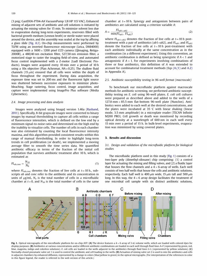
Fig. 1. Optical micrographs of the microfluidic platform for on-chip AST: (A) The devicedisplay purposes. (B) Antibiotics at various concentrations and/or different antibiotic comblue, magenta, indigo and colorless solutions). E. coli cells are loaded in the wells adjacNormally-closed pneumatic valve set 1 is used to control the antibiotic and cell loading inin adjacent chambers by enhanced diffusion, represented by a change in colors (blue/yelloin this figure legend, the reader is referred to the web version of this article.)
chamber at t¼10 h. Synergy and antagonism between pairs ofantibiotics are calculated using a criterion variable A:
A¼ FLab1þab2
minðFLab1; FLab2Þð2Þ
where FLab1+ab2 denotes the fraction of live cells at t¼10 h post-treatment with a pair of antibiotics (ab1+ab2), and FLab1 and FLab2denote the fraction of live cells at t¼10 h post-treatment witheach antibiotic individually at the same concentration as in thecombination (in a different experiment). Using this convention, anantibiotic combination is defined as being synergistic if Ao1 andantagonistic if A41. For experiments involving combinations ofthree or four antibiotics, this definition of A was extended toaccount for combinatorial treatment methods (Eqs (A.(1) and A.2)in Appendix A).
2.5. Antibiotic susceptibility testing in 96-well format (macroscale)
To benchmark our microfluidic platform against macroscalemethods for antibiotic screening, we performed antibiotic suscept-ibility testing on E. coli using 96-well plates. E. coli cell cultureswere prepared as described above and added to the wells of a127.8 mm�85.5 mm flat-bottom 96-well plate (Nunclon). Anti-biotics were added to each well at the desired concentrations, andthe plates were incubated at 35 1C with linear shaking (linearmode, 3.5 mm amplitude) in a microplate reader (TECAN InfiniteM200 PRO). Cell growth or death was monitored by recordingoptical density at a wavelength of 600 nm in each well every15 min over a period of 15 h. In bulk-level experiments, evapora-tion was minimized by using covered plates.
3. Results and discussion
3.1. Design and validation of the microfluidic platform for biologicalstudies
The microfluidic platform used in this study (Fig. 1) consists of atwo-layer poly (dimethyl-siloxane) chip comprising: (1) a controllayer for actuating the mixing and filling valves, and (2) a fluidic layerthat houses the flow channels and a 4�6-array of wells. Each wellconsists of two half wells that house the cells and antibiotic solutions,respectively. Each half well is 400 μm wide, 15 μm tall and 500 μmlong. In this way, the 4�6 array design facilitates the treatment ofone microbial cell sample with six distinct antibiotic solutions,
features a 4�6 array of 3 nL volume wells, which are loaded with colored dyes forbinations are loaded in each well through fluid lines A–F (represented by green, red,ent to the antibiotics through fluid lines G-L (represented by yellow solutions). (C)the wells. (D) Actuation of mixing valve set 2 is used to mix the antibiotics and cellsw to green) in the optical micrographs. (For interpretation of the references to color

R. Mohan et al. / Biosensors and Bioelectronics 49 (2013) 118–125 121
thereby enabling the execution of up to six unique screens per chipalong with four replicates for each screen (Fig. 1). Antibiotic solutionsand cell samples are readily loaded and mixed by actuating therespective filling and mixing valves using a vacuum pump. Theincorporation of normally closed valves improves platform portabil-ity by obviating the need for continuous actuation using positivepressure (Mohan et al., 2011; Schudel et al., 2009; Schudel et al.,2011; Thorson et al., 2011). To validate the feasibility of the micro-fluidic platform for biological studies, we determined the doublingtimes of E. coli cultured on-chip and in 96-well plates using twodifferent kinds of media: nutrient-rich Lennox medium and minimalglucose-based M9 medium. We verified that the on-chip growthprofiles of E. coli cells were in close agreement with the growthprofiles observed in the 96-well plate based experiments (Fig. A.2).Specifically, the on-chip doubling times of E. coli were approximately35 min and 70 min in Lennox broth and M9 medium, respectively.The corresponding doubling times in the same media when using96-well plates were 30 min and 73 min, respectively. The generalscheme of the sensing system is shown in Fig. A.3.
3.2. Effects of individual antibiotics on E. coli cells
We employed the microfluidic screening chip to investigate theeffects of four widely prescribed antibiotics on E. coli expressingGFP. The use of GFP as a genetically encodable indicator of cellviability has been previously reported (Keymer et al., 2006).Transformation of bacteria to express GFP is a standard procedureroutinely used in microbiology or for applications in monitoringcell growth over an extended period of time. Antibiotics were
Fig. 2. Effects of individual antibiotics on E. coli growth using an on-chip assay. Time traand bacteriostatic antibiotics: (C) tetracycline and (D) chloramphenicol on cell growth. Cover a period of 10 hours. Cell numbers are normalized to the initial (t¼0) value. Each drepresent the standard error of the mean (SEM) and are depicted for every fifth data p
selected to comprise bactericidal (ampicillin, cefalexin) and bac-teriostatic (tetracycline, chloramphenicol) classes. We treatedearly log-phase cultures of E. coli with varying concentrations ofeach antibiotic on-chip and quantified cell numbers using TLFMover a period of 10 h (Fig. 2). Representative optical imagesshowing action of an antibiotic on bacteria over a period of 10 hare shown in Fig. A.4. Although cell numbers are quantified over aperiod of 10 h, antibiotic susceptibility information is discerniblewithin 2–4 h (Fig. A.5). We estimated antibiotic efficiency in termsof the fraction of the initial cell population in a chamber thatsurvives antibiotic treatment at the end of an experiment (FLab, conc)as defined by Eq. (1). FLab, conc values are tabulated in Table 1. In caseof unperturbed cell growth (no antibiotics added), FL typically has avalue close to 166, which corresponds to 6–8 population doublingevents over a period of 10 h. Bactericidal (cell killing) antibioticaction is expected to result in a value of FLab,conco1, whereasbacteriostatic (growth arresting) antibiotic action will lead toFLab,conc≈1. In this way, FLab,conc serves as a robust measure ofantibiotic potency.
We observed that all four antibiotics failed to inhibit cellgrowth when employed at low concentrations (0.5 mg/mL),although the final cell densities (at t¼10 h.) are considerablylower than in the case where cells are not treated with antibiotics(FLamp,0.5≈15.4, FLcef,0.5≈23.5, FLchl,0.5≈29.2, and FLtet,0.5≈32.9). At a10-fold higher concentration (5 mg/mL), ampicillin results in asubstantial amount of cell death by lysis (FLamp,5≈0.55), whereastetracycline and chloramphenicol almost completely abrogatecell division without causing significant lysis (FLtet,5≈1.21, andFLchl,5≈1.24). These results are consistent with the bactericidal
ces represent the effects of bactericidal antibiotics: (A) ampicillin and (B) cefalexinell growth and death were monitored by counting cells in each well, every 10 min,ata point represents the mean of measurements from three experiments. Error barsoint for visual clarity.

R. Mohan et al. / Biosensors and Bioelectronics 49 (2013) 118–125122
and bacteriostatic mode of action of the respective antibiotics.Interestingly, cell proliferation is observed in the case of cefalexin(FLcef,5≈33.2). At the highest concentration (500 mg/mL), weobserved significant cell death for all antibiotics (FLamp,500≈0.16and FLcef,500≈0.06; FLtet,500≈0.33 and FLchl,500≈0.37).
Cell growth data obtained using the on-chip microfluidic assayare generally in good agreement with those obtained in the96-well plate assays, albeit with a few notable differences(Fig. A.6). In particular, 96-well plate based assays revealed robustcell growth upon treatment with cefalexin at 50 mg/mL, whereascomplete growth arrest was observed using the on-chip assay forthe same antibiotic concentration (FLcef,50≈0.58). This differencecan be explained by the different ways in which cell density isdetermined using the microfluidic on-chip assay versus the bulk-level 96-well plate assay. At a concentration of 50 mg/mL, cefalexincauses massive cell filamentation because it inhibits cell wallsynthesis in E. coli. Indeed, in on-chip experiments, we observeda nearly 20-fold increase in cell length within 4 h of cefalexintreatment (Fig. A.7). Cell elongation contributes to increasedabsorbance at 600 nm; however, in bulk assays that rely on opticalabsorbance to quantify cell growth, cell filamentation is perceivedas an increase in cell density. In an analogous fashion, in the earlystages (t≈1.5 h.) of ampicillin treatment (5 mg/mL) and cefalexintreatment (50 and 500 mg/mL), cell filamentation is misconstruedas rapid growth by the bulk absorbance based assay performed inthe 96-well plates (See Figs. A.7 and A.8). In this way, themicrofluidic-based assay relies on single cell measurements todetermine the efficacy of treatment, which intrinsically results in amore accurate measure of antibiotic potency.
A second important difference between bulk-level AST andmicrofluidic-based AST is the possibility of antibiotic precipitationat high concentrations, which can obscure absorbance measure-ments in bulk-level analysis. We observed this phenomenon in thecase of tetracycline. Based on 96-well plate measurements, thehighest concentration of tetracycline (500 mg/mL) fails to inhibit cellgrowth initially (up to t≈1.5 h.), whereas the same concentration
Table 1Efficacy of individual antibiotics quantified in terms of FL values.
Conc (lg/mL) FLamp FLcef FLchl FLtet
500 0.1670.05 0.0670.00 0.3770.01 0.3370.0150 0.1270.01 0.5870.06 0.6670.02 1.0070.085 0.5570.04 33.1673.82 1.2470.08 1.2170.040.5 15.4471.41 23.4573.93 29.1975.99 32.8672.57
Fig. 3. Synergistic and antagonistic effects of antibiotic combinations on E. coli. Time tr4 antibiotics on E. coli cell growth. All values are normalized to the initial (t¼0) cell numrepresent the standard error of the mean (SEM) and are depicted for every fifth data p
results in cell lysis using the on-chip assay (FLtet,500≈0.33). More-over, inhibition of cell growth by tetracycline appears to be moreeffective at lower concentrations of the antibiotic (5 and 50 mg/mL). We attribute this discrepancy to the rapid precipitation oftetracycline in aqueous solution at concentrations exceeding500 mg/mL (Martindale, 1989). Precipitation of tetracycline fromsolution in the bulk-level assays results in an erroneous ∼3-foldincrease in optical absorbance at 600 nm (Abs600, 0.5 mg/L tet¼0.25versus Abs600, blank¼0.08). In addition, it is well known thathydrophobic small molecules can be absorbed by PDMS. Therefore,we performed a series of control experiments to ensure that theon-chip antibiotic concentrations in our microfluidic platformwere not significantly perturbed during the course of our experi-ments (Table A.2). We quantified antibiotic concentrations beforeand after on-chip incubation for 10 h using liquid chrom-atography-mass spectrometry (LC-MS). Based on mass spectro-metry, ampicillin, cefalexin, and chloramphenicol showed nosignificant absorption by PDMS. However, we observed somedegree of absorption in case of tetracycline at the endpointof prolonged 10-hour incubation in PDMS devices. Nevertheless,the on-chip AST results followed the same general trends com-pared to bulk-level growth experiments, including growth inhibi-tion at low concentrations of tetracycline (5 mg/mL), which isconsistent with 96-well plate measurements (Fig. 2).
3.3. Effects of antibiotics tested in pairs
The development of novel antimicrobials has lagged in pacerelative to the rapid emergence of microbial drug resistanceagainst several existing antibiotics (Keith et al., 2005). In theabsence of new potent pharmaceuticals, multidrug resistantpathogens are frequently treated with combinations of two ormore antibiotics. Combination therapy offers a potential way tomitigate the emergence of drug resistance traits because microbialpathogens are less likely to simultaneously develop mutations thatrender them resistant to multiple antibiotics (Chait et al., 2007;Mouton, 1999). However, interactions between multiple antibio-tics may exhibit either synergistic or antagonistic behavior,wherein the combination shows improved or decreased efficiencycompared to each antibiotic applied individually. To evaluate theeffects of antibiotic combinations on E. coli cell growth, we treatedcells with the aforementioned antibiotics administered in pairs ata concentration of 5 mg/mL per antibiotic (Fig. 3). In this way, weselected an antibiotic concentration that is lower than the anti-biotic minimal inhibitory concentration (MIC) that we determined
aces represent the effects of (A) pairs of antibiotics and (B) combinations of 3 andber. Each data point represents the mean of at least three experiments. Errors barsoint for visual clarity.

R. Mohan et al. / Biosensors and Bioelectronics 49 (2013) 118–125 123
using 96-well plates (Table A.3). Discernible changes in cellnumbers were obtained within 4 h of antibiotic treatment (Fig.A.9). We quantify synergistic or antagonistic behavior of a pair ofantibiotics based on its ability to eradicate bacterial cells relative tothe action of each antibiotic applied individually. For each anti-biotic pair, we calculate the value of a criterion variable A asdefined above in Eq. (2). Synergistic combinations of antibioticssuccessfully eradicate E. coli cells more effectively than anyindividual antibiotic (Ao1). In contrast, a combination is deemedantagonistic if it eliminates fewer cells relative to the most potentsingle antibiotic used in the combination (A41).
Table 2 lists the FL and A values that we determined for eachantibiotic pair after performing the corresponding on-chip experi-ment. This data indicates that the ampicillin-cefalexin pair exhib-ited the best antibacterial activity (FLamp+cef ≈0.06). In contrast,neither ampicillin nor cefalexin exhibit appreciable cell killingactivity when used individually at 5 mg/mL. Our results indicatethat a high degree of synergy occurs when the two antibiotics areused in combination (A¼0.11). Indeed, synergism between beta-lactam antibiotics, like ampicillin and cefalexin, constitutes theclinical basis for their widespread paired application in treatingrecalcitrant infections (Allewelt et al., 2004; Dejace and Klastersky,1986).
In contrast, combinations of the bacteriostatic antibiotics, thechloramphenicol-tetracycline pair resulted in antagonistic beha-vior. Slow cell growth was observed in this case (FLtet+chl≈7.9, FLtet,5≈1.21 and FLchl,5≈1.24). Tetracycline and chloramphenicol exerttheir bacteriostatic effects by inhibiting protein synthesis throughtheir interactions with the bacterial ribosome. As both antibioticsaim for very similar target sites in the ribosome, antagonism mayresult from mutual exclusion of the antibiotics from their pre-ferred target sites in cells (Lorian, 1986).
Paired combinations of ampicillin with tetracycline or chloram-phenicol proved to be moderately synergistic (FLamp+tet≈0.46, FLamp
+chl≈0.31) (Cottarel and Wierzbowski, 2007). However, cephalexinresulted in significant antagonism with regards to cell proliferationwhen combined with tetracycline or chloramphenicol and observedup to 10 h (FLcef+chl≈6.34, FLcef+tet≈8.85). Antagonism of growthinhibitory effects of the bacteriostatic antibiotics by cefalexin canbe attributed to cefalexin's activity on the bacterial cell wall.Consistent with the mode of action of cephalosporin antibiotics,
Table 2Synergistic/antagonistic interactions of antibiotic combinations.
Combinations FL A Interaction
Amp+Cef0.0670.01
0.11 synergistic
Amp+Chl0.3170.04
0.52 synergistic
Amp+Tet0.4670.03
0.77 synergistic
Cef+Chl6.3470.41
5.12 antagonistic
Cef+Tet8.8570.44
7.29 antagonistic
Chl+Tet7.9470.45
6.54 antagonistic
Amp+Cef+Chl0.2470.04
3.44 antagonistic
Amp+Cef+Tet0.2970.12
4.17 antagonistic
Amp+Chl+Tet0.2270.01
0.70 synergistic
Cef+Chl+Tet10.072.63
8.23 antagonistic
Amp+Cef+Chl+Tet0.3870.16
5.61 antagonistic
cefalexin interferes with cell wall peptidoglycan synthesis andcompromises the integrity of the bacterial cell wall (Rolinson,1980). Enhanced cell wall permeability may hinder intracellularretention of chloramphenicol and tetracycline to concentrationssufficient for antibacterial activity (Lorian, 1986). Although ampi-cillin exerts an analogous effect on the bacterial cell wall, antagon-ism is likely to be less apparent due to the greater potency ofampicillin relative to cefalexin (Table A.3). Specifically, as mentionedabove, 5 mg/mL ampicillin is sufficient to prevent cell growth,whereas higher concentrations of cefalexin need to be used toaccomplish the same effect. Interestingly, E. coli cells respond to thecefalexin-chloramphenicol pair in a biphasic manner; herein, aninitial phase of cell elongation and rapid lysis (up to t¼5 h.) isfollowed by a period of steady growth. Finally, the microfluidic-based on-chip results agree well with those obtained from 96-wellplate assays with similar differences as described in the previoussection (Fig. A.10). In particular, cell elongation in the case ofantibiotic pairs involving ampicillin or cefalexin is misconstruedas rapid initial growth by the absorbance-based bulk assay.
3.4. Effect of antibiotics tested in combinations of three and four
Moving beyond simple pairs of antibiotics, we also testedcombinations of three and four antibiotics together to assesswhether higher order antibiotic combinations could significantlyenhance the antibacterial activity compared to antibiotic pairs(Fig. 3). Synergistic or antagonistic interactions between combina-tions of three or four antibiotics are defined based on the ability ofthe combination to inhibit cell proliferation relative to the effect ofeach antibiotic in isolation, as well as to that of sub-groups ofantibiotics (pairs and triplets) included in the higher order combi-nation. We calculated the criterion variable A using Eq. (3) in amanner analogous to antibiotic pairs (Table 2). Interestingly, alltested combinations of three or four antibiotics performed worsethan the ampicillin-cefalexin pair (FLamp+cef+chl≈0.24, FLamp+cef+tet
≈0.29, FLamp+chl+tet≈0.22, FLcef+chl+tet≈10, FLamp+cef+chl+tet≈0.38). Thiskey observation suggests that a high degree of antagonism resultswhen combinations of beta lactam antibiotics (ampicillin, cefalexin)are supplemented with one or more bacteriostatic antibiotics(chloramphenicol, tetracycline), with the exception of ampicillin–chloramphenicol–tetracycline, which was the only higher ordercombination that exhibits synergistic effects (A¼0.70). Beta lactamantibiotics rely on active cell division with concomitant synthesis ofthe bacterial cell wall to exert their effects (Tipper, 1985). Bacterio-static antibiotics have been known to antagonize the effects of betalactam antibiotics through their inhibition of protein synthesis andconsequent stalling of cell wall synthesis (Winslow et al., 1983).Consistent with our earlier observations, combinations of cefalexin,chloramphenicol, and tetracycline proved to be highly antagonistic(FLcef+chl+tet≈10.0). Finally, most of the combinations of three andfour antibiotics caused substantial cell elongation, which wasanomalously interpreted as cell growth by 96-well plate assays(Fig. A.10).
3.5. Effect of cell density on the killing efficacy of antibiotics
The efficiency of an antibiotic in eradicating pathogen popula-tions is critically dependent on the density of the infecting cells(pathogen load) at the site of infection (Udekwu et al., 2009). Densebacterial populations often successfully counter antibacterial activ-ities of several potent antibiotics through the initiation of complexgenotypic responses based on quorum sensing (Butler et al., 2010;Dorr et al., 2009). Bacterial cells colonizing infection sites canrapidly reach very high densities, because these cells are confinedin the constrained space of the host organism. In our work, themicrofluidic chambers (dimensions 500 μm�400 μm�15 μm and
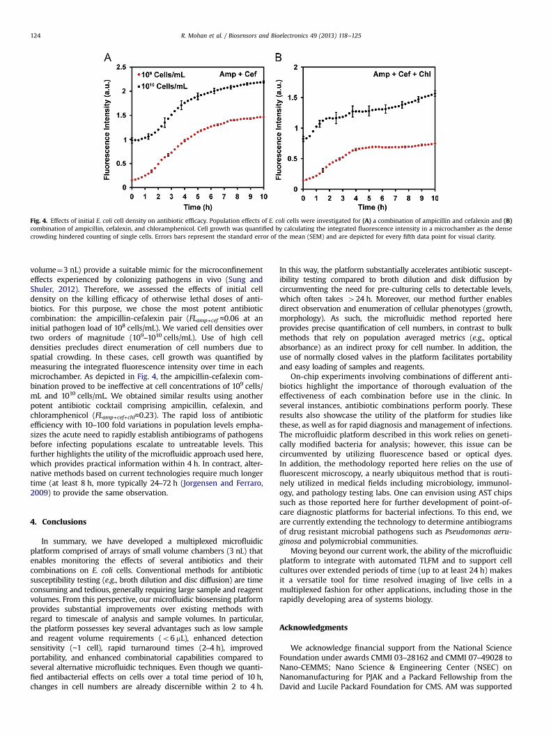
Fig. 4. Effects of initial E. coli cell density on antibiotic efficacy. Population effects of E. coli cells were investigated for (A) a combination of ampicillin and cefalexin and (B)combination of ampicillin, cefalexin, and chloramphenicol. Cell growth was quantified by calculating the integrated fluorescence intensity in a microchamber as the densecrowding hindered counting of single cells. Errors bars represent the standard error of the mean (SEM) and are depicted for every fifth data point for visual clarity.
R. Mohan et al. / Biosensors and Bioelectronics 49 (2013) 118–125124
volume¼3 nL) provide a suitable mimic for the microconfinementeffects experienced by colonizing pathogens in vivo (Sung andShuler, 2012). Therefore, we assessed the effects of initial celldensity on the killing efficacy of otherwise lethal doses of anti-biotics. For this purpose, we chose the most potent antibioticcombination: the ampicillin-cefalexin pair (FLamp+cef ≈0.06 at aninitial pathogen load of 108 cells/mL). We varied cell densities overtwo orders of magnitude (109–1010 cells/mL). Use of high celldensities precludes direct enumeration of cell numbers due tospatial crowding. In these cases, cell growth was quantified bymeasuring the integrated fluorescence intensity over time in eachmicrochamber. As depicted in Fig. 4, the ampicillin-cefalexin com-bination proved to be ineffective at cell concentrations of 109 cells/mL and 1010 cells/mL. We obtained similar results using anotherpotent antibiotic cocktail comprising ampicillin, cefalexin, andchloramphenicol (FLamp+cef+chl≈0.23). The rapid loss of antibioticefficiency with 10–100 fold variations in population levels empha-sizes the acute need to rapidly establish antibiograms of pathogensbefore infecting populations escalate to untreatable levels. Thisfurther highlights the utility of the microfluidic approach used here,which provides practical information within 4 h. In contract, alter-native methods based on current technologies require much longertime (at least 8 h, more typically 24–72 h (Jorgensen and Ferraro,2009) to provide the same observation.
4. Conclusions
In summary, we have developed a multiplexed microfluidicplatform comprised of arrays of small volume chambers (3 nL) thatenables monitoring the effects of several antibiotics and theircombinations on E. coli cells. Conventional methods for antibioticsusceptibility testing (e.g., broth dilution and disc diffusion) are timeconsuming and tedious, generally requiring large sample and reagentvolumes. From this perspective, our microfluidic biosensing platformprovides substantial improvements over existing methods withregard to timescale of analysis and sample volumes. In particular,the platform possesses key several advantages such as low sampleand reagent volume requirements (o6 mL), enhanced detectionsensitivity (∼1 cell), rapid turnaround times (2–4 h), improvedportability, and enhanced combinatorial capabilities compared toseveral alternative microfluidic techniques. Even though we quanti-fied antibacterial effects on cells over a total time period of 10 h,changes in cell numbers are already discernible within 2 to 4 h.
In this way, the platform substantially accelerates antibiotic suscept-ibility testing compared to broth dilution and disk diffusion bycircumventing the need for pre-culturing cells to detectable levels,which often takes 424 h. Moreover, our method further enablesdirect observation and enumeration of cellular phenotypes (growth,morphology). As such, the microfluidic method reported hereprovides precise quantification of cell numbers, in contrast to bulkmethods that rely on population averaged metrics (e.g., opticalabsorbance) as an indirect proxy for cell number. In addition, theuse of normally closed valves in the platform facilitates portabilityand easy loading of samples and reagents.
On-chip experiments involving combinations of different anti-biotics highlight the importance of thorough evaluation of theeffectiveness of each combination before use in the clinic. Inseveral instances, antibiotic combinations perform poorly. Theseresults also showcase the utility of the platform for studies likethese, as well as for rapid diagnosis and management of infections.The microfluidic platform described in this work relies on geneti-cally modified bacteria for analysis; however, this issue can becircumvented by utilizing fluorescence based or optical dyes.In addition, the methodology reported here relies on the use offluorescent microscopy, a nearly ubiquitous method that is routi-nely utilized in medical fields including microbiology, immunol-ogy, and pathology testing labs. One can envision using AST chipssuch as those reported here for further development of point-of-care diagnostic platforms for bacterial infections. To this end, weare currently extending the technology to determine antibiogramsof drug resistant microbial pathogens such as Pseudomonas aeru-ginosa and polymicrobial communities.
Moving beyond our current work, the ability of the microfluidicplatform to integrate with automated TLFM and to support cellcultures over extended periods of time (up to at least 24 h) makesit a versatile tool for time resolved imaging of live cells in amultiplexed fashion for other applications, including those in therapidly developing area of systems biology.
Acknowledgments
We acknowledge financial support from the National ScienceFoundation under awards CMMI 03–28162 and CMMI 07–49028 toNano-CEMMS; Nano Science & Engineering Center (NSEC) onNanomanufacturing for PJAK and a Packard Fellowship from theDavid and Lucile Packard Foundation for CMS. AM was supported

R. Mohan et al. / Biosensors and Bioelectronics 49 (2013) 118–125 125
in part by FMC Technologies Graduate Fellowship. We thankDr. Amit Desai and Dr. Ashtamurthy Pawate for helpful discussionsand for proof-reading the manuscript. In addition, we acknowl-edge Dr. Desai's assistance in designing the antibiotic absorptionstudies. Finally, we thank Kevin B. Weyant for assistance withbacterial cell cloning.
Appendix A. Supporting information
Supplementary data associated with this article can be found inthe online version at http://dx.doi.org/10.1016/j.bios.2013.04.046.
References
Alanis, A.J., 2005. Archives of Medical Research 36 (6), 697–705.Allewelt, M., Schuler, P., Bolcskei, P.L., Mauch, H., Lode, H., Dalhoff, K., Loos, U.,
Vogel, F., Wendel, H., von Eiff, C., 2004. Clinical Microbiology and Infection 10(2), 163–170.
Ang, B.S.P., 2001. Annals of the Academy of Medicine Singapore 30 (2), 199–202.Balaban, N.Q., Merrin, J., Chait, R., Kowalik, L., Leibler, S., 2004. Science 305 (5690),
1622–1625.Boedicker, J.Q., Li, L., Kline, T.R., Ismagilov, R.F., 2008. Lab on a Chip 8 (8),
1265–1272.Butler, M.T., Wang, Q., Harshey, R.M., 2010. Proceedings of the National Academy of
Sciences of the United States of America 107 (8), 3776–3781.Chait, R., Craney, A., Kishony, R., 2007. Nature 446 (7136), 668–671.Chen, C.H., Lu, Y., Sin, M.L.Y., Mach, K.E., Zhang, D.D., Gau, V., Liao, J.C., Wong, P.K.,
2010. Analytical Chemistry 82 (3), 1012–1019.Chiang, Y.-L., Lin, C.-H., Yen, M.-Y., Su, Y.-D., Chen, S.-J., Chen, H.-f., 2009. Biosensors
and Bioelectronics 24 (7), 1905–1910.Choi, J., Jung, Y.-G., Kim, J., Kim, S., Jung, Y., Na, H., Kwon, S., 2013. Lab on a Chip 13
(2), 280–287.Churski, K., Kaminski, T.S., Jakiela, S., Kamysz, W., Baranska-Rybak, W., Weibel, D.B.,
Garstecki, P., 2012. Lab on a Chip 12 (9), 1629–1637.Cira, N.J., Ho, J.Y., Dueck, M.E., Weibel, D.B., 2012. Lab on a Chip 12 (6), 1052–1059.Cottarel, G., Wierzbowski, J., 2007. Trends in Biotechnology 25 (12), 547–555.Dawson, C.C., Intapa, C., Jabra-Rizk, M.A., 2011. PLoS Pathogens 7 (7).Dejace, P., Klastersky, J., 1986. American Journal of Medicine 80 (6 B), 29–38.Dorr, T., Lewis, K., Vulic, M., 2009. PLoS Genetics 5 (12).Gales, A.C., Reis, A.O., Jones, R.N., 2001. Journal of Clinical Microbiology 39 (1),
183–190.Goldstein, F.W., Ly, A., Kitzis, M.D., 2007. Journal of Antimicrobial Chemotherapy 59
(5), 1039–1040.Ho, J.Y., Cira, N.J., Crooks, J.A., Baeza, J., Weibel, D.B., 2012. PLoS ONE 7 (7).Jorgensen, J.H., Ferraro, M.J., 2009. Clinical Infectious Diseases 49 (11), 1749–1755.Kalashnikov, M., Lee, J.C., Campbell, J., Sharon, A., Sauer-Budge, A.F., 2012. Lab on a
Chip.Karasinski, J., White, L., Zhang, Y., Wang, E., Andreescu, S., Sadik, O.A., Lavine, B.K.,
Vora, M., 2007. Biosensors and Bioelectronics 22 (11), 2643–2649.
Keith, C.T., Borisy, A.A., Stockwell, B.R., 2005. Nature Reviews Drug Discovery 4 (1),71–78.
Keymer, J.E., Galajda, P., Muldoon, C., Park, S., Austin, R.H., 2006. Proceedings of theNational Academy of Sciences of the United States of America 103 (46),17290–17295.
Kinnunen, P., Sinn, I., McNaughton, B.H., Newton, D.W., Burns, M.A., Kopelman, R.,2011. Biosensors and Bioelectronics 26 (5), 2751–2755.
Koydemir, H.C., Kulah, H., Ozgen, C., Alp, A., Hascelik, G., 2011. Biosensors andBioelectronics 29 (1), 1–12.
Lazcka, O., Campo, F.J.D., Munoz, F.X., 2007. Biosensors and Bioelectronics 22 (7),1205–1217.
Lewis, K., 2010. Persister cells. Annual Review of Microbiology. 64, 357–372.Lo-Ten-Foe, J.R., De Smet, A.M.G.A., Diederen, B.M.W., Kluytmans, J.A.J.W., Van
Keulen, P.H.J., 2007. Antimicrobial Agents and Chemotherapy 51 (10),3726–3730.
Lorian, V., 1986. Antibiotics in Laboratory Medicine. Williams & Wilkins, Philadelphia.Lutz, R., Bujard, H., 1997. Nucleic Acids Research 25 (6), 1203–1210.Mancini, N., Carletti, S., Ghidoli, N., Cichero, P., Burioni, R., Clementi, M., 2010.
Clinical Microbiology Reviews 23 (1), 235–251.Martindale, W., 1989. Martindale: The Extra Pharmacopoeia. Pharmaceutical Press,
London.Mohan, R., Schudel, B.R., Desai, A.V., Yearsley, J.D., Apblett, C.A., Kenis, P.J.A., 2011.
Sensors and Actuators B Chemical 160 (1), 1216–1223.Mouton, J.W., 1999. Infection 27 (SUPPL. 2), S24–S28.Nakamura, N., Shigematsu, A., Matsunaga, T., 1991. Biosensors and Bioelectronics 6
(7), 575–580.Nicodemo, A.C., Araujo, M.R.E., Ruiz, A.S., Gales, A.C., 2004. Journal of Antimicrobial
Chemotherapy 53 (4), 604–608.Rasband, W., 2011. Image J.Rice, L.B., 2010. Infection Control and Hospital Epidemiology 31 (SUPPL. 1), S7–S10.Rolinson, G.N., 1980. Journal of General Microbiology 120 (2), 317–323.Schudel, B.R., Choi, C.J., Cunningham, B.T., Kenis, P.J.A., 2009. Lab on a Chip 9 (12),
1676–1680.Schudel, B.R., Tanyeri, M., Mukherjee, A., Schroeder, C.M., Kenis, P.J.A., 2011. Lab on a
Chip 11 (11), 1916–1923.Sia, S.K., Whitesides, G.M., 2003. Electrophoresis 24 (21), 3563–3576.Sun, P., Liu, Y., Sha, J., Zhang, Z., Tu, Q., Chen, P., Wang, J., 2011. Biosensors and
Bioelectronics 26 (5), 1993–1999.Sung, J., Shuler, M., 2012. Annals of Biomedical Engineering 40 (6), 1289–1300.Tan, T.Y., Ng, S.Y., 2007. Clinical Microbiology and Infection 13 (5), 541–544.Theberge, A.B., Courtois, F., Schaerli, Y., Fischlechner, M., Abell, C., Hollfelder, F.,
Huck, W.T.S., 2010. Angewandte Chemie - International Edition 49 (34),5846–5868.
Thorson, M.R., Goyal, S., Gong, Y., Zhang, G.G.Z., Kenis, P.J.A., 2011. CrystEngComm.14 (7), 2404–2412.
Tipper, D.J., 1985. Pharmacology and Therapeutics 27 (1), 1–35.Traub, W.H., 1970. Applied Microbiology 20 (1), 98–102.Tsou, P.-H., Sreenivasappa, H., Hong, S., Yasuike, M., Miyamoto, H., Nakano, K.,
Misawa, T., Kameoka, J., 2010. Biosensors and Bioelectronics 26 (1), 289–294.Udekwu, K.I., Parrish, N., Ankomah, P., Baquero, F., Levin, B.R., 2009. Journal of
Antimicrobial Chemotherapy 63 (4), 745–757.White, R.L., Burgess, D.S., Manduru, M., Bosso, J.A., 1996. Antimicrobial Agents and
Chemotherapy 40 (8), 1914–1918.Winslow, D.L., Damme, J., Dieckman, E., 1983. Antimicrobial Agents and Che-
motherapy 23 (4), 555–558.Xia, Y., Whitesides, G.M., 1998. Annual Review of Materials Science 28 (1), 153–184.