bk virus-plasmid expression vector that persists episomally in
TRANSCRIPT
MOLECULAR AND CELLULAR BIOLOGY, Aug. 1984. p. 1551-1560 Vol. 4. No. 80270-7306/84/081551-10$02.00/0Copyright © 1984, American Society for Microbiology
BK Virus-Plasmid Expression Vector That Persists Episomally inHuman Cells and Shuttles into Escherichia coli
GABRIELE MILANESI,' GIUSEPPE BARBANTI-BRODANO,' MASSIMO NEGRINI,' DOUGLAS LEE,3tALFREDO CORALLINI,2 ANTONELLA CAPUTO,2 MARIA P. GROSSI,2 AND ROBERT P. RICCIARDI3*
Institute of Biochemical Genetics, National Research Couincil, I-27100 Pavia, 1 and Institiute of Microbiology, School ofMedicine, University of Ferrara, 144100 Ferrara,2 Ital', and The Wistar Institiute of Anatomy and Biology, Philadelphia,
Pennsvlvania 191043
Received 16 February 1984/Accepted 14 May 1984
We describe a novel expression vector, pBK TK-1, that persists episomally in human cells that can beshuttled into bacteria. This vector includes sequences from BK virus (BKV), the thymidine kinase (TK) gene ofherpes simplex virus type 1, and plasmid pML-1. TK+-transformed HeLa and 143 B cells containedpredominantly full-length episomes. There were typically 20 to 40 (HeLa) and 75 to 120 143 B vector copies percell, although some 143 B transformants contained hundreds. Low-molecular-weight DNA from TK+-transformed cells introduced into Escherichia coli were recovered as plasmids that were indistinguishable fromthe input vector. Removal of selective pressure had no apparent effect upon the episomal status of pBK TK-1molecules in TK+-transformed cells. BKV T antigen may play a role in episomal replication of pBK TK-1 sincethis viral protein was expressed in TK+ transformants and since a plasmid that contained only the BKV originof replication was highly amplified in BKV-transformed human cells that synthesize BKV T antigen.
Development of eucaryotic vectors has provided a directand convenient means of introducing additional and novelgenetic information into cultured cells. Essentially, portionsof all eucaryotic vectors include sequences derived fromanimal viruses (for extensive reviews, see references 12, 39,and 40). Expression systems vary widely in the way they are
used. For example, infection of permissive cells occurs withparticular vectors of simian virus 40 (SV40) (13, 17, 34),adenovirus (47, 53), herpes simplex virus (50), vaccinia virus(37), and retroviruses (46, 57). However, short-term or
transient expression, which is noninfectious, has been de-scribed with SV40 vectors that become highly amplified inCOS cells (11), e.g., 200,000 episomal copies per cell within48 h of posttransfection (31). In contrast, permanent celllines which integrate the vector DNA have been obtained intwo ways by using selectable marker genes. The first hasbeen by "per-force" selection, using the thymidine kinase(TK) gene of herpes simplex virus type 1 (HSV-1) in TK-deficient mutant cells (44, 58). The second has been bydominant selection in which expression of a dominant mark-er gene (e.g., Escherichia coli xanthine-guanine phosphori-bosyltransferase) is extended to nonmutated cells (33, 55). Inaddition, permanent mouse cell lines have been obtained bytransformation with bovine papillomavirus vectors which donot integrate into host cell DNA but replicate episomally andwhich can be shuttled into bacteria (6, 43).We report here the development of a virus-plasmid
expression vector, pBK TK-1, that has the unusual featureof being able to persist episomally in human cells. Thisexpression vector contains DNA sequences which originatefrom the human papovavirus, BK virus (BKV). BKV wasisolated from the urine of a renal transplant recipient onimmunosuppressive therapy (9) and is ubiquitous in thehuman population (2, 8). BKV efficiently infects human cellsand transforms hamster, mouse, rat, rabbit, and monkeycells in tissue culture (36). The genomic organization of the
* Corresponding author.t Present address: Temple Medical School, Philadelphia, PA
19104.
1551
circular double-stranded DNA of BKV is remarkably similarto that of SV40, even though these viruses are infectious fordifferent hosts. In fact, an homology of more than 80% isfound in the nucleotide sequences of the two viruses (45, 59).As in SV40, both large T and small t antigens of BKV aretranslated from mRNAs which initiate their transcription onone strand near the putative origin of replication (20). Inparallel to SV40, capsid proteins of BKV (VP1, VP2, andVP3) are produced after DNA synthesis from late mRNAswhich initiate transcription proximal to the 5' end of earlymRNAs on the opposite strand (20). One notable distinctionbetween the genomes of SV40 and BKV is the extensivedivergence in the DNA sequences of their tandem repeatswhich constitute transcriptional enhancer regions (41).Human cells persistently infected with BKV as well as
transformed human cells have been isolated (16, 35). In someof these viral host cell relationships, the BKV DNA residesas apparent full-length episomes, whereas in others, theBKV DNA may be free but may contain deletions or beintegrated (16. 35). Although the mechanisms for episomalpersistence of BKV DNA are not understood, we haveexploited this basic feature to produce a recombinant DNAvector which will persist as an amplified episomal resident inhuman cells. The potential usefulness of this vector foranalyzing the expression of artificially acquired genes inhuman cells is discussed.
MATERIALS AND METHODSPlasmids, virus, and recombinant DNA constructions. Plas-
mid pML-1 (referred to here as pML) is a deletion derivativeof pBR322 (25). Plasmid pHSV106 (30) was used as thesource of the HSV-1 TK gene, inserted at the BamHI site.Propagation of BKV (Gardner strain) and extraction of viralDNA were by the procedure of Meneguzzi et al. (32). Detailsof the construction of the viral plasmids pBK TK-1 andpBODE are presented in the text. The source of restrictionenzymes was New England Biolaboratories and BethesdaResearch Laboratories, and T4 DNA ligase was from P-LBiochemicals. DNA fragments were isolated by the methodof Vogelstein and Gillespie (56). The above recombinant
Dow
nloa
ded
from
http
s://j
ourn
als.
asm
.org
/jour
nal/m
cb o
n 31
Dec
embe
r 20
21 b
y 12
1.23
3.14
0.20
7.

1552 MILANESI ET AL.
molecules were introduced by transformation and screenedand propagated in E. coli HblO1, C600, and MC-1061 (10,28), followed by standard isolation procedures (21, 38).
Cell cultures, DNA transfer, and selection of transformants.Human HeLa TK-deficient (TK-) cells and human 143 BTK- cells were used in these experiments. HeLa cells arederived from a human carcinoma, whereas 143 B cells arefrom a human osteosarcoma (4). TK- cells were maintainedin minimal essential medium (MEM) with 8% calf serumcontaining 30 jg of bromodeoxyuridine per ml. Beforetransfection, the HeLa TK- and 143 B TK- cells weregrown for one passage in the same medium without bromo-deoxyuridine. The BKV-transformed human embryonic fi-broblast cell line L603 (16) was maintained in MEM with10% fetal bovine serum at all times. Cells at ca. 60%confluency were supplied with fresh medium 4 h beforetransfection with supercoiled plasmid DNA (20 ,ug for about107 cells, unless otherwise indicated, and in the absence ofcarrier DNA) by the calcium phosphate precipitation proce-dure of Graham and van der Eb (14) as modified by Wigler etal. (58). In some instances, cells were shocked by 20%glycerol (7) 4 h after transfection; otherwise, the transfectionmedium was replaced directly with fresh nonselective medi-um. To select for TK+ HeLa and TK+ 143B transformants,cells were split 1:2 after 20 h and maintained in MEM with8% calf serum and HAT medium (100 ,uM hypoxanthine, 0.4,uM aminopterin, 16 puM thymidine; 24). Resulting cellcolonies were either individually cloned and expanded orpooled and grown as mass cultures.
Preparation and analysis of cellular DNA. Total cellularDNA was extracted as previously described (3, 15). Separa-tion of DNA into supernatant (nonchromosomal) and pellet(chromosomal) fractions was by the procedure of Hirt (18).Extraction of DNA from both fractions was by standardmethods, in which proteinase K (Boehringer MannheimBiochemicals) treatment (50 ,ug/ml, overnight at 37°C) wasfollowed by phenol extraction and ethanol precipitation. Thedrained pellets were dissolved in 10 mM Tris-hydrochloride(pH 7.8) and treated with RNase (25 ,ug/ml at room tempera-ture for 1 h), followed by two extractions in phenol andtwo in chloroform-isoamyl alcohol (24:1, vol/vol) and precip-itation in ethanol. The pellets were finally dissolved in 15mM NaCI-1.5 mM sodium citrate.
Cellular DNA was fractionated in agarose gels and trans-ferred to nitrocellulose filters by the Southern procedure(48). Probe DNA was labeled by nick translation (29) with[a-32P]dXTP (Amersham Corp.) and DNA polymerase I(Boehringer Mannheim Biochemicals). The hybridizationreactions were performed either (i) in 50% formamide with1x Denhardt solution (5), 4x SSC (0.6 M NaCl plus 0.06 Msodium citrate), and 4% sodium dodecyl sulfate at 37°C for36 h, followed by a series of aqueous washes at 68°C with thelast in 0.2x SCC-0.2% sodium dodecyl sulfate, or (ii)according to conditions described by Chenciner et al. (3).Filters were air dried and exposed to XRP-film (usually for 2to 24 h). The amount of free vector DNA in TK+ transform-ants was determined by reconstruction experiments, inwhich serial dilutions of cellular DNA were electrophoresedadjacent to known amounts ofpBK TK-1 DNA, blotted, andprobed with vector DNA. The copy number was calculatedfrom densitometric analysis of the autoradiograms carriedout with a Corning 750 scanner.Transformation of bacteria with cellular DNA and analysis.
E. coli HblOl or C600 was transformed with cellular DNAsby the calcium chloride procedure (27). E. coli MC-1061 wastransformed by the calcium chloride-rubidium chloride pro-
cedure (23). Plasmid DNA from transformants was isolatedby a rapid small-scale extraction procedure (19).
Immunoprecipitation and immunofluorescence. Labeling,immunoprecipitation, and fluorescent antibody staining ofBKV T antigen in TK+-transformed human cell lines wereperformed as described by Grossi et al. (16) with serum fromhamsters bearing tumors induced by BKV-transformed ham-ster cells.
RESULTSVector construction. Vector pBK TK-1 includes DNA
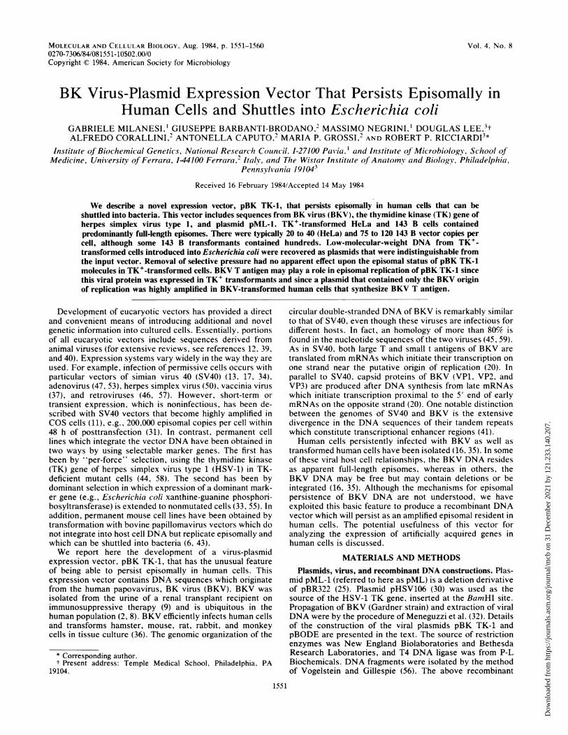
sequences originating from plasmid pML, BKV, and the TKgene of HSV-1. The location of these sequences within thisvector and its construction are depicted in Fig. 1. The pBKTK-1 vector was derived by digesting both plasmid pMLDNA and BKV DNA with EcoRI and BamHI (Fig. 1A). Thelarger of the respective EcoRI-BamHI fragments of pMLDNA (2,617 base pairs [bp]) aqd BKV DNA (5,089 bp) wereligated to produce pBK-1. A BamHI fragment of HSV-1DNA (3,600 bp) which contains the TK gene was introducedinto the single BamHI site of pBK-1 to generate vector pBKTK-1 (Fig. 1A). The pBK TK-1 molecule thus includesalmost the entire BKV genome (5,196 bp), except for 107 bpeliminated by digestion of BKV DNA with BamHI andEcoRI. This 107-bp fragment contained sequences from eachof the late viral capsid transcripts Vpl, Vp2, and Vp3 (54).Since BKV DNA is infectious for human cells, deletion ofthese late viral sequences was intended to obviate potentialrecombination and release of the complete viral genomefrom a vector containing full-length BKV DNA.The topographical arrangement of the transcripts and
pertinent coding regions of the genes contained within pBKTK-1 are depicted in Fig. 1B. The essential features of pBKTK-1, in addition to encoding the ampicillin resistance geneof pML and the TK gene of HSV-1, are that it contains theentire early region sequences which encode BKV large Tand small t antigens (45, 59). In addition, pBK TK-1 alsoincludes the BKV origin of replication based upon relatedsequence homology to SV4Q and the transcriptional en-hancer sequences (45, 59). The late genes of BKV in pBKTK-1 are not only missing 107 bp as described above but arealso disrupted by ligation ofpML DNA at the EcoRI site andof TK DNA at the BamHI site. Plasmid pBODE, shown inFig. 1A, is discussed later.pBK TK-1 vector persists episomally in human cells. Hu-
man TK- HeLa cells or 143 B cells were transfected withpBK TK-1 vector DNA and subjected to selection in HATmedium. Individually cloned or mass culture cell lines grownin HAT medium were then analyzed. Either total cellularDNA was extracted from TK+-transformed cells, or theDNA was separated into Hirt supernatant (HS) and Hirtpellet (HP) fractions. Essentially, HS DNA contains almostentirely free, low-molecular-weight DNA, whereas HP DNAcontains high-molecular-weight DNA that is most oftencontaminated with free DNA.The DNA from several TK+ clones of HeLa cells was
extracted and examined by Southern blot analysis (48) forthe presence of pBK TK-1 molecules. Analysis of HS DNAfrom two HeLa clones H-303 and H-384 (Fig. 2A, lanes 1 and2, respectively) revealed sequences that comigrated with thesupercoiled (form I), circular (form II), or linear (form III)structures of pBK TK-1. When this same DNA from cloneH-303 was digested with Sall (Fig. 2B, lane 2), which cutspBK TK-1 once, a single band comigrated with Sall-linear-ized pBK TK-1 DNA (Fig. 2B, lane 1). These resultsindicated that full-length pBK TK-1 molecules persist as free
MOL. CELL. BIOL.
Dow
nloa
ded
from
http
s://j
ourn
als.
asm
.org
/jour
nal/m
cb o
n 31
Dec
embe
r 20
21 b
y 12
1.23
3.14
0.20
7.

BKV EXPRESSION VECTOR PERSISTS EPISOMALLY 1553
A
H H
B
!5'5"
pBK TK-lFIG. 1. Construction of vectors. (A) pBK TK-1 was constructed from BKV, the TK gene of HSV-1, and plasmid pML. pBODE was
constructed from BKV and pML. The putative replication origin of BKV (0.67 map units) is contained in the HindIll C fragment designatedby the black box. The early and late transcription regions of BKV are designated by the dotted lines. The restriction sites are EcoRI (E),BamHI (B), and HindlIl (H). (B) Genomic landmarks in pBK TK-1. Locations of DNAs derived from pML, BKV, and TK (described above)in pBK TK-1 are represented in the double circle. Their respective transcripts are indicated by the thin lines, of which the dotted portionrepresents introns. Where pertinent, the orientation of the transcripts is shown 5' to 3'. Coding regions (thick black lines) contained withinspecific BKV transcripts indicate early proteins, large T and small t antigens, and late capsid proteins VP1 and VP2/3. The putative BKVreplication origin is represented by the black box within the circles. The bacterial ampicillin resistance gene of pML is designated by Ar. Therepresented restriction sites are EcoRI (E) and BamHI (B). Details are discussed in the text.
molecules in HeLa cells. To further characterize the state ofvector DNA in HeLa cells, total DNA from six differentTK+ cloned lines was digested with BamHI, which cutspBK TK-1 DNA twice to generate 3,600- and 7,706-bpfragments. Five of the HeLa clones (Fig. 2C, lanes 1, 2, 3, 5,and 6) contained only two bands which comigrated with the3,600 and 7,706-bp marker fragments (Fig. 2C, lane 7), whichwas expected if pBK TK-1 was episomal. Only one HeLacell clone (Fig. 2C, lane 4) contained an additional bandwhich may have originated from either integrated or rear-ranged episomal molecules ofpBK TK-1 DNA; the addition-al bands in lane 3 represent incompletely digested free pBKTK-1 DNA, since they comigrated with undigested pBK TK-1 DNA (lane 8). It is noted that although the autoradiogramshown in Fig. 2C had been overexposed, minor bands ofpBK TK-1 which could represent integrated sequences arenot detected. These results again suggested that in HeLacells pBK TK-1 molecules exist as full-length episomes.The transfected 143 B TK+ cell clones were also analyzed
for the presence of pBK TK-1 molecules. Undigested HS
DNA from each of several clones (Fig. 3A, lanes 2 to 5)revealed molecules which comigrated with pBK TK-1 mark-er DNA (Fig. 3A, lane 1). The HS DNA from two of thesecell clones, after digestion with Sall which cuts pBK TK-1once, yielded single bands (Fig. 3B, lanes 2 and 3), whichcomigrated with SalI-linearized pBK TK-1 DNA (Fig. 3B,lane 1). Free forms of pBK TK-1 (50 to 100 genomeequivalents per diploid cell genome) were similarly observedwhen total cellular DNA from 143 B TK+ cell clones wasdigested with HpaI, a noncutter for pBK TK-1 (data notshown). Total cellular DNA from nine 143 B clones wasdigested with BamHI and analyzed for sequences of theexpected 7,706- and 3,600-bp fragments that would be gener-ated from nonintegrated molecules of pBK TK-1. Indeed, asseen in Fig. 3C (lanes 1 to 9), each 143 B clone containedboth expected BamHI fragments. However, some 143 B cellclones revealed novel bands which hybridized to the pBKTK-1 probe (Fig. 3C, lanes 1, 3, 4, and 8); the band below the7,706-bp fragment of clone B4 (lane 2) is probably undigestedvector DNA, since it comigrates with form I pBK TK-1
VOL. 4, 1984
Dow
nloa
ded
from
http
s://j
ourn
als.
asm
.org
/jour
nal/m
cb o
n 31
Dec
embe
r 20
21 b
y 12
1.23
3.14
0.20
7.

1554 MILANESI ET AL.
A B C
12 12 1234 5678
E_- -
mPI. .
39
_- 4
7.7- MOdwa__406. b
310* I
Ii..
FIG. 2. Southern blots of DNA from TK+-transformed HeLacell clones hybridized with a pBK TK-1 DNA probe. HS DNA ortotal cellular DNA was extracted from various individual TK+HeLa cell clones that had been transformed by pBK TK-1. HSDNAs (corresponding to 105 cells) and total cellular DNAs (10,Ug)were digested with restriction endonucleases as indicated, electro-phoresed through an 0.8% agarose gel, blotted onto nitrocellulose,and probed with nick-translated pBK TK-1 DNA. (A) UndigestedHS DNA from HeLa cell clones H-303 (lane 1) and H-384 (lane 2).The positions of forms I, II, and III of pBK TK-1 marker DNA areindicated. (B) HS DNA of HeLa clone H-303 digested with Sall(lane 2), and Sall-digested pBK TK-1 DNA (4.8 ng) as marker (lane1). (C) Total cellular DNA was digested with BamHI. Lanes 1 to 6:HeLa clones H-311; H-303; H-383; H-301; H-385; H-386, respec-tively. pBK TK-1 DNA marker (50 genome equivalents): digestedwith BamHl (lane 7) and undigested (lane 8).
DNA (lane 10). These novel sequences likely representpolymeric and deleted episomal forms of pBK TK-1 orpossibly integrated forms of the vector.We examined whether the property of pBK TK-1 mole-
cules to persist as episomes in individual cell clones wouldalso extend to a mass culture prepared by pooling hundredsof individual TK+ cell colonies. The DNA extracted fromsuch a mass culture of 143 B cells was separated into HS andHP fractions, which, respectively, were digested with five
A1 2345
"'p
m !'
I-
B1 23
restriction endonucleases that cut pBK TK-1 DNA twice(BamHI, XbaI, and KpnI) or three times (EcoRI and BglII)(Fig. 4A). As seen in Fig. 4B, in all instances, sequences ofpBK TK-1 found in both the HS (lanes 1, 4, 7, 10, and 13)and HP (lanes 2, 5, 8, 11, and 14) fractions after digestion bythese enzymes were those which comigrated with the corre-sponding digestions of marker pBK TK-1 DNA (lanes 3, 6, 9,12, and 15). The observation that most of the hybridizedsequences from each HP fraction comigrated with the re-spective restriction fragments of marker pBK TK-1 DNAindicated that most of these molecules originated from pBKTK-1 episomes and that only negligible amounts, if any, ofthis vector DNA integate into chromosomal DNA. Addition-al bands, particularly in the HS fraction, could representincompletely digested pBK TK-1 DNA molecules, freemolecules containing deletions, or degraded forms of pBKTK-1 DNA. Since these results were obtained with a massculture, they are representative of hundreds of individuallytransfected cells. This finding demonstrates clearly that inthe majority of independently transfected human cells pBKTK-1 molecules persist in the free state.The average number of free pBK TK-1 copies maintained
per cell was between 75 and 120 in 143 B cells and 20 and 40in HeLa cells. It is important to note that in some 143 Bclones (e.g., clones B-23 and B-36; Fig. 3C, lanes 7 and 9),many times this number of copies persisted.pBK TK-1 DNA can be shuttled from human cells into E.
coli. To further substantiate that pBK TK-1 DNA moleculespersist as stable replicating episomes in human cells, HSDNAs from both HeLa and 143 B cell clones were used totransform E. coli to an ampicillin-resistant phenotype. In oneexperiment, half of a sample of the HS DNA from two clonesof HeLa cells and six clones of 143 B cells was directly usedto transform E. coli MC-1061, whereas the other half wastreated with the restriction endonuclease MboI (which has17 restriction sites within pBK TK-1 DNA) before transfor-mation. DNA is not cut when the adenine within therecognition sequence of MboI (GATC) is methylated, asoccurs in most E. coli strains including HblOl and MC-1061.
C1 2 3 4 5 6 7 8 9 10
I:~~ -
_|S"--|+| I
FIG. 3. Southern blots ofDNA from TK+-transformed 143 B cell clones hybridized with pBK TK-1DNA. HS DNA or total cellular DNAwas extracted from various individual TK+ 143 B cell clones that had been transformed by pBK TK-1 DNA. HS DNAs (corresponding to 105cells) and total cellular DNAs (10 JLg) were digested with the indicated restriction endonucleases, electrophoresed through a 0.6% agarose gel,blotted onto nitrocellulose, and probed with nick-translated pBK TK-1 DNA. (A) HS DNA from 143 B clones B-6, B-27, B-31, and B-20 (lanes2 to 5, respectively); lane 1, 4.8 ng of pBK TK-1 DNA marker. (B) Sall digestion of HS DNA of 143 B clones B-31 (lane 2) and B-27 (lane 3);lane 1, 4.8 ng of SalI-digested pBK TK-1 marker DNA (arrow). (C) Total cellular DNA from 143 B clones digested with BamHI; lanes 1 to 9,143 B clones B-1, B-4, B-6, B-8, B-18, B-20, B-23, B-31, and B-36, respectively; lane 10, undigested pBK TK-1 DNA marker (100 genomeequivalents). The upper and lower arrows indicate the positions of the 7,706 and 3,600-bp fragments, respectively, generated by BamHIdigestion of pBK TK-1 DNA.
MOL. CELL. BIOL.
Dow
nloa
ded
from
http
s://j
ourn
als.
asm
.org
/jour
nal/m
cb o
n 31
Dec
embe
r 20
21 b
y 12
1.23
3.14
0.20
7.

BKV EXPRESSION VECTOR PERSISTS EPISOMALLY 1555
TABLE 1. Transformation of E. coli MC-1061 with HS DNAsfrom HeLa and 143 B cell clones transformed to the TK+
phenotype by pBK TK-1
No. of Ampr bacterialcolonies'
Cell clonesUntreated Treated with
Untreated MboIHeLa H-303 28 NDHeLa H-384 114 ND143 B 656 95 0143 B 657 233 0143 B F6-D4 30 0143 B 633 28 0143 B B-27 25 0143 B B-3 150 0pBK TK-1 DNA (control, 10 ng) 26,400 21,000
a Ampr bacterial colonies were produced by transformation of E. coli withsamples of HSs obtained from 105 cells. ND, Not done.
9 10 11 12 13 14 15
FIG. 4. (A) Positions ofBamHI (B), BglII (Bg), EcoRI (E), KpnI(K), and Xbal (X) restriction sites in pBK TK-1. The unique Sall (S)site is suitable for insertion of additional genes. (B) Southern blot ofDNA from TK+-transformed 143 B mass culture cells hybridized topBK TK-1 DNA. HS and HP DNAs were extracted from TK+ 143 Bmass culture cells, digested with restriction endonucleases BamHI,Xbal, EcoRI, BglII, and KpnI, electrophoresed through a 1.0%agarose gel, blotted onto nitrocellulose, and probed with nick-translated pBK TK-1 DNA. Lanes 1, 4, 7, 10, and 13 show HS DNAfrom 10W cells; lanes 2, 5, 8, 11, and 14 show HP DNA (10 ,ug); lanes3, 6, 9, 12, and 15 show pBK TK-1 marker DNA (1 ng or 50 genomeequivalents).
However, the DNA becomes sensitive to MboI digestionafter replication in eucaryotic cells, where the adenine is notmethylated. The untreated HS DNA from each HeLa and143 B cell clone did transform E. coli MC-1061, whereasdigestion by MboI rendered these same DNAs incapable oftransformation (Table 1). The transforming ability of pBKTK-1 DNA directly isolated from E. coli HblOl was unaf-fected by MboI treatment. These results indicate that pBKTK-1 does replicate as a free molecule in human cells andthat the gene for ampicillin resistance remains functional.
Replication of pBK TK-1 in TK+ transformants was
further supported by blot hybridization analysis. The HSDNA from each of seven 143 B TK+ individual clones wascompletely digested by MboI (lanes 3 to 9), whereas thecontrol pBK TK-1 was not (lanes 1 and 2) (Fig. 5).
In another experiment, plasmids recovered from E. coliMC-1061 transformed by HS DNAs from either HeLa or 143B TK+ cells were compared to input DNA. The plasmidsfrom ampicillin-resistant colonies were digested with EcoRI,
which cuts pBK TK-1 three times (Fig. 4A), and the frag-ments thus obtained were analyzed in agarose gels. As seenin Fig. 6A (lanes 1 to 12), plasmid DNA from each of 12ampicillin-resistant colonies transformed by the HS DNA ofHeLa cell line H-303 contained three EcoRI restrictionfragments that comigrated with those of pBK TK-1 DNA(lane 15). No other DNA bands were seen; the higher-molecular-weight bands seen in lanes 6 and 12 (Fig. 6A)probably represent incomplete digestions. A similar analysiswith the HS of an individual 143 B cell clone revealed thatthe plasmid DNAs from 11 of 12 bacterial transformantswere indistinguishable from that of the input vector (data notshown). In addition, the HS DNA from a mass culture of 143B cells was also used to transform E. coli C600. Plasmidsextracted from 18 ampicillin-resistant colonies were ana-
1 2 3 4 5 6 7 8 9
1ff
mI-
20
FIG. 5. Digestion with MboI and blot hybridization analysis of143 B cell clones transformed to the TK+ phenotype by pBK TK-1.HS DNA from 105 cells was restricted with MboI, migrated in a 1%agarose gel, transferred to nitrocellulose, and hybridized to a pBKTK-1 probe. Lanes 3 to 9, 143 B clones B-18, B-28, B-6, B-23, B-36,B-31, and B-2, respectively; lane 1, pBK TK-1 marker digested withMboI (2.4 ng); lane 2, pBK TK-1 digested with BamHI and MboI(2.4 ng). Forms I, II, and III of marker PBK TK-1 are indicated.
AE
Bg
BBam Hi Xba I Eco RI Bgi 11 Kpn I
r !
1 2 3 4 5 6 7 8
VOL. 4, 1984
Dow
nloa
ded
from
http
s://j
ourn
als.
asm
.org
/jour
nal/m
cb o
n 31
Dec
embe
r 20
21 b
y 12
1.23
3.14
0.20
7.

1556 MILANESI ET AL.
A1 2 3 4 5 6 7 8 9 10 1112 13 1415
B1 2 3 4 5 6 7 8 9 10 1112 1314 1516 17 181920
.4
.4
FIG. 6. pBK TK-1 from TK+-transformed HeLa and 143 B cellsis shuttled into E. coli and reestablished as plasmids. HS DNA fromTK+ HeLa and 143 B cells that had been transformed by pBK TK-1was used to transform E. coli MC-1061 (A) or C600 (B). PlasmidDNAs were prepared from E. coli ampicillin-resistant (Ampr) colo-nies, electrophoresed in 1% agarose gels, and visualized by ethidiumbromide staining at 280 nm. (A) EcoRI digestion of plasmids fromAmpr colonies, transformed by HS DNA of HeLa cell clone H-303(lanes 1 to 12). Marker pBK TK-1 was uncut (lane 14) or digestedwith EcoRl (lane 15). The arrows point to the three fragmentsgenerated by EcoRI digestion (Fig. 4A). Lane 13 is blank. (B)Plasmids (undigested) from Ampr colonies transformed by HS DNAfrom 143 B mass culture cells (lanes 2 to 19). Marker pBK TK-1DNA is in lanes 1 and 20. The lower arrow indicates the position ofsupercoiled (form I) pBK TK-1 plasmid DNA, and the upper arrowmarks its relaxed circular form (form II).
lyzed directly in agarose gels. As seen in Fig. 6B, 15 of 18colonies (lanes 2 to 19) contained supercoiled and relaxedcircular molecules that comigrated with form I and form IIDNA ofpBK TK-1 (lanes 1 and 20). Two plasmids containedan insertion (Fig. 6B, lanes 5 and 17) and a third plasmidcontained a deletion (lane 7), as confirmed by restriction-enzyme analysis (data not shown). These results show thatin human cell lines originating either from a single transfect-ed cell or from a mixture of hundreds of independentlytransfected cells, pBK TK-1 DNA persists as stable full-length episomes for many cell generations.pBK TK-1 vector continues to persist in the episomal state
after removal of selective growth medium. In all the experi-ments thus far described, the TK- human cells were placedunder conditions of selective growth after transfection bypBK TK-1 DNA. Since only TK+ cells are able to survive inHAT medium, it was not apparent whether selective pres-sure was required for maintenance of stable episomes.Therefore, an individual clone of 143 B cells (B-27) and amass culture of 143 B cells which contained pBK TK-1 DNAwere removed from the selective HAT medium and placedonto nonselective Eagle MEM.
Surprisingly, pBK TK-1 DNA molecules from the HS of
clone B-27 cells maintained in HAT medium for 200 genera-tions (Fig. 7A, lane 2) appeared qualitatively unchangedafter 25 generations of growth in nonselective MEM (Fig.7A, lane 3). Furthermore, replacement of HAT medium foran additional 25 generations after growth in MEM did notalter the persistence of full-length pBK TK-1 episomes; thehigh-molecular-weight comigrating bands probably repre-sent polymers of pBK TK-1 DNA (Fig. 7A, lane 4). Noapparent loss in the number of pBK TK-1 molecules wasdetected in B-27 cells upon removal or reintroduction ofHAT medium, since in each case (Fig. 7A, lanes 2 to 4) theamount of HS DNA analyzed was from the same number ofcells.A similar analysis of the 143 B mass culture also proved
that maintenance of stable pBK TK-1 episomal DNA inthese cells is not dependent upon selection in HAT medium.In this experiment, 143 B mass culture cells that had beenmaintained in HAT medium for 5 months were switched togrowth in nonselective MEM for 3 months (48 generations).As seen in Fig. 7B, the Southern blot of HS and HP fractionsdigested with BamHI reveals no obvious change in pBK TK-1 DNA after many cell doublings in nonselective medium(lane 1, HS; lane 2, HP) as compared with cells grown inHAT medium (lane 3, HS; lane 4, HP).pBK TK-1 synthesizes BKV T antigen. It is possible that
persistence of amplified episomes of pBK TK-1 in humancells may depend upon expression of BKV large T antigenencoded by the vector. This hypothesis seemed reasonable,since large T antigen of the closely related SV40 stimulatesits replication (for review, see reference 54). Both 143 B- andHeLa TK+-transformed cell lines were found to expressBKV T antigen. As seen in Fig. 8, immunoprecipitation ofthe 95,000-dalton BKV large T antigen from one of theselines occurs with a specific hamster serum to BKV T antigen
A
1 234B
1 234I .A -
.FwA
2-_j " ff > e& t
I--a-U-
FIG. 7. Blot hybridization of HS DNA from TK+-transformed143 B cells maintained with and without selective HAT medium.Undigested or BamHI-restricted DNA was electrophoresed througha 0.6% agarose gel (A) or a 1% agarose gel (B), blotted ontonitrocellulose, and probed with nick-translated pBK TK-1 DNA. (A)HS DNA (undigested and corresponding to 105 cells) from 143 B cellclone B-27, maintained first in HAT medium for more than 200generations (lane 2), then for 25 generations in nonselective MEM(lane 3), and then replaced into HAT medium for 25 generations(lane 4). pBK TK-1 marker DNA (2.4 ng), indicating forms I and II,is shown in lane 1. (B) BamHI-digested DNA from 143 B massculture cells that had been maintained for 5 months in HAT medium:HS DNA (lane 3) and HP DNA (lane 4). DNA from the same cellsafter 3 months in nonselective MEM: HS DNA (lane 1) and HPDNA (lane 2). Uppermost bands represent partial DNA digests.Positions of the two fragments generated by BamHI digestion ofpBK TK-1 are shown by the arrows.
MOL. CELL. BIOL.
Dow
nloa
ded
from
http
s://j
ourn
als.
asm
.org
/jour
nal/m
cb o
n 31
Dec
embe
r 20
21 b
y 12
1.23
3.14
0.20
7.

BKV EXPRESSION VECTOR PERSISTS EPISOMALLY 1557
1 234
95_**
45--
31- *
21-14 -S
FIG. 8. Immunoprecipitation of BKV T antigen from 143 B cellstransformed by pBK TK-1. TK+ 143 B cells (clone B-633) werelabeled with 32Pi, and anti-T hamster serum was used to immunopre-cipitate BKV T antigen as described in the text. The immunoprecipi-tated products were fractioned in a 10% polyacrylamide gel. La-beled cell extracts were reacted with normal hamster serum (lane 2),15 p.l of anti-T serum (lane 3), or 30 pul of anti-T serum (lane 4).Protein molecular weight standards in kilodaltons are in lane 1:phosphorylase b, 95; ovalbumin, 45; carbonic anhydrase, 31; trypsininhibitor, 21; lysozyme, 14. The arrow indicates the position of the95,000 molecular-weight BKV large T antigen.
(lanes 3 and 4), but not with a normal serum (lane 2). Inaddition, BKV T antigen was also detected by immunofluo-rescence in 143 B cells stably transformed to the TK+phenotype by pBK TK-1 (Fig. 9).pBK TK.1 contains an active origin of replication. As a
corollary to these findings, it was important to demonstratedirectly that pBK TK-1 DNA contains viral sequencesresponsible for replication in the presence of BKV T antigen.The BKV origin of replication, in fact, is predicted to belocated at 0.67 map units, based upon extensive sequencehomology to the known origin of replication in SV40 (45, 59).Therefore, the HindIll C fragment (555 bp) of BKV whichcontains sequences including the origin of replication wascloned into pML (Fig. 1A). The resulting recombinant,
referred to as pBODE (Fig. 1A), was transfected into L603cells. These cells are human embryonic fibroblasts that hadbeen transformed by a BKV DNA fragment containing theentire early region and are 100% positive for BKV T antigen(16). The intent was to see whether the pBODE molecule,containing the predicted BKV origin of replication but voidof any complete viral genes, would be amplified in cells thatconstitutively produce BKV T antigen. The HS DNA wasextracted at 16, 50, and 81 h posttransfection, linearized withBamHI, and analyzed by Southern blotting, using pBODE asthe labeled probe. The pBODE molecule is greatly ampli-fied, since densitometric analysis of the autoradiogramsshowed that the amount of pBODE DNA increases from0.04 ng at 16 h posttransfection (Fig. 10, lane 1) to 29 ng at 50h (lane 2) and then decreases to 3.7 ng at 81 h (lane 3).Therefore, reduction in copy number at 81 h posttransfectiondefines this amplification as transient. The pBODE mole-cules were not amplified in 143 B cells (data not shown).These results indicate that the 0.67-map-unit region of BKVpresent in the pBK TK-1 molecule serves as an activereplication origin which is stimulated by BKV T antigen.
DISCUSSIONWe describe an expression vector, pBK TK-1, which is
able to persist episomally in human cells. This vectorincludes sequences derived from BKV, a human papovavi-rus; pML, a deletion derivative of plasmid pBR322 (25); andthe TK gene of HSV-1, which transforms TK- mutant cellsto the TK+ phenotype (58). Free molecules of pBK TK-1were detected in the low-molecular-weight DNA fraction ofestablished TK+-transformed cells. Enzymatic expressionof the TK gene of HSV-1 was also detected in the TK+transformants (unpublished data). The origin of the humancell into which pBK TK-1 was introduced influenced to acertain extent its episomal status both quantitatively andqualitatively. In HeLa and 143 B TK+ cell transformants,the numbers of extrachromosomal copies of pBK TK-1 per
FIG. 9. Fluorescent antibody staining for BKV T antigen in 143 B TK+ cells transformed by pBK TK-1. Cells were first reacted with aspecific hamster serum to BKV T antigen and then with fluorescein-conjugated goat antibodies to hamster immunoglobulin G. (A) Clone B-28and (B) clone B-31.
VOL. 4, 1984
Dow
nloa
ded
from
http
s://j
ourn
als.
asm
.org
/jour
nal/m
cb o
n 31
Dec
embe
r 20
21 b
y 12
1.23
3.14
0.20
7.

1558 MILANESI ET AL.
1 2345
FIG. 10. Amplification of pBODE, a BKV origin recombinantplasmid, in BKV-transformed human cells positive for BKV Tantigen, as analyzed by Southern blot hybridization. The BKV-transformed human cells, L603, in which the BKV T-antigen gene isintegrated and expressed, were transfected with pBODE (10 jig ofDNA per 107 cells). At indicated times after transfection, cells were
rigorously washed, and HS DNA was prepared. Samples of HSDNA corresponding to 105 cells were digested with BamHl, electro-phoresed through a 0.6% agarose gel, blotted onto nitrocellulose,and probed with nick-translated pBODE DNA. Lane 1. 16 hposttransfection; lane 2, 50 h posttransfection; lane 3, 81 h post-transfection. Marker pBODE DNA cut with BamHI is shown in lane4 (3.5 ng) and lane 5 (0.07 ng). The arrow points to linear form III ofpBODE DNA.
cell were generally 20 to 40 and 75 to 120, respectively. InHeLa cells, pBK TK-1 molecules almost exclusively ap-peared as full-length episomes with undetectable rearrange-ments. Although all 143 B transformants contained full-length episomes, additional free forms that probablyincluded polymeric and deleted molecules were observed insome clones. The nature of these rearranged molecules isunknown. In this connection, it must be pointed out that inBKV-transformed human cells, where BKV DNA replicatesepisomally, free unintegrated polymeric forms of the viralgenome were constantly detected in considerable amounts(16). In all cases, however, full-length pBK TK-1 moleculeswere the predominant episomal form as specifically demon-strated in 143 B mass culture, cells, which represented a poolof hundrds of individual TK+ colonies. In both HeLa and143 B cells, integration of pBK TK-1 into chromosomalDNA appeared infrequent and certainly negligible in propor-tion to the number of amplified free molecules.The pBK TK-1 molecules from TK+-transformed human
cells can be readily shuttled into bacteria. Surprisingly,almost 85% of the Ampr bacterial colonies transformed byDNA from the 143 B TK+ mass culture cells containedplasmid DNA that was indistinguishable from the input pBKTK-1 vector DNA; this value was near 100% when DNAfrom a TK+ HeLa or 143 B cell clone was used. Further-more, plasmid DNA could not be rescued in bacteria whenthese cellular DNAs were digested with the endonucleaseMboI, which selectively restricts DNA that has replicated inanimal cells. Not only do these results demonstrate defini-tively that pBK TK-1 persists as stable replicons in humancells, but they also show that a resident bacterial gene,which confers no selective advantage to the eucaryotic cell,retains its capacity to function, i.e., the ,B-lactamase gene ofpML which confers ampicillin resistance in E. coli.
Importantly, episomal maintenance of pBK TK-1 in hu-man cells does not appear to depend upon forced expressionof the TK gene under selection for the TK+ phenotype, sincethe vector is not drastically affected by removal of HAT
medium. This is in contrast to a report which describes theloss of TK-plasmid recombinant episomes in transformedrodent cells after removal of selective medium (51). Ourexperiments indicate that the sequences inherent in the BKviral moiety of pBK TK-1 dictate the copy level and stabilityof the vector. This further suggests that any dominantselection marker gene (33) may be substituted for the TKgene of HSV-1 to thereby extend the spectrum of human celltypes into which this novel expression vector can be intro-duced and amplified. The possibility that an origin of replica-tion in HSV-1 TK gene could be responsible for the episomalstate of the vector, as suggested by others (51), was ruled outbecause only 0.2 copies of free plasmid DNA per cell weredetected in HS DNA of 143 B cells transformed to the TK+phenotype by a recombinant molecule containing only pMLand the HSV-1 TK gene. In this case, essentially all vectorDNA sequences were found to be integrated into high-molecular-weight DNA of the TK+ transformants (G. Bar-banti-Brodano, unpublished data).The features which control the episomal replication func-
tion of pBK TK-1 are unknown. It seems unlikely that thelate BK viral genes are involved in episomal maintenance ofpBK TK-1 since they were partially deleted to circumventpotential infection. However, the early viral large T- andsmall t-antigen genes are completely encoded in pBK TK-1.It is likely that the immunoreactive BKV large T antigen inthe TK+-transformed cells stimulates the BKV origin ofreplication in pBK TK-1 analogous to SV40 T-antigen-dependent replication (54). The supportive argument for thismechanism is demonstrated by the great amplification ofplasmid pBODE, which contains the putative BKV origin ofreplication (0.67 viral map units; 42, 45, 59), but is void ofcomplete early and late BKV genes, in BKV-transformedhuman cells that constitutively synthesize BKV T antigen.
This BKV T-antigen-dependent replication of pBODE issimilar to transient replication of SV40 vectors in COS cells(31). It is intriguing to consider that in TK+ transformantsthe copy level of pBK TK-1 may be held constant byautoregulation of BKV T-antigen synthesis, similar to con-trol of SV40 T-antigen production during lytic infection (1,22, 52). We have begun constructing deletion mutants in theT-antigen gene of pBK TK-1 to define potential elementsnecessary for episomal replication. Alternatively, mainte-nance of the episomal state of pBK TK-1 in human cells maydepend on specific viral sequences, not necessarily relatedto the T-antigen coding region, similar to what has beenshown for vectors based on bovine papillomavirus DNA(26). A search for such sequences in the BKV genome isunder way. Finally, it cannot be ruled out that replication ofthe vector is also under the control of some host cellcomponents.The pBK TK-1 molecule represents the first stable expres-
sion vector that persists episomally in human cells and thatcan be shuttled into bacteria. These features are also sharedby the bovine papillomavirus-plasmid vectors that functionin mouse cells (6, 43). Key advantages and potential uses ofthe episomal pBK TK-1 expression vector are the following.(i) The inserted gene of interest cannot be interrupted orsubjected to regulatory constraints that frequently occurfrom integration into cellular DNA. (ii) The gene of interestis amplified with the vector. (iii) The vector can be used tostudy genes which are normally expressed or require expres-sion in human cells. For example, we have produced humancell lines in which the episomal vector contains adenovirustype 12 transforming genes and expresses functional andimmunoreactive adenovirus type 12 ElA proteins (R. Vasa-
MOL. CELL. BIOL.
Dow
nloa
ded
from
http
s://j
ourn
als.
asm
.org
/jour
nal/m
cb o
n 31
Dec
embe
r 20
21 b
y 12
1.23
3.14
0.20
7.

BKV EXPRESSION VECTOR PERSISTS EPISOMALLY 1559
vada, G. Barbanti-Brodano, and R. P. Ricciardi, unpub-lished data). The pBK TK-1 vector contains restriction sitesconvenient for different cloning strategies. (v) Substitutionof the TK gene with a dominant selection gene such as Tn5aminoglycoside phosphotransferase (49) or E. coli xanthine-guanine phosphoribosyltransferase (33) in the vector predict-ably will expand the variety of human cells that can be used.(vi) The system may be applied in transient assays as
suggested from this study with pBODE in L603 cells.Indeed, we have already prepared a pBODE-CAT (chloram-phenicol acetyltransferase) recombinant that transiently ex-
presses high enzyme activity in 143 B TK+ transformantscontaining pBK TK-1 (A. Caputo, G. Barbanti-Brodano, andR. P. Ricciardi, unpublished data). (vii) The pBK TK-1recombinant may be modified for the insertion and expres-
sion of cDNAs.ACKNOWLEDGMENTS
We are grateful to and acknowledge support of Carlo M. Croce(The Wistar Institute) in assisting in this project.
This work was supported by Public Health Service grant CA-29797 from the National Cancer Institute and a Ruth Estrin Gold-berg Memorial for Cancer Research grant to R.P.R. This work was
also supported by Consiglio Nazionale delle Ricerche-ProgettoFinalizzato 'Ingegneria Genetica e Basi Molecolari delle MalattieEreditarie" and by North Atlantic Treaty Organization researchgrant 284.81.
LITERATURE CITED1. Alwine, J. C., S. I. Reed, and G. R. Stark. 1977. Characteriza-
tion of the autoregulation of simian virus 40 gene A. J. Virol.24:22-27.
2. Brown, P., T. Tsai, and D. C. Gajdusek. 1975. Seroepidemiologyof human papovaviruses: discovery of virgin populations andsome unusual patterns of antibody prevalence among remotepeoples of the world. Am. J. Epidemiol. 102:331-340.
3. Chenciner, N., M. P. Grossi, G. Meneguzzi, A. Corallini, R.Manservigi, G. Barbanti-Brodano, and G. Milanesi. 1980. Stateof viral DNA in BK virus-transformed rabbit cells. Virology103:138-148.
4. Croce, C. M., J. Barrick, A. Linnenbach, and H. Koprowski.1979. Expression of malignancy in hybrids between normal andmalignant cells.J. Cell Physiol. 99:279-286.
5. Denhart, D. T. 1966. A membrane-filter technique for thedetection of complementary DNA. Biochem. Biophys. Res.Commun. 23:641-646.
6. Dimaio, D., R. Treisman, and T. Maniatis. 1982. Bovine papillo-ma-virus that propagates as a plasmid in both mouse andbacterial cells. Proc. Natl. Acad. Sci. U.S.A. 79:4030-4034.
7. Frost, E., and J. Williams. 1978. Mapping temperature-sensitiveand host-range mutations of adenovirus type 5 by markerrescue. Virology 91:39-50.
8. Gardner, S. D. 1973. Prevalence in England of antibodies tohuman polyomavirus (BK). Br. Med. J. 1:77-78.
9. Gardner, S. D., A. M. Field, D. V. Coleman, and B. Hulme.1971. New human papovavirus (BK) isolated from urine afterrenal transplantation. Lanceti:1253-1257.
10. Gergen, J. P., R. H. Stern, and P. C. Wensink. 1979. Filterreplicas and permanent collections of recombinant DNA plas-mids. Nucleic Acids Res. 7:2115-2136.
11. Gluzman, Y. 1981.SV40-transformed simian cells support thereplication of earlySV40 mutants. Cell 23:175-182.
12. Gluzman, Y. 1982. Eukaryotic viral vectors. Cold Spring Har-bor Laboratory, Cold Spring Harbor, N.Y.
13. Goff, S. P., and P. Berg. 1976. Construction of hybrid virusescontainingSV40 and X phage DNA segments and their propaga-tion in cultured monkey cells. Cell 9:965-705.
14. Graham, F. L., and A. J. van der Eb. 1973. A new technique forthe assay of infectivity of human adenovirus5 DNA. Virology52:456-467.
15. Gross-Bellard, M., P. Oudet, and P. Chambon. 1973. Isolation of
high molecular weight DNA from mammalian cells. Eur. J.Biochem. 36:32-38.
16. Grossi, M. P., A. Caputo, G. Meneguzzi, A. Corallini, L. Carra,M. Portolani, M. Borgatti, G. Milanesi, and G. Barbanti-Bro-dano. 1982. Transformation of human embryonic fibroblasts byBK virus, BK virus DNA and a subgenomic BK virus DNAfragment. J. Gen. Virol. 63:393-403.
17. Hamer, D. H., D. Davoli, C. A. Thomas, and G. C. Fareed. 1977.Simian virus 40 carrying an Escherichia coli suppressor gene. J.Mol. Biol. 112:155-182.
18. Hirt, B. 1967. Selective extraction of polyoma DNA frominfected mouse cell cultures. J. Mol. Biol. 26:365-369.
19. Holmes, D. S., and M. Quigley. 1981. A rapid method for thepreparation of bacterial plasmids. Anal. Biochem. 114:193-197.
20. Howley, P. M. 1980. Molecular biology of SV40 and the humanpolyomaviruses BK and JC, p. 489-550. In G. Klein (ed.), Viraloncology. Raven Press, New York.
21. Katz, L., D. T. Kingsbury, and D. R. Helinski. 1973. Stimulationby cyclic adenosine monophosphate of plasmid deoxyribonucle-ic acid replication and catabolite repression of the plasmiddeoxyribonucleic acid-protein relaxation complex. J. Bacteriol.114:577-591.
22. Khoury, G., and E. May. 1977. Regulation of early and latesimian virus 40 transcription: overproduction of early viral RNAin the absence of a functional T-antigen. J. Virol. 23:167-176.
23. Kushner, S. R. 1978. An improved method for transformation ofEscherichia coli with Col El-derived plasmids, p. 17. In H.Boyer and S. Nicosia (ed.), Genetic engineering. Elsevier/North-Holland, Amsterdam.
24. Littlefield, J. W. 1965. The use of drug-resistant markers tostudy the hybridization of mouse fibroblasts. Exp. Cell Res.41:190-196.
25. Lusky, M., and M. Botchan. 1981. Inhibition ofSV40 replicationin simian cells by specific pBR322 DNA sequences. Nature(London) 293:79-81.
26. Lusky, M., and M. Botchan. 1984. Characterization of thebovine papilloma virus plasmid maintenance sequences. Cell36:391-401.
27. Mandel, M., and A. Higa. 1970. Calcium-dependent bacterio-phage DNA infection. J. Mol. Biol. 53:159-162.
28. Maniatis, T., E. F. Fritsch, and J. Sambrook. 1982. Molecularcloning: a laboratory manual. Cold Spring Harbor Laboratory,Cold Spring Harbor, N.Y.
29. Maniatis, T., S. G. Kee, A. Efstratiadis, and F. C. Kafatos. 1976.Amplification and characterization of a,B-globin gene synthe-sized in vitro. Cell 8:163-182.
30. McKnight, S. L., C. M. Croce, and R. Kingsbury. 1979. Intro-duction of isolated DNA sequences into cultured eukaryoticcells. Carnegie Inst. Wash. Year Book 78:56-61.
31. Mellon, P., V. Parker, Y. Gluzman, and T. Maniatis. 1981.Identification of DNA sequences required for transcription ofthe humanal-globin gene in a newSV40 host-vector system.Cell 27:279-288.
32. Meneguzzi, G., G. Barbanti-Brodano, and G. Milanesi. 1978.Transcription of BK virus DNA by Esclherichlia c oli RNApolymerase: size and sequence analysis of RNA. J. Virol.25:940-943.
33. Mulligan, R. C., and P. Berg. 1980. Expression of a bacterialgene in mammalian cells. Science 209:1422-1427.
34. Mulligan, R. C., B. H. Howard, and P. Berg. 1979. Synthesis ofrabbit P-globin in cultured monkey kidney cells following infec-tion with a SV40-,B-globin recombinant genome. Nature (Lon-don) 277:108-114.
35. Norkin, L.C. 1982. Papovaviral persistent infections. Microbiol.Rev. 46:384-425.
36. Padgett, B. 1980. Human papovaviruses, p. 339-370. In J.Tooze (ed.), Molecular biology of tumor viruses.II. DNA tumorviruses. Cold Spring Harbor Laboratory, Cold Spring Harbor,N.Y.
37. Panicali, D., and E. Paoletti. 1982. Construction of poxviruses ascloning vectors: insertion of the thymidine kinase gene fromherpes simplex virus into the DNA of infectious vaccinia virus.Proc. Natl. Acad. Sci. U.S.A. 79:4927-4931.
VOL. 4, 1984
Dow
nloa
ded
from
http
s://j
ourn
als.
asm
.org
/jour
nal/m
cb o
n 31
Dec
embe
r 20
21 b
y 12
1.23
3.14
0.20
7.

1560 MILANESI ET AL.
38. Radloff, F., W. Bauer, and J. Vinograd. 1967. A dye-buoyant-density method for the detection and isolation of closed circularduplex DNA: the closed circular DNA in HeLa cells. Proc.Natl. Acad. Sci. U.S.A. 57:1514-1521.
39. Rigby, P. W. J. 1982. Expression of cloned genes in eukaryoticcells using vector systems derived from viral replicons, p. 83-141. In R. Williamson (ed.). Genetic engineering, vol. 3. Aca-demic Press, Inc., New York.
40. Rigby, P. W. J. 1983. Cloning vectors derived from animalviruses. J. Gen. Virol. 64:255-266.
41. Rosenthal, N., M. Kress, P. Gruss, and G. Khoury. 1983. BKviral enhancer element and a human cellular homolog. Science222:749-755.
42. Ryder, K., A. L. DeLucia, and P. Tegtmeyer. 1983. Binding ofSV40 A protein to the BK virus origin of DNA replication.Virology 129:239-245.
43. Sarver, N., P. Gruss, M.-F. Law, G. Khoury, and P. M. Howley.1981. Bovine papilloma virus deoxyribonucleic acid: a noveleucaryotic cloning vector. Mol. Cell. Biol. 1:486-496.
44. Scangos, G., and F. H. Ruddle. 1981. Mechanisms and applica-tions of DNA-mediated gene transfer in mammalian cells: areview. Gene 14:1-10.
45. Seif, I., G. Khoury, and R. Dhar. 1979. The genome of humanpapovavirus BKV. Cell 18:963-977.
46. Shimotohno, K., and H. M. Temin. 1981. Formation of infec-tious progeny virus after insertion of herpes simplex thymidinekinase gene into DNA of an avian retrovirus. Cell 26:67-77.
47. Solnick, D. 1981. Construction of an adenovirus SV40 recombi-nant producing SV40 T-antigen from an adenovirus late promot-er. Cell 24:135-143.
48. Southern, E. 1975. Detection of specific sequences among DNAfragments separated by gel electrophoresis. J. Mol. Biol.98:503-517.
49. Southern, P. J., and P. Berg. 1982. Transformation of mammali-an cells to antibiotic resistance with a bacterial gene undercontrol of the SV40 early region promoter. J. Mol. AppI. Genet.
1:327-341.50. Spaete, R. R., and N. Frenkel. 1982. The herpes simplex virus
amplicon: a new eukaryotic defective virus cloning amplifyingvector. Cell 30:295-304.
51. Spandidos, D. A., P. R. Harrison, and J. Paul. 1982. Replicationand amplification of recombinant plasmid molecules as extra-chromosomal elements in transformed mammalian cells. Exp.Cell Res. 141:149-158.
52. Tegtmeyer, P., M. Schwartz, J. K. Collins, and K. Rundell. 1975.Regulation of tumor antigen synthesis by simian virus 40 geneA. J. Virol. 16:168-178.
53. Thummel, C., R. Tjian, and T. Grodzicker. 1981. Expression ofSV40 T-antigen under control of adenovirus promoters. Cell23:825-836.
54. Tooze, J. (ed.). 1981. Molecular biology of tumor viruses,revised edition 2. Cold Spring Harbor Press, Cold SpringHarbor, N.Y.
55. Tsui, L.-C., M. L. Breitman, L. Siminovitch, and M. Buchwald.1982. Persistence of freely replicating SV40 recombinant mole-cules carrying a selectable marker in permissive simian cells.Cell 30:499-508.
56. Vogelstein, B., and D. Gillespie. 1979. Preparative and analyticalpurification of DNA from agarose. Proc. Natl. Acad. Sci.U.S.A. 76:615-619.
57. Wei, C.-M., M. Gibson, P. G. Spear, and E. M. Scolnick. 1981.Construction and isolation of a transmissible retrovirus contain-ing the src gene of Harvey murine sarcoma virus and thethymidine kinase gene of herpes simplex virus type 1. J. Virol.39:935-944.
58. Wigler, M., R. Sweet, G. K. Sim, B. Wold, A. Pellicer, E. Lacy,T. Maniatis, S. Silverstein, and R. Axel. 1979. Tranformation ofmammalian cells with genes from prokaryotes and eukaryotes.Cell 16:777-785.
59. Yang, R. C. A., and R. Wu. 1979. BK virus DNA: completenucleotide sequence of a human tumor virus. Science 206:456-462.
MOL. CELL. BIOL.
Dow
nloa
ded
from
http
s://j
ourn
als.
asm
.org
/jour
nal/m
cb o
n 31
Dec
embe
r 20
21 b
y 12
1.23
3.14
0.20
7.