bmc pregnancy and childbirth biomed central many of these techniques is controversial and their...
TRANSCRIPT

BioMed CentralBMC Pregnancy and Childbirth
ss
Open AcceReview of interventionsReducing stillbirths: screening and monitoring during pregnancy and labourRachel A Haws1, Mohammad Yawar Yakoob2, Tanya Soomro2, Esme V Menezes2, Gary L Darmstadt1 and Zulfiqar A Bhutta*2Address: 1Department of International Health, Bloomberg School of Public Health, Johns Hopkins University, Baltimore, Maryland, USA and 2Division of Maternal and Child Health, the Aga Khan University, Karachi, Pakistan
Email: Rachel A Haws - [email protected]; Mohammad Yawar Yakoob - [email protected]; Tanya Soomro - [email protected]; Esme V Menezes - [email protected]; Gary L Darmstadt - [email protected]; Zulfiqar A Bhutta* - [email protected]
* Corresponding author
AbstractBackground: Screening and monitoring in pregnancy are strategies used by healthcare providersto identify high-risk pregnancies so that they can provide more targeted and appropriate treatmentand follow-up care, and to monitor fetal well-being in both low- and high-risk pregnancies. The useof many of these techniques is controversial and their ability to detect fetal compromise oftenunknown. Theoretically, appropriate management of maternal and fetal risk factors andcomplications that are detected in pregnancy and labour could prevent a large proportion of theworld's 3.2 million estimated annual stillbirths, as well as minimise maternal and neonatal morbidityand mortality.
Methods: The fourth in a series of papers assessing the evidence base for prevention of stillbirths,this paper reviews available published evidence for the impact of 14 screening and monitoringinterventions in pregnancy on stillbirth, including identification and management of high-riskpregnancies, advanced monitoring techniques, and monitoring of labour. Using broad and specificstrategies to search PubMed and the Cochrane Library, we identified 221 relevant reviews andstudies testing screening and monitoring interventions during the antenatal and intrapartumperiods and reporting stillbirth or perinatal mortality as an outcome.
Results: We found a dearth of rigorous evidence of direct impact of any of these screeningprocedures and interventions on stillbirth incidence. Observational studies testing someinterventions, including fetal movement monitoring and Doppler monitoring, showed someevidence of impact on stillbirths in selected high-risk populations, but require larger rigourous trialsto confirm impact. Other interventions, such as amniotic fluid assessment for oligohydramnios,appear predictive of stillbirth risk, but studies are lacking which assess the impact on perinatalmortality of subsequent intervention based on test findings. Few rigorous studies ofcardiotocography have reported stillbirth outcomes, but steep declines in stillbirth rates have beenobserved in high-income settings such as the U.S., where cardiotocography is used in conjunctionwith Caesarean section for fetal distress.
Conclusion: There are numerous research gaps and large, adequately controlled trials are stillneeded for most of the interventions we considered. The impact of monitoring interventions on
Published: 7 May 2009
BMC Pregnancy and Childbirth 2009, 9(Suppl 1):S5 doi:10.1186/1471-2393-9-S1-S5<supplement> <title> <p>Stillbirths – the global picture and evidence-based solutions</p> </title> <editor>Zulfiqar A Bhutta, Gary L Darmstadt, Joy E Lawn. Guest review editor: Robert L Goldenberg.</editor> <sponsor> <note>Review and publication were supported by the Saving Newborn Lives programme of Save the Children-US through a grant from the Bill & Melinda Gates Foundation.</note> </sponsor> <note>Systematic reviews</note> <url>http://www.biomedcentral.com/content/pdf/1471-2393-9-S1-info.pdf</url> </supplement>
This article is available from: http://www.biomedcentral.com/1471-2393/9/S1/S5
© 2009 Haws et al; licensee BioMed Central Ltd. This is an open access article distributed under the terms of the Creative Commons Attribution License (http://creativecommons.org/licenses/by/2.0), which permits unrestricted use, distribution, and reproduction in any medium, provided the original work is properly cited.
Page 1 of 48(page number not for citation purposes)

BMC Pregnancy and Childbirth 2009, 9(Suppl 1):S5 http://www.biomedcentral.com/1471-2393/9/S1/S5
stillbirth relies on use of effective and timely intervention should problems be detected. Numerousstudies indicated that positive tests were associated with increased perinatal mortality, but whilesome tests had good sensitivity in detecting distress, false-positive rates were high for most tests,and questions remain about optimal timing, frequency, and implications of testing. Few studiesincluded assessments of impact of subsequent intervention needed before recommendingparticular monitoring strategies as a means to decrease stillbirth incidence. In high-incomecountries such as the US, observational evidence suggests that widespread use of cardiotocographywith Caesarean section for fetal distress has led to significant declines in stillbirth rates. Efforts toincrease availability of Caesarean section in low-/middle-income countries should be coupled withintrapartum monitoring technologies where resources and provider skills permit.
IntroductionAlthough most pregnancies progress normally, some aremore complex because of antenatal or intrapartum condi-tions that place the mother, the developing fetus, or bothat a higher risk for complications than pregnancies with-out these conditions. Pre-existing chronic conditions, aswell as conditions that arise during pregnancy, canthreaten the life and health of the fetus or the mother.Maternal hypertension, diabetes mellitus, renal disease,and autoimmune disorders, as well as placentation abnor-malities and congenital anomalies, are examples of condi-tions that can place the pregnancy at high risk of fetalcompromise. Fetal growth restriction arising from placen-tal insufficiency is a significant cause of perinatal mortal-ity (stillbirth or neonatal death) and morbidity(complications of prematurity) internationally [1]. Addi-tionally, if not detected and addressed promptly, fetalhypoxia resulting from placental dysfunction or poor fetaltolerance of labour can cause stillbirth, neonatal death, orphysical and developmental disabilities in the child [2].
Relatively non-invasive techniques exist to screen for anumber of these conditions during the antenatal andintrapartum periods. These screening tools can also beused to monitor fetal well-being via assessment of fetalmovement, heart rate, and/or growth; and feto-placentaland/or uteroplacental circulatory dynamics, whether rou-tinely at antenatal care (ANC) visits or via more complexscreening tests in high-risk and post-term pregnancies [3].Despite widespread clinical use of many of these tech-niques, the sensitivity and predictive value of these testsand methods are often too poor to reliably detect prob-lems. Prompt detection of risk factors and complicationsis also critical, as measures of fetal distress or compromiseassociated with certain high-risk conditions may rapidlylead to fetal demise. Certain maternal or fetal problemsmay prompt the need for pharmacological intervention,early delivery, or surgical delivery (Caesarean section)rather than vaginal delivery. Optimising gestational age atdelivery and judicious timing of corticosteroid adminis-tration are key challenges in responding to fetal compro-mise arising pre-term. The appropriate use of accurate
screening and monitoring technologies can facilitatetimely referral to facilities capable of providing operativedelivery or other interventions for complications prior toor during labour. On the other hand, screening and mon-itoring techniques during pregnancy and the intrapartumperiod could inadvertently result in avoidable perinataldeaths, either because the technique itself is harmful orbecause it increases the risk of inappropriate or unneces-sary use of drugs, induction of labour, early delivery, orCaesarean section.
Most studies of fetal screening and monitoring to datehave been conducted in high-resource settings. Theoreti-cally, evidence-based screening and monitoring tech-niques that are already in widespread use in high-incomecountries could be promoted to prevent stillbirth andother adverse pregnancy outcomes in low-/middle-income countries. We focus here on monitoring methodsduring pregnancy and the intrapartum periods, includingidentification and care of high-risk pregnancies andadvanced monitoring techniques, with attention givenwhere relevant to the feasibility and potential impact ofimplementing these techniques in low-resource settingswhere most stillbirths occur.
MethodsThis is the fourth in a series of papers on the evidence forinterventions that impact stillbirths. Details of the searchstrategy and review procedures for this paper are describedin detail in Paper 1 of this series [4]. Each study wasassigned a level of evidence (LOE) based on its designstrength, size, and findings. The cumulative strength ofthe body of evidence for each intervention was thengraded as A, B, C, or D using the SIGN grading system;impact estimates for each intervention were further cumu-latively assessed as having no/negative, uncertain, someor clear evidence of benefit.
We reviewed 14 screening and monitoring interventionsfor evidence of impact which are included in this paper(Table 1). For most of these interventions, we firstreviewed studies reporting how effectively a given screen-
Page 2 of 48(page number not for citation purposes)
BMC Pregnancy and Childbirth 2009, 9(Suppl 1):S5 http://www.biomedcentral.com/1471-2393/9/S1/S5
ing or monitoring test detected potential risk to the fetus(primarily observational studies), followed by studies thatassessed the utility and/or impact of screening or monitor-ing interventions in preventing adverse outcomes, forwhich randomised controlled trials (RCTs) were mostinformative.
ResultsIdentification and care of high-risk pregnanciesPregnancy risk screeningBackgroundEarly identification of high-risk pregnancies can theoreti-cally facilitate monitoring, referral and prompt initiationof therapy. Multiple screening and scoring systems havebeen developed to assess obstetric risk generally [5,6], aswell as the risk of preterm labour, Caesarean delivery, andother maternal and fetal outcomes. Risk scores using thesesystems can range from simple additive scores to the prod-ucts of more complex multivariable models that quantifyrisk factors according to their association with adverseoutcomes [7]. An effective risk screening system, particu-larly if convenient to implement and relatively non-dependent on diagnostic technologies, would be particu-larly useful in low-resource settings to help providersidentify high-risk pregnancies and refer them for appro-priate facility-based care, and to help facilities allocatescarce resources.
Literature-based evidenceTen observational studies met our inclusion criteria; nonetested interventions for pregnancies scored as high-risk(Table 2). Most risk scoring systems were originally devel-
oped and tested in high-resource settings. At a nationalhospital in New Zealand, Pattison et al. [8] developed andtested an antepartum risk scoring system (N = 29,101 con-secutive pregnancies) using prior obstetric history andcurrent pregnancy risk factors, where a fetal risk score 3denoted high risk. One-third of the total population (N =10,859) was scored as high-risk, and 90% of those whohad a perinatal death were identified using the scoring sys-tem. Women with an antepartum risk score of 7 or more(very high risk) had a perinatal mortality rate of 200/1000, whereas the low risk group of 18,242 (63%) had aperinatal mortality rate of 4.1/1000. The system clearlyidentified the population at risk of fetal or early neonatalloss, but could not effectively predict the need for inter-vention, as 60% of the low-risk group had a complicatedpregnancy requiring intervention [LOE: 2-]. The sameresearch group later used this dataset to develop a statisti-cally derived antenatal risk scoring system using data on27 antenatal variables from 20,985 pregnancies [9].Tested on 3120 subsequent pregnancies, the scoring sys-tem had a positive predictive value of 0.73 in early preg-nancy and 0.91 at onset of labour. Although only 16% ofpregnancies were classified as high-risk at onset of labour,87% of adverse outcomes occurred within this group. Thepositive predictive value of this system was higher thanany previously reported statistically derived score, butrequires that clinicians be able to sum logistic coefficients(basic statistical analysis), which requires more trainingthan some other systems [LOE: 2-].
In the UK, an effort by Chard et al. [10] to calculate obstet-ric risk scores from individual risk factors (N = 2029 preg-
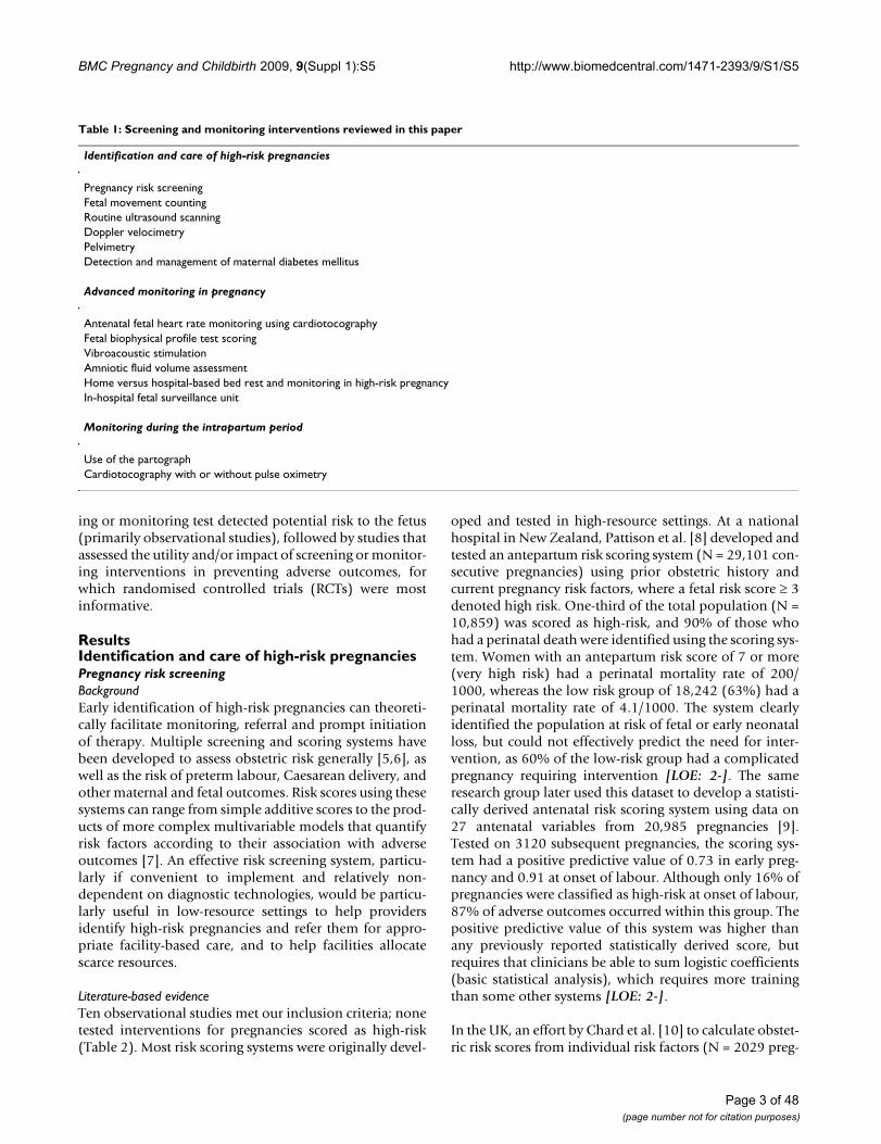
Table 1: Screening and monitoring interventions reviewed in this paper
Identification and care of high-risk pregnancies
Pregnancy risk screeningFetal movement countingRoutine ultrasound scanningDoppler velocimetryPelvimetryDetection and management of maternal diabetes mellitus
Advanced monitoring in pregnancy
Antenatal fetal heart rate monitoring using cardiotocographyFetal biophysical profile test scoringVibroacoustic stimulationAmniotic fluid volume assessmentHome versus hospital-based bed rest and monitoring in high-risk pregnancyIn-hospital fetal surveillance unit
Monitoring during the intrapartum period
Use of the partographCardiotocography with or without pulse oximetry
Page 3 of 48(page number not for citation purposes)

BMC Pregnancy and Childbirth 2009, 9(Suppl 1):S5 http://www.biomedcentral.com/1471-2393/9/S1/S5
Table 2: Impact of pregnancy risk screening on stillbirth and perinatal mortality
Source Location and Type of Study Intervention Stillbirths/Perinatal Outcomes
Observational studies
Abraham et al. 1991 [18] India. Health centre setting.Prospective cohort study. Health workers at 6 primary health centres used a home-based mothers card with pregnant, mostly illiterate women (N = 2446).
Assessed the association of perinatal mortality with risk factors recorded on a home-based mother's card to pregnant women on which risk factors and ANC attendance were documented.
PMR directly related to # of risk factors:0 risk factors: PMR = 25.9/10001 risk factor: PMR = 39.7/10002 risk factors: PMR = 56.5/10003 risk factors: PMR 122.5/1000)
Chard et al. 1992 [10] UK.N = 994 pregnant women (470 primiparae; 524 multiparae)
Used receiver-operating characteristic curves (ROC) to compare the use of weighted and unweighted risk scores in estimating an overall risk score based on individual risk factors, and relating this score to fetal outcome.
Weighted risk factor method clearly superior to unweighted risk factor method in primiparae. No difference in multiparae.
Cho et al. 1991 [163] Korea. Chung Ang Medical Center.Cross-sectional study to test scoring system. N = 1300 pregnant women (N = 1313 infants) admitted from 1988–1990.
Assessed the utility of Edwards' scoring system adapted to a Korean setting in identifying high-risk pregnancy. Risk scoring included demographic, obstetric, medical, and miscellaneous factors.
560 infants (42.7%) were born to mothers with risk-scores greater than 7, and 753 infants (57.3%) were born to mothers with risk-scores less than 7.
Lefevre et al. 1989 [15] USA. Rural primary care setting.Prospective study. N = 635 women. N = 47 (8.3%) adverse outcomes.
Tested the predictive value of Coopland's obstetric risk in anticipating adverse outcome (perinatal death, birthweight < 2500 g, 5-min Apgar score < 7, or newborn transferred to a level 2 or level 3 nursery.
There was a clear relationship between risk score and probability of adverse outcome. Good sensitivity could be achieved only at the expense of a very high false-positive rate, however. Risk scoring no more effective than a policy that would refer all women with standard obstetric risk factors; majority of adverse outcomes occurred in women identified as low-risk.
Majoko et al. 2002 [12] Zimbabwe. Rural setting.Evaluation of screening test; sub-study of ANC trial. N = 5223 women who received traditional care from nurse-midwives in 12 rural health centres (N = 2890 high risk).
Used traditional risk scoring at ANC booking to group women into low- and high-risk groups. High-risk women were encouraged to deliver in facilities.
Complications: 924 (17.7%) of women; 62.4% had had risk markers identified at booking. 20% (577/2890) of women classified as high risk developed complications.Predictive ability of risk allocation: Likelihood ratio = 1.16.
Mikulandra 1986 [164] Croatia.Prospective study.
Assessed the associations of a risk factor scale (low, moderate, and high risk) for pregnancy and delivery on perinatal outcomes.High pregnancy risk: 10.9% of cases.High intrapartum risk: 14.02% of cases.
Severe asphyxia (Apgar 3): 0.37%, 0.81%, and 4.36% in low, moderate, and high-risk groups, respectively (P < 0.001).SBR: 0.76% vs. 34.48% in low vs. high-risk groups (P < 0.01)
Morrison 1980 [165] USA.Retrospective analysis. N = 1994 consecutive parturients, N = 472 (23%) high-risk (risk score 3).
Assessed the association of high-risk (risk score 3) pregnancy with adverse perinatal outcomes.
PMR: Significantly higher in high-risk group (P > 0.001).Abnormal intrapartum outcome: 71% of high-risk group (P < 0.0001).
Morrison 1979 [11] USA.N = 16,733 deliveries. Women scored during pregnancy using a simplified, numerical form for antepartum risk scoring.
Tested the predictive value of a simplified risk scoring system in anticipating the risk of perinatal mortality.
19% of group was high-risk (score 3).PMR: 69/1000 vs. 7/1000 in high- vs. low-risk groups, respectively (P < 0.0001).70% of perinatal deaths occurred in high-risk group.
Page 4 of 48(page number not for citation purposes)
BMC Pregnancy and Childbirth 2009, 9(Suppl 1):S5 http://www.biomedcentral.com/1471-2393/9/S1/S5
nant women) found that risk scores were useful only foridentifying the small group of women at particularly highrisk of adverse fetal outcomes. For most women, riskscores were uninformative [LOE: 2-].
In the USA, Morrison et al. [11] found that perinatal mor-tality was significantly higher in the high- versus the low-risk groups identified with the application of a simplifiedrisk scoring system, where high risk was a score of 3 orgreater (69/1000 versus 7/1000, respectively, P < 0.0001).Seventy percent of perinatal deaths occurred in the high-risk group, which was 19% of the total group screened[LOE: 2-].
Other studies implemented risk scoring systems in moreremote or low-resource settings in low-/middle-incomecountries. Attempting to predict intrapartum complica-tions in rural Zimbabwe where most women receive carefrom nurse-midwives, Majoko et al. [12] employed ante-natal risk assessment at the first antenatal visit based onmedical and demographic measures and obstetric history(N = 5223 women at 12 health centres). All high-riskwomen (N = 2890) were encouraged to seek hospitaldelivery. Of the 924 (17.7%) women who experiencedcomplications, 577 (62.4%) had had risk markers identi-fied at booking; however, only 20% (577/2890) classifiedas high risk developed intrapartum complications. Thisrisk screening system had a likelihood ratio of 1.16, indi-cating it was ineffective in identifying women at risk ofpregnancy complications and generated too large a riskgroup for referral [LOE: 2-].
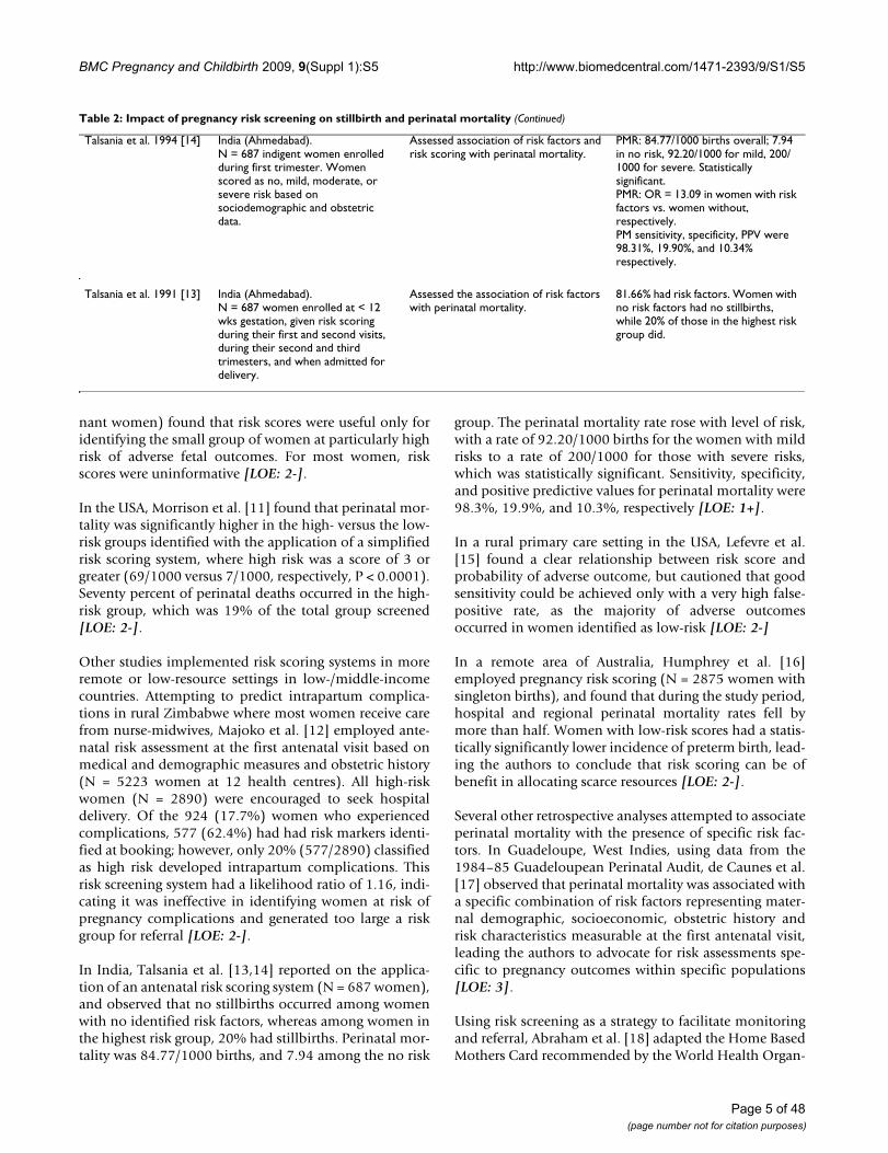
In India, Talsania et al. [13,14] reported on the applica-tion of an antenatal risk scoring system (N = 687 women),and observed that no stillbirths occurred among womenwith no identified risk factors, whereas among women inthe highest risk group, 20% had stillbirths. Perinatal mor-tality was 84.77/1000 births, and 7.94 among the no risk
group. The perinatal mortality rate rose with level of risk,with a rate of 92.20/1000 births for the women with mildrisks to a rate of 200/1000 for those with severe risks,which was statistically significant. Sensitivity, specificity,and positive predictive values for perinatal mortality were98.3%, 19.9%, and 10.3%, respectively [LOE: 1+].
In a rural primary care setting in the USA, Lefevre et al.[15] found a clear relationship between risk score andprobability of adverse outcome, but cautioned that goodsensitivity could be achieved only with a very high false-positive rate, as the majority of adverse outcomesoccurred in women identified as low-risk [LOE: 2-]
In a remote area of Australia, Humphrey et al. [16]employed pregnancy risk scoring (N = 2875 women withsingleton births), and found that during the study period,hospital and regional perinatal mortality rates fell bymore than half. Women with low-risk scores had a statis-tically significantly lower incidence of preterm birth, lead-ing the authors to conclude that risk scoring can be ofbenefit in allocating scarce resources [LOE: 2-].
Several other retrospective analyses attempted to associateperinatal mortality with the presence of specific risk fac-tors. In Guadeloupe, West Indies, using data from the1984–85 Guadeloupean Perinatal Audit, de Caunes et al.[17] observed that perinatal mortality was associated witha specific combination of risk factors representing mater-nal demographic, socioeconomic, obstetric history andrisk characteristics measurable at the first antenatal visit,leading the authors to advocate for risk assessments spe-cific to pregnancy outcomes within specific populations[LOE: 3].
Using risk screening as a strategy to facilitate monitoringand referral, Abraham et al. [18] adapted the Home BasedMothers Card recommended by the World Health Organ-
Talsania et al. 1994 [14] India (Ahmedabad).N = 687 indigent women enrolled during first trimester. Women scored as no, mild, moderate, or severe risk based on sociodemographic and obstetric data.
Assessed association of risk factors and risk scoring with perinatal mortality.
PMR: 84.77/1000 births overall; 7.94 in no risk, 92.20/1000 for mild, 200/1000 for severe. Statistically significant.PMR: OR = 13.09 in women with risk factors vs. women without, respectively.PM sensitivity, specificity, PPV were 98.31%, 19.90%, and 10.34% respectively.
Talsania et al. 1991 [13] India (Ahmedabad).N = 687 women enrolled at < 12 wks gestation, given risk scoring during their first and second visits, during their second and third trimesters, and when admitted for delivery.
Assessed the association of risk factors with perinatal mortality.
81.66% had risk factors. Women with no risk factors had no stillbirths, while 20% of those in the highest risk group did.
Table 2: Impact of pregnancy risk screening on stillbirth and perinatal mortality (Continued)
Page 5 of 48(page number not for citation purposes)
BMC Pregnancy and Childbirth 2009, 9(Suppl 1):S5 http://www.biomedcentral.com/1471-2393/9/S1/S5
isation (WHO) for a rural Indian setting. Perinatal mortal-ity was directly associated with number of risk factors:perinatal mortality rates (PMRs) were higher amongwomen with 3 or 4 risk factors than those with 1 or 2 riskfactors [LOE: 2-].
ConclusionA number of studies reviewed were able to successfullyidentify women at high risk of obstetric complications.However, despite good sensitivity, risk scoring systems [7]have poor positive predictive value in anticipating adversebirth outcomes, particularly when used well before termor in populations significantly different from the popula-tion in which the system was developed [19]. This limita-tion of risk scoring systems limits the impact of their use.No studies were found that effectively incorporated riskscreening with appropriate interventions to demonstrate apossible impact on stillbirth or perinatal mortality ratescompared to a control group. The evidence for risk screen-ing at the community level yielded a Grade C assessment.
Fetal movement countingBackgroundMonitoring fetal movements using counting strategies isan indirect measure of central nervous system integrityand fetal responsiveness. Commonly employed in clinicalpractice, fetal movement counting is a simple and inex-pensive means of monitoring fetal well-being [20]. Therationale for fetal movement counting is that decreasedfetal movements signal decreased oxygenation, whichoften precedes fetal demise [1]. Kick charts or otherrecording strategies involve a pregnant woman in the sec-ond half of pregnancy monitoring fetal movements, doc-umenting the frequency of movements she feels, andreporting these counts to her physician. Changes in thesecounts, particularly decreases, indicate possible fetal com-promise, and thus alert care providers to the need for fur-ther diagnostic tests such as non-stress testing or thebiophysical profile. Cessation of movement can indicateimpending fetal death, while gradual diminishment ofactivity can indicate chronic fetal compromise [21]. Fetalmovement monitoring may be used routinely, or only inhigh-risk pregnancies. There are many different countingmethods, and fetal movement monitoring has a wide fol-lowing among clinicians, who perceive the practice toserve as an early warning system for fetal compromise. Acriticism of the practice is that it may cause undue worryfor the pregnant woman [22].
Literature-based evidenceThe literature search identified one Cochrane review com-prised of three RCTs; and seven observational and inter-vention studies (Table 3).
Several trials in high-risk pregnancies documented anassociation between poor fetal movements and stillbirth/perinatal mortality rates. A trial among high-risk women(N = 110) by Lema et al. [23] found that fetal movementwas a predictor of stillbirth rate, documenting differentialrates [12/1000 (1/83) versus 185/1000 (5/27) in thegroups with good versus poor fetal movements, respec-tively] [LOE: 2-]. De Muylder [24] evaluated the use of akick chart to monitor the fetus in high-risk pregnancies,finding that both stillbirth rates (SBR) and PMR increasedsignificantly if a previously normal chart kick chartbecame abnormal (antepartum SBR = 194/1000 versus 7/1000; and PMR = 222/1000 vs. 27/1000 in charts thatbecame abnormal versus normal, respectively, P < 0.001)[LOE: 2-]. Two other observational studies found no dif-ference in perinatal mortality between groups with goodversus poor fetal movements measured using kick charts[25,26].
Other observational studies, RCTs, and reviews assessedimpact on perinatal mortality of interventions using kickcharts. A before-after study by Moore et al [27] introducedformal fetal movement monitoring into clinical practice,and stillbirth rates declined from 8.7 to 2.1/1000 over thecourse of the study (2 = 6.8; P < 0.01). A Cochrane reviewby Mangesi et al [22] included 3 trials that tested strategiesof routine kick counting, but varied study designs pre-cluded outcome pooling (Additional file 1). Unfortu-nately, no included trials compared fetal movementcounting with no fetal movement counting, and all stud-ies showed nonsignificant impact on perinatal outcomes,including stillbirth incidence. [LOE: 1++].
The largest RCT testing kick charts [28] assessed theimpact of the use of kick charts on unexplained stillbirthin normally-formed singleton pregnancies (N = 68654),and found no difference in rates of fetal death betweenintervention and control groups (59/31993 [2.9/1000]versus 58/36661 [2.7/1000], respectively [NS]). Thesefindings alone largely shaped the UK National Institutefor Health and Clinical Excellence (NICE) evidence-basedroutine antenatal care guidelines, which do not recom-mend the use of kick charts in uncomplicated pregnancy[29][LOE: 1+]. However, of the 17 women in the studyrandomised to kick charts who alerted their providerabout decreased fetal movement and subsequently deliv-ered a stillborn baby, none had an emergency delivery, asfollow-up testing using cardiotocography resulted in falsenegatives for all 17 women[30].
ConclusionThe existence of a Cochrane review of multiple RCTs [22]yields a Grade B evidence rating. In keeping with the NICEguidelines based largely on 1 study [28], evidence fromthese studies indicates a lack of impact of fetal movement
Page 6 of 48(page number not for citation purposes)

BMC Pregnancy and Childbirth 2009, 9(Suppl 1):S5 http://www.biomedcentral.com/1471-2393/9/S1/S5
Page 7 of 48(page number not for citation purposes)
Table 3: Impact of fetal movement counting on stillbirth and perinatal mortality
Source Location and Type of Study Intervention Stillbirths/Perinatal outcomes
Reviews and meta-analyses
Mangesi et al. 2007 [22] Peru, Denmark.Meta-analysis (Cochrane). 3 RCTs included (N = 66 women).
Routine fetal movement counting (intervention) versus mixed or undefined fetal movement counting (controls).
SBR: weighted mean difference = 0.23 [95% confidence interval (CI): -0.61–1.07) [NS][Mean (SD) = 2.90 (1.90) vs. 2.67 (1.55) in intervention vs. control groups, respectively].
Intervention studies
Gomez et al. 2007 [166] Peru. Hospital setting.RCT. Pregnant women (N = 1400).
Compared two different charting methods: a novel fetal movement chart proposed by the Latin American Center for Perinatology (CLAP) (intervention) vs. the count-to-ten Cardiff chart method (comparison).
Fetal death (miscarriage+SB): Relative risk (RR) not estimable.[0/700 in both groups].
Grant et al. 1989 [28] UK, USA, Ireland, Sweden, Belgium.Cluster RCT. 66 clusters. Pregnant women (N = 68654 women; N = 31993 intervention, N = 36661 controls).
Compared the impact on birth outcomes of asking mothers to keep routine kick charts (intervention) vs. not keeping kick charts (controls).
Unexplained late antepartum fetal death: 59/31993 (2.9/1000) vs. 58/36661 (2.7/1000) in intervention vs. control groups, respectively [NS].
Moore 1989 [27] USA. Hospital setting.Before-after pilot study (N = 2519 deliveries before intervention, N = 1864 after introduction of intervention.)
Assessed the impact of introducing formal fetal movement assessment (intervention) compared to no monitoring before the intervention (controls).
Fetal death (miscarriage+SB): 2.1/1000 vs. 8.7/1000 after vs. before, respectively. (2 = 6.8; P < 0.01)
Observational studies
De Muylder 1988 [24] Zimbabwe. Hospital setting.Prospective cohort study. High-risk pregnant women (N = 200).
Compared the obstetrical outcome among the patients with a normal kick chart (unexposed), compared to those with an abnormal count (exposed).
SBR: 19.4% vs. 0.7% in charts that went from normal to being abnormal vs. unexposed. (P < 0.001)PMR: 22.2% vs. 2.7% for previously normal charts that became abnormal vs. unexposed (P < 0.001)
Lema et al. 1988 [23] Kenya. Urban hospital setting.Prospective cohort study. High-risk pregnant women (N = 110).
Compared birth outcomes among women with good fetal movements vs. poor fetal movements.
SBR: 12/1000 (1/83) vs 185/1000 (5/27) in the good vs. poor fetal movements group, respectively. No statistical significance data.
Sinha et al. 2007 [25] UK. Hospital setting.Retrospective cohort study. N = 180 case reports.
Compared the impact of reduced fetal movements (exposed) to women without reduced fetal movements (unexposed) on PMR.
PMR: RR not estimable.[0/90 in the exposed vs. 0/90 in the control groups, respectively].Intervention needed solely due to fetal compromise: 29/90 (32%) in the study vs. 19/90 (21%) in the control groups, respectively.
Romero Gutiérrez et al. 1994 [26] Mexico. Hospital setting.Prospective cohort study. Pregnant women (N = 200; N = 100 intervention, N = 100 controls) 32–41 wks gestation without risk factors.
Compared the impact of decreased fetal movement (exposed) vs. normal fetal movement (unexposed) on PMR.
PMR: No difference [NS]

BMC Pregnancy and Childbirth 2009, 9(Suppl 1):S5 http://www.biomedcentral.com/1471-2393/9/S1/S5
monitoring on stillbirth or perinatal mortality. Despiteindirect evidence that formal movement monitoringusing a counting method is more effective than mothers'subjective assessments of fetal movement in identifyingbabies at risk of intrauterine death, false negatives on sub-sequent fetal assessment tests and clinical error may beresponsible for the lack of impact on perinatal mortality,as suggested by Del Mar et al [30]. Monitoring doesappear to be of some value in high-risk pregnancies[23,24], particularly those in which there is suspicion ofplacental insufficiency. Routine fetal movement monitor-ing is currently recommended only for high-risk pregnan-cies, particularly those in which there is clinical suspicionof restricted fetal growth or placental dysfunction revealedthrough ultrasonographic or Doppler studies. Furtherstudies are warranted to determine which methods of fetalmovement counting prove most effective in identifyingcomplications (sensitivity and specificity) early enoughfor interventions to prevent stillbirth, as well as accepta-bility to and feasibility for women. Universal fetal move-ment monitoring for all pregnancies is unsupported byscientific evidence.
Routine use of ultrasound scanningBackgroundUltrasound scans during pregnancy are widely used, evenin many resource-poor settings, but availability and qual-ity of ultrasound machines vary, and ultrasound operatorsin some settings may lack the ability to accurately inter-pret imaging. Diagnostic ultrasound examination may beemployed to date pregnancies, identify multiple pregnan-cies, document placental location, identify fetal anoma-lies (particularly when the fetus is suspected to be at highrisk of malformation), identify fetal growth restriction orabnormal amniotic fluid volume, or to investigate clinicalcomplications (e.g., bleeding). Some clinicians have pos-tulated that routine use of ultrasound in all pregnanciescould identify problems in asymptomatic pregnancies,whether early or late in gestation [31].
Literature-based evidenceWe identified two Cochrane reviews and five other inter-vention/observational studies; we also conducted anindependent meta-analysis incorporating 9 RCTs (Table4).
One observational study of routine ultrasound use sug-gested that ultrasound may help to identify some high-risk pregnancies. In Egypt, Mahran et al [32] reported thatroutine ultrasound was superior to fundal palpation inidentifying fetal growth restriction (89.7% versus 34.7%of growth-restricted infants identified accurately with eachmethod, respectively). No observational studies reportedany statistically significant impact of routine ultrasoundscanning on subsequent stillbirth rates. A Cochrane
review on the impact of ultrasound during pregnancy is inprogress [33].
Another Cochrane review by Neilson [31] reviewed ade-quately controlled trials of routine ultrasound imaging inearly pregnancy (N = 9) (Additional file 2). The studyfound that routine ultrasound examination was associ-ated with earlier detection of multiple pregnancies andreduced rates of induction of labour for post-term preg-nancy, but ultrasound had no impact on PMR [Odds ratio(OR) = 0.86, 95% confidence interval (CI): 0.67–1.12],even in twin pregnancies, despite generally earlier diagno-sis in the ultrasound-screened pregnancies. Where detec-tion of fetal abnormality was a specific aim of theexamination, ultrasound was associated with increasedterminations of pregnancy [LOE: 1+]
A second Cochrane review by Bricker et al. [34] assessedthe impact of ultrasound in late pregnancy (8 RCTs, N =27,024 women) and found no difference in antenatal,intrapartum and neonatal intervention or morbidity inthose undergoing ultrasound screening versus those notscreened (Additional file 3). The Caesarean section ratewas slightly higher among the screened group, but thisdifference did not reach statistical significance. Routinelate pregnancy ultrasound was not associated withimprovements in overall perinatal mortality.
An additional RCT (N = 1528 women) from New Zealandby Duff et al. [35] documented a statistically non-signifi-cant increase in SBR among women scanned twice duringpregnancy, at 16–24 weeks and again at 32–36 weeks ges-tation [LOE: 2+]. In another RCT by Proud et al [36]where placental grading information from ultrasoundscreening was either given to a clinician (intervention) orwithheld (controls), the antepartum SBR (excludinglethal malformations) was 0/1014 versus 9/1011 amongintervention versus controls, respectively (P < 0.05) [LOE:2+].
New meta-analysisWe also conducted an independent, new meta-analysis forthe purposes of this review, as we identified 9 RCTs (N =35,049 women) reporting an impact on perinatal mortal-ity rate of ultrasound in early pregnancy versus no orselective use of ultrasound in early pregnancy (before 24weeks) (Figure 1). We found no significant differencebetween the 2 groups when the results were pooled (OR =0.89, 95% CI: 0.70–1.14).
ConclusionThere is no clear evidence that ultrasound examinationduring pregnancy is harmful [31], and its assumed bene-fits include (1) better gestational age assessment; (2) ear-lier detection of multiple pregnancies; (3) determination
Page 8 of 48(page number not for citation purposes)

BMC Pregnancy and Childbirth 2009, 9(Suppl 1):S5 http://www.biomedcentral.com/1471-2393/9/S1/S5
of placental location to rule out placenta praevia; (4)detection of clinically unsuspected fetal malformationwhen termination of pregnancy is more feasible, and (5)
monitoring of fetal growth for pregnancies at increasedrisk of fetal growth restriction or macrosomia. Thereduced incidence of induction of labour for apparent
Table 4: Impact of use of routine ultrasound scanning on stillbirth and perinatal mortality
Source Location and Type of Study Intervention Stillbirths/Perinatal Outcomes
Reviews and meta-analyses
Bricker et al. 2008 [34] New Zealand), Norway (Trondheim), Australia, UK (Peterborough), USA.Meta-analysis (Cochrane). 8 RCTs included (N = 21,708 women).
Assessed the effects of routine ultrasound > 24 wks (intervention) vs. no/concealed/selective ultrasound > 24 wks (control).
SBR: RR = 1.11 (95% CI: 0.29–4.26) [NS][45/10894 vs. 38/10814 in intervention vs. control groups, respectively].PMR: R = 0.94 (95% CI: 0.55-1.64) [NS][79/12198 vs. 75/12078 in intervention vs. control groups, respectively.]
Neilson 1998 [31] Finland, UK, USA, Sweden, Trondheim, South Africa.Meta-analysis (Cochrane). 8 RCTs included (N = 34,245).
Assessed the effects of routine ultrasound (intervention) vs. the selective use of ultrasound (control) in early pregnancy (i.e. < 24 wks).
PMR: OR = 0.86 (95% CI: 0.67–1.12)
Intervention studies
van Dyk et al. 2007 [167] South Africa.Open cluster RCT. Pregnant women (N = 804).
Compared the impact of ultrasound screening (intervention) vs. no ultrasound (controls).
PMR: RR = 1.05 (95% CI: 0.54–2.03, P = 0.88.) [NS][18/416 (4.3%) vs. 16/388 (4.1%) in intervention vs. control groups, respectively].
Observational studies
Cristina et al. 2005 [168] Spain.Retrospective (case-control) review of all obstetric ultrasounds. Pregnant patients (N = 5,987 examined by ultrasound scan at 20 wks; N = 40 cases with a single umbilical artery, N = 82 controls).
Compared the impact of having a single umbilical artery (cases) vs. not having this condition (controls) as diagnosed by ultrasound scan on PMR.
PMR: 5% (2/40) among single uterine artery cases (10× greater than overall patient rate). No statistical significance data.
Mahran et al. 1992 [32] Egypt. Tertiary care setting.Comparison of diagnostic tests. Pregnant women (N = 828), of whom a proportion had growth-restricted neonates (N = 98).
Compared the effectiveness of diagnostic ultrasound (intervention) vs. fundal palpation (controls) in predicting growth restriction.
Growth restriction: 89.7% (88/98) vs. 34.7% (34/98) detection rate in ultrasound vs. fundal palpation groups, respectively.
Sylvan et al. 2005 [169] Sweden. University clinics.Observational cohort study. Deliveries from 1985–1996; stored data (N = 209,726).
Compared the impact of routine ultrasound screening (exposed group) vs. no routine screening (unexposed) in third trimester on PMR.
PMR: [NS][160/56,371 vs. 488/153,355 in exposed vs. unexposed, respectively.]
Viero et al. 2004 [170] Canada.Observational study. Structurally and chromosomally normal singleton pregnancies (N = 60) with abnormal fetoplacental blood flow < 32 wks of gestation; N = 21 of these resulted in stillbirth and were delivered vaginally.
To assess the ability of grayscale placental ultrasound to detect pathological lesions in the placentas of pre-term pregnancies.
SB: charts with both abnormal uterine artery Doppler and abnormal grayscale findings strongly predictive of stillbirth (17/21 SBs; sensitivity 81%, PPV 52%, P = 0.006).
Page 9 of 48(page number not for citation purposes)

BMC Pregnancy and Childbirth 2009, 9(Suppl 1):S5 http://www.biomedcentral.com/1471-2393/9/S1/S5
post-term pregnancy in the routinely scanned groups pre-sumably results from better gestational age dating, andtwin pregnancies being detected earlier. Neither of theseeffects has been shown to improve fetal outcome, butmuch larger numbers of participants would be required toaccurately measure this outcome.
Based on the results of 2 Cochrane reviews, our meta-anal-ysis, and other RCTs (overall Grade B evidence), there isno evidence that routine ultrasonography has any impacton perinatal mortality compared to the selective use ofultrasonography based on clinician judgement. It may bethat routine ultrasound cannot reliably detect complica-tions, or that high rates of false positives expose highernumbers of babies to iatrogenic intervention (particularlythe risk of iatrogenic preterm birth in the event of inaccu-rate gestational age dating). The routine use of early ultra-sonography in pregnancy cannot be recommended toprevent stillbirth, as there is no evidence of its benefit inpreventing stillbirth or perinatal mortality. There is a needfor decision analysis studies subsequent to diagnosticultrasound, like the study of indicated Caesarean sectionfor fetal macrosomia diagnosed by ultrasound by Rouse etal [37], but which report perinatal mortality outcomes.Clinics and hospitals, particularly those in resource-con-strained settings, must assess whether the potential bene-fits are worth the cost of routine ultrasound screening forall pregnant women.
Doppler velocimetryBackgroundIn many high-income countries, Doppler ultrasoundstudies are used as a non-invasive means to assess the suf-
ficiency of uterine and umbilical cord blood flow. Thesevelocimetry studies can improve management of pregnan-cies by aiding identification of fetuses at highest risk ofadverse outcomes associated with pre-eclampsia, fetalgrowth restriction, and congenital malformations. Man-agement of pre-term pregnancies with signs of fetalgrowth restriction and pre-eclampsia is complex, espe-cially before 32 weeks gestation. The risk of prolongedhypoxia and acidaemia leading to stillbirth or neonataldeath if the pregnancy is allowed to progress must be bal-anced against the risks of neonatal morbidity and mortal-ity associated with prematurity if early delivery is chosen.Doppler ultrasonographic evaluation may aid determina-tions of the degree to which the fetus may be or becomecompromised.
Early in normal pregnancy, trophoblasts invade thematernal uterine spiral arteries and reduce resistance touterine blood flow. Impeded flow measured by uterineartery Doppler suggests a failure of this trophoblasticinvasion, which is associated with subsequent pre-eclampsia, fetal growth restriction, and stillbirth [38].Uterine artery Doppler studies could therefore be helpfulin identifying women likely to develop pre-eclampsia orto have a growth-restricted fetus [39].
Observational Doppler studies of the umbilical artery,first conducted in the 1970s, consistently showed a corre-lation between extremely abnormal waveforms andadverse outcomes, including fetal growth restriction andstillbirth [40-42]. In growth-restricted fetuses, results ofumbilical and fetal Doppler waveform analyses suggestprogressive severity of fetal compromise [38,43]. Initially,
Forest plot of results of meta-analysis of perinatal mortality rates in women examined by routine vs. selective ultrasound in early pregnancyFigure 1Forest plot of results of meta-analysis of perinatal mortality rates in women examined by routine vs. selective ultrasound in early pregnancy.
Study or Subgroup
Bakketeig LS 1984Bennett MJ 1982Eik-Nes SH 1984Ewigman B 1990Geerts LTGM 1996LeFevre ML 1993Saari-Kemppainen A 1990van Dyk B 2007Waldenstrom U 1988
Total (95% CI)
Total eventsHeterogeneity: Chi² = 9.46, df = 8 (P = 0.30); I² = 15%Test for overall effect: Z = 0.92 (P = 0.36)
Events
55629
52201812
129
Total
516531794404460
76854389416
2413
17608
Events
53
104
1341391612
143
Total
507531765420455
75964347
3882432
17441
Weight
3.8%2.9%5.8%2.1%8.0%
35.3%20.3%12.6%9.2%
100.0%
Peto, Fixed, 95% CI
0.98 [0.28, 3.41]1.67 [0.40, 7.04]0.57 [0.21, 1.59]0.52 [0.09, 2.84]0.68 [0.29, 1.60]1.26 [0.83, 1.89]0.51 [0.29, 0.87]1.05 [0.53, 2.09]1.01 [0.45, 2.25]
0.89 [0.70, 1.14]
Ultrasound No or selective U/S Odds Ratio Odds RatioPeto, Fixed, 95% CI
0.01 0.1 1 10 100Favours experimental Favours control
Page 10 of 48(page number not for citation purposes)

BMC Pregnancy and Childbirth 2009, 9(Suppl 1):S5 http://www.biomedcentral.com/1471-2393/9/S1/S5
umbilical artery velocity waveforms show increased resist-ance; subsequent deterioration is indicated by absent oreven reversed end diastolic flow in the umbilical artery.Later, fetal middle cerebral artery flow shows decreasedresistance, indicating brain sparing, and eventually,abnormal venous Doppler results (ductus venosus wave-forms and umbilical vein pulsatility) suggest fetal cardiacdysfunction. Consequent central nervous system damagethen manifests as non-reactive results to fetal tests of well-being, but there is wide variability in the timeline of fetalprogression to severe compromise [38,43].
Despite these well-established markers of fetal compro-mise in Doppler testing, it is not clear whether abnormalresults of different modalities of Doppler ultrasound leadto improved perinatal outcomes and prevention of still-births, nor are the most appropriate indications and tim-
ing of testing known. Additionally, if a fetus is notseriously compromised, Doppler ultrasound may poten-tially cause iatrogenic harm in suggesting the need forinappropriate early delivery.
Literature-based evidenceThe literature search identified three systematic reviews,one Cochrane protocol, and 10 other intervention/obser-vational studies (Table 5 and Table 6).
Uterine artery Doppler waveform analysisSeveral studies assessed whether uterine artery Dopplervelocimetry in unselected populations could identifyhigh-risk pregnancies, particularly those at risk of still-birth. In a systematic review, Papageorghiou et al [44]reviewed 15 studies of routine Doppler assessments inpregnancy in unselected populations, and found that
Table 5: Impact of uterine artery Doppler velocimetry on stillbirth and perinatal mortality
Source Location and Type of Study Intervention Stillbirths/Perinatal Outcomes
Reviews and meta-analyses
Papageorghiou et al. 2002 [44] Multiple sites.Review. 15 studies of routine Doppler assessments in pregnancy in unselected populations.
Sought to relate the risk of antepartum stillbirth to uterine artery Doppler flow velocimetry at 22–24 weeks.
Fetal growth restriction and perinatal death associated with impeded uterine artery flow.Positive Doppler diagnosis appropriately identified ~40% of women who subsequently developed pre-eclampsia (6-fold increased risk with positive Doppler) and ~20% of fetal growth restriction cases (3.5-fold increased risk)
Intervention studies
Subtil et al; Essai Régional Aspirine Mère-Enfant (ERASME) Collaborative Group 2003. [46]
France and Belgium.Multicentre RCT. Nulliparous women (N = 1853; N = 1253 intervention, N = 617 controls) 14–20 wks gestation.
Compared the impact of uterine Doppler (intervention) versus placebo (controls) on PMR. Women with abnormal Doppler waveforms received 100 mg of aspirin daily from Doppler exam until 36 wks.
PMR: RR = 4.02 (95% CI: 0.5–32.0) [NS][8/1249 (0.6%) vs. 1/327 (0.2%) in intervention vs. control groups, respectively].
Observational studies
Smith et al. 2007 [45] UK.Observational study. Unselected women (N = 30,519) who had uterine artery Doppler performed 22–24 wks of gestation.
Studied the relationship between abnormal (mean pulsatility index in the top decile and a bilateral notch) vs. normal Doppler flow on the risk of antepartum stillbirth.
Antepartum SBR: adj. HR = 5.5 (95% CI: 2.8–10.6) in Doppler with mean pulsatility index in the top decile vs. controls.Antepartum SBR: adj. HR = 3.9 (95% CI: 2.0–7.8) in Doppler with a bilateral notch versus controls.Unexplained SBR: adj. HR 2.5 (95% CI: 1.1–5.6) in Doppler with mean pulsatility index in the top decile vs. controls. No association between Doppler with a bilateral notch and SB.
Page 11 of 48(page number not for citation purposes)
BMC Pregnancy and Childbirth 2009, 9(Suppl 1):S5 http://www.biomedcentral.com/1471-2393/9/S1/S5
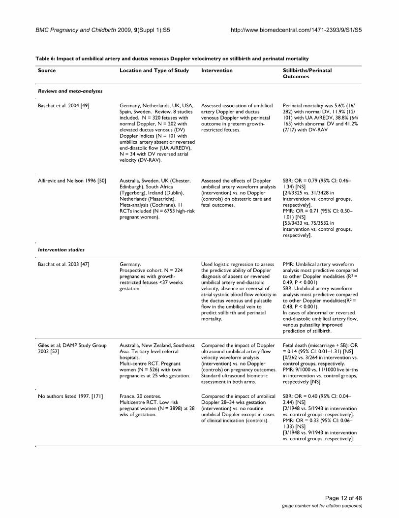
Table 6: Impact of umbilical artery and ductus venosus Doppler velocimetry on stillbirth and perinatal mortality
Source Location and Type of Study Intervention Stillbirths/Perinatal Outcomes
Reviews and meta-analyses
Baschat et al. 2004 [49] Germany, Netherlands, UK, USA, Spain, Sweden. Review. 8 studies included. N = 320 fetuses with normal Doppler, N = 202 with elevated ductus venosus (DV) Doppler indices (N = 101 with umbilical artery absent or reversed end-diastolic flow (UA A/REDV), N = 34 with DV reversed atrial velocity (DV-RAV).
Assessed association of umbilical artery Doppler and ductus venosus Doppler with perinatal outcome in preterm growth-restricted fetuses.
Perinatal mortality was 5.6% (16/282) with normal DV, 11.9% (12/101) with UA A/REDV, 38.8% (64/165) with abnormal DV and 41.2% (7/17) with DV-RAV
Alfirevic and Neilson 1996 [50] Australia, Sweden, UK (Chester, Edinburgh), South Africa (Tygerberg), Ireland (Dublin), Netherlands (Maastricht).Meta-analysis (Cochrane). 11 RCTs included (N = 6753 high-risk pregnant women).
Assessed the effects of Doppler umbilical artery waveform analysis (intervention) vs. no Doppler (controls) on obstetric care and fetal outcomes.
SBR: OR = 0.79 (95% CI: 0.46–1.34) [NS][24/3325 vs. 31/3428 in intervention vs. control groups, respectively].PMR: OR = 0.71 (95% CI: 0.50–1.01) [NS][53/3433 vs. 75/3532 in intervention vs. control groups, respectively].
Intervention studies
Baschat et al. 2003 [47] Germany.Prospective cohort. N = 224 pregnancies with growth-restricted fetuses <37 weeks gestation.
Used logistic regression to assess the predictive ability of Doppler diagnosis of absent or reversed umbilical artery end-diastolic velocity, absence or reversal of atrial systolic blood flow velocity in the ductus venosus and pulsatile flow in the umbilical vein to predict stillbirth and perinatal mortality.
PMR: Umbilical artery waveform analysis most predictive compared to other Doppler modalities (R2 = 0.49, P < 0.001)SBR: Umbilical artery waveform analysis most predictive compared to other Doppler modalities(R2 = 0.48, P < 0.001).In cases of abnormal or reversed end-diastolic umbilical artery flow, venous pulsatility improved prediction of stillbirth.
Giles et al; DAMP Study Group 2003 [52]
Australia, New Zealand, Southeast Asia. Tertiary level referral hospitals.Multi-centre RCT. Pregnant women (N = 526) with twin pregnancies at 25 wks gestation.
Compared the impact of Doppler ultrasound umbilical artery flow velocity waveform analysis (intervention) vs. no Doppler (controls) on pregnancy outcomes. Standard ultrasound biometric assessment in both arms.
Fetal death (miscarriage + SB): OR = 0.14 (95% CI: 0.01–1.31) [NS][0/262 vs. 3/264 in intervention vs. control groups, respectively.PMR: 9/1000 vs. 11/1000 live births in intervention vs. control groups, respectively [NS]
No authors listed 1997. [171] France. 20 centres.Multicentre RCT. Low risk pregnant women (N = 3898) at 28 wks of gestation.
Compared the impact of umbilical Doppler 28–34 wks gestation (intervention) vs. no routine umbilical Doppler except in cases of clinical indication (controls).
SBR: OR = 0.40 (95% CI: 0.04–2.44) [NS][2/1948 vs. 5/1943 in intervention vs. control groups, respectively].PMR: OR = 0.33 (95% CI: 0.06–1.33) [NS][3/1948 vs. 9/1943 in intervention vs. control groups, respectively].
Page 12 of 48(page number not for citation purposes)

BMC Pregnancy and Childbirth 2009, 9(Suppl 1):S5 http://www.biomedcentral.com/1471-2393/9/S1/S5
increased impedance to uterine artery flow was associatedwith increased risk of pre-eclampsia, fetal growth restric-tion and perinatal death, and that Doppler diagnosis ofimpeded artery flow appropriately identified ~40% ofwomen who subsequently developed pre-eclampsia (6-fold increased risk with positive Doppler) and ~20% offetal growth restriction cases (3.5-fold increased risk)[LOE: 1+] (Additional file 4).
Smith et al [45] reported a statistically significantlyincreased risk of antepartum stillbirth among womenwhose uterine artery Doppler flow velocimetry at 22–24weeks had a pulsatility index in the top decile [adjusted
Hazard Ratio (HR) = 5.5, 95% CI: 2.8–10.6] and amongthose with a bilateral notch (adjusted HR = 3.9, 95% CI:2.0–7.8), compared with controls with normal Dopplerflow [LOE: 2-].
An intervention RCT attempting to use Doppler in inter-ventions to reduce rates of pre-eclampsia in France andBelgium by the Essai Régional Aspirine Mère-Enfant(ERASME) Collaborative Group [46] randomised womento either uterine Doppler (N = 1253) or placebo (N =617). Women with abnormal waveforms in the interven-tion group were given low-dose aspirin to prevent pre-eclampsia; but despite the aspirin prescription, rates of
Davies et al. 1992 [172] UK (London). Single centre; unselected population.RCT. Singleton pregnancies (N = 2600) > 20 wks gestation.
Compared the impact of routine umbilical and uterine artery Doppler ultrasound to assess placental perfusion (intervention) vs. no Doppler (controls) on pregnancy outcomes. Standard ANC in both arms.
SBR: 11/1246 vs. 4/1229 in intervention vs. control groups, respectively.PMR (uncorrected): RR = 2.4 (95% CI: 1.00–5.76) [NS][17/1246 vs. 7/1229 in intervention vs. control groups, respectively].PMR (normally formed): RR = 3.95 (95% CI: 1.32–11.77).[16/1246 vs. 4/1229 in intervention vs. control groups, respectively].
Whittle et al. 1994 [173] UK (Glasgow).RCT. Singleton pregnancies (N = 2986) < 26 wks gestation at 1st ANC visit. Doppler ultrasound at 26–30 wks and 34–36 wks gestation in all women.
Compared the impact of umbilical artery Doppler ultrasound revealed to clinician (intervention) vs. concealed from clinician (controls).
SBR: OR = 0.34 (95% CI: 0.10–1.07) [NS][3 vs. 8 in intervention vs. control groups, respectively.]
Observational studies
Hugo et al. 2007 [48] South Africa (Cape Town). Secondary hospital.Case series. Singleton pregnant women (N = 572) referred for suspected poor fetal growth.
Investigated the use of a personal computer- based, continuous-wave Doppler machine by a trained midwife to assess umbilical artery flow velocity waveforms with respect to the resistance indices (RIs).
PMR:[RIs < P75]: 13.2[RIs: P75-95]: 39.1[RIs > P95]: 41.7SGA (%):[RIs < P75]: 27.2%[RIs: P75-95]: 41.2%[Ris > P95]: 55.6%
Theron et al. 1992 [41] South Africa.Prospective cohort study. Pregnant women (N = 127) with poor symphysis fundal growth (N = 39 abnormal Doppler flow velocimetry, N = 88 normal velocimetry).
Compared the impact of poor Doppler flow velocimetry of umbilical artery (exposed) with normal flow (unexposed).
PMR: OR = 33.2 (95% CI: 6.6–109.6; P < 0.000001).[43.6% vs. 2.3% in exposed vs. unexposed groups, respectively].Fetal death (miscarriage + SB):[28.2% vs. 0% in exposed vs. unexposed groups, respectively; (P < 0.0005)].
Torres et al. 1995 [42] Spain (Barcelona). Hospital Clinic.Prospective observational study over a 2-year period. Hypertensive pregnant women (N = 172; N = 166 with live births, N = 6 fetal deaths).
Assessed the use of umbilical artery Doppler in predicting SB. Compared the impact of absent (exposed) vs. normal end-diastolic velocity (unexposed).
SB: All had absence of end-diastolic velocity (sensitivity 100%).Fetal death (miscarriage + SB): 6/9 vs. 0/163 in absent vs. normal flow.Absent end-diastolic velocity in predicting fetal death: sensitivity: 100%, specificity: 98.2%, positive predictive value 66.7%, negative predictive value 100%.
Table 6: Impact of umbilical artery and ductus venosus Doppler velocimetry on stillbirth and perinatal mortality (Continued)
Page 13 of 48(page number not for citation purposes)

BMC Pregnancy and Childbirth 2009, 9(Suppl 1):S5 http://www.biomedcentral.com/1471-2393/9/S1/S5
pre-eclampsia were similar in the Doppler and placebogroups (28/1237 [2.3%] versus 9/616 [1.5%], respec-tively; RR = 1.55, 95% CI: 0.7–3.3 [NS]). The study founda non-significant elevated impact on perinatal mortalityamong Doppler-assessed fetuses compared to thosewhose mothers were given placebo (RR = 4.02; 95% CI:0.5–32.0 [NS]).
Umbilical artery and venous Doppler waveform analysisBaschat et al [47] performed umbilical artery and venousDoppler velocimetry in preterm growth-retarded fetuses(N = 224) to evaluate whether absent or reversed umbili-cal end-diastolic velocity, absence or reversed atrial systo-lic blood flow velocity in the ductus venosus, or pulsatileflow in the umbilical vein could predict stillbirth or peri-natal death before 37 weeks' gestational age. Logisticregression analysis showed that umbilical artery wave-form analysis was the Doppler application most predic-tive of perinatal mortality (R2 = 0.49, P < 0.001) andstillbirth (R2 = 0.48, P < 0.001). In fetuses with absent orreversed umbilical end flow, prediction of asphyxia andstillbirth was significantly enhanced by venous Doppler.Umbilical artery waveform analysis offered the highestsensitivity and negative predictive value, whereas ductusvenosus and umbilical vein flow studies had the best spe-cificity and positive predictive values for perinatal death[LOE: 1+].
Using personal computer-based, continuous-wave Dop-pler machines to assess umbilical artery flow velocitywaveforms in women referred for suspected fetal growthrestriction, Hugo et al. [48] found a direct associationbetween resistance indices (RIs) and perinatal mortalityrate (PMR) (PMRs of 13.2, 39.1 and 41.7 for women withRIs < P75, P75-95 and > P95, respectively) [LOE: 3].
Baschat et al. [49] conducted a review of studies (N = 8)where umbilical artery and ductus venosus Doppler stud-ies were used to make decisions regarding delivery timingin pre-term growth-restricted fetuses (Additional file 5).One analysis in the review compared outcomes amongfetuses with normal ductus venosus indices (N = 302)with fetuses with elevated ductus venosus index (N =202), of whom N = 101 had absent or reversed umbilicalend-diastolic flow and N = 34 had absence or reversal ofatrial velocity in the ductus venosus. Perinatal mortalitywas 5.6% (16/282) among fetuses with normal Doppler,versus 11.9% (12/101) with absent or reversed umbilicalend-diastolic flow, 38.8% (64/165) with abnormal duc-tus venosus index and 41.2% (7/17) with reversed atrialvelocity in the ductus venosus. Abnormal ductus venosusresults (N = 3 studies) effectively identified fetuses at riskof stillbirth at least 1 week prior to delivery, independentof umbilical artery waveform results, though questions
remain about optimal delivery timing in growth-restrictedpre-term fetuses [LOE: 1+].
Recently, researchers have focused on downstream impacton perinatal mortality of the use of umbilical artery Dop-pler ultrasound followed by appropriate interventions forthe identified conditions. A Cochrane review by Neilsonand Alfirevic [50] reviewed 11 RCTs (~7000 women) ofDoppler ultrasound investigating umbilical artery wave-forms in high-risk pregnancies. Compared to no Dopplerultrasound, evaluation with umbilical artery Dopplerultrasound in complicated pregnancies (especially withhypertension or presumed growth restriction) was associ-ated with a non-significant trend toward lower perinatalmortality (OR = 0.71, 95% CI: 0.50–1.01) and stillbirthrisk (OR = 0.79, 95% CI: 0.46–1.34), as well as signifi-cantly fewer inductions of labour (OR = 0.83, 95% CI:0.74–0.93) and hospital admissions (OR = 0.56, 95% CI:0.43–0.72) (Additional file 6). Whether a woman hadreceived Doppler ultrasound was unassociated with fetaldistress in labour or Caesarean section rates [LOE: 1+]. ACochrane protocol by Alfirevic et al indicates that a reviewis in progress to assess whether the use of umbilical artery,middle cerebral artery, and ductus venosus Dopplervelocimetry improves subsequent obstetric care and fetaloutcome [51].
Umbilical artery Doppler alone may not be any moreeffective than routine ultrasonography in some diagnosticassessments of fetal growth. An RCT of Doppler in twinpregnancies in New Zealand, Australia and Southeast Asiawhere all pregnancies were studied ultrasonographicallyto monitor fetal growth showed no statistically significantdifference in perinatal mortality between groups on whichumbilical artery Doppler was performed versus those withno Doppler [52].
ConclusionDoppler ultrasound is a relatively new technique that hasbeen applied to the study of fetal, placental and uterinecirculatory dynamics. Despite its novelty, it has been eval-uated by more RCTs than has any other biophysical test offetal growth or well-being. In low-risk or unselected pop-ulations, there is little evidence that any form of routineDoppler velocimetry contributes to reductions in stillbirthrates (overall Grade C evidence). This lack of impact maybe complex: Doppler ultrasound may not identify a suffi-cient proportion of flow abnormalities to measurablyimpact stillbirth incidence; Doppler-detected abnormali-ties may not be subsequently monitored appropriatelywith other tests of fetal well-being and serial Doppler test-ing; intervention based on abnormal Doppler may notwork; or high rates of false-positives may unnecessarilyexpose the fetus to the risk of preterm birth, particularly ifgestational age dating is inaccurate. Additionally, most
Page 14 of 48(page number not for citation purposes)

BMC Pregnancy and Childbirth 2009, 9(Suppl 1):S5 http://www.biomedcentral.com/1471-2393/9/S1/S5
existing studies are underpowered to detect small impactson perinatal or maternal outcomes.
Uterine artery Doppler waveform analysis accurately iden-tifies compromised fetuses at risk of stillbirth, especiallyin cases of placental underperfusion associated with pre-eclampsia and/or growth restriction, but no studies haveshown any ability of subsequent intervention to preventstillbirth. More studies are needed into the optimal timingof monitoring and intervention in cases of abnormal uter-ine artery waveforms.
Of all Doppler diagnostic studies of the fetus, umbilicalartery and ductus venosus Doppler velocimetry are mostpredictive of fetal compromise associated with fetalgrowth restriction, and there is some evidence that timelyand appropriate intervention for abnormal umbilicalartery or ductus venosus waveforms can prevent stillbirths[53]. More evidence should soon be available: in additionto the Cochrane review of umbilical and fetal Dopplervelocimetry, results are forthcoming of the multi-centreTrial of Umbilical and Fetal Flow in Europe (TRUFFLE)group, an RCT of timing of delivery in early pre-term fetalgrowth restriction based on early and late fetal Dopplervenous changes versus cardiotocography. Such studiesmay shed light on the most appropriate and effectivemethods of fetal surveillance and optimal uses of Dopplervelocimetry, including multi-vessel analysis [54].
Further studies are needed to assess whether such decisionanalytical models based on Doppler and other fetal sur-veillance findings for fetal growth restriction, pre-eclamp-sia, or congenital abnormalities could have any impact onstillbirths. Further research might also investigate otherapplications of Doppler velocimetry, including identify-
ing women who should be given other screening tests, thecomparative efficacy of Doppler versus other fetal surveil-lance methods, and contributing to the study of thepathophysiology of impaired placentation, uteroplacentaland fetoplacental haemodynamics, and pre-eclampsia.
PelvimetryBackgroundPelvimetry, or pelvic measurement in pregnant womenwith the intention of predicting likely cephalopelvic dis-proproportion of cephalic presentations (and thus theirsubsequent need for Caesarean section), can be per-formed by clinical manual examination, or with imagingtechniques including conventional x-ray, computerisedtomography scanning, or magnetic resonance imaging.Successful detection and management of cephalopelvicdisproportion is thought to reduce the risk of obstructedlabour and intrapartum stillbirth.
Literature-based evidenceWe identified 1 Cochrane review and 1 other observa-tional study of pelvimetry reporting perinatal outcomes(Table 7). The observational study by Fine et al [55] retro-spectively analysed studies of x-ray pelvimetry (N = 100trials) in cephalic presentations, comparing 3 prognosticindicators for vaginal delivery: the Thoms method, themodified Ball technique, and manual assessment. Neitherpelvimetric method was significantly more accurate thanmanual assessment of the pelvis in predicting obstetricoutcome, nor was any one method superior to the other.
When implemented as part of a strategy of pregnancymanagement, pelvimetry appears to have no impact onstillbirth. A Cochrane review by Pattinson and Farrell [56]assessed RCTs implementing x-ray pelvimetry in cephalic
Table 7: Impact of pelvimetry on stillbirth and perinatal outcomes
Source Location and Type of Study Intervention Stillbirths/Perinatal Outcomes
Reviews and meta-analyses
Pattinson and Farrell 1997 [56]
South Africa, U.S.A.Meta-analysis (Cochrane). 4 RCTs included (N = 895 women).
Assessed the effects of pelvimetry performed antenatally, intrapartum or postpartum (intervention) vs. no pelvimetry (controls) on PMR.
PMR: OR = 0.51 (95% CI: 0.18–1.42) [NS].[5/449 vs. 10/446 in intervention vs. control groups, respectively].
Observational studies
Fine et al. 1980 [55] Retrospective study. N = 100 X-ray pelvimetry studies of cephalic presentations.
Compared the Thoms method of interpretation to the modified Ball technique for x-ray pelvimetry (comparing both to manual assessment of the pelvis) as prognostic indicators for safe vaginal delivery.
Uneventful nonoperative vaginal deliveries: 28.6% of patients with either inlet or midpelvic disproportion by the Thoms method, and in 22.5% of women with absolute disproportion in either plane by the modified Ball method.Prediction of obstetric outcome: Neither technique significantly more accurate than manual assessment, or than the other.
Page 15 of 48(page number not for citation purposes)

BMC Pregnancy and Childbirth 2009, 9(Suppl 1):S5 http://www.biomedcentral.com/1471-2393/9/S1/S5
presentations (N = 4 trials, N>1000 women). Pelvimetrywas associated with increased rates of Caesarean section(OR = 2.17; 95% CI = 1.63–2.88) but had no impact onPMR (4 RCTs; OR = 0.51, 95% CI = 0.18–1.42 [NS])[LOE: 1+] (Additional file 7).
ConclusionThere is little support for the use of pelvimetry to predictthe need for Caesarean section in women with fetuseswith cephalic presentations, as the dynamic and individ-ual nature of maternal tissue changes during labour andfetal head moulding in the birth canal make antenatal pel-vimetry a poor predictor of cephalopelvic disproportion.The practice may result in inadvertent harm to the motherby significantly increasing her risk of having a Caesareansection, without increasing the benefit to fetus or neonate,as pelvimetry shows no impact on stillbirth incidence.However, deficiencies in the existing studies included inPattinson's meta-analysis should be noted: Parsons [57]attributed the increased perinatal mortality and birthasphyxia reported by Crichton [58] to lack of availabilityof electronic fetal monitoring, not to cephalopelvic dis-proportion. Even more problematic, the 2 deaths docu-mented by Richards [59] occurred in utero prior to labour,which imply that cephalopelvic disproportion was notimplicated. Treatment allocation strategies in all trialswere of poor quality.
Given the deficiencies in existing studies, it remains plau-sible that other forms of imaging could effectively diag-nose true cephalopelvic disproportion and avert stillbirthvia Caesarean section in these cases. We classified theoverall evidence as Grade C and found no evidence insupport of using pelvimetry for preventing stillbirths.
Detection and management of maternal diabetes mellitusBackgroundIn pregnant women, pre-existing diabetes mellitus cancause severe complications for both mother and childduring pregnancy and delivery, including congenital mal-formations, hypertension, pre-eclampsia, macrosomiaand intrauterine fetal death [60-63]. Macrosomia (largesize for gestational age) increases the risk to the fetus ofbirth trauma, including shoulder dystocia, bone fracturesand brachial plexus injury, in addition to obstructedlabour. Good metabolic control in the mother from priorto conception through the postpartum period reduces therisk of these complications [64-66]. Because pre-gesta-tional diabetes is a known risk factor for stillbirth, womenwith this condition are usually offered intensive surveil-lance and management during pregnancy, which mayinclude glycaemic control efforts through diet, exercise,and/or insulin therapy with glucose monitoring, frequentfetal surveillance using tests of fetal well-being, and/orinduction at or before term. Despite widespread practice
of this protocol, a recent UK study demonstrated a 4-foldhigher rate of stillbirth among women with pre-gesta-tional diabetes compared to non-diabetic women, with83% of stillbirths unrelated to congenital malformations[60].
There is also substantial confusion surrounding optimalscreening for and management of glucose intolerance andgestational diabetes. Gestational diabetes and impairedglucose tolerance are relatively common and, like pre-ges-tational diabetes, have been linked with adverse perinataloutcomes including stillbirth and shoulder dystocia as aconsequence of fetal macrosomia [67,63,68]. However,how to best screen for gestational diabetes is controver-sial. The American Diabetes Association, the AmericanCollege of Obstetricians and Gynecologists, and theWorld Health Organization recommend universal screen-ing for gestational diabetes based on the conclusion thatselective screening is inadequate [69-72], while the USPreventive Services Task Force concluded that there isinsufficient evidence of benefit to justify universal screen-ing for gestational diabetes. As with management of pre-gestational diabetes, intensive management of gestationaldiabetes includes glucose monitoring, dietary regulation,and tests of fetal well-being. The need for insulin therapyis usually less for gestational diabetes than with pre-gesta-tional diabetes.
Literature-based evidenceOur literature search identified 4 reviews and 12 otherintervention and observational studies reporting perinataloutcomes after management of women with any form ofdiabetes mellitus (Table 8).
Intensive managementTuffnell et al. [73] undertook a systematic review of RCTs(N = 3) of strategies for intensive management of womenwith gestational diabetes and/or impaired glucose toler-ance in pregnancy (N = 223), including obstetric monitor-ing, dietary regulation, and insulin therapy in some cases.No trials of treatments for gestational diabetes wereincluded, however, and of the trials of treatments forimpaired glucose tolerance that reported perinatal out-comes, only 1 trial (N = 68) of mostly Hispanic patientsreported birth trauma incidence. This study found no sig-nificant difference between the group receiving intensivemanagement versus any minimal treatment (RR = 0.37,95% CI: 0.02–8.86 [NS]). Caesarean section rates werenot significantly different [LOE: 1+].
A number of studies compared different strategies of man-agement of diabetic pregnant women, including glycae-mic monitoring and control, diet and exercise regimens,and insulin treatments. In a prospective population-basedstudy of intensive management of women with diabetes
Page 16 of 48(page number not for citation purposes)

BMC Pregnancy and Childbirth 2009, 9(Suppl 1):S5 http://www.biomedcentral.com/1471-2393/9/S1/S5
Table 8: Impact of detection and management of maternal diabetes mellitus on stillbirth and perinatal mortality
Source Location and Type of Study Intervention Stillbirths/Perinatal Outcomes
Reviews and meta-analyses
Russell et al. 2007 [67] USA.Review. 3 RCTs included, 1 reported perinatal outcomes.
Assessed the impact of treatment of gestational diabetes on perinatal outcomes.
Serious perinatal complication (shoulder dystocia, nerve injury, fracture, or perinatal death): 67% reduction (1 RCT).Macrosomia: 53% reduction (1 RCT).2 of 3 studies lacked power to detect a difference in perinatal outcomes.
Tuffnell et al. 2003 [73]. US, UK.Review (Cochrane). 3 RCTs and quasi-RCTs included (N = 223 pregnant women with impaired glucose tolerance)
Assessed the effect of treatments for impaired glucose tolerance on perinatal outcome.
PMR: Insufficient data to assess.Reduction in BW > 90% percentile: RR = 0.55, 95% CI: 0.19–1.61) [NS]
Boulvain et al. 2001 [84] USA.Review (Cochrane). 1 RCT included (N = 200 term diabetic women).
Assessed the effects of a policy of elective delivery (intervention) vs. expectant management (controls) on maternal and perinatal mortality and morbidity.
PMR: RR not estimable[0/100 in both groups].
Mukhopadhyay et al. [81] United Kingdom.Review (non-Cochrane). 5 RCTs included (N = 182 diabetic pregnant women).
Compared the impact of continuous subcutaneous insulin infusion (CSII) (intervention) with multiple daily insulin injections (MDI)/intensive conventional therapy (ICT) (controls).
SBR: OR = 2.50 (95% CI: 0.53 – 11.77) [NS]; P = 0.25[6/94 (6.4%) vs. 1/88 (1.1%) in intervention and control groups, respectively].
Intervention studies
Hod et al. 2008 [82] Multicentre, multinational. 63 study sites in 18 countries.Open-label RCT. N = 322 pregnant type I diabetic women (N = 157 intervention group, N = 165 controls).
Assessed the impact of mealtime insulin Aspart (IAsp) (intervention) with human insulin (HI) (controls), both in combination with basal neutral protamine Hagedorn (NPH) insulin.
SBR: one in each group.PMR: 14 vs. 22/1000 births in intervention and control groups, respectively.
Bancroft et al. 2000 [174] UK (West Yorkshire). Hospital-based study (district general hospital and large teaching hospital).RCT. Pregnant women (N = 68) with impaired glucose tolerance.
Compared the effects in a group that monitored plasma glucose up to 4× daily (intervention) vs. an unmonitored group (controls). Median plasma glucose measurements in intervention group = 118 (range: 0–500); 19% of women in the monitored group treated with insulin.
PMR: 0/36 vs. 0/32 in intervention vs. control groups, respectively [NS]NMR: 0/36 vs. 0/32 in intervention vs. control groups, respectively [NS]
Karmon et al. 2009 [175] Israel.Retrospective cohort study.
Measured pregnancy outcomes in women with diet-controlled gestational diabetes subject to a routine policy of labour induction at 40 weeks.
Rates of dystocia, congenital malformation, and macrosomia higher in offspring of diet-controlled diabetic patients than non-diabetic patients.Perinatal mortality: no difference when adjusted for confounders.SBR: Higher in non-diabetic women after 40 weeks (likely due to policy of induction of diabetic women at 40 weeks).
Page 17 of 48(page number not for citation purposes)
BMC Pregnancy and Childbirth 2009, 9(Suppl 1):S5 http://www.biomedcentral.com/1471-2393/9/S1/S5
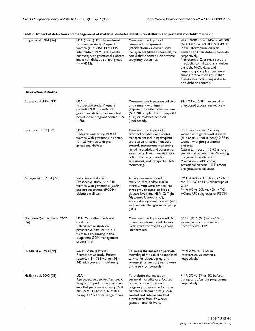
Langer et al. 1994 [74] USA (Texas). Population-based.Prospective study. Pregnant women (N = 2461; N = 1145 intervention, N = 1316 diabetic controls) with gestational diabetes and a non-diabetic control group (N = 4922).
Compared the impact of intensified management (intervention) vs. conventional management (diabetic controls) vs. non-diabetic controls on adverse pregnancy outcomes.
SBR: 1/1000 (N = 1145) vs. 4/1000 (N = 1316) vs. 4/1000 (N = 4922) in the intervention, diabetic controls and non-diabetic controls, respectively.Macrosomia, Caesarean section, metabolic complications, shoulder dystocia, NICU days, and respiratory complications lower among intervention group than diabetic controls; comparable to non-diabetic controls.
Observational studies
Aucott et al. 1994 [83] USA.Prospective study. Pregnant patients (N = 78) with pre-gestational diabetes vs. matched non-diabetic pregnant controls (N = 78).
Compared the impact on stillbirth of treatment with insulin (exposed) by either infusion pump (N = 20) or split-dose therapy (N = 58) vs. matched controls (unexposed).
SB: 1/78 vs. 0/78 in exposed vs. unexposed groups, respectively.
Fadel et al. 1982 [176] USA.Observational study. N = 84 women with gestational diabetes, N = 23 women with pre-gestational diabetes.
Compared the impact of a protocol of intensive diabetes management including frequent prenatal visits, strict metabolic control, antepartum monitoring including estriols and contraction stress tests, liberal hospitalization policy, fetal lung maturity assessment, and intrapartum fetal monitoring.
SB: 1 antepartum SB among women with gestational diabetes (due to true knot in cord); 0 SB in women with pre-gestational diabetes.Caesarean section: 15.4% among gestational diabetics, 56.5% among pre-gestational diabetics.Macrosomia: 20% among gestational diabetics, 13% among pre-gestational diabetics.
Banerjee et al. 2004 [77] India. Antenatal clinic.Prospective study. N = 240 women with gestational (GDM) and pre-gestational (PGDM) diabetes mellitus.
All women were placed on exercise, diet, and/or insulin therapy. And were divided into three groups based on blood glucose levels and HbA1C: Tight Glycaemic Control (TC), Acceptable glycaemic control (AC) and uncontrolled glycaemic group (UC).
PMR: 4.16% vs. 18.2% vs. 22.2% in the TC, AC and UC subgroups of GDM.PMR: 0% vs. 20% vs. 40% in TC, AC and UC subgroups of PGDM.
Gonzalez-Quintero et al. 2007 [76]
USA. Centralised perinatal database.Retrospective study on prospective data. N = 3,218 women participating in the outpatient GDM management programme.
Compared the impact on stillbirth of women whose blood glucose levels were controlled vs. those uncontrolled.
SBR (n,%): 2 (0.1) vs. 4 (0.3) in women with controlled vs. uncontrolled GDM.
Huddle et al 1993 [79] South Africa (Soweto).Retrospective study. Patient records (N = 733 women; N = 348 with gestational diabetes).
To assess the impact on perinatal mortality of the use of a specialised service for diabetic pregnant women (intervention) vs. non-use of the service (controls).
PMR: 3.7% vs. 15.6% in intervention vs. controls, respectively.
McElvy et al. 2000 [78] USA.Retrospective before-after study. Pregnant Type-1 diabetic women enrolled peri-conceptionally (N = 306; N = 111 before, N = 103 during, N = 92 after programme).
To evaluate the impact on perinatal mortality of a focused preconceptional and early pregnancy programme for Type 1 diabetes including strict glucose control and antepartum fetal surveillance from 32 weeks gestation until delivery.
PMR: 3% vs. 2% vs. 0% before, during, and after the programme, respectively.
Table 8: Impact of detection and management of maternal diabetes mellitus on stillbirth and perinatal mortality (Continued)
Page 18 of 48(page number not for citation purposes)

BMC Pregnancy and Childbirth 2009, 9(Suppl 1):S5 http://www.biomedcentral.com/1471-2393/9/S1/S5
Landon et al. 1992 [80] USA (Ohio). 2 teaching hospitals.Prospective cohort study. Pregnant women (N = 114) with insulin-dependent diabetes. Non-stress testing was begun weekly at 28–30 wks and 2× weekly at 32 wks (N = 1676 NSTs performed (14.7+/-3.2 tests per patient)).8% of tests (N = 134) non-reactive, necessitating a biophysical profile.
To determine whether maternal vascular disease and/or glycaemic control are associated with fetal condition in diabetic pregnancies by comparing the effect of reactive (exposed) vs. non-reactive (unexposed) NST on perinatal outcomes.
Fetal death (miscarriage + SB): 1/114.N = 10 deliveries among patients with abnormal test results.No significant differences in ambulatory glucose profile data in exposed vs. unexposed groups.No significant differences in glycaemic parameters in women delivered for suspected fetal compromise vs. nonintervention group.8/20 (40%) women with nephropathy or hypertension required delivery for fetal well-being, vs. 2/94 women (2%) without nephropathy or hypertension but with abnormal test results (P < 0.001).
Nachum et al. 2001 [177] Israel.Prospective controlled study. Pre-gestationally and gestationally diabetic women (N = 681 women; N = 801 pregnancies) recruited 1986–1989.
Compared the impact of diabetic pregnancies managed by hospitalisation vs. those managed by ambulatory care.
NMR: 1/394 vs. 0/407 in hospitalised vs. ambulatory care groups, respectively.
Table 8: Impact of detection and management of maternal diabetes mellitus on stillbirth and perinatal mortality (Continued)
mellitus (N = 1145), Langer et al [74] observed that anintensively managed group of women with diabetes hadrates of stillbirth and neonatal complications similar tonon-diabetic controls (N = 4922), while diabetic womengiven conventional management (N = 1316) had higherrates of these adverse outcomes [LOE: 2+]. Crowther et al.[75] randomly assigned women with gestational diabetesto receive either dietary advice, blood glucose monitoring,and insulin therapy as needed, or routine care, andreported decreased perinatal mortality associated with theintervention compared to the routine care group (0 versus5 perinatal deaths in intervention versus routine caregroups, respectively, P = 0.07) [LOE: 1-] (Additional file8).
In the US, Gonzalez-Quintero et al. [76] conducted anobservational study to assess the impact on perinatal out-comes of glycaemic control among women with gesta-tional diabetes attending an outpatient gestationaldiabetes management programme. Over one-third of theparticipants in the group with poor glycaemic control hadat least one of the factors in the composite study outcomevariable (macrosomia, large-for-gestational-age, hypogly-caemia, jaundice, or stillbirth) compared with only 24%in the well-controlled group (P < 0.001) [LOE: 2-]. Asomewhat less rigorous intervention study in India (N =240 mothers) by Banerjee et al. [77] sought to assess theincidence of fetal complications in both gestational andpre-gestational diabetic pregnant women with tight versusacceptable or uncontrolled glycaemic control. In womenwith pre-gestational diabetes, the tight glycaemic control
group had the lowest perinatal mortality (4.16% versus18.8% versus 22.2% in tight, acceptable, and uncon-trolled glycaemic control groups, respectively). In womenwith gestational diabetes, the same trend was noted witheven greater impact of tight glycaemic control (0% versus20% versus 40% in tight, acceptable, and uncontrolledglycaemic control groups, respectively). No statistical sig-nificance measures were provided [LOE: 1-].
In the US, McElvy et al. [78] conducted an evaluation of aprogramme that worked to achieve strict glycaemic con-trol before and during early pregnancy in women withpre-gestational diabetes (N = 306), with intensiveantepartum surveillance from 32 weeks gestation untildelivery. Perinatal mortality declined steadily from 3%before the programme to a rate comparable to generalpopulation levels after completion of the programme[LOE: 1-]. In Soweto, South Africa, Huddle et al [79]reported that a programme involving gestational diabeticwomen given special services during pregnancy observedlower rates of perinatal mortality among women partici-pating in the service compared to non-users of the service(3.7% versus 15.6%, respectively), but no statistical datawas furnished [LOE: 3].
A descriptive study by Landon et al. [80] of non-stress test-ing (with delivery for non-reactive tests) in women withpre-gestational diabetes found no differences in glycaemicparameters in women delivered for suspected fetal com-promise compared to a non- intervention group, suggest-
Page 19 of 48(page number not for citation purposes)
BMC Pregnancy and Childbirth 2009, 9(Suppl 1):S5 http://www.biomedcentral.com/1471-2393/9/S1/S5
ing the possibility that glycaemic control was unrelated tofetal compromise [LOE: 3].
Several studies assessed the impact on perinatal outcomesof mode of insulin treatment. Mukhopadhyay et al. [81]conducted a meta-analysis of RCTs (6 trials) comparingthe impact of continuous subcutaneous insulin infusionversus multiple-dose insulin on glyacemic control andpregnancy outcome in diabetic women. Neither glycae-mic control nor pregnancy outcomes were differentbetween treatment groups [LOE: 1++] (Additional file 9).In Israel, Hod et al. [82] conducted a small RCT (N = 322women) comparing the impact on fetal and perinatal out-comes of insulin aspart, a fast-acting insulin analogue,versus human insulin therapy in women with pre-gesta-tional diabetes. A slight trend toward lower perinatal mor-tality was observed in the insulin aspart group comparedto the human insulin group (14/1000 versus 22/1000,respectively), but no statistical significance data wasreported [LOE: 1-]. A study of insulin therapy using eitherpump infusion or split-dose therapy found no differencein rates of perinatal mortality in the treated groups com-pared to non-diabetic controls [83].
Elective deliveryA Cochrane review by Boulvain et al. [84] assessed trials ofexpectant management versus elective delivery in termwomen with pre-existing diabetes mellitus; only 1 trialwas included (N = 200), which documented no perinatalmortality in either group [LOE: 1+] (Additional file 10).
ConclusionWhile data from RCTs is scarce, the limited evidence,largely from clinical management of diabetic patients(Grade C evidence), suggests that pregnant women whodo not have vascular complications and with good glycae-mic control do not as a group have an increased risk ofstillbirth above the general population [85]. In settingswith poor access to care and high prevalence of gestationaldiabetes, perinatal mortality has been shown to bereduced with diabetes screening and treatment [79].Although retrospective studies reveal declines in the inci-dence of stillbirth among diabetic women in low-preva-lence populations where diabetes care is available [86],few prospective trials have been able to show any impacton stillbirths or perinatal mortality, largely owing to thehigh sample sizes required for such studies [63]. Adequateglycaemic control and monitoring during pregnancy are areasonable means for reducing stillbirths that has beenshown to reduce many complications that may be relatedto stillbirth, including congenital anomalies and macro-somia. Where feasible, particularly in high-prevalence set-tings, large studies with rigorous designs may helpconfirm this recommendation.
Advanced monitoring in pregnancyAntenatal fetal heart rate monitoring using cardiotocographyBackgroundAntenatal fetal surveillance studies are widely used toassess fetal well-being and to identify the compromisedfetus. Fetal heart rate patterns are a principal componentof the majority of tests of fetal well-being, and cardioto-cography, where it is available, is the method frequentlyused to electronically record the fetal heart rate. In high-income countries, beginning in the third trimester, cardi-otocographic monitoring (electronic fetal monitoring)has largely replaced intermittent auscultation (periodiclistening to the fetal heart rate using a stethoscope orhandheld Doppler device) for monitoring fetal heart rate.During the antenatal period, external cardiotocography isused, which employs a Doppler ultrasound transducer;the mother's uterine contractions are also monitoredusing a pressure transducer, with both transducersstrapped to the mother's abdomen while recording is inprocess.
Antenatal fetal heart monitoring includes both non-stresstests (NST) and contraction stress tests (CST). Fetal heartrate patterns are classified as either reassuring (reactive),non-reassuring (non-reactive) or abnormal, consideringheart rate baseline, variability, and decelerations. In high-income countries, particularly the US, the NST is widelyaccepted as a primary fetal surveillance tool in high-riskpregnancies. It may be used in combination with ultra-sound testing to observe fetal movement and amnioticfluid indices. When an NST is non-reactive, its poor pre-dictive value (< 50%) warrants confirmatory testing withCST or a complete BPP [87]. During a CST, uterine con-tractions are induced (usually with intravenous oxytocin,but contractions can be generated using nipple stimula-tion) and subsequent fetal heart accelerations and decel-erations are monitored to detect possible cases ofuteroplacental insufficiency. It is important to note thatantenatal fetal heart monitoring is gestational-agedependent; at 28 weeks' gestation, only about 60% of nor-mally formed fetuses will have reactive non-stress testingdue to immaturity of the fetal autonomic nervous systemrather than placental insufficiency.
Variation between practitioners in interpretation of cardi-otocographic tracings can potentially lead to inappropri-ate intervention, or false reassurance without appropriateintervention. Tracings are notoriously difficult to inter-pret, as fetal sleep patterns, labour progress, external stim-uli, and opiate administration to the mother can alter fetalheart rate changes in the absence of fetal hypoxia or dis-tress [2]. Additionally, as indications of fetal distress oftenindicate early delivery, the gestational age threshold atwhich antepartum testing is initiated will depend on an
Page 20 of 48(page number not for citation purposes)

BMC Pregnancy and Childbirth 2009, 9(Suppl 1):S5 http://www.biomedcentral.com/1471-2393/9/S1/S5
institution's ability to care for very preterm neonates,which, in general, is earlier in high-income settings thanin lower-resource settings [21].
Literature-based evidenceWe identified 1 Cochrane review and 4 observationalstudies testing the impact of antepartum cardiotocogra-
phy (including both NST and CST) on perinatal outcomes(Table 9).
Several studies described how well NST and CST resultscorrelated with subsequent perinatal outcome. An RCT byFlynn et al [88] identified a significant association of NSTtracings (N = 569 tracings, N = 300 patients) with still-births, neonatal deaths, fetal growth restriction, complica-
Table 9: Impact of antepartum cardiotocography on stillbirth and perinatal outcomes
Source Location and Type of Study Intervention Stillbirths/Perinatal Outcomes
Reviews and meta-analyses
Pattison and McCowan 1999 [91] UK, Australia.Meta-analysis (Cochrane). 3 RCTs included (N = 1279).
Compared the impact of electronic fetal monitoring with an antenatal CTG (intervention) vs. a control group where the results of the CTG were withheld from the caregiver or no monitoring was done on perinatal morbidity and mortality and maternal morbidity.
PMR (excluding lethal abnormalities): OR = 2.65 (95% CI: 0.99–7.12) [NS].[12/651 vs. 4/628 in intervention and control groups, respectively].
Observational studies
Evertson et al. 1978 [90] USA.Case series. N = 746 pregnant women undergoing 1119 CSTs.
Assessed the incidence of fetal deaths within one week of a negative CST.
SBR: 7/680 patients (1%) within 1 week of a negative CST.Fetal death in most cases resulted from factors other then uteroplacental insufficiency (umbilical cord accident, malformations, and placental abruption)
Flynn et al. 1982 [88] UK.RCT. Pregnant patients (N = 300) with non-stress antepartum cardiotocography tracings (N = 569; N = 144 intervention, N = 156 controls).
Compared impact on pregnancy outcomes of revealing cardiotocography results to clinician (intervention) vs. concealing results (controls).
SBR+NMR: Significant association with 'non-reactive' traces.Significant association of nonreactive traces with rates of fetal growth restriction, admission to special care baby unit, Apgar scores at 1 and 5 min.
Freeman et al. 1982 [92] USA and Canada. 18 institutions.Comparison of diagnostic tests. Pregnant women (N = 6168; N = 4626 CST, N = 1542 NST) with increased risk for uteroplacental insufficiency.
Compared the impact on fetal outcomes of CST (intervention) vs. NST (comparison) for primary fetal surveillance.
PMR: 8.4/1000 vs. 21.4/1000 in intervention vs. comparison groups, respectively (P < 0.05). After correction for congenital anomalies and unrelated causes: 3.5/1000 vs. 7.1/1000 intervention vs. comparison groups, respectively (P < 0.05).Fetal death (miscarriage+SB): 1.1/1000 vs. 7.8/1000 in intervention vs. comparison groups, respectively (P < 0.05). After correction: 0.4/1000 vs. 3.2/1000 in intervention vs. comparison groups, respectively (P < 0.05).
Rayburn et al. 1980 [89] USA (Lexington). University hospital.Prospective cohort study. High-risk clinic patients (N = 561) who had undergone NST within one week prior to delivery.
Compared the association of reactive vs. non-reactive NSTs vs. no testing with fetal outcomes.
SB: 1/509 in reactive non-stress group (cord accident).Corrected PMR: 1/509 vs. 2/22 vs. 20/1000 in reactive, non-reactive and no testing groups, respectively (P < 0.05).Fetal compromise: 4%, 36%, and 13% in reactive, non-reactive and no testing groups, respectively (P < 0.001)
Page 21 of 48(page number not for citation purposes)
BMC Pregnancy and Childbirth 2009, 9(Suppl 1):S5 http://www.biomedcentral.com/1471-2393/9/S1/S5
tions of intrauterine hypoxia, and low Apgar scores [LOE:1+]. An observational study of high-risk pregnant women(N = 561) by Rayburn et al [89] assessed NST tracingswithin 1 week of delivery along with subsequent perinataloutcome. Perinatal mortality in patients with reactiveNSTs was comparable to that of patients with no apparentantepartum complications (1/509 versus 6/1408, respec-tively), and significantly lower than among untestedpatients or patients with nonreactive NSTs (20/1000 and2/22, respectively, P < 0.05) [LOE: 2-]. Evertson et al [90]assessed the rates of fetal death within 1 week of negativeCST results in a cohort of patients (N = 680), identifyingonly 7 deaths, of which most were due to factors otherthan placental insufficiency, suggesting that the test hadlow rates of false negatives.
A review by Pattison and McCowan [91] included 4 stud-ies (N = 1,588 pregnancies) of the impact of cardiotocog-raphy use on perinatal mortality in high or intermediaterisk pregnancies (Additional file 11). The trial reported atrend toward increased perinatal mortality in the cardioto-cography group versus controls receiving no monitoringor whose cardiotocography results were concealed fromthe clinician; (3 trials, N = 1279 pregnancies, OR = 2.85,95% CI: 0.99–7.12). Four of the 7 deaths in the largesttrial reporting perinatal deaths were associated with fetalinfection and prematurity, suggesting these deaths were not associated with the intervention. There was noincrease in the incidence of elective Caesarean section orinduction of labour [LOE: 1++].
Freeman et al [92] compared results from the NST (N =1542 women) to the CST (N = 4626) for primary fetal sur-veillance and found a nearly 8-fold higher risk of antepar-tum fetal death in the NST group (7.8/1000 versus 1.1/1000, respectively, P < 0.05). Corrected for congenitalanomalies and unrelated causes, the differential inantepartum fetal death remained 8-fold higher in the NSTgroup (3.2/1000 versus 0.4/1000, respectively, P < 0.05)[LOE: 2+].
ConclusionThere are relatively few RCTs of antepartum fetal heart ratesurveillance (Grade C evidence), though observationalstudies clearly indicate a correlation between non-reassur-ing cardiotocographic traces and adverse perinatal out-comes, including stillbirth. Although there is a Cochranereview on antepartum cardiotocography [91] showing atrend toward increased risk of perinatal mortality, thestudy was underpowered to detect such an impact, and thecauses of the deaths observed appeared unlikely to havebeen preventable. While RCTs are lacking to evaluate animpact of NSTs in reducing stillbirth, apparent reductionsin stillbirth rates have followed the incorporation of theNST into protocols for management of high-risk preg-
nancy in the United States [21]. As with fetal movementmonitoring and umbilical Doppler velocimetry, the rateof false-positives with a non-reactive NST is high enoughthat additional testing, whether CST or BPP, is advised toguard against unnecessary intervention.
When used experimentally as a primary fetal surveillancetool, CST use was associated with lower rates of antepar-tum stillbirth [92], suggesting that it could be useful forprimary fetal surveillance testing in pregnancies at highestrisk of stillbirth, including those of women with hyperten-sion, diabetes, and intrauterine growth restriction. As ittypically uses intravenous oxytocin to stimulate uterinecontractions, the CST is more technologically intensivethan the NST and can induce labour; thus, it is contraindi-cated for patients with placenta previa, placental abrup-tion, previous classical Caesarean section, and prematurerupture of membranes [87]. The CST should be conductedcautiously in cases of multiple pregnancy, incompetentcervix, and polyhydramnios.
Fetal biophysical profile (BPP) test scoring for assessing high-risk pregnancyBackgroundThe BPP was conceptually derived from the Apgar scorethat is used to rate the condition of the newborn [93].Using NST monitoring with cardiotocography and fetalultrasound, the BPP collects 5 indicators of fetal well-being: fetal heart rate reactivity, breathing movements,gross body movements, muscular tone and qualitativeamniotic fluid volume. Indications of fetal compromiseoften suggest the need for early delivery by induction oflabour or Caesarean section.
Literature-based evidenceWe identified one Cochrane review and four other obser-vational studies of BPP reporting perinatal mortality out-comes (Table 10). A multi-institutional study in the US byFreeman et al [94] compared antepartum BPP versus NST,and reported that the fetal death rate among false nega-tives was several times lower among fetuses tested withBPP compared to the NST, though rates of false positiveresults with BPP were higher than with NST. Several otherobservational clinical studies were identified but hadinsufficient rigor or size to contribute meaningfully to theevidence base associating BPP results with stillbirth inci-dence [95-97].
Lalor et al. [1] undertook a Cochrane review of RCTs ofvariable quality (5 trials, N = 2839 women) comparingBPP with other forms of fetal assessment in women withhigh risk pregnancies (Additional file 12). There were nosignificant differences among pregnancies evaluated bythe various methods of fetal assessment in perinatal mor-
Page 22 of 48(page number not for citation purposes)

BMC Pregnancy and Childbirth 2009, 9(Suppl 1):S5 http://www.biomedcentral.com/1471-2393/9/S1/S5
tality (RR = 1.33, 95% CI: 0.60–2.98) or Apgar score <7 at5 minutes (RR = 1.27, 95% CI: 0.85–1.92) [LOE: 1+].
ConclusionCompared with conventional fetal monitoring, which isbased primarily on cardiotocography/NST, BPP appearsto offer no improvement in pregnancy outcomes (GradeC evidence). Although rates of false negative test results ofthe BPP are low, rates of false positives are high. The total
number of subjects included in the meta-analysis remainssmall (N = 2974), and some of the studies carry a high riskof allocation bias. Although there is no consensus on opti-mal protocol for fetal monitoring, studies demonstratethat it is reasonable to implement the modified BPP(which consists of an NST and amniotic fluid volumemeasurement only), with full BPP or other tests such asthe CST reserved for abnormal test results [98]. This pas-sive and rapid approach to monitoring has been shown to
Table 10: Impact of fetal BPP scoring on stillbirth and perinatal outcomes
Source Location and Type of Study Intervention Stillbirths/Perinatal Outcomes
Reviews and meta-analyses
Lalor et al. 2008 [1] USA, UK.Meta-analysis (Cochrane). 4 RCTs included (N = 2839 pregnant women).
Compared the effects of complex (BPP; intervention) vs. simple fetal monitoring (cardiotocography and maximum pool depth) (controls).
PMR (including major malformations): RR = 1.33 (95% CI: 0.60–2.98) [NS][13/1405 vs. 10/1434 in intervention vs. control groups, respectively].PMR: RR = 1.30 (95% CI: 0.58–2.92) [NS][13/1405 vs. 11/1434 in intervention vs. control groups, respectively].
Observational studies
Awad 1991 [101] Egypt. Al Fayrouz Hospital.Before-after study. N = 319 women (N = 160 intervention, N = 159 controls). Routine BPP introduced in 1990; compared to historical controls without BPP at same hospital prior to 1990.
Assessed the impact on perinatal mortality of introduction of routine BPP (intervention) vs. historical controls.
SBR: 0/1000 vs. 6/29/1000 in intervention vs. control groups, respectively.PMR (excluding malformations and alloimmunization disorders): 6.25/1000 vs. 25.16/1000 in intervention vs. control groups, respectively.
Golde et al. 1984 [97] USA. University of Southern California Medical Centre and Women's Hospital, Los Angeles,Case series. Pregnant diabetic women (N = 107) vs. historic controls (N = 140) undergoing antepartum fetal surveillance.
Compared the impact of a package of nonstress heart rate testing, backed up by either fetal BPP or CST twice weekly (intervention), vs. weekly NSTs and daily plasma estriols (controls).
SB: 3/107 vs. 1/140 in intervention vs. control groups, respectively. 0 unexplained losses in either group.
de la Vega A 2002 [95] Puerto Rico. Private clinic.Case series. Pregnancies (N = 1810) 20 wks gestation. High-resolution sonograph was performed in each trimester; BPP in 3rd trimester if risk factor was identified.
To assess the impact of testing fetal well-being using sonography and BPP in clinic cases (intervention) compared with the US average.
SBR: 14/1810 (7.7/1000 births) in this series vs. the U.S. national average of 6.7–7.8/1000 births.
Kennelly et al. 2007 [96] UK. Single tertiary centre.Retrospective study. Records from Fetal Medicine Database, 2000–2005. Pregnant women (N = 39) with SGA twins (19 monochorionic sets, 13 dichorionic sets) with absent or reversed end diastolic flow in the umbilical artery.
To assess the impact of active monitoring with daily BPP after estimated fetal weight 500 g in both twins and gestational age 24 wks. Delivery was timed based on abnormal BPP, two equivocal BPP within 12 h or gestational age 32 wks.
Fetal death (miscarriage+SB): None.
Page 23 of 48(page number not for citation purposes)

BMC Pregnancy and Childbirth 2009, 9(Suppl 1):S5 http://www.biomedcentral.com/1471-2393/9/S1/S5
be more cost-effective without diminishing its predictivevalue [99]. For reasons of cost and simplicity, the modi-fied BPP may also prove to be more implementable thanthe full BPP in low-/middle-income country contexts withultrasound and cardiotocographic capacity [100,101].
Vibroacoustic stimulationBackgroundAdjunctive vibroacoustic stimulation with the NST is usedless often than CST or BPP after non-reassuring NSTresults. Vibroacoustic stimulation provokes fetal responseusing a sound-emitting device placed on the maternalabdomen near the fetal head, along with simultaneous
cardiotocography to document fetal movements andheart traces. Theoretically, the resultant startle reflex in thefetus and subsequent fetal heart rate acceleration or tran-sient tachycardia following vibroacoustic stimulation pro-vide reassurance of fetal well-being and minimiseunnecessary intervention for nonreactive tests [102].
Literature-based evidenceOur literature search identified one Cochrane review onvibroacoustic stimulation and four other observational/interventional studies (Table 11). In a Cochrane review ofvibroacoustic stimulation in conjunction with tests offetal well-being (9 RCTs, N = 4838 women), Tan and
Table 11: Impact of vibroacoustic stimulation on stillbirth and perinatal outcomes
Source Location and Type of Study Intervention Stillbirths/Perinatal Outcomes
Reviews and meta-analyses
Tanand Smyth 2001 [102] Australia, USA, Mexico, Greece, Turkey, Thailand.Meta-analysis (Cochrane). 8 RCTs included (N = 4838 women).
Compared the impact of fetal vibratory acoustic stimulation (intervention) vs. mock or no stimulation (controls).
Non-reactive antenatal cardiotocography test (7 RCTs): RR = 0.62 (95% CI: 0.52–0.74)[176/2244 vs. 286/2239 in intervention vs. control groups, respectively.]PMR (2 RCTs): RR = 0.32 (95% CI: 0.01–7.78) [NS][0/476 vs. 1/464 in intervention vs. control groups, respectively].
Intervention studies
Papadopoulos et al. 2007 [103] Greece.RCT. Pregnant women (N = 2833).
Compared the effect of vibroacoustic stimulation with a 3-s stimulus with an artificial larynx (repeated if BPP remained abnormal for 30 min; intervention) vs. no stimulation (controls).
Fetal death (miscarriage+SB): 10/1349 (0.74%) vs. 9/1484 (0.6%) in intervention vs. control groups, respectively [NS]
Petrovic 1998. [178] Croatia.RCT. Singleton pregnancies (N = 494; N = 168 intervention, N = 326 controls).Groups were monitored using the modified BPP; given vibroacoustic stimulation in absence of fetal activity at the start of the BPP.
Compared the impact of vibroacoustic stimulation (intervention) vs. no stimulation (controls).
SBR: 2/168 vs. 4/326 in intervention vs. control groups, respectively.
Sood 2007 [104] India.RCT. Singleton high-risk pregnancies (N = 214).
Compared the effect of vibroacoustic stimulation (intervention) vs. mock stimulation (controls). Both arms also had modified BPP conducted.
PMR: OR = 0.62 (95% CI: 0.05–5.57) [NS][2/110 (1.8%) vs. 3/104 (2.9%) in intervention vs. control groups, respectively.
Observational studies
Salamalekis et al. 1994 [179] Greece.Case series. High-risk pregnancies (N = 180).
Compared the association with fetal deaths of reactive (study group) vs. non reactive (controls) NST results for tests conducted 24 hrs before delivery.
Fetal death (miscarriage+SB): 1 (0.67%) vs. 2 (6.25%) in intervention vs. control groups, respectively.
Page 24 of 48(page number not for citation purposes)
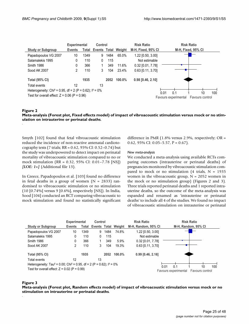
BMC Pregnancy and Childbirth 2009, 9(Suppl 1):S5 http://www.biomedcentral.com/1471-2393/9/S1/S5
Smyth [102] found that fetal vibroacoustic stimulationreduced the incidence of non-reactive antenatal cardioto-cography tests (7 trials; RR = 0.62, 95% CI: 0.52–0.74) butthe study was underpowered to detect impact on perinatalmortality of vibroacoustic stimulation compared to no ormock stimulation (RR = 0.32, 95% CI: 0.01–7.78 [NS])[LOE: 1+] (Additional file 13).
In Greece, Papadopoulos et al. [103] found no differencein fetal deaths in a group of women (N = 2833) ran-domised to vibroacoustic stimulation or no stimulation(10 [0.74%] versus 9 [0.6%], respectively [NS]). In India,Sood [104] conducted an RCT comparing vibroacoustic tomock stimulation and found no statistically significant
difference in PMR (1.8% versus 2.9%, respectively; OR =0.62, 95% CI: 0.05–5.57, P = 0.67).
New meta-analysisWe conducted a meta-analysis using available RCTs com-paring outcomes (intrauterine or perinatal deaths) ofpregnancies monitored by vibroacoustic stimulation com-pared to mock or no stimulation (4 trials, N = 1935women in the vibroacoustic group, N = 2052 women inthe mock or no stimulation group) (Figures 2 and 3).Three trials reported perinatal deaths and 1 reported intra-uterine deaths, so the outcome of the meta-analysis wasexpanded and renamed as 'intrauterine or perinataldeaths' to include all 4 of the studies. We found no impactof vibroacoustic stimulation on intrauterine or perinatal
Meta-analysis (Forest plot, Random effects model) of impact of vibroacoustic stimulation versus mock or no stimulation on intrauterine or perinatal deathsFigure 3Meta-analysis (Forest plot, Random effects model) of impact of vibroacoustic stimulation versus mock or no stimulation on intrauterine or perinatal deaths.
Study or Subgroup
Papadopoulos VG 2007Salamalekis 1995Smith 1986Sood AK 2007
Total (95% CI)
Total eventsHeterogeneity: Tau² = 0.00; Chi² = 0.95, df = 2 (P = 0.62); I² = 0%Test for overall effect: Z = 0.02 (P = 0.99)
Events
10002
12
Total
1349110366110
1935
Events
9013
13
Total
1484115349104
2052
Weight
74.8%
5.9%19.3%
100.0%
M-H, Random, 95% CI
1.22 [0.50, 3.00]Not estimable
0.32 [0.01, 7.78]0.63 [0.11, 3.70]
0.99 [0.46, 2.16]
Experimental Control Risk Ratio Risk RatioM-H, Random, 95% CI
0.01 0.1 1 10 100Favours experimental Favours control
Meta-analysis (Forest plot, Fixed effects model) of impact of vibroacoustic stimulation versus mock or no stimulation on intra-uterine or perinatal deathsFigure 2Meta-analysis (Forest plot, Fixed effects model) of impact of vibroacoustic stimulation versus mock or no stim-ulation on intrauterine or perinatal deaths.
Study or Subgroup
Papadopoulos VG 2007Salamalekis 1995Smith 1986Sood AK 2007
Total (95% CI)
Total eventsHeterogeneity: Chi² = 0.95, df = 2 (P = 0.62); I² = 0%Test for overall effect: Z = 0.06 (P = 0.96)
Events
10002
12
Total
1349110366110
1935
Events
9013
13
Total
1484115349104
2052
Weight
65.0%
11.6%23.4%
100.0%
M-H, Fixed, 95% CI
1.22 [0.50, 3.00]Not estimable
0.32 [0.01, 7.78]0.63 [0.11, 3.70]
0.98 [0.46, 2.10]
Experimental Control Risk Ratio Risk RatioM-H, Fixed, 95% CI
0.01 0.1 1 10 100Favours experimental Favours control
Page 25 of 48(page number not for citation purposes)
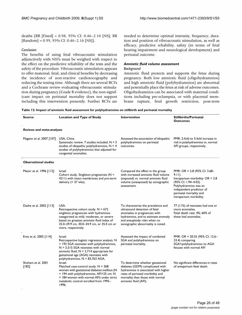
BMC Pregnancy and Childbirth 2009, 9(Suppl 1):S5 http://www.biomedcentral.com/1471-2393/9/S1/S5
deaths (RR [Fixed] = 0.98, 95% CI: 0.46–2.10 [NS]; RR[Random] = 0.99, 95% CI: 0.46–2.16 [NS]).
ConclusionThe benefits of using fetal vibroacoustic stimulationadjunctively with NSTs must be weighed with respect tothe effect on the predictive reliability of the tests and thesafety of the procedure. Vibroacoustic stimulation appearsto offer maternal, fetal, and clinical benefits by decreasingthe incidence of non-reactive cardiotocography andreducing the testing time. Although there are several RCTsand a Cochrane review evaluating vibroacoustic stimula-tion during pregnancy (Grade B evidence), the non-signif-icant impact on perinatal mortality does not supportincluding this intervention presently. Further RCTs are
needed to determine optimal intensity, frequency, dura-tion and position of vibroacoustic stimulation, as well asefficacy, predictive reliability, safety (in terms of fetalhearing impairment and neurological development) andperinatal outcome.
Amniotic fluid volume assessmentBackgroundAmniotic fluid protects and supports the fetus duringpregnancy. Both low amniotic fluid (oligohydramnios)and high amniotic fluid (polyhydramnios) are abnormaland potentially place the fetus at risk of adverse outcomes.Oligohydramnios can be associated with maternal condi-tions including pre-eclampsia, or with placental mem-brane rupture, fetal growth restriction, post-term
Table 12: Impact of amniotic fluid assessment for polyhydramnios on stillbirth and perinatal mortality
Source Location and Type of Study Intervention Stillbirths/Perinatal Outcomes
Reviews and meta-analyses
Magann et al. 2007 [107] USA, ChinaSystematic review. 7 studies included. N = 3 studies of idiopathic polyhydramnios, N = 4 studies of polyhydramnios that adjusted for congenital anomalies.
Assessed the association of idiopathic polyhydramnios on perinatal outcomes.
PMR: 2-fold to 5-fold increase in risk in polyhydramnios vs. normal AFI groups, respectively.
Observational studies
Mazor et al. 1996 [112] Israel.Cohort study. Singleton pregnancies (N = 4211) with intact membranes and pre-term delivery (< 37 wks).
Compared the effect in the group with increased amniotic fluid volume (exposed) vs. normal amniotic fluid volume (unexposed) by sonographic assessment.
PMR: OR = 5.8 (95% CI: 3.68–9.11).Intrapartum morbidity: OR = 2.8 (95% CI: 1.94–4.03).Polyhydramnios was an independent predictor of perinatal mortality and intrapartum morbidity.
Dashe et al. 2002 [113] USA.Retrospective cohort study. N = 672 singleton pregnancies with hydramnios categorised as mild, moderate, or severe based on greatest amniotic fluid index of 25.0–29.9 cm, 30.0–34.9 cm, or 35.0 cm or more, respectively.
To characterise the prevalence and ultrasound detection of fetal anomalies in pregnancies with hydramnios, and to estimate anomaly and aneuploidy risks when no sonographic abnormality is noted.
77 (11%) of neonates had one or more anomalies.Fetal death rate: 4%; 60% of these had anomalies.
Erez et al. 2005 [114] Israel.Retrospective logistic regression analysis. N = 192 SGA neonates with polyhydramnios, N = 5,515 SGA neonates with normal amniotic fluid, N = 3,714 appropriate for gestational age (AGA) neonates with polyhydramnios, N = 83,763 AGA.
Assessed the impact of combined SGA and polyhydramnios on perinatal mortality.
PMR: OR = 20.55 (95% CI: 12.6–33.4) comparing SGA+polyhydramnios to AGA fetuses with normal AFI
Shoham et al. 2001 [182]
Israel.Matched case-control study. N = 368 women with gestational diabetes mellitus (N = 194 with polyhydramnios, AFI>25 cm; N = 184 women with normal AFI) under strict metabolic control enrolled from 1995–1996.
To determine whether gestational diabetes (GDM) complicated with hydramnios is associated with higher rates of perinatal morbidity and mortality than those with normal amniotic fluid (AFI).
No significant differences in rates of antepartum fetal death.
Page 26 of 48(page number not for citation purposes)

BMC Pregnancy and Childbirth 2009, 9(Suppl 1):S5 http://www.biomedcentral.com/1471-2393/9/S1/S5
pregnancy, fetal kidney problems, or fetal or placentalabnormalities [105,106]. Polyhydramnios is frequentlyassociated with maternal diabetes mellitus, maternal car-diac problems, twin-twin transfusion syndrome, or fetalor placental malformations, but in 50–60% of cases, it isidiopathic [107-110]. Until ultrasound became available,the invasive nature of amniotic fluid testing limited itsclinical usefulness [111]. Using ultrasound, multiplemethods for measuring amniotic fluid have been devel-oped and are used to identify at-risk pregnancies. Theamniotic fluid index (AFI) is a measure of the amount ofamniotic fluid which is calculated by summing centime-ters of depth of 4 different pockets of fluid; alternatively,the single deepest vertical pocket or maximum pool depthmay be used. At term, many clinicians will induce labouror perform Caesarean section after diagnosis of decreasedamniotic fluid volume to prevent an adverse pregnancyoutcome. However, using assessments of amniotic fluidvolume to predict fetal complications is controversial, andthe utility of amniotic fluid assessment is different whenused prior to versus after the onset of labour. Here, weexamine the potential for assessment of amniotic fluidvolume, or interventions to achieve normal amniotic fluidvolume, to effectively detect high-risk pregnancy or fetaldistress and subsequent adverse perinatal outcomes. Wealso examined the impact of interventions to achieve nor-mal amniotic fluid volume on perinatal outcomes
Literature-based evidenceOur literature search identified two Cochrane reviews,one other review, and 15 other interventional or observa-tional trials of amniotic fluid assessment to identify poly-hydramnios or oligohydramnios that reported perinataloutcomes (Table 12 and Table 13).
PolyhydramniosSeveral observational studies explored the association ofpolyhydramnios with perinatal mortality, with inconsist-ent findings. Several observational studies [112] observedthat polyhydroamnios was an independent risk factor forboth perinatal mortality and intrapartum morbidity[LOE: 2-]. A retrospective cohort study by Dashe et al.[113] documented a fetal death rate of 4% in pregnancieswith polyhydramnios diagnosed by AFI, of which 40%were unrelated to fetal malformations [LOE: 2-]. Magannet al. conducted a review of idiopathic polyhydramnios[107] including 4 studies that evaluated perinatal mortal-ity with polyhydramnios after correcting for congenitalanomalies. In the larger studies, idiopathic polyhyram-nios was associated with macrosomia and a statisticallysignificant 2- to 5-fold increase in the risk of perinatalmortality [LOE: 2+] (Additional file 14). A retrospectivelogistic regression analysis of SGA and appropriate-weightfetuses with and without polyhydramnios determinedthat the combination of SGA and polyhydramnios was an
independent risk factor for perinatal mortality comparedto normal weight-for-age infants with normal AFI values(OR = 20.55; CI: 12.6–33.4) [114]. The association maynot apply to certain subsets of high-risk pregnancies, as asimilar study among women with gestational diabeteswho had AFI performed antenatally found no elevatedrisk of perinatal morbidity or mortality among pregnan-cies complicated by polyhydramnios compared to gesta-tional diabetics without polyhydramnios [LOE: 2-]. Nointervention studies subsequent to diagnosis of polyhy-dramnios were identified.
OligohydramniosObservational studies of oligohydramnios cases demon-strated a consistent elevated risk of poor perinatal out-comes. Anandakumar et al. [115] studied how AFI usedwith non-stress cardiotocography and fetal acoustic stim-ulation for non-reactive NSTs might predict adverse preg-nancy outcome in high-risk pregnancies (N = 565women). Of the 4 perinatal deaths in the group with lowAFI, 3 had had a reactive NST within 7 days of fetal death[LOE: 2-]. In Italy, Locatelli et al. [116] reported rates ofoligohydramnios twice as high in pregnancies with poorperinatal outcome than pregnancies with no adverse out-come [LOE: 2-]. However, oligohydramnios identifiedduring the intrapartum period did not appear associatedwith adverse perinatal outcome in high-risk pregnancies:Baron et al [117] observed that women with low AFI weremore likely to have Caesarean section for fetal distressthan women with normal AFI, but the rates of neonatalcomplications were similar [LOE: 2+].
One study found that in addition to the absolute volumeof AFI, the distribution of amniotic fluid by intrauterinequadrants is predictive of fetal outcome. Myles et al. [118]observed higher rates of meconium staining, poor 1-minute Apgar scores, fetal heart decelerations, and fetalacidaemia in pregnancies where fluid volume was higherin the upper quadrants than in pregnancies where fluidvolume was higher in lower quadrants [LOE: 2+].
A number of observational studies, RCTs and a systematicreview compared AFI measurement to other methods ofmeasuring amniotic fluid in ability to predict poor perina-tal outcome. In an observational study in Egypt, Youssefet al. [119] (N = 174 women) found that the AFI was moresensitive than the single largest vertical pocket measure-ment in predicting perinatal mortality and multiple meas-ures of perinatal morbidity. Morris et al. [120]documented similar findings, but the sensitivity of AFI <5 cm for major adverse fetal outcome was only 28 percent.A Cochrane review [121] compared the evidence for thepredictive value of 2 methods of amniotic fluid assess-ment (AFI versus single deepest vertical pocket) onadverse pregnancy outcomes (Additional file 15). Com-
Page 27 of 48(page number not for citation purposes)
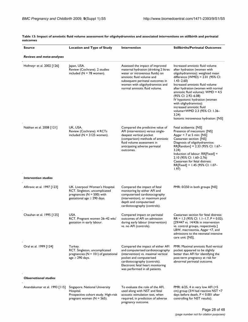
BMC Pregnancy and Childbirth 2009, 9(Suppl 1):S5 http://www.biomedcentral.com/1471-2393/9/S1/S5
Table 13: Impact of amniotic fluid volume assessment for oligohydramnios and associated interventions on stillbirth and perinatal outcomes
Source Location and Type of Study Intervention Stillbirths/Perinatal Outcomes
Reviews and meta-analyses
Hofmeyr et al. 2002 [126] Japan, USA.Review (Cochrane). 2 studies included (N = 78 women).
Assessed the impact of improved maternal hydration (drinking 2 litres water or intravenous fluids) on amniotic fluid volume and subsequent perinatal outcomes in women with oligohydramnios and normal amniotic fluid volume.
Increased amniotic fluid volume after hydration (women with oligohydramnios): weighted mean difference (WMD) = 2.01 (95% CI: 1.43–2.60)Increased amniotic fluid volume after hydration (women with normal amniotic fluid volume): WMD = 4.5 (95% CI: 2.92–6.08)IV hypotonic hydration (women with olighydramnios):increased amniotic fluid volume=WMD 2.3 (95% CI: 1.36–3.24)Isotonic intravenous hydration: [NS]
Nabhan et al. 2008 [121] UK, USA.Review (Cochrane). 4 RCTs included (N = 3125 women).
Compared the predictive value of AFI (intervention) versus single-deepest vertical pocket (comparison) methods of amniotic fluid volume assessment in anticipating adverse perinatal outcomes.
Fetal acidaemia: [NS]Presence of meconium: [NS]Apgar < 7 at 5 min: [NS]Caesarean section: [NS]Diagnosis of oligohydramnios: RR[Random] = 2.33 (95% CI: 1.67–3.24)Induction of labour: RR[Fixed] = 2.10 (95% CI: 1.60–2.76)Caesarean for fetal distress: RR[Fixed] = 1.45 (95% CI: 1.07–1.97)
Intervention studies
Alfirevic et al. 1997 [123] UK. Liverpool Women's Hospital.RCT. Singleton, uncomplicated pregnancies (N = 500) with gestational age 290 days.
Compared the impact of fetal monitoring by either AFI and computerised cardiotocography (intervention), or maximum pool depth and computerised cardiotocography (controls).
PMR: 0/250 in both groups [NS]
Chauhan et al. 1995 [125] USA.RCT. Pregnant women 26–42 wks' gestation in early labour.
Compared impact on perinatal outcomes of AFI on admission during early labour (intervention) vs. no AFI (controls).
Caesarean section for fetal distress: RR = 1.3 (95% CI: 1.1–1.7, P = 0.02).[29/447 vs. 14/436 in intervention vs. control groups, respectively.]LBW, macrosomia, Apgar <7, and admissions to the neonatal intensive care unit: [NS].
Oral et al. 1999 [124] Turkey.RCT. Singleton, uncomplicated pregnancies (N = 101) of gestational age 290 days.
Compared the impact of either AFI and computerised cardiotocography (intervention) vs. maximal vertical pocket and computerised cardiotocography (controls). Electronic fetal heart monitoring was performed in all patients.
PMR: Maximal amniotic fluid vertical pocket appeared to be slightly better than AFI for identifying the post-term pregnancy at risk for abnormal perinatal outcome.
Observational studies
Anandakumar et al. 1993 [115] Singapore. National University Hospital.Prospective cohort study. High-risk pregnant women (N = 565).
To evaluate the role of the AFI, used along with NST and fetal acoustic stimulation test, when required, in prediction of adverse pregnancy outcome.
PMR: 6/25, 4 in very low AFI (<5 cm) group (3/4 had reactive NST <7 days before death, P < 0.001 after controlling for NST results).
Page 28 of 48(page number not for citation purposes)

BMC Pregnancy and Childbirth 2009, 9(Suppl 1):S5 http://www.biomedcentral.com/1471-2393/9/S1/S5
Baron et al. 1995 [117] USA.Prospective cohort study. Pregnant women > 26 wks gestation who had an intrapartum AFI measurement.
Compared rates of adverse fetal and neonatal outcomes in women diagnosed with oligohydramnios via AFI (cases) vs. women with normal AFI (controls).
Meconium staining: RR = 0.67 (95% CI: 0.49–0.92) in cases vs. controls, respectively.Variable decelerations: RR = 1.44 (95% CI: 1.12–1.87) in cases vs. controls, respectively.C-section for fetal distress: RR = 6.83 (95% CI: 1.55–30.4). cases vs. controls, respectively.Neonatal complications: No difference between groups.Sensitivity and specificity of oligohydramnios diagnosis for Caesarean delivery for fetal distress: 78% and 74%, respectively.
Kreiser et al. 2001 [180] USA.Retrospective study. Low-risk singleton pregnancies (N = 150) > 30 wks' gestation with decreased AFI. Pregnancies (N = 57) with very low AFI ( 5 cm); N = 93 with borderline AFI (>5 cm but < 2.5th
percentile).
Compared the impact in pregnancies with low AFI (intervention) vs. those with borderline AFI (controls).
PMR: 0 in both groups [NS]
Locatelli et al. 2004 [116] Italy.Prospective study. Uncomplicated, singleton pregnant women (N = 3050) with a non-anomalous fetus reaching 40 wks' gestation recruited from 1997–2000. All women underwent semi-weekly monitoring of AFI until delivery. Oligohydramnios (N = 341).
Compared the rate of oligohydramnios in gestations with adverse perinatal outcome, including 5-min Apgar score < 7; umbilical artery pH < 7.0; Caesarean section for fetal distress; or fetal death (cases) vs. favorable outcome (controls).
Oligohydramnios: 33/167 (19.8%) vs. 308/2883 (10.7%) in cases and controls, respectively; P = 0.001).
Morris et al. 2003 [120] UK. University teaching hospital.Prospective, double-blind cohort study. Pregnant women (N = 1584) 40 wks of gestation were subjected to ultrasound assessment.
To compare predictive ability single ultrasound scan to detect a single deepest pool of AFI<2 cm (exposed) vs. AFI<5 cm (unexposed) in anticipating subsequent adverse pregnancy outcome.
PMR: 0 in both groups.An AFI <5 cm but not a single deepest pool <2 cm was significantly associated with birth asphyxia or meconium aspiration.Sensitivity of AFI < 5 cm for major adverse outcome: 28.6%
Myles et al. 1992 [118] USA.Prospective cohort study. N = 218 pregnant women on whom AFI was performed (N = 125 with greater volume in upper quadrants; N = 93 with greater volume in lower quadrants).
Assessed the predictive value of distribution of amniotic fluid measured by the 4-quadrant method, comparing perinatal outcomes among women with greater amniotic fluid volume in upper quadrants (intervention) vs. lower quadrants (comparison).
Meconium staining: 32.8% vs. 9.7% in intervention vs. comparison groups, respectively (P < 0.0001).1-min Apgar <7: 12.0% vs. 2.2% in intervention vs. comparison groups, respectively (P < 0.007).Umbilical arterial pH<7.20: 29.6% vs. 8.9% in intervention vs. comparison groups, respectively (P < 0.0105).Umbilical venous pH <7.20: 8.9 vs. 0% in intervention vs. comparison groups, respectively (P < 0.0398).
Sherer et al. 1996 [181] USA.Retrospective database study. N = 352 nonhypertensive, nondiabetic pregnant women delivering at < 32 wks' gestation with amniotic fluid measurement performed as part of BPP <24 hours before delivery.
Assessed association of low AFI with fetal movements.
Low AFI associated with reduced fetal movements (P < 0.0001).Higher incidence of chorioamnionitis in patients with no fetal movements (P < 0.005)
Table 13: Impact of amniotic fluid volume assessment for oligohydramnios and associated interventions on stillbirth and perinatal outcomes (Continued)
Page 29 of 48(page number not for citation purposes)

BMC Pregnancy and Childbirth 2009, 9(Suppl 1):S5 http://www.biomedcentral.com/1471-2393/9/S1/S5
Youssef et al. 1993 [119] Egypt.Observational study. Fetuses (N = 174) within one wk of delivery.
Compared the impact of the single largest vertical pocket (oligohydramnios = depth < 1 cm) (study group) vs. the 4-quadrant amniotic fluid index (oligohydramnios 5 cm) (controls).
The AFI was more sensitive in predicting mortality (87.5%) and the following measures of perinatal morbidity: low 5-minute Apgar score (88.8%), fetal distress during labour (86.6%), meconium-stained amniotic fluid (63.6%), and the presence of fetal growth restriction (79.4%).
Table 13: Impact of amniotic fluid volume assessment for oligohydramnios and associated interventions on stillbirth and perinatal outcomes (Continued)
piling the evidence from available RCTs (4 trials, N = 3125women), the authors found that AFI versus single deepestvertical pocket led to significantly more cases of oligohy-dramnios being diagnosed (RR [Random] = 2.33, 95% CI:1.67–3.24), more frequent labour induction (RR [Fixed] =2.10, 95% CI: 1.60–2.76) and higher rates of Caesareandelivery for fetal distress (RR [Fixed] = 1.45, 95% CI:1.07–1.97). They recommended that the single deepestvertical pocket measurement be used because AFI resultedin increased diagnoses of oligohydramnios and rates oflabour induction with no improvement in perinatal out-comes compared to single deepest vertical pocket, suggest-ing better predictive value of single deepest verticalpocket. However, others have expressed concern thatinter-observer reliability is lower with single deepest verti-cal pocket measurement (kappa = 0.33) compared to AFI(kappa = 0.72) [122]. An RCT by Alfirevic et al [123] ran-domised women to fetal monitoring by either AFI withcomputerised cardiotocography, or maximum pool depthdetermined by computerised cardiotocography, butfound no statistically significant difference in perinataloutcome in the 2 groups. Another RCT by Oral et al [124](N = 101 pregnancies) compared fetal monitoring (withcardiotocography) by either AFI or maximal verticalpocket. Measurement of maximal amniotic fluid verticalpocket proved slightly better than AFI in identifying post-term pregnancies with abnormal perinatal outcomes.
Only one intervention RCT compared AFI to no amnioticfluid assessment. Comparing AFI on admission (interven-tion) to no AFI (controls), Chauhan et al [125] found thatwomen in the intervention group with a diagnosis of lowAFI (measured as 5 cm or 5th percentile) were no morelikely to have Caesarean section for fetal distress, neonatalacidosis, or Apgar score < 7 at 5 minutes than untestedcontrols or women with normal AFI results; no perinatalmortality statistics were reported [LOE: 1+].
One intervention strategy to improve perinatal outcomesis to augment amniotic fluid volume in cases of oligohy-droamnios. Hofmeyr and Gülmezoglu [126] undertook asystematic review of RCTs (2 trials, N = 78 women) assess-ing the impact of maternal hydration status (requestingthat women drink 2 litres of water prior to a repeat ultra-
sound) on AFI (Additional file 16). In women with andwithout oligohydramnios, drinking water was associatedwith an increase in amniotic volume (WMD for womenwith oligohydramnios = 2.01, 95% CI: 1.43–2.60; WMDfor women with normal AFI = 4.5, 95% CI: 2.92–6.08).Intravenous hypotonic hydration in women with oligohy-dramnios was associated with an increase in amnioticfluid volume (WMD = 2.3, 95% CI: 1.36–3.24), but isot-onic intravenous hydration had no measurable effect.
ConclusionPolyhydramnios is a clear risk factor for perinatal mortal-ity, whether associated with congenital malformations,placental insufficiency, or of idiopathic origin. However,no studies reported the impact of interventions subse-quent to a diagnosis of polyhydramnios after amnioticfluid assessment, so the impact on stillbirth of amnioticfluid screening for polyhydramnios remains unclear.Although robust RCTs are limited, very low AFI values(oligohydramnios) are frequently associated with poorpregnancy outcomes, and in these cases a reactive NSTloses its usual reassuring value (Grade C evidence). Wherefeasible, amniotic fluid volume estimation may be helpfulfor identifying severe oligohydramnios, but furtherresearch is needed to document subsequent interventionand perinatal mortality outcomes to determine the cost-benefit ratio of utilizing amniotic fluid assessment proce-dures. Amniotic fluid assessment is complicated by highvariability of sequential measurements and use of differ-ent measurement methods, which can compromise theaccuracy of the test. No particular method of amnioticfluid volume assessment appears superior to another[123,124], though using maximum vertical pool depthrather than AFI appears to limit unnecessary inductions oflabour and Caesarean section by reducing diagnoses ofoligohydramnios. There is a need for further research totest the impact of interventions to prevent or treat oligo-hydroamnios, particularly in the antepartum period withintact membranes, on perinatal outcomes.
Page 30 of 48(page number not for citation purposes)

BMC Pregnancy and Childbirth 2009, 9(Suppl 1):S5 http://www.biomedcentral.com/1471-2393/9/S1/S5
Home versus hospital bed rest and monitoring for high-risk pregnanciesBackgroundWomen with high-risk pregnancies, especially multiplepregnancies and pregnancies complicated by hypertensivedisorders, are frequently admitted to hospital for bed restand monitoring. However, in some instances, more lim-ited monitoring in facilities and home-based bed rest orreduced physical activity may be as effective as hospital-based monitoring. Home-based activity modification,accompanied by outpatient surveillance and hospitaladmissions only for complications, would offer cost sav-ings over hospital admission, reduced burden on hospitalresources and personnel, and reduced disruption to thelife of the mother posed by lengthy hospital stays.
Literature-based evidenceThe literature search identified two Cochrane reviews andtwo other interventional studies comparing home-basedversus hospital-based methods of care for high-risk preg-nancies (Table 14). The first Cochrane review [127]assessed the effectiveness, in management of multiple ges-tations, of hospital-based bed rest versus home bed rest(reduced physical activity) with admission only for com-plications (6 RCTs, N>600 women, N>1400 infants)(Additional file 17). Hospital bed rest had no advantagecompared to domiciliary bed rest in reducing perinatalmortality (OR = 0.89, 95% CI: 0.43–1.85), but birthweights were slightly higher in the hospitalised group. Inthe subset of trials of uncomplicated twin [128] and tri-plet [129] pregnancies, there was no advantage of hospi-tal-based bed rest compared to non-hospitalised patientsin preventing stillbirths (OR = 0.82, 95% CI: 0.38–1.77;
Table 14: Impact of home versus hospital-based bed rest and monitoring in high-risk pregnancy on stillbirth and perinatal mortality
Source Location and Type of Study Intervention Stillbirths/Perinatal Outcomes
Reviews and meta-analyses
Crowther 2001 [127] Zimbabwe, Finland, Australia.Meta-analysis (Cochrane). 6 RCTs included (N = 1431 women).
Compared pregnancy outcomes among women with a multiple pregnancy and their babies who were offered bed rest in hospital (intervention) vs. admission to hospital only if complications occurred (controls).
SBR: OR = 0.89 (95% CI: 0.43–1.85) [NS].[14/698 vs. 16/733 in intervention and control groups, respectively].PMR: OR = 1.14 (95% CI: 0.65–2.01) [NS].[26/694 vs. 24/733 in intervention and control groups, respectively].
Meher et al. 2005 [130] Zimbabwe.Cochrane review. 1 RCT included (N = 218 women).
To assess the effects on the mother and the baby of some bed rest in hospital (intervention) vs. routine activity at home (controls) for primary treatment of hypertension during pregnancy.
SBR: RR = 4.91 (95% CI: 0.24–101.10) [NS].[2/110 vs. 0/108 in intervention and control groups, respectively].PMR: RR = 1.96 (95% CI: 0.18–21.34) [NS].[2/110 vs. 1/108 in intervention and control groups, respectively].
Intervention studies
Monincx et al. 1997 [131] The Netherlands (Amsterdam). The Academic Medical Centre.RCT. N = 150 women recruited between September 1992 and June 1994 (N = 76 intervention group, N = 74 controls).
To compare the outcomes of high-risk pregnancy monitored antenatally at home via domiciliary care (intervention) vs. hospital admission (controls).
SBR: 1/77 (1%) vs. 0/74 in intervention and control groups, respectively.PMR: 1/77 vs. 1/74 in intervention and control groups, respectively.
[No authors listed] 1990 [132] China (Shanhai). 6 hospitals.RCT. Women (N = 13006) of at least 30 wks' gestation (N = 6,506 intervention group, N = 6,500 controls).
Compared the impact on PMR of self-monitoring at home (intervention) vs. controls (without self-monitoring).
PMR: 6.30% vs. 10.92% in intervention and control groups, respectively (statistically significant).Fetal death (miscarriage + SB): 3.23% vs. 6.34% in intervention and control groups, respectively (statistically significant).
Page 31 of 48(page number not for citation purposes)

BMC Pregnancy and Childbirth 2009, 9(Suppl 1):S5 http://www.biomedcentral.com/1471-2393/9/S1/S5
and OR = 6.69, 95% CI: 0.13–338.79; for twin and tripletpregnancies, respectively) [LOE: 1++].
Another Cochrane review [130] evaluated the impact ofdifferent degrees of bed rest compared with routine activ-ity, as well as the effects of hospital-based versus home-based bed rest, in hypertensive pregnant women bothwith and without proteinuria (4 RCTs, N = 449 women)(Additional file 18). Three studies were of good quality,but only 1 reported stillbirth as outcome, reporting noimpact of hospital-based bed rest versus routine activity athome on stillbirth (RR = 4.91, 95% CI: 0.24–101.10).Only 1 RCT (N = 218 women) of the 2 studies comparinghospital-based bed rest with routine activity at home incases of non-proteinuric hypertension reported stillbirthand neonatal death as a combined measure. There was noevidence of impact of hospital-based bed rest on this com-posite measure (RR = 1.96, 95% CI: 0.18–21.34), thoughthey did report a reduced risk of severe hypertension (RR= 0.58, 95% CI: 0.38–0.89) and a borderline reduction inrisk of pre-term birth (RR = 0.53, 95% CI: 0.29–0.99)compared to normal domiciliary activity [LOE: 1++]
In The Netherlands, an RCT [131] compared domiciliarycare to hospital-based care in high-risk pregnant women(N = 150). There was 1 perinatal death in each group (1/77 versus 1/74 in home- versus hospital-based groups,respectively), and the only stillbirth occurred in the domi-cilary care group (1/77 versus 0/74 in home- versus hospi-tal-based groups, respectively); the sample was too smallto assess statistical significance [LOE: 1-]. A large RCTfrom China [132] (N = 13,006 women) compared a pro-
tocol of self-monitoring at home to a control group man-aged at hospitals. Both perinatal mortality and fetaldeaths were lower among the self-monitoring group (peri-natal mortality: 6.30% versus 10.92% in self-monitoringversus control groups, respectively, P < 0.05; fetal deaths:3.23% versus 6.34%, respectively, P < 0.05).
ConclusionThe limited evidence available (Grade D evidence) sug-gests that home-based care for certain subsets of womenwith uncomplicated high-risk pregnancy has no apparentdisadvantage compared with hospital-based bed rest andmonitoring in terms of impact on stillbirths and perinatalmortality. The number of studies on this subject is small,however, and limited to the conditions of multiple gesta-tion and hypertensive disorders of pregnancy, exclusivelyin high-resource settings. As one RCT above indicated,bed rest in hospital for non-proteinuric hypertension maybe superior to home-based care [133], but insufficient evi-dence exists to recommend hospitalization in this case, asthe study was underpowered. The Cochrane review foundthat in multiple pregnancy, bed rest (in hospital) appearsto confer little advantage over no modification of physicalactivity; bed rest had no impact on rates of perinatal mor-tality, stillbirth, or pre-term birth, but may improve fetalgrowth [127]. The potential cost savings and increasedconvenience to women of home-based bed rest and mon-itoring indicates that further large studies of the efficacy ofbed rest as an intervention as well as monitoring at homeversus in hospital are needed, particularly studies thatinclude economic analyses of costs to mothers and hospi-tals.
Table 15: Impact of a fetal surveillance unit on stillbirth and perinatal outcomes
Source Location and Type of Study Intervention Stillbirths/Perinatal Outcomes
Reviews and meta-analyses
Kröner et al. 2001 [135] UK.Cochrane review. 1 RCT included (N = 54 women).
Compared the impact on perinatal mortality of antenatal day care units (intervention) vs. inpatient care (controls) for women with complicated pregnancy.
PMR: OR not estimable.[0/30 vs. 0/24 in intervention and control groups, respectively].
Intervention studies
Menzies et al. 2007 [138] Canada. Tertiary perinatal unit.Before-after design. Pregnant women (N = 700) admitted to hospital with pre-eclampsia.
Compared the impact after (intervention) and before (controls) introducing standardised assessment and surveillance for pre-eclampsia.
SBR: OR = 1.47 (95% CI: 0.50–4.34, P = 0.602) [NS]10/405 vs. 5/295 in the intervention and control groups, respectively.
Soothill et al. 1991 [134] UK (London). Kings College.Before-after design. Pregnancies (N = 2666).
Compared the number and length of antenatal admissions for 6 months before compared to 5 months after the opening of a fetal surveillance unit.
SB: 6/1294 vs. 8/1372 in before and after periods, respectively; (P = 0.336) [NS]
Page 32 of 48(page number not for citation purposes)
BMC Pregnancy and Childbirth 2009, 9(Suppl 1):S5 http://www.biomedcentral.com/1471-2393/9/S1/S5
In-hospital fetal surveillance unitsBackgroundA fetal surveillance unit provides a wide range of maternaland fetal diagnostic tests, often on an outpatient basis, ofparticular benefit in identifying and monitoring high-riskpregnancies. Antenatal fetal surveillance protocols, whichmay include multiple surveillance methods at a specifiedfrequency, have the potential to impact perinatal mortal-ity, neonatal morbidity, birth weight in cases of fetalgrowth problems, rates of prematurity, and length of hos-pital admission. Assessment of fetal condition can usuallybe performed on an outpatient basis, with admissionreserved for delivery, which is usually less disruptive to thepregnant woman's family [134]. Specially designatedantenatal surveillance units that offer predominantly out-patient services also offer operating cost savings comparedwith standard hospital admission.
Literature-based evidenceOur literature search identified 1 Cochrane review, 1Cochrane protocol and 2 other intervention studies(Table 15). A Cochrane review [135] evaluated the use ofantenatal outpatient day care units as an alternative toinpatient care for women with complicated pregnancy (1RCT, N = 54 women) [136] (Additional file 19). Day careunit assessment for non-proteinuric hypertensionreduced subsequent inpatient stay (difference in meanstay: 4.0 days; 95% CI: 2.1–5.9 days). The rate of induc-tion of labour was much higher in the hospital-basedinpatient care group compared to the day care unit group(OR = 4.9, 95% CI: 1.6–13.8). There were no perinataldeaths in either group [LOE: 1+]. The Cochrane protocol,for a review currently in progress, proposes to assess theimpact of different specified regimens of fetal surveillancefor impaired fetal growth on maternal and perinatal out-comes and length of hospital admission [137]. The 2intervention studies adopted different approaches.Soothill et al [134] conducted a before-after interventionstudy comparing the number and length of antenataladmissions in the 5 months after the opening of a perina-tal care unit providing largely outpatient-based services torecords from the 6 months prior to the opening. Theyfound no significant change in stillbirth rate (6/1294 ver-sus 8/1372 after versus before, respectively; rate difference(RD) = 0.0012, 95% CI: -0.0043–0.0067) [LOE: 2+], sug-gesting that fetal surveillance on an outpatient basis wasequally effective as hospital admission in managing high-risk pregnancy. Menzies et al. [138] performed a similarstudy of all women admitted with pre-eclampsia to a ter-tiary-level perinatal unit before and after introducingstandardised assessment and surveillance, and reportedthat perinatal outcomes did not change [LOE: 2-].
ConclusionIf patients could be consistently monitored on an outpa-tient basis, obstetricians would be more willing to admitpatients only for delivery, and to perform more fetalassessments and maternal tests as outpatient services.Outpatient fetal surveillance offers benefits in terms ofcost and convenience, and could improve health facilityorganisation, and improved record-keeping wouldstreamline obstetric and perinatal audit, data quality forresearch, and teaching opportunities [135]. However, theevidence base for the benefits of outpatient surveillance ofhigh-risk pregnancies is relatively limited, as the singleCochrane review on the subject included 1 small RCT thatreported no perinatal deaths (Grade C evidence). Thereduced rates of induction of labour are of interest andrequire further trials reporting labour induction rates withsufficient power to detect differences in subsequent peri-natal outcomes. At this time, even in high-resource set-tings, in-hospital fetal surveillance units cannot berecommended as an intervention to prevent stillbirths.
Monitoring in labourUse of the partographBackgroundA partograph, alternatively called a partogram, is a simplepre-printed paper form on which midwives and obstetri-cians record labour observations. The tool provides a con-tinuous pictorial overview of the progress of labour, whilemonitoring maternal and fetal well-being. The partographdistinguishes between the latent and active phases oflabour. The active labour section has 2 straight lines calledthe alert and action lines. The alert line reflects a modifi-cation of the mean rate of cervical dilatation of the slowest10% of primigravid women in the active phase of labour(1 cm per hour). Slower progress than this crosses the alertline on the partograph, which may prompt initiation ofthe process of transfer to a facility with emergency obstet-ric capacity in preparation for intervention for prolongedlabour. Depending on the partograph version, the 'actionline' is 2 to 4 hours to the right of the alert line. Labourcrossing this line suggests primary inefficient uterine activ-ity and prompts immediate appropriate management ofslow progress of labour, usually via amniotomy, oxytocininfusion, or both.
Some evidence suggests that midwives and physiciansfind the partograph practical in terms of ease of use, timeresourcefulness, continuity of care and educational assist-ance [139], which may contribute to positive maternaland fetal outcomes. Partographs are also inexpensive andrelatively simple to use, making their use attractive in low-resource settings where other intrapartum monitoringtechnologies are unavailable or prohibitively expensive.In higher-resource settings, the partograph can be imple-mented alongside other fetal surveillance tests such as car-
Page 33 of 48(page number not for citation purposes)

BMC Pregnancy and Childbirth 2009, 9(Suppl 1):S5 http://www.biomedcentral.com/1471-2393/9/S1/S5
diotocography to provide more information for decision-making. However, some practitioners view the partographas inappropriately restrictive and formulaic, promptingintervention prematurely [139], factors which couldimpact both clinical and maternal psychological out-comes. Various versions of the partograph are marked bydifferent slope and position of the action line, which islikely to impact labour augmentation interventions, Cae-sarean section rates, and maternal satisfaction.
Literature-based evidenceWe identified 1 Cochrane review including 5 RCTs andquasi-RCTs (N = 6963 women), and 3 intervention/obser-vational studies (Table 16). The Cochrane review [140]compared the impact of use of the partograph in compar-ison with no partograph, as well as different versions ofthe partograph, for monitoring the progress of spontane-ous labour at term (Additional file 20). None of theincluded studies reported intrapartum stillbirth rates,though several reported neonatal outcomes. The ran-domised controlled trials (RCTs) in the review that com-pared partograph use to no partograph use (2 trials; N =1590 women)[141,142] showed no impact of partographuse on Caesarean section rate (risk ratio (RR) = 0.64, 95%confidence interval (CI): 0.24–1.70), instrumental vagi-nal delivery (RR = 1.00, 95% CI: 0.85–1.17) or Apgarscore < 7 at 5 minutes (RR = 0.77, 95% CI: 0.29–2.06).
The Cochrane review [140] also compared the impact ofdifferent versions of the partograph with different actionline placement on labour outcomes. Two RCTs, both con-ducted in high-resource settings, compared different par-tograph versions with the action line either 2 hours or 4hours after the alert line (N = 3601 women) [143,144],and found no difference in Caesarean section ratesbetween labours monitored with the 2 different parto-graphs (RR = 1.06, 95% CI: 0.85–1.32 [NS]). Women inthe 2-hour action line group were significantly more likelyto receive oxytocin augmentation (RR = 1.14, 95% CI:1.05–1.22). One RCT, also in a high-resource setting,compared a 2-hour versus a 3-hour action line (N = 617women)[143]. There was no difference in Caesarean sec-tion (RR = 0.78, 95% CI: 0.51–1.18). Maternal satisfac-tion with care was higher in the 2-hour action line groupthan the 3-hour action line group (RR = 0.49, 95% CI:0.27–0.90). There was no difference in neonatal out-comes. One RCT in a high-resource setting compared theimpact of partographs with a 3-hour versus a 4-houraction line (N = 613) [143]. The Caesarean section ratewas significantly lower in the 4-hour action line group(RR = 1.70, 95% CI: 1.07–2.70) but there was no differ-ence in neonatal outcomes. One RCT in a low-resourcesetting compared a partograph with no action line (alertline only) to a partograph with an action line (N = 694women) [145]. The Caesarean section rate was lower in
the group with no action line (RR = 0.68, 95% CI: 0.50–0.93), but there was no difference in neonatal outcomes.
The review also pooled the results from 3 RCTs to com-pare shorter (2-hour action line or alert line only) versuslonger time-to-intervention (4-hour action line or alertplus action lines) and found no differences between thegroups for Caesarean section rate, Apgar score or instru-mental delivery, but early intervention in the low-resourcesetting reduced the Caesarean section rate.
A large multicentre study [146] compared the impact ofpartograph use in multiple hospitals in Southeast Asia (N= 35,484 women). The reported stillbirth rate was 0.3% inthe group for which the partograph was used, versus 0.5%in the control group; this small difference was notassessed for statistical significance because partograph usewas introduced in stages. A sub-study using the sameWHO dataset of breech birth management using the par-tograph [147] found a non-significant decrease in intra-partum stillbirth after the introduction of the partographcompared with before (1.1% versus 1.9% after versusbefore, respectively).
In Indonesia, Fahdhy et al [148] compared the use of mid-wives trained to employ the partograph versus standardmidwifery care without the partograph. Seventy-one of304 labours plotted on the partograph progressed beyondthe alert line; the study reported significant decreases inobstructed labour, oxytocin use, and Apgar score less than7 at 1 minute, but there were only non-significant reduc-tions in fetal death and early neonatal death [adjusted ORfor fetal death = 0.62, 95% CI: 0.17–2.19 (NS); adjustedOR for neonatal death = 0.70, 95% CI: 0.16–3.11(NS)].
ConclusionOverall, there were no significant differences in maternalor perinatal outcomes with the use of partograph versusno partograph, and no evidence that any particular ver-sion of the partograph is better than another in preventingperinatal mortality. Partographs may be comparativelymore effective in low-resource settings, as the studies fromAfrica and Mexico in the Lavender review [140], as well asdata from Southeast Asia [146] that showed reduced Cae-sarean section rates with use of the partograph and earlyintervention for slow progress of labour. The data fromSoutheast Asia and Indonesia also showed trends towardimproved birth outcomes [146]. Our overall assessmentof the grade of evidence of studies of partograph is GradeC. Given the limitations of the studies included and thepotential impact of organisational issues, e.g. guidelineson partograph use, a large cluster-RCT in low-resource set-tings is recommended to compare partograph versus nopartograph use, specifically including stillbirths as areported outcome.
Page 34 of 48(page number not for citation purposes)
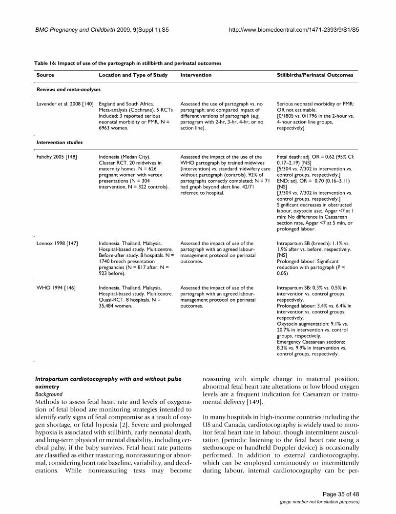
BMC Pregnancy and Childbirth 2009, 9(Suppl 1):S5 http://www.biomedcentral.com/1471-2393/9/S1/S5
Intrapartum cardiotocography with and without pulse oximetryBackgroundMethods to assess fetal heart rate and levels of oxygena-tion of fetal blood are monitoring strategies intended toidentify early signs of fetal compromise as a result of oxy-gen shortage, or fetal hypoxia [2]. Severe and prolongedhypoxia is associated with stillbirth, early neonatal death,and long-term physical or mental disability, including cer-ebral palsy, if the baby survives. Fetal heart rate patternsare classified as either reassuring, nonreassuring or abnor-mal, considering heart rate baseline, variability, and decel-erations. While nonreassuring tests may become
reassuring with simple change in maternal position,abnormal fetal heart rate alterations or low blood oxygenlevels are a frequent indication for Caesarean or instru-mental delivery [149].
In many hospitals in high-income countries including theUS and Canada, cardiotocography is widely used to mon-itor fetal heart rate in labour, though intermittent auscul-tation (periodic listening to the fetal heart rate using astethoscope or handheld Doppler device) is occasionallyperformed. In addition to external cardiotocography,which can be employed continuously or intermittentlyduring labour, internal cardiotocography can be per-
Table 16: Impact of use of the partograph in stillbirth and perinatal outcomes
Source Location and Type of Study Intervention Stillbirths/Perinatal Outcomes
Reviews and meta-analyses
Lavender et al. 2008 [140] England and South Africa.Meta-analysis (Cochrane). 5 RCTs included; 3 reported serious neonatal morbidity or PMR. N = 6963 women.
Assessed the use of partograph vs. no partograph; and compared impact of different versions of partograph (e.g. partogram with 2-hr, 3-hr, 4-hr, or no action line).
Serious neonatal morbidity or PMR: OR not estimable.[0/1805 vs. 0/1796 in the 2-hour vs. 4-hour action line groups, respectively].
Intervention studies
Fahdhy 2005 [148] Indonesia (Medan City).Cluster RCT. 20 midwives in maternity homes. N = 626 pregnant women with vertex presentations (N = 304 intervention, N = 322 controls).
Assessed the impact of the use of the WHO partograph by trained midwives (intervention) vs. standard midwifery care without partograph (controls). 92% of partographs correctly completed; N = 71 had graph beyond alert line. 42/71 referred to hospital.
Fetal death: adj. OR = 0.62 (95% CI: 0.17–2.19) [NS][5/304 vs. 7/302 in intervention vs. control groups, respectively.]END: adj. OR = 0.70 (0.16–3.11) [NS] [3/304 vs. 7/302 in intervention vs. control groups, respectively.]Significant decreases in obstructed labour, oxytocin use:, Apgar <7 at 1 min: No difference in Caesarean section rate, Apgar <7 at 5 min, or prolonged labour.
Lennox 1998 [147] Indonesia, Thailand, Malaysia. Hospital-based study. Multicentre.Before-after study. 8 hospitals. N = 1740 breech presentation pregnancies (N = 817 after, N = 923 before).
Assessed the impact of use of the partograph with an agreed labour-management protocol on perinatal outcomes.
Intrapartum SB (breech): 1.1% vs. 1.9% after vs. before, respectively. [NS]Prolonged labour: Significant reduction with partograph (P < 0.05)
WHO 1994 [146] Indonesia, Thailand, Malaysia. Hospital-based study. Multicentre.Quasi-RCT. 8 hospitals. N = 35,484 women.
Assessed the impact of use of the partograph with an agreed labour-management protocol on perinatal outcomes.
Intrapartum SB: 0.3% vs. 0.5% in intervention vs. control groups, respectively.Prolonged labour: 3.4% vs. 6.4% in intervention vs. control groups, respectively.Oxytocin augmentation: 9.1% vs. 20.7% in intervention vs. control groups, respectively.Emergency Caesarean sections: 8.3% vs. 9.9% in intervention vs. control groups, respectively.
Page 35 of 48(page number not for citation purposes)
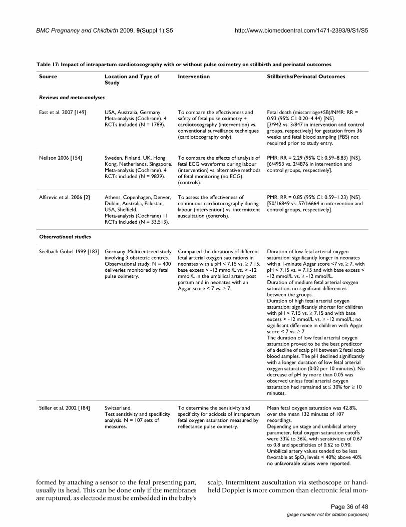
BMC Pregnancy and Childbirth 2009, 9(Suppl 1):S5 http://www.biomedcentral.com/1471-2393/9/S1/S5
Page 36 of 48
formed by attaching a sensor to the fetal presenting part,usually its head. This can be done only if the membranesare ruptured, as electrode must be embedded in the baby's
scalp. Intermittent auscultation via stethoscope or hand-held Doppler is more common than electronic fetal mon-
Table 17: Impact of intrapartum cardiotocography with or without pulse oximetry on stillbirth and perinatal outcomes
Source Location and Type of Study
Intervention Stillbirths/Perinatal Outcomes
Reviews and meta-analyses
East et al. 2007 [149] USA, Australia, Germany.Meta-analysis (Cochrane). 4 RCTs included (N = 1789).
To compare the effectiveness and safety of fetal pulse oximetry + cardiotocography (intervention) vs. conventional surveillance techniques (cardiotocography only).
Fetal death (miscarriage+SB)/NMR: RR = 0.93 (95% CI: 0.20–4.44) [NS].[3/942 vs. 3/847 in intervention and control groups, respectively] for gestation from 36 weeks and fetal blood sampling (FBS) not required prior to study entry.
Neilson 2006 [154] Sweden, Finland, UK, Hong Kong, Netherlands, Singapore.Meta-analysis (Cochrane). 4 RCTs included (N = 9829).
To compare the effects of analysis of fetal ECG waveforms during labour (intervention) vs. alternative methods of fetal monitoring (no ECG) (controls).
PMR: RR = 2.29 (95% CI: 0.59–8.83) [NS].[6/4953 vs. 2/4876 in intervention and control groups, respectively].
Alfirevic et al. 2006 [2] Athens, Copenhagen, Denver, Dublin, Australia, Pakistan, USA, Sheffield.Meta-analysis (Cochrane) 11 RCTs included (N = 33,513).
To assess the effectiveness of continuous cardiotocography during labour (intervention) vs. intermittent auscultation (controls).
PMR: RR = 0.85 (95% CI: 0.59–1.23) [NS].[50/16849 vs. 57/16664 in intervention and control groups, respectively].
Observational studies
Seelbach Gobel 1999 [183] Germany. Multicentreed study involving 3 obstetric centres.Observational study. N = 400 deliveries monitored by fetal pulse oximetry.
Compared the durations of different fetal arterial oxygen saturations in neonates with a pH < 7.15 vs. 7.15, base excess < -12 mmol/L vs. > -12 mmol/L in the umbilical artery post partum and in neonates with an Apgar score < 7 vs. 7.
Duration of low fetal arterial oxygen saturation: significantly longer in neonates with a 1-minute Apgar score <7 vs. 7, with pH < 7.15 vs. = 7.15 and with base excess < -12 mmol/L vs. -12 mmol/L.Duration of medium fetal arterial oxygen saturation: no significant differences between the groups.Duration of high fetal arterial oxygen saturation: significantly shorter for children with pH < 7.15 vs. 7.15 and with base excess < -12 mmol/L vs. -12 mmol/L; no significant difference in children with Apgar score < 7 vs. 7.The duration of low fetal arterial oxygen saturation proved to be the best predictor of a decline of scalp pH between 2 fetal scalp blood samples. The pH declined significantly with a longer duration of low fetal arterial oxygen saturation (0.02 per 10 minutes). No decrease of pH by more than 0.05 was observed unless fetal arterial oxygen saturation had remained at 30% for 10 minutes.
Stiller et al. 2002 [184] Switzerland.Test sensitivity and specificity analysis. N = 107 sets of measures.
To determine the sensitivity and specificity for acidosis of intrapartum fetal oxygen saturation measured by reflectance pulse oximetry.
Mean fetal oxygen saturation was 42.8%, over the mean 132 minutes of 107 recordings.Depending on stage and umbilical artery parameter, fetal oxygen saturation cutoffs were 33% to 36%, with sensitivities of 0.67 to 0.8 and specificities of 0.62 to 0.90.Umbilical artery values tended to be less favorable at SpO2 levels < 40%; above 40% no unfavorable values were reported.
(page number not for citation purposes)
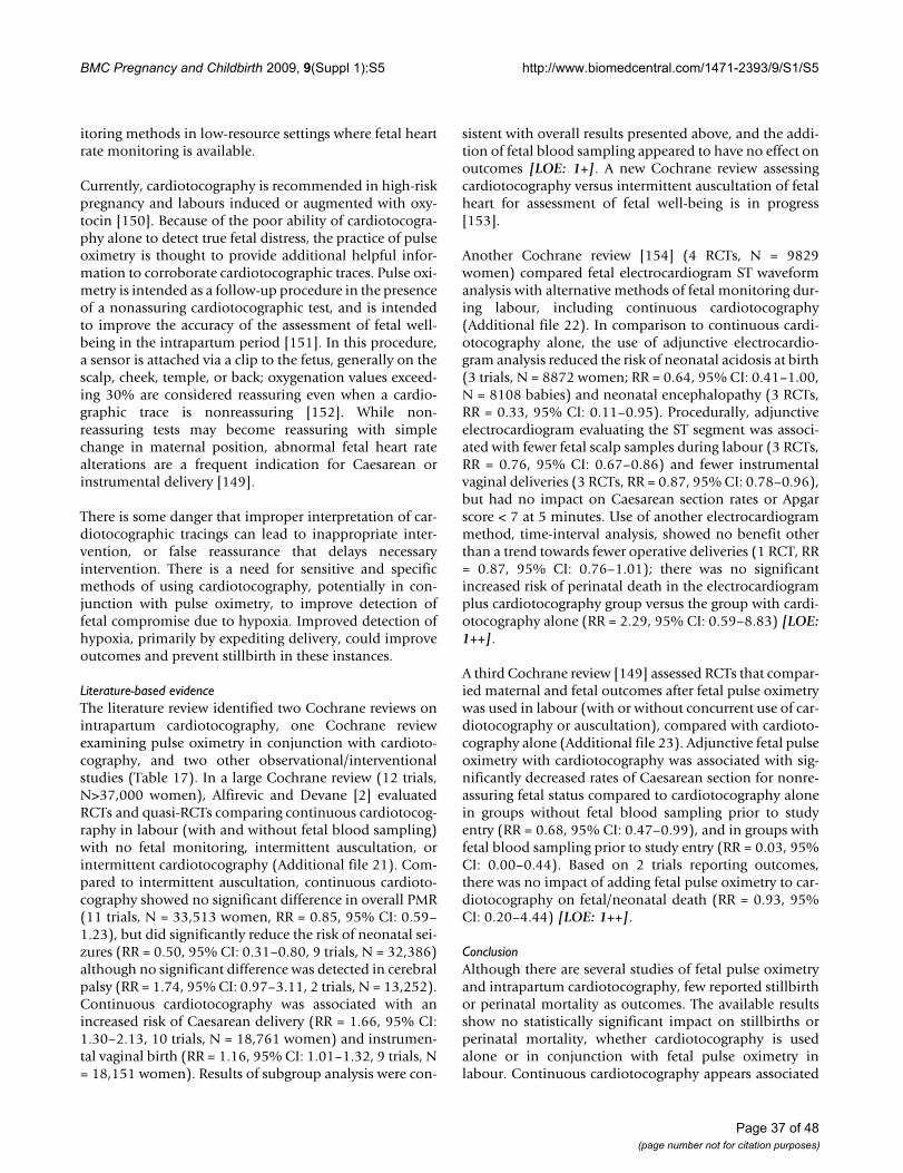
BMC Pregnancy and Childbirth 2009, 9(Suppl 1):S5 http://www.biomedcentral.com/1471-2393/9/S1/S5
itoring methods in low-resource settings where fetal heartrate monitoring is available.
Currently, cardiotocography is recommended in high-riskpregnancy and labours induced or augmented with oxy-tocin [150]. Because of the poor ability of cardiotocogra-phy alone to detect true fetal distress, the practice of pulseoximetry is thought to provide additional helpful infor-mation to corroborate cardiotocographic traces. Pulse oxi-metry is intended as a follow-up procedure in the presenceof a nonassuring cardiotocographic test, and is intendedto improve the accuracy of the assessment of fetal well-being in the intrapartum period [151]. In this procedure,a sensor is attached via a clip to the fetus, generally on thescalp, cheek, temple, or back; oxygenation values exceed-ing 30% are considered reassuring even when a cardio-graphic trace is nonreassuring [152]. While non-reassuring tests may become reassuring with simplechange in maternal position, abnormal fetal heart ratealterations are a frequent indication for Caesarean orinstrumental delivery [149].
There is some danger that improper interpretation of car-diotocographic tracings can lead to inappropriate inter-vention, or false reassurance that delays necessaryintervention. There is a need for sensitive and specificmethods of using cardiotocography, potentially in con-junction with pulse oximetry, to improve detection offetal compromise due to hypoxia. Improved detection ofhypoxia, primarily by expediting delivery, could improveoutcomes and prevent stillbirth in these instances.
Literature-based evidenceThe literature review identified two Cochrane reviews onintrapartum cardiotocography, one Cochrane reviewexamining pulse oximetry in conjunction with cardioto-cography, and two other observational/interventionalstudies (Table 17). In a large Cochrane review (12 trials,N>37,000 women), Alfirevic and Devane [2] evaluatedRCTs and quasi-RCTs comparing continuous cardiotocog-raphy in labour (with and without fetal blood sampling)with no fetal monitoring, intermittent auscultation, orintermittent cardiotocography (Additional file 21). Com-pared to intermittent auscultation, continuous cardioto-cography showed no significant difference in overall PMR(11 trials, N = 33,513 women, RR = 0.85, 95% CI: 0.59–1.23), but did significantly reduce the risk of neonatal sei-zures (RR = 0.50, 95% CI: 0.31–0.80, 9 trials, N = 32,386)although no significant difference was detected in cerebralpalsy (RR = 1.74, 95% CI: 0.97–3.11, 2 trials, N = 13,252).Continuous cardiotocography was associated with anincreased risk of Caesarean delivery (RR = 1.66, 95% CI:1.30–2.13, 10 trials, N = 18,761 women) and instrumen-tal vaginal birth (RR = 1.16, 95% CI: 1.01–1.32, 9 trials, N= 18,151 women). Results of subgroup analysis were con-
sistent with overall results presented above, and the addi-tion of fetal blood sampling appeared to have no effect onoutcomes [LOE: 1+]. A new Cochrane review assessingcardiotocography versus intermittent auscultation of fetalheart for assessment of fetal well-being is in progress[153].
Another Cochrane review [154] (4 RCTs, N = 9829women) compared fetal electrocardiogram ST waveformanalysis with alternative methods of fetal monitoring dur-ing labour, including continuous cardiotocography(Additional file 22). In comparison to continuous cardi-otocography alone, the use of adjunctive electrocardio-gram analysis reduced the risk of neonatal acidosis at birth(3 trials, N = 8872 women; RR = 0.64, 95% CI: 0.41–1.00,N = 8108 babies) and neonatal encephalopathy (3 RCTs,RR = 0.33, 95% CI: 0.11–0.95). Procedurally, adjunctiveelectrocardiogram evaluating the ST segment was associ-ated with fewer fetal scalp samples during labour (3 RCTs,RR = 0.76, 95% CI: 0.67–0.86) and fewer instrumentalvaginal deliveries (3 RCTs, RR = 0.87, 95% CI: 0.78–0.96),but had no impact on Caesarean section rates or Apgarscore < 7 at 5 minutes. Use of another electrocardiogrammethod, time-interval analysis, showed no benefit otherthan a trend towards fewer operative deliveries (1 RCT, RR= 0.87, 95% CI: 0.76–1.01); there was no significantincreased risk of perinatal death in the electrocardiogramplus cardiotocography group versus the group with cardi-otocography alone (RR = 2.29, 95% CI: 0.59–8.83) [LOE:1++].
A third Cochrane review [149] assessed RCTs that compar-ied maternal and fetal outcomes after fetal pulse oximetrywas used in labour (with or without concurrent use of car-diotocography or auscultation), compared with cardioto-cography alone (Additional file 23). Adjunctive fetal pulseoximetry with cardiotocography was associated with sig-nificantly decreased rates of Caesarean section for nonre-assuring fetal status compared to cardiotocography alonein groups without fetal blood sampling prior to studyentry (RR = 0.68, 95% CI: 0.47–0.99), and in groups withfetal blood sampling prior to study entry (RR = 0.03, 95%CI: 0.00–0.44). Based on 2 trials reporting outcomes,there was no impact of adding fetal pulse oximetry to car-diotocography on fetal/neonatal death (RR = 0.93, 95%CI: 0.20–4.44) [LOE: 1++].
ConclusionAlthough there are several studies of fetal pulse oximetryand intrapartum cardiotocography, few reported stillbirthor perinatal mortality as outcomes. The available resultsshow no statistically significant impact on stillbirths orperinatal mortality, whether cardiotocography is usedalone or in conjunction with fetal pulse oximetry inlabour. Continuous cardiotocography appears associated
Page 37 of 48(page number not for citation purposes)

BMC Pregnancy and Childbirth 2009, 9(Suppl 1):S5 http://www.biomedcentral.com/1471-2393/9/S1/S5
with increased rates of operative delivery and lower ratesof neonatal seizures compared to no or intermittent cardi-otocography, but has no demonstrated impact on rates ofperinatal mortality acidosis, dystocia, or long-term physi-cal or developmental outcomes. There is currently no evi-dence of benefit from randomised or quasi-randomisedstudies for intrapartum cardiotocography alone or in con-junction with electrocardiogram or fetal pulse oximetry inpreventing stillbirth.
One drawback of intrapartum cardiotocography is that itspoor predictive value of true fetal distress and hypoxia isnot sufficiently enhanced by any adjunctive technologiesat this time. Pulse oximetry appears safe, though long-term developmental studies have not been performed, butit does not have any clear impact on stillbirths or perinatalmortality [149]. Adjunctive electrocardiogram possiblyreduces neonatal encephalopathy, academia, and instru-mental delivery, but these findings need additionalresearch to verify. Minimally invasive technologies and
Table 18: Collective grading of evidence for impact of monitoring interventions in pregnancy on stillbirth and related perinatal outcomes
Evidence of no or negative impact
(leave out of programmes)
Uncertain evidence(need for additional
research before including in programmes)
Some evidence(may include in
programmes, but further evaluation is warranted)
Clear evidence(merits inclusion in
programmes)
Pregnancy risk screening X
Fetal movement counting X X(for high-risk pregnancies)
Ultrasound scanning X
Doppler velocimetry X (uterine artery and unselected populations)
X (umbilical artery and ductus venosus in high-risk
pregnancies)
Pelvimetry X
Detection and management of diabetes mellitus
X
Antepartum fetal heart rate monitoring with cardiotocography (NST, CST)
X
Fetal BPP scoring X
Vibroacoustic stimulation X
Amniotic fluid volume assessment
X (oligohydramnios clear risk factor)
Home vs. hospital bed rest and monitoring for high-risk pregnancies
X
In-hospital fetal surveillance unit
X
Use of the partograph X
Intrapartum cardiotocography and pulse oximetry
X (based largely on observational evidence)
Page 38 of 48(page number not for citation purposes)

BMC Pregnancy and Childbirth 2009, 9(Suppl 1):S5 http://www.biomedcentral.com/1471-2393/9/S1/S5
tests are needed in addition to intrapartum cardiotocogra-phy to more accurately identify intrapartum hypoxia anddystocia.
There are robust studies and Cochrane reviews on fetalpulse oximetry and intrapartum cardiotocography (GradeB evidence). The non-significant impact of cardiotocogra-phy with or without fetal pulse oximetry on stillbirth/peri-natal mortality in these studies suggests no evidence ofbenefit. In high-income countries, documented stillbirthrates, particularly stillbirths associated with intrauterineasphyxia, in high-income countries have followed increas-ing prevalence of cardiotocographic fetal monitoring withCaesarean section for fetal distress [155-157], though thisassociation may be confounded. Electronic fetal monitor-ing (with access to operative delivery) may an importanttool in the arsenal of strategies to prevent stillbirth,though this is unproven. More precise tests for fetal dis-tress and indications for Caesarean section are needed.
SummaryAlthough a reasonable number of Cochrane reviews andRCTs were available which assessed the impact of one ormore implementation strategies for many screening andmonitoring interventions, none of the interventions wereviewed demonstrated convincing evidence of impact onstillbirths or perinatal mortality. A number of studies thatdid report statistically significant impact on stillbirth orperinatal mortality, or which suggested large magnitudedifferences between intervention and control/comparisongroups, lacked sufficient rigor to justify recommendingthe routine use of the interventions evaluated.
The evidence for all interventions reviewed in this paper issummarised in Table 18. Of the range of monitoring inter-ventions evaluated, fetal movement counting and Dop-pler monitoring were promising for further evaluation inhigh-risk pregnancies in low-resource settings. Low amni-otic fluid measurements were strongly predictive of still-birth, but interventions to restore adequate amniotic fluidvolume or to deliver the baby based on identification ofoligohydramnios have not been systematically tested toconclude whether amniotic fluid assessment is a usefuldiagnostic tool that leads to actions which prevent still-birth.
Research gapsDespite the existence of many tools, devices, and tech-niques for monitoring pregnancy for complications, thereis a dearth of rigorous evidence that any screening andmonitoring intervention has a direct impact on stillbirthrates in unselected and low-risk populations. This stemsfrom several common shortcomings in the design andinterpretation of studies investigating the use of screeningand monitoring techniques. First, the small size and insuf-
ficient rigor of many screening and monitoring studies todate renders them underpowered to detect significant dif-ferences in perinatal mortality subsequent to their imple-mentation. Many studies assessing the impact of theinterventions covered in this review did not report still-births or perinatal mortality at all. For virtually all theinterventions included in this review (with the exceptionof x-ray pelvimetry), there is a need for RCTs sufficientlypowered to detect statistically significant impact on still-birth or perinatal mortality, if such differences exist. Sec-ond, for screening and monitoring interventions to reducestillbirth incidence, they must effectively identify womenat higher risk of stillbirth in time for an appropriate andeffective intervention to be provided. Most studies wereviewed limited their analysis to the first step of success-ful screening or monitoring: detection of women atincreased risk of stillbirth or perinatal death. A number ofstudies showed that positive test results correspondedfairly well with increased risk of adverse pregnancy out-come. However, a trial of an otherwise effective screeningmethod will show no impact on stillbirths if the subse-quent intervention fails to prevent stillbirth, eitherbecause intervention is ineffective or provided too late. Inmost of the studies we reviewed, few women whoscreened positive were treated according to an establishedprotocol. This flaw precludes assessment of whether thelack of impact of any given screening technique is attrib-utable to a failed screening method or a failed interven-tion. This fact highlights the need for time-to-decision andtime-to-intervention studies, as well as analyses that con-sider which interventions are employed, and protocols foremploying them, subsequent to adverse findings ofscreening and monitoring studies. The quality, appropri-ateness, and timeliness of the intervention care providedmust also be considered along with the effectiveness ofscreening or monitoring in detecting true complications.
In addition to these tests of diagnostic accuracy and inter-vention effectiveness, other types of studies are alsoneeded, including innovative pilot studies, safety studies,and effectiveness trials, to bolster the weak evidence basefor screening and monitoring interventions during labour(Table 19). In high-income countries, there is a particularneed for better understanding of placental pathophysiol-ogy, as up to half of unexplained stillbirths show signs ofgrowth restriction, much of which is attributed to placen-tal insufficiency [158]. Placental dysfunction is oftenobserved in other major known causes of stillbirth includ-ing pre-eclampsia and abruption, suggesting the need foraccurate placental function or placental biomarker screen-ing tests, especially those appropriate for use in unselectedpopulations. One such test in early pregnancy whichmeasures pregnancy associated plasma protein-A hasbeen associated with a 40–50-fold increase in risk of still-birth due to placental dysfunction in a selected popula-
Page 39 of 48(page number not for citation purposes)
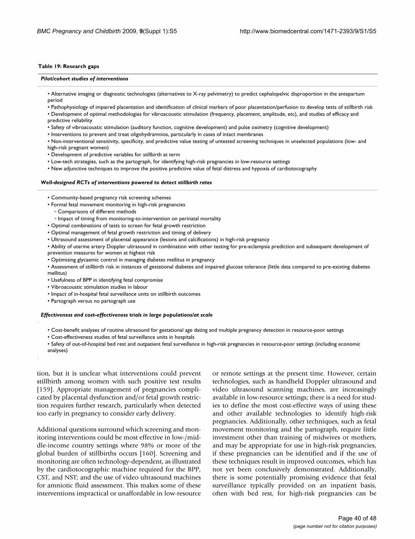
BMC Pregnancy and Childbirth 2009, 9(Suppl 1):S5 http://www.biomedcentral.com/1471-2393/9/S1/S5
tion, but it is unclear what interventions could preventstillbirth among women with such positive test results[159]. Appropriate management of pregnancies compli-cated by placental dysfunction and/or fetal growth restric-tion requires further research, particularly when detectedtoo early in pregnancy to consider early delivery.
Additional questions surround which screening and mon-itoring interventions could be most effective in low-/mid-dle-income country settings where 98% or more of theglobal burden of stillbirths occurs [160]. Screening andmonitoring are often technology-dependent, as illustratedby the cardiotocographic machine required for the BPP,CST, and NST; and the use of video ultrasound machinesfor amniotic fluid assessment. This makes some of theseinterventions impractical or unaffordable in low-resource
or remote settings at the present time. However, certaintechnologies, such as handheld Doppler ultrasound andvideo ultrasound scanning machines, are increasinglyavailable in low-resource settings; there is a need for stud-ies to define the most cost-effective ways of using theseand other available technologies to identify high-riskpregnancies. Additionally, other techniques, such as fetalmovement monitoring and the partograph, require littleinvestment other than training of midwives or mothers,and may be appropriate for use in high-risk pregnancies,if these pregnancies can be identified and if the use ofthese techniques result in improved outcomes, which hasnot yet been conclusively demonstrated. Additionally,there is some potentially promising evidence that fetalsurveillance typically provided on an inpatient basis,often with bed rest, for high-risk pregnancies can be
Table 19: Research gaps
Pilot/cohort studies of interventions
• Alternative imaging or diagnostic technologies (alternatives to X-ray pelvimetry) to predict cephalopelvic disproportion in the antepartum period• Pathophysiology of impaired placentation and identification of clinical markers of poor placentation/perfusion to develop tests of stillbirth risk• Development of optimal methodologies for vibroacoustic stimulation (frequency, placement, amplitude, etc), and studies of efficacy and predictive reliability• Safety of vibroacoustic stimulation (auditory function, cognitive development) and pulse oximetry (cognitive development)• Interventions to prevent and treat oligohydramnios, particularly in cases of intact membranes• Non-interventional sensitivity, specificity, and predictive value testing of untested screening techniques in unselected populations (low- and high-risk pregnant women)• Development of predictive variables for stillbirth at term• Low-tech strategies, such as the partograph, for identifying high-risk pregnancies in low-resource settings• New adjunctive techniques to improve the positive predictive value of fetal distress and hypoxia of cardiotocography
Well-designed RCTs of interventions powered to detect stillbirth rates
• Community-based pregnancy risk screening schemes• Formal fetal movement monitoring in high-risk pregnancies
� Comparisons of different methods� Impact of timing from monitoring-to-intervention on perinatal mortality
• Optimal combinations of tests to screen for fetal growth restriction• Optimal management of fetal growth restriction and timing of delivery• Ultrasound assessment of placental appearance (lesions and calcifications) in high-risk pregnancy• Ability of uterine artery Doppler ultrasound in combination with other testing for pre-eclampsia prediction and subsequent development of prevention measures for women at highest risk• Optimising glycaemic control in managing diabetes mellitus in pregnancy• Assessment of stillbirth risk in instances of gestational diabetes and impaired glucose tolerance (little data compared to pre-existing diabetes mellitus)• Usefulness of BPP in identifying fetal compromise• Vibroacoustic stimulation studies in labour• Impact of in-hospital fetal surveillance units on stillbirth outcomes• Partograph versus no partograph use
Effectiveness and cost-effectiveness trials in large populations/at scale
• Cost-benefit analyses of routine ultrasound for gestational age dating and multiple pregnancy detection in resource-poor settings• Cost-effectiveness studies of fetal surveillance units in hospitals• Safety of out-of-hospital bed rest and outpatient fetal surveillance in high-risk pregnancies in resource-poor settings (including economic analyses)
Page 40 of 48(page number not for citation purposes)
BMC Pregnancy and Childbirth 2009, 9(Suppl 1):S5 http://www.biomedcentral.com/1471-2393/9/S1/S5
offered on an outpatient basis with bed rest or reducedactivity at home without deleterious impact on pregnancyoutcomes; this evidence requires confirmatory effective-ness trials in low-resource settings before programmaticadoption of this approach could be recommended.
Practicality, logistical feasibility, and cost-effectiveness arechief concerns in many low-resource areas. Where ANCservices are available and widely utilised, the prospects foridentifying and monitoring high-risk pregnancy improve.Along with interventions that improve detection of riskfactors and complications, improved management andreferral of women with complications are critical to man-age obstetric risk in low-/middle-income countries.Unfortunately, in areas where ANC attendance is poor,few of these screening and monitoring techniques arelikely to be implementable.
Implications for programmes and clinical practiceA few monitoring approaches showed promise for use inhigh-risk pregnancies, including fetal movement monitor-ing and umbilical Doppler velocimetry. Before recom-mending these interventions for widespread use, furtherlarge RCTs of sufficient rigour to detect differences in still-birth and perinatal outcomes are needed to determine theutility and effectiveness of these monitoring approachesand interventions used after positive tests.
This review identified several interventions – for example,amniotic fluid measurement for signs of oligohydram-nios, especially as pregnancy approaches or exceeds term;routine ultrasound scanning, and intrapartum cardioto-cography – that are in widespread use in high-resource set-tings, but for which rigorous evidence is lacking. For theseinterventions, their continued use is reasonable eventhough the evidence base for impact on stillbirths is lack-ing. Intrapartum cardiotocography in particular has beenshown in RCTs to elevate the risk of operative delivery.Prevailing wisdom credits cardiotocography and availableCaesarean section with the diminution of stillbirth ratesin high-income countries in recent decades; however, therole of cardiotocography in reducing stillbirth should beconfirmed with more rigorous evidence. Efforts to achieveglycaemic control in cases of maternal diabetes mellitusare also encouraged, despite the lack of strong evidenceshowing an impact of such interventions on stillbirth, onthe grounds that women with good glycaemic controlexperience fewer complications and negative outcomes ofpregnancy.
Particularly in pregnancies deemed high-risk based onscreening test results, clinical progression of pregnancy, orreproductive history, multiple antepartum testing modal-ities can minimise deficiencies in sensitivity and predic-tive value when used in combination to assess fetal well-
being and abnormalities. The BPP reflects one standard-ised strategy by which tests of fetal well-being have beenpackaged to permit a more comprehensive assessment offetal well-being. In cases where one test raises the possibil-ity that a pregnancy is high-risk, multiple tests of fetalwell-being should be considered if time allows, alongwith the patient's overall clinical history, to determine theappropriateness of intervention. Multiple tests are recom-mended in many protocols for identifying and makingclinical decisions about management of pregnancies withsuspected fetal growth restriction [161]. In these cases thecollective results of a battery of tests including all the com-ponents of the BPP and Doppler velocimetry, as well asultrasound biometry, can be informative in decision-mak-ing to maximise gestational age and inform the strategictiming of corticosteroid administration and early deliveryvia induction or Caesarean section. However, conflictingtest results – a recent study showed only 44.5% concord-ance between BPP and Doppler assessments of compro-mise in growth-restricted fetuses – can complicateinterpretation [162].
Recommendations for clinical decision-making protocolsand introduction of interventions in low-/middle-incomecountries are more difficult. Resource constraints precludethe implementation of many of the interventionsreviewed in this paper. The interventions requiring thelowest investment in resources and training – fetal move-ment monitoring and the partograph – show some evi-dence of benefit in high-risk pregnancies, but morestudies are needed. Where safe Caesarean section is avail-able, ensuring that intermittent or continuous intrapar-tum fetal heart rate monitoring is performed may improvetargeting of Caesarean section while improving birth out-comes.
Screening and monitoring interventions cannot be effec-tive without responsive, quality care and careful clinicalmanagement, particularly for high-risk pregnancies.Health systems quality improvement activities, includingboosting provider skill in interpreting test results anddetermining appropriate intervention, timely responseincluding swift referral, and evaluative strategies to docu-ment outcomes after intervention are essential comple-mentary activities to ensure that an effective screening ormonitoring technique translates to measurable improve-ments in perinatal mortality outcomes.
ConclusionScreening pregnancies to identify risk factors, complica-tions, or indications of fetal distress and providing appro-priate surveillance for identified risk factors theoreticallyencourages appropriate use of interventions, includinginduction of labour, Caesarean section, and pharmaco-logical treatment, to prevent fetal loss and adverse mater-
Page 41 of 48(page number not for citation purposes)

BMC Pregnancy and Childbirth 2009, 9(Suppl 1):S5 http://www.biomedcentral.com/1471-2393/9/S1/S5
Page 42 of 48
nal or neonatal outcomes. Screening and monitoring arefutile, however, in the absence of effective interventions toact promptly and appropriately to manage identified riskfactors and complications. The weak evidence in thisreview for impact of screening and monitoring interven-tions on stillbirth incidence is due at least in part to limi-tations in study designs that result in uninterpretable data.Screening tests may be ineffective, inappropriate, or occurtoo late to prevent adverse outcomes, but more problem-atically, most studies did not report the impact of inter-vention (particularly standardised intervention)following positive screening test results. Effective inter-ventions exist to prevent stillbirth associated with manymaternal infections or conditions (e.g., syphilis, malaria,pre-eclampsia) and placental dysfunction (e.g., oligohy-dramnios, placental insufficiency, abruption). Becausescreening and monitoring techniques during pregnancyalso pose the risk of inappropriate or unnecessary use ofdrugs, induction of labour, iatrogenic preterm birth, orCaesarean section, it is practically, ethically, and econom-ically important to validate the sensitivity, specificity, andpositive predictive value of available screening and moni-toring techniques to protect women and their babies fromiatrogenic harm; and to ensure the wise use of scarce med-ical resources in low- and middle-income settings.
List of abbreviations usedAFI: amniotic fluid index; ANC: antenatal care; BPP: bio-physical profile; CI: confidence interval; CST: contractionstress test; CTG: cardiotocography; ECG: electrocardio-gram; FHR: fetal heart rate; HR: hazard ratio; IU: interna-tional units; LOE: level of evidence; NS: non-significant;NST: non-stress test; OR: odds ratio; PMR: perinatal mor-tality rate; PPV: positive predictive value; PROM: prema-ture rupture of membranes; PPROM: pre-term prematurerupture of membranes; RCT: randomised clinical/control-led trial; RD: risk difference; RI: resistance index; RR: rela-tive risk/risk ratio; SB: stillbirth; SBR: stillbirth rate; SD:standard deviation; SGA: small for gestational age; TBA:traditional birth attendant; UK: United Kingdom; USA:United States of America; WHO: World Health Organisa-tion; WMD: weighted mean difference.
Competing interestsThe authors declare that they have no competing interests.
Authors' contributionsThe paper was written and reviewed by all the authors.
Additional material
Additional file 1Web Table 1. Component studies in Mangesi et al. 2007 meta-analy-sis: Impact of fetal movement monitoring. Component studies in Man-gesi et al. 2007 meta-analysis showing impact on stillbirths/perinatal mortalityClick here for file[http://www.biomedcentral.com/content/supplementary/1471-2393-9-S1-S5-S1.doc]
Additional file 2Web Table 2. Component studies in Neilson 1998 meta-analysis: Impact of routine ultrasound in pregnancy. Component studies in Neil-son 1998 meta-analysis showing impact on stillbirths/perinatal mortalityClick here for file[http://www.biomedcentral.com/content/supplementary/1471-2393-9-S1-S5-S2.doc]
Additional file 3Web Table 3. Component studies in Bricker et al. 2008 meta-analysis: Impact of ultrasound in late pregnancy. Component studies in Bricker et al. 2008 meta-analysis showing impact on stillbirths/perinatal mortal-ityClick here for file[http://www.biomedcentral.com/content/supplementary/1471-2393-9-S1-S5-S3.doc]
Additional file 4Web Table 4. Component studies in Papageorghiou et al. 2002: Impact of second trimester uterine artery Doppler on perinatal mor-tality. Component studies in Papageorghiou et al. 2002 meta-analysis showing impact on stillbirths/perinatal mortalityClick here for file[http://www.biomedcentral.com/content/supplementary/1471-2393-9-S1-S5-S4.doc]
Additional file 5Web Table 5. Component studies in Baschat 2004: impact of Doppler velocimetry on stillbirth and perinatal mortality in pre-term growth-restricted fetuses. Component studies in Baschat 2004 meta-analysis showing impact on stillbirths/perinatal mortalityClick here for file[http://www.biomedcentral.com/content/supplementary/1471-2393-9-S1-S5-S5.doc]
Additional file 6Web Table 6. Component studies in Neilson et al. 2000 meta-analy-sis: Impact of Doppler screening during pregnancy. Component studies in Neilson et al. 2000 meta-analysis showing impact on stillbirths/perina-tal mortalityClick here for file[http://www.biomedcentral.com/content/supplementary/1471-2393-9-S1-S5-S6.doc]
Additional file 7Web Table 7. Component studies in Pattinson et al. 1997 meta-anal-ysis: Impact of pelvimetry during pregnancy. Component studies in Pat-tinson et al. 1997 meta-analysis showing impact on stillbirths/perinatal mortalityClick here for file[http://www.biomedcentral.com/content/supplementary/1471-2393-9-S1-S5-S7.doc]
(page number not for citation purposes)

BMC Pregnancy and Childbirth 2009, 9(Suppl 1):S5 http://www.biomedcentral.com/1471-2393/9/S1/S5
Page 43 of 48
Additional file 8Web Table 8. Component studies in Russell et al. 2007 meta-analysis: Impact of management of gestational diabetes on stillbirth and peri-natal outcomes. Component studies in Russell et al. 2007 showing impact on stillbirths/perinatal mortalityClick here for file[http://www.biomedcentral.com/content/supplementary/1471-2393-9-S1-S5-S8.doc]
Additional file 9Web Table 9. Component studies in Mukhopadhyay et al. 2007: Impact of continuous subcutaneous insulin infusion on intrauterine fetal death. Component studies in Mukhopadhyay et al. 2007 showing impact on stillbirths/perinatal mortalityClick here for file[http://www.biomedcentral.com/content/supplementary/1471-2393-9-S1-S5-S9.doc]
Additional file 10Web Table 10. Component studies in Boulvain et al. 2001 meta-anal-ysis: Impact of elective delivery on perinatal mortality. Component studies in Boulvain et al. 2001 showing impact on stillbirths/perinatal mortalityClick here for file[http://www.biomedcentral.com/content/supplementary/1471-2393-9-S1-S5-S10.doc]
Additional file 11Web Table 11. Component studies in Pattison and McCowan 1999 meta-analysis: Impact of cardiotocography for antepartum fetal assessment on perinatal mortality. Component studies in Pattison and McCowan 1999 showing impact on stillbirths/perinatal mortalityClick here for file[http://www.biomedcentral.com/content/supplementary/1471-2393-9-S1-S5-S11.doc]
Additional file 12Web Table 12. Component studies in Lalor et al. 2008 meta-analysis: Impact of fetal biophysical profile. Component studies in Lalor et al. 2008 showing impact on stillbirths/perinatal mortalityClick here for file[http://www.biomedcentral.com/content/supplementary/1471-2393-9-S1-S5-S12.doc]
Additional file 13Web Table 13. Component studies in Tan et al. 2001 meta-analysis: Impact of vibroacoustic stimulation. Component studies in Tan et al. 2001 meta-analysis showing impact on stillbirths/perinatal mortalityClick here for file[http://www.biomedcentral.com/content/supplementary/1471-2393-9-S1-S5-S13.doc]
Additional file 14Web Table 14. Component studies in Magann et al 2007 review: impact of idiopathic polyhydramnios on stillbirth and perinatal mor-tality. Component studies in Magann et al. 2007 review showing impact on stillbirths/perinatal mortalityClick here for file[http://www.biomedcentral.com/content/supplementary/1471-2393-9-S1-S5-S14.doc]
Additional file 15Web Table 15. Component studies in Nabhan and Abdelmoula 2008 [185]meta-analysis: comparison of single deepest vertical pocket vs. AFI in predicting perinatal outcome. Component studies in Nabhan and Abdelmoula 2008 review showing impact on stillbirths/perinatal mortal-ityClick here for file[http://www.biomedcentral.com/content/supplementary/1471-2393-9-S1-S5-S15.doc]
Additional file 16Web Table 16. Component studies in Hofmeyr and Gulmezoglu 2002 [126]meta-analysis: impact of maternal hydration on amniotic fluid volume and perinatal outcomes. Component studies in Hofmeyr and Gulmezoglu 2002 review showing impact on stillbirths/perinatal mortal-ityClick here for file[http://www.biomedcentral.com/content/supplementary/1471-2393-9-S1-S5-S16.doc]
Additional file 17Web Table 17. Component studies in Crowther 2001 meta-analysis: Impact of hospitalisation for bed rest for women with a multiple preg-nancy on stillbirth and perinatal mortality. Component studies in Crowther 2001 review showing impact on stillbirths/perinatal mortalityClick here for file[http://www.biomedcentral.com/content/supplementary/1471-2393-9-S1-S5-S17.doc]
Additional file 18Web Table 18. Component studies in Meher et al. 2005: Impact of some rest in hospital vs. routine activity at home on stillbirth and peri-natal mortality. Component studies in Meher et al. 2005 review showing impact on stillbirths/perinatal mortalityClick here for file[http://www.biomedcentral.com/content/supplementary/1471-2393-9-S1-S5-S18.doc]
Additional file 19Web Table 19. Component studies in Kroner et al. 2001: Impact of antenatal day care units on perinatal mortality. Component studies in Kroner et al. 2001 review showing impact on stillbirths/perinatal mortal-ityClick here for file[http://www.biomedcentral.com/content/supplementary/1471-2393-9-S1-S5-S19.doc]
Additional file 20Web Table 20. Component studies in Lavender et al. 2008 meta-anal-ysis: Impact of different designs of partogram on neonatal morbidity or perinatal mortality. Component studies in Lavender et al. 2008 review showing impact on stillbirths/perinatal mortalityClick here for file[http://www.biomedcentral.com/content/supplementary/1471-2393-9-S1-S5-S20.doc]
Additional file 21Web Table 21. Component studies in Alfirevic and Devane 2006 meta-analysis: Impact of continuous cardiotocography on stillbirth and perinatal mortality. Component studies in Lavender et al. 2008 review showing impact on stillbirths/perinatal mortalityClick here for file[http://www.biomedcentral.com/content/supplementary/1471-2393-9-S1-S5-S21.doc]
(page number not for citation purposes)

BMC Pregnancy and Childbirth 2009, 9(Suppl 1):S5 http://www.biomedcentral.com/1471-2393/9/S1/S5
Page 44 of 48
AcknowledgementsThis series was supported by the Saving Newborn Lives programme of Save the Children-US through a grant from the Bill & Melinda Gates Foundation.
This article has been published as part of BMC Pregnancy and Childbirth Vol-ume 9 Supplement 1, 2009: Stillbirths – the global picture and evidence-based solutions. The full contents of the supplement are available online at http://www.biomedcentral.com/1471-2393/9?issue=S1.
References1. Lalor JG, Fawole B, Alfirevic Z, Devane D: Biophysical profile for
fetal assessment in high risk pregnancies. Cochrane DatabaseSyst Rev 2008:CD000038.
2. Alfirevic Z, Devane D, Gyte GM: Continuous cardiotocography(CTG) as a form of electronic fetal monitoring (EFM) forfetal assessment during labour. Cochrane Database Syst Rev 2006,3:CD006066.
3. Katz M, Meizner I, Insler V: Fetal Well-being: Physiological Basisand Methods of Clinical Assessment. CRC Press; 1990.
4. Lawn JE, Yakoob MY, Haws RA, Soomro T, Darmstadt GL, BhuttaZA: 3.2 million stillbirths: epidemiology and overview of theevidence review. BMC Pregnancy and Childbirth 2009, 9(Suppl1):S2.
5. Coopland AT, Peddle LJ, Baskett TF, Rollwagen R, Simpson A, ParkerE: A simplified antepartum high-risk pregnancy scoring form:statistical analysis of 5459 cases. Can Med Assoc J 1977,116(9):999-1001.
6. Hobel CJ, Hyvarinen MA, Okada DM, Oh W: Prenatal and intra-partum high-risk screening. I. Prediction of the high-riskneonate. Am J Obstet Gynecol 1973, 117(1):1-9.
7. Lilford RJ, Chard T: Problems and pitfalls of risk assessment inantenatal care. Br J Obstet Gynaecol 1983, 90(6):507-510.
8. Pattison NS, Sadler L, Mullins P: Obstetric risk factors: can theypredict fetal mortality and morbidity? N Z Med J 1990,103(891):257-259.
9. Knox AJ, Sadler L, Pattison NS, Mantell CD, Mullins P: An obstetricscoring system: its development and application in obstetricmanagement. Obstet Gynecol 1993, 81(2):195-199.
10. Chard T, Learmont J, Carroll S, Hudson C, Lloyd DS, Sloan D: Eval-uation of a fetal risk-scoring system. Am J Perinatol 1992, 9(5–6):388-393.
11. Morrison I, Olsen J: Perinatal mortality and antepartum riskscoring. Obstet Gynecol 1979, 53(3):362-366.
12. Majoko F, Nystrom L, Munjanja S, Lindmark G: Usefulness of riskscoring at booking for antenatal care in predicting adversepregnancy outcome in a rural African setting. J Obstet Gynaecol2002, 22(6):604-609.
13. Talsania NJ, Lala MK: Scoring of high risk pregnant women andrelated outcome. Indian J Matern Child Health 1991, 2(3):92-94.
14. Talsania NJ, Lala MK: Evaluation of antenatal risk scoring in apreterm birth prevention and perinatal loss. Indian J MaternChild Health 1994, 5(1):5-9.
15. LeFevre M, Williamson HA, Hector M Jr: Obstetric risk assess-ment in rural practice. J Fam Pract 1989, 28(6):691-695. discus-sion 695–696
16. Humphrey MD: The beneficial use of risk scoring in a remoteand high-risk pregnant population. J Perinat Med 1990,18(1):23-29.
17. de Caunes F, Alexander GR, Berchel C, Guengant JP, Papiernik E:Anamnestic pregnancy risk assessment. Int J Gynaecol Obstet1990, 33(3):221-227.
18. Abraham S, Joshi S, Kumar V, Patwary A, Pratinidhi A, Saxena VB, Mai-tra K, Singh KK, Saxena NC, Saxena BN: Indian experience ofhome based mothers' card: ICMR task force study. Indian JPediatr 1991, 58:795-804.
19. Wall EM: Assessing obstetric risk. A review of obstetric risk-scoring systems. J Fam Pract 1988, 27(2):153-163.
20. Velazquez MD, Rayburn WF: Antenatal evaluation of the fetususing fetal movement monitoring. Clin Obstet Gynecol 2002,45(4):993-1004.
21. Boehm FH, Gabbe SG: Putting it all together. Clin Obstet Gynecol2002, 45(4):1063-1068.
22. Mangesi L, Hofmeyr GJ: Fetal movement counting for assess-ment of fetal wellbeing. Cochrane Database of Systematic Reviews2007, 1:CD004909.
23. Lema VM, Rogo KO, Mwalali PN: Fetal movements: value inmonitoring high-risk pregnancies. East Afr Med J 1988,65:785-792.
24. De Muylder X: The kick chart in high-risk pregnancies: a two-year experience in Zimbabwe. Int J Gynaecol Obstet 1988,27(3):353-357.
25. Sinha D, Sharma A, Nallaswamy V, Jayagopal N, Bhatti N: Obstetricoutcome in women complaining of reduced fetal move-ments. J Obstet Gynaecol 2007, 27(1):41-43.
26. Romero Gutierrez G, Sanchez Cortes R, Soto Pompa V, RodriguezFlores P: [Perinatal morbidity and mortality associated withfetal hypomotility]. Ginecol Obstet Mex 1994, 62:222-225.
27. Moore TR, Piacquadio K: A prospective evaluation of fetalmovement screening to reduce the incidence of antepartumfetal death. Am J Obstet Gynecol 1989, 160(5 Pt 1):1075-1080.
28. Grant A, Elbourne D, Valentin L, Alexander S: Routine formal fetalmovement counting and risks of antepartum late deaths innomally formed singletons. Lancet 1989, 2:345-347.
29. National Institute for Health and Clinical Excellence: CG62: Ante-natal care – Routine care for the healthy pregnant woman,full guideline. 2008 [http://www.nice.org.uk/Guidance/CG62/NiceGuidance/pdf/English]. (accessed 31 Jan 2009)
30. Del Mar C, O'Connor V: Should we stop telling well pregnantwomen to monitor fetal movements? How to use and inter-pret guidelines. Br J Gen Pract 2004, 54(508):810.
31. Neilson J: Ultrasound for fetal assessment in early pregnancy.Cochrane Database of Systematic Reviews 1998:CD000182.
32. Mahran M, Omran M: The impact of diagnostic ultrasound onthe prediction of intrauterine growth retardation in develop-ing countries. Int J Gynaecol Obstet 1988, 26(3):375-378.
33. Whitworth M, Bricker L, Neilson JP: Ultrasound for fetalassess-ment in early pregnancy (Protocol). Cochrane Database of Sys-tematic Reviews 2008, 1(2):CD007058.
34. Bricker L, Neilson JP, Dowswell T: Routine ultrasound in latepregnancy (after 24 weeks' gestation). Cochrane Database SystRev 2008:CD001451.
35. Duff GB: A randomized controlled trial in a hospital popula-tion of ultrasound measurement screening for the small fordates baby. Aust N Z J Obstet Gynaecol 1993, 33(4):374-378.
36. Proud J, Grant AM: Third trimester placental grading by ultra-sonography as a test of fetal wellbeing. Br Med J (Clin Res Ed)1987, 294(6588):1641-1644.
37. Rouse DJ, Owen J, Goldenberg RL, Cliver SP: The effectivenessand costs of elective cesarean delivery for fetal macrosomiadiagnosed by ultrasound. JAMA 1996, 276(18):1480-1486.
38. Reddy UM, Filly RA, Copel JA: Prenatal imaging: ultrasonogra-phy and magnetic resonance imaging. Obstet Gynecol 2008,112(1):145-157.
39. Papageorghiou AT, Leslie K: Uterine artery Doppler in the pre-diction of adverse pregnancy outcome. Curr Opin Obstet Gynecol2007, 19(2):103-109.
Additional file 22Web Table 22. Component studies in Neilson 2006 meta-analysis: Impact of fetal electrocardiogram (ECG) on perinatal mortality. Com-ponent studies in Neilson meta-analysis showing impact on stillbirths/perinatal mortalityClick here for file[http://www.biomedcentral.com/content/supplementary/1471-2393-9-S1-S5-S22.doc]
Additional file 23Web Table 23. Component studies in East et al. 2007 meta-analysis: Impact of fetal pulse oximetry on fetal/neonatal death. Component studies in East et al. 2007 meta-analysis showing impact on stillbirths/perinatal mortalityClick here for file[http://www.biomedcentral.com/content/supplementary/1471-2393-9-S1-S5-S23.doc]
(page number not for citation purposes)

BMC Pregnancy and Childbirth 2009, 9(Suppl 1):S5 http://www.biomedcentral.com/1471-2393/9/S1/S5
40. FitzGerald DE, Drumm JE: Non-invasive measurement ofhuman fetal circulation using ultrasound: a new method. BrMed J 1977, 2(6100):1450-1451.
41. Theron GB, Pattinson RC: Management of patients with poorsymphysis pubis-fundus growth by Doppler flow velocimetryof the umbilical artery – an effective method to detect thefetus at risk. Int J Gynaecol Obstet 1992, 39(2):93-98.
42. Torres PJ, Gratacos E, Alonso PL: Umbilical artery Dopplerultrasound predicts low birth weight and fetal death inhypertensive pregnancies. Acta Obstet Gynecol Scand 1995,74(5):352-355.
43. Turan OM, Turan S, Gungor S, Berg C, Moyano D, Gembruch U,Nicolaides KH, Harman CR, Baschat AA: Progression of Dopplerabnormalities in intrauterine growth restriction. UltrasoundObstet Gynecol 2008, 32(2):160-167.
44. Papageorghiou AT, Yu CK, Cicero S, Bower S, Nicolaides KH: Sec-ond-trimester uterine artery Doppler screening in unse-lected populations: a review. J Matern Fetal Neonatal Med 2002,12(2):78-88.
45. Smith GC, Yu CK, Papageorghiou AT, Cacho AM, Nicolaides KH:Maternal uterine artery Doppler flow velocimetry and therisk of stillbirth. Obstet Gynecol 2007, 109(1):144-151.
46. Subtil D, Goeusse P, Houfflin-Debarge V, Puech F, Lequien P, BreartG, Uzan S, Quandalle F, Delcourt YM, Malek YM: Randomisedcomparison of uterine artery Doppler and aspirin (100 mg)with placebo in nulliparous women: the Essai Regional Aspir-ine Mere-Enfant study (Part 2). BJOG 2003, 110(5):485-491.
47. Baschat AA, Gembruch U, Weiner CP, Harman CR: Qualitativevenous Doppler waveform analysis improves prediction ofcritical perinatal outcomes in premature growth-restrictedfetuses. Ultrasound Obstet Gynecol 2003, 22(3):240-245.
48. Hugo EJ, Odendaal HJ, Grove D: Evaluation of the use of umbili-cal artery Doppler flow studies and outcome of pregnanciesat a secondary hospital. J Matern Fetal Neonatal Med 2007,20(3):233-239.
49. Baschat AA: Doppler application in the delivery timing of thepreterm growth-restricted fetus: another step in the rightdirection. Ultrasound Obstet Gynecol 2004, 23(2):111-118.
50. Neilson JP, Alfirevic Z: Doppler ultrasound for fetal assessmentin high risk pregnancies. Cochrane Database of Systematic Reviews1996:CD000073.
51. Alfirevic Z, Stampalija T, Gyte GML, Neilson JP: Fetal and umbilicalDoppler ultrasound in high-risk pregnancies (Protocol).Cochrane Database of Systematic Reviews 2009:CD007529.
52. Giles W, Bisits A, O'Callaghan S, Gill A: The Doppler assessmentin multiple pregnancy randomised controlled trial of ultra-sound biometry versus umbilical artery Doppler ultrasoundand biometry in twin pregnancy. BJOG 2003, 110(6):593-597.
53. Divon MY, Ferber A: Doppler evaluation of the fetus. Clin ObstetGynecol 2002, 45(4):1015-1025.
54. Lees C, Baumgartner H: The TRUFFLE study – a collaborativepublicly funded project from concept to reality: how to nego-tiate an ethical, administrative and funding obstacle coursein the European Union. Ultrasound Obstet Gynecol 2005,25(2):105-107.
55. Fine EA, Bracken M, Berkowitz RL: An evaluation of the useful-ness of x-ray pelvimetry: comparison of the Thoms and mod-ified Ball methods with manual pelvimetry. Am J Obstet Gynecol1980, 137(1):15-20.
56. Pattinson RC, Farrell E: Pelvimetry for fetal cephalic presenta-tions at or near term. Cochrane Database of Systematic Reviews1997:CD000161.
57. Parsons MT, Spellacy WN: Prospective randomized study of x-ray pelvimetry in the primigravida. Obstet Gynecol 1985,66(1):76-79.
58. Crichton D: The accuracy and value of cephalopelvimetry.Journal of Obstetrics and Gynaecology of the British Commonwealth 1962,69:366-378.
59. Richards A, Strang A, Moodley J, Philpott H: Vaginal delivery fol-lowing caesarean section – is X-ray pelvimetry a reliable pre-dictor? Proceedings of 4th Conference on Priorities in Perinatal Care inSouth Africa: 1985.; Natal, South Africa 1985:62-65.
60. Macintosh MC, Fleming KM, Bailey JA, Doyle P, Modder J, Acolet D,Golightly S, Miller A: Perinatal mortality and congenital anom-alies in babies of women with type 1 or type 2 diabetes in
England, Wales, and Northern Ireland: population basedstudy. BMJ 2006, 333(7560):177.
61. Enkin M, Keirse MJNC, Neilson J, Crowther C, Duley L, Hodnett E,Hofmeyr GJ, editors: A guide to effective care in pregnancy andchildbirth. 3rd edition. Oxford: Oxford University Press; 2000.
62. Casson IF, Clarke CA, Howard CV, McKendrick O, Pennycook S,Pharoah PO, Platt MJ, Stanisstreet M, van Velszen D, Walkinshaw S:Outcomes of pregnancy in insulin dependent diabeticwomen: results of a five year population cohort study. BMJ1997, 315(7103):275-278.
63. Jarrett RJ, Castro-Soares J, Dornhorst A, Beard RW: Should wescreen for gestational diabetes? BMJ 1997, 315(7110):736-739.
64. Kinsley B: Achieving better outcomes in pregnancies compli-cated by type 1 and type 2 diabetes mellitus. Clin Ther 2007,29(Suppl D):S153-160.
65. Delgado Del Rey M, Herranz L, Martin Vaquero P, Janez M, Juan Loz-ano Garcia J, Darias R, Felipe Pallardo L: [Effect of preconcep-tional metabolic control in the course of pregnancy indiabetic patients]. Med Clin (Barc) 2001, 117(2):45-48.
66. Darias R, Herranz L, Garcia-Ingelmo MT, Pallardo LF: Pregnancy ina patient with type 1 diabetes mellitus and prior ischaemicheart disease. Eur J Endocrinol 2001, 144(3):309-310.
67. Russell MA, Carpenter MW, Coustan DR: Screening and diagno-sis of gestational diabetes mellitus. Clin Obstet Gynecol 2007,50(4):949-958.
68. Broughton Pipkin F: Risk factors for preeclampsia. N Engl J Med2001, 344(12):925-926.
69. Alberti KG, Zimmet PZ: Definition, diagnosis and classificationof diabetes mellitus and its complications. Part 1: diagnosisand classification of diabetes mellitus provisional report of aWHO consultation. Diabet Med 1998, 15(7):539-553.
70. Metzger BE, Buchanan TA, Coustan DR, de Leiva A, Dunger DB, Had-den DR, Hod M, Kitzmiller JL, Kjos SL, Oats JN, et al.: Summary andrecommendations of the Fifth International Workshop-Con-ference on Gestational Diabetes Mellitus. Diabetes Care 2007,30(Suppl 2):S251-260.
71. American Diabetes Association.: Diagnosis and classification ofdiabetes mellitus. Diabetes Care 2007, 29(Suppl 1):S43-48.
72. ACOG Practice Bulletin. Clinical management guidelinesfor obstetrician-gynecologists. Number 30, September 2001(replaces Technical Bulletin Number 200, December 1994).Gestational diabetes. Obstet Gynecol 2001, 98(3):525-538.
73. Tuffnell DJ, West J, Walkinshaw SA: Treatments for gestationaldiabetes and impaired glucose tolerance in pregnancy.Cochrane Database Syst Rev 2003:CD003395.
74. Langer O, Rodriguez DA, Xenakis EM, McFarland MB, Berkus MD,Arrendondo F: Intensified versus conventional management ofgestational diabetes. Am J Obstet Gynecol 1994, 170(4):1036-1046.discussion 1046-1037
75. Crowther CA, Hiller JE, Moss JR, McPhee AJ, Jeffries WS, RobinsonJS: Effect of treatment of gestational diabetes mellitus onpregnancy outcomes. N Engl J Med 2005, 352(24):2477-2486.
76. Gonzalez-Quintero VH, Istwan NB, Rhea DJ, Rodriguez LI, Cotter A,Carter J, Mueller A, Stanziano GJ: The impact of glycemic controlon neonatal outcome in singleton pregnancies complicatedby gestational diabetes. Diabetes Care 2007, 30(3):467-470.
77. Banerjee S, Ghosh US, Banerjee D: Effect of tight glycaemic con-trol on fetal complications in diabetic pregnancies. J Assoc Phy-sicians India 2004, 52:109-113.
78. McElvy SS, Miodovnik M, Rosenn B, Khoury JC, Siddiqi T, Dignan PS,Tsang RC: A focused preconceptional and early pregnancyprogram in women with type 1 diabetes reduces perinatalmortality and malformation rates to general population lev-els. J Matern Fetal Med 2000, 9(1):14-20.
79. Huddle KR: Audit of the outcome of pregnancy in diabeticwomen in Soweto, South Africa, 1992 – 2002. S Afr Med J 2005,95(10):789-794.
80. Landon MB, Langer O, Gabbe SG, Schick C, Brustman L: Fetal sur-veillance in pregnancies complicated by insulin-dependentdiabetes mellitus. Am J Obstet Gynecol 1992, 167(3):617-621.
81. Mukhopadhyay A, Farrell T, Fraser RB, Ola B: Continuous subcu-taneous insulin infusion vs intensive conventional insulintherapy in pregnant diabetic women: a systematic reviewand metaanalysis of randomized, controlled trials. Am J ObstetGynecol 2007, 197(5):447-456.
Page 45 of 48(page number not for citation purposes)

BMC Pregnancy and Childbirth 2009, 9(Suppl 1):S5 http://www.biomedcentral.com/1471-2393/9/S1/S5
82. Hod M, Damm P, Kaaja R, Visser GH, Dunne F, Demidova I, HansenAS, Mersebach H: Fetal and perinatal outcomes in type 1 dia-betes pregnancy: a randomized study comparing insulinaspart with human insulin in 322 subjects. Am J Obstet Gynecol2008, 198(2):186. e181-187
83. Aucott SW, Williams TG, Hertz RH, Kalhan SC: Rigorous manage-ment of insulin-dependent diabetes mellitus during preg-nancy. Acta Diabetol 1994, 31(3):126-129.
84. Boulvain M, Stan C, Irion O: Elective delivery in diabetic preg-nant women. Cochrane Database Syst Rev 2001:CD001997.
85. Dudley DJ: Diabetic-associated stillbirth: incidence, patho-physiology, and prevention. Clin Perinatol 2007, 34(4):611-626.
86. O'Sullivan JB, Charles D, Mahan CM, Dandrow RV: Gestational dia-betes and perinatal mortality rate. Am J Obstet Gynecol 1973,116(7):901-904.
87. Solt I, Divon MY: Fetal surveillance tests. In The Embryo: ScientificDiscovery and Medical Ethics Edited by: Blazer S, Zimmer EZ. Basel:Karger; 2005:291-308.
88. Flynn AM, Kelly J, Mansfield H, Needham P, O'Conor M, Viegas O: Arandomized controlled trial of non-stress antepartum cardi-otocography. Br J Obstet Gynaecol 1982, 89(6):427-433.
89. Rayburn W, Greene J Jr, Donaldson M: Nonstress testing andperinatal outcome. J Reprod Med 1980, 24(5):191-196.
90. Evertson LR, Gauthier RJ, Collea JV: Fetal demise following neg-ative contraction stress tests. Obstet Gynecol 1978,51(6):671-673.
91. Pattison N, McCowan L: Cardiotocography for antepartumfetal assessment. Cochrane Database of Systematic Reviews1999:CD001068.
92. Freeman RK, Anderson G, Dorchester W: A prospective multi-institutional study of antepartum fetal heart rate monitor-ing. II. Contraction stress test versus nonstress test for pri-mary surveillance. Am J Obstet Gynecol 1982, 143(7):778-781.
93. Manning FA, Lange IR, Morrison I, Harman CR: Fetal biophysicalprofile score and the nonstress test: a comparative trial.Obstet Gynecol 1984, 64(3):326-331.
94. Freeman RK, Anderson G, Dorchester W: A prospective multi-institutional study of antepartum fetal heart rate monitor-ing. I. Risk of perinatal mortality and morbidity according toantepartum fetal heart rate test results. Am J Obstet Gynecol1982, 143(7):771-777.
95. de la Vega A, Verdiales M: Failure of intensive fetal monitoringand ultrasound in reducing the stillbirth rate. P R Health Sci J2002, 21(2):123-125.
96. Kennelly MM, Sturgiss SN: Management of small-for-gesta-tional-age twins with absent/reversed end diastolic flow inthe umbilical artery: outcome of a policy of daily biophysicalprofile (BPP). Prenat Diagn 2007, 27(1):77-80.
97. Golde SH, Montoro M, Good-Anderson B, Broussard P, Jacobs N,Loesser C, Trujillo M, Walla C, Phelan J, Platt LD: The role of non-stress tests, fetal biophysical profile, and contraction stresstests in the outpatient management of insulin-requiring dia-betic pregnancies. Am J Obstet Gynecol 1984, 148(3):269-273.
98. Nageotte MP, Towers CV, Asrat T, Freeman RK: Perinatal out-come with the modified biophysical profile. Am J Obstet Gynecol1994, 170(6):1672-1676.
99. Manning FA: Fetal biophysical profile. Obstet Gynecol Clin North Am1999, 26(4):557-577.
100. Tongprasert F, Jinpala S, Srisupandit K, Tongsong T: The rapid bio-physical profile for early intrapartum fetal well-being assess-ment. Int J Gynaecol Obstet 2006, 95(1):14-17.
101. Awad MM: The fetal biophysical profile score: a routinescreening technique for pregnant women. J Egypt Soc ObstetGynecol 1991, 17(1):27-32.
102. Tan KH, Smyth R: Fetal vibroacoustic stimulation for facilita-tion of tests of fetal wellbeing. Cochrane Database Syst Rev2001:CD002963.
103. Papadopoulos VG, Decavalas GO, Kondakis XG, Beratis NG:Vibroacoustic stimulation in abnormal biophysical profile:verification of facilitation of fetal well-being. Early Hum Dev2007, 83(3):191-197.
104. Sood A: Vibroacoustic stimulation and modified fetal bio-physical profile in high risk pregnancy. Journal of Obstetrics andGynaecology of India 2007, 57(1):27-36.
105. Harman CR: Amniotic fluid abnormalities. Semin Perinatol 2008,32(4):288-294.
106. Flack NJ, Fisk NM: Oligohydramnios and associated fetal com-plications. Fetal and Maternal Medicine Review. Volume 5.Cambridge University Press; 1993.
107. Magann EF, Chauhan SP, Doherty DA, Lutgendorf MA, Magann MI,Morrison JC: A review of idiopathic hydramnios and preg-nancy outcomes. Obstet Gynecol Surv 2007, 62(12):795-802.
108. Ventolini G, Neiger R: Placental dysfunction: pathophysiologyand clinical considerations. J Obstet Gynaecol 2006,26(8):728-730.
109. Stoll CG, Roth MP, Dott B, Alembik Y: Study of 290 cases of pol-yhydramnios and congenital malformations in a series of225,669 consecutive births. Community Genet 1999, 2(1):36-42.
110. Phelan JP, Park YW, Ahn MO, Rutherford SE: Polyhydramnios andperinatal outcome. J Perinatol 1990, 10(4):347-350.
111. Magann EF, Isler CM, Chauhan SP, Martin JN Jr: Amniotic fluid vol-ume estimation and the biophysical profile: a confusion ofcriteria. Obstet Gynecol 2000, 96(4):640-642.
112. Mazor M, Ghezzi F, Maymon E, Shoham-Vardi I, Vardi H, HershkowitzR, Leiberman JR: Polyhydramnios is an independent risk factorfor perinatal mortality and intrapartum morbidity in pre-term delivery. Eur J Obstet Gynecol Reprod Biol 1996, 70(1):41-47.
113. Dashe JS, McIntire DD, Ramus RM, Santos-Ramos R, Twickler DM:Hydramnios: anomaly prevalence and sonographic detec-tion. Obstet Gynecol 2002, 100(1):134-139.
114. Erez O, Shoham-Vardi I, Sheiner E, Dukler D, Bashiri A, Mazor M:Hydramnios and small for gestational age are independentrisk factors for neonatal mortality and maternal morbidity.Arch Gynecol Obstet 2005, 271(4):296-301.
115. Anandakumar C, Biswas A, Arulkumaran S, Wong YC, Malarvishy G,Ratnam SS: Should assessment of amniotic fluid volume forman integral part of antenatal fetal surveillance of high riskpregnancy? Aust N Z J Obstet Gynaecol 1993, 33(3):272-275.
116. Locatelli A, Zagarella A, Toso L, Assi F, Ghidini A, Biffi A: Serialassessment of amniotic fluid index in uncomplicated termpregnancies: prognostic value of amniotic fluid reduction. JMatern Fetal Neonatal Med 2004, 15(4):233-236.
117. Baron C, Morgan MA, Garite TJ: The impact of amniotic fluidvolume assessed intrapartum on perinatal outcome. Am JObstet Gynecol 1995, 173(1):167-174.
118. Myles TD, Strassner HT: Four-quadrant assessment of amnioticfluid volume: distribution's role in predicting fetal outcome.Obstet Gynecol 1992, 80(5):769-774.
119. Youssef AA, Abdulla SA, Sayed EH, Salem HT, Abdelalim AM, DevoeLD: Superiority of amniotic fluid index over amniotic fluidpocket measurement for predicting bad fetal outcome. SouthMed J 1993, 86(4):426-429.
120. Morris JM, Thompson K, Smithey J, Gaffney G, Cooke I, ChamberlainP, Hope P, Altman D, MacKenzie IZ: The usefulness of ultrasoundassessment of amniotic fluid in predicting adverse outcomein prolonged pregnancy: a prospective blinded observationalstudy. BJOG 2003, 110(11):989-994.
121. Nabhan AF, Abdelmoula YA: Amniotic fluid index versus singledeepest vertical pocket as a screening test for preventingadverse pregnancy outcome. Cochrane Database of SystematicReviews 2007:CD006593.
122. Williams K, Wittmann B, Dansereau J: Intraobserver reliability ofamniotic fluid volume estimation by two techniques: amni-otic fluid index vs. maximum vertical pocket. Ultrasound ObstetGynecol 1993, 3(5):346-349.
123. Alfirevic Z, Luckas M, Walkinshaw SA, McFarlane M, Curran R: Arandomised comparison between amniotic fluid index andmaximum pool depth in the monitoring of post-term preg-nancy. Br J Obstet Gynaecol 1997, 104(2):207-211.
124. Oral B, Gocen C, Ozbasar D: A comparison between two differ-ent ultrasonographic methods for assessing amniotic fluidvolume in postterm pregnancies. Ondokuz Mayis Universitesi TipDergisi 1999, 16(3):180-186.
125. Chauhan SP, Washburne JF, Magann EF, Perry KG Jr, Martin JN Jr,Morrison JC: A randomized study to assess the efficacy of theamniotic fluid index as a fetal admission test. Obstet Gynecol1995, 86(1):9-13.
126. Hofmeyr GJ, Gulmezoglu AM: Maternal hydration for increasingamniotic fluid volume in oligohydramnios and normal amni-otic fluid volume. Cochrane Database Syst Rev 2002:CD000134.
127. Crowther CA: Hospitalisation and bed rest for multiple preg-nancy. Cochrane Database Syst Rev 2001:CD000110.
Page 46 of 48(page number not for citation purposes)

BMC Pregnancy and Childbirth 2009, 9(Suppl 1):S5 http://www.biomedcentral.com/1471-2393/9/S1/S5
128. Saunders MC, Dick JS, Brown IM, McPherson K, Chalmers I: Theeffects of hospital admission for bed rest on the duration oftwin pregnancy: a randomised trial. Lancet 1985,2(8459):793-795.
129. Crowther CA, Verkuyl DA, Ashworth MF, Bannerman C, AshurstHM: The effects of hospitalization for bed rest on duration ofgestation, fetal growth and neonatal morbidity in tripletpregnancy. Acta Genet Med Gemellol (Roma) 1991, 40(1):63-68.
130. Meher S, Abalos E, Carroli G: Bed rest with or without hospital-isation for hypertension during pregnancy. Cochrane DatabaseSyst Rev 2005:CD003514.
131. Monincx WM, Zondervan HA, Birnie E, Ris M, Bossuyt PM: High riskpregnancy monitored antenatally at home. Eur J Obstet GynecolReprod Biol 1997, 75(2):147-153.
132. [A method of evaluating self-monitoring at home duringpregnancy]. Zhonghua Fu Chan Ke Za Zhi 1990, 25(3):130-133.
133. Crowther CA, Bouwmeester AM, Ashurst HM: Does admission tohospital for bed rest prevent disease progression or improvefetal outcome in pregnancy complicated by non-proteinurichypertension? Br J Obstet Gynaecol 1992, 99(1):13-17.
134. Soothill PW, Ajayi R, Campbell S, Gibbs J, Chandran R, Gibb D, Nico-laides KH: Effect of a fetal surveillance unit on admission ofantenatal patients to hospital. BMJ 1991, 303(6797):269-271.
135. Kroner C, Turnbull D, Wilkinson C: Antenatal day care units ver-sus hospital admission for women with complicated preg-nancy. Cochrane Database Syst Rev 2001:CD001803.
136. Tuffnell DJ, Lilford RJ, Buchan PC, Prendiville VM, Tuffnell AJ, HolgateMP, Jones MD: Randomised controlled trial of day care forhypertension in pregnancy. Lancet 1992, 339(8787):224-227.
137. Grivell RM, Wong L, Bhatia V: Regimens of fetal surveillance forimpaired fetal growth (Protocol). Cochrane Database ofSystem-atic Reviews 2008:CD007113.
138. Menzies J, Magee LA, Li J, MacNab YC, Yin R, Stuart H, Baraty B, LamE, Hamilton T, Lee SK, et al.: Instituting surveillance guidelinesand adverse outcomes in preeclampsia. Obstet Gynecol 2007,110(1):121-127.
139. Lavender T, Malcolmson L: Is the partogram a help or a hin-drance? An exploratory study of midwives' views. Pract Mid-wife 1999, 2(8):23-27.
140. Lavender T, Hart A, Smyth RM: Effect of partogram use onout-comes for women in spontaneous labour at term. CochraneDatabase Syst Rev 2008:CD005461.
141. Walss Rodriguez RJ, Gudino Ruiz F, Tapia Rodriguez S: [Trial oflabor. A comparative study between Friedman's partogramand the conventional descriptive partogram]. Ginecol ObstetMex 1987, 55:318-322.
142. Windrim R, Seaward G, Hodnett E, Akoury H, Kingdom J, SalenieksME, et al.: A randomized controlled trial of a bedside parto-gram in the active management of primiparous labour. Jour-nal of Obstetrics and Gynaecology Canada 2006, 29(1):27-34.
143. Lavender T, Alfirevic Z, Walkinshaw S: Partogram action linestudy: a randomised trial. Br J Obstet Gynaecol 1998,105(9):976-980.
144. Lavender T, Alfirevic Z, Walkinshaw S: Effect of different parto-gram action lines on birth outcomes: a randomized control-led trial. Obstet Gynecol 2006, 108(2):295-302.
145. Pattinson RC, Howarth GR, Mdluli W, Macdonald AP, Makin JD, FunkM: Aggressive or expectant management of labour: a ran-domised clinical trial. BJOG 2003, 110(5):457-461.
146. World Health Organization partograph in management oflabour. World Health Organization Maternal Health andSafe Motherhood Programme. Lancet 1994,343(8910):1399-1404.
147. Lennox C: Active management of labour. WHO partogramhelps. BMJ 1994, 309(6960):1016-1017.
148. Fahdhy M, Chongsuvivatwong V: Evaluation of World HealthOrganization partograph implementation by midwives formaternity home birth in Medan, Indonesia. Midwifery 2005,21(4):301-310.
149. East CE, Chan FY, Colditz PB, Begg LM: Fetal pulse oximetry forfetal assessment in labour. Cochrane Database Syst Rev2007:CD004075.
150. Royal College of Obstetricians and Gynaecologists: The use of elec-tronic fetal monitoring. The use and interpretation of cardi-otocography in intrapartum fetal surveillance. Evidence-based Clinical Guideline Number 8. London: RCOG Press; 2001.
151. Colditz PB, Begg LM, East CE: Fetal pulse oximetry. Instrumen-tation and recent clinical experience. Clin Perinatol 1999,26(4):869-880.
152. Kuhnert M, Seelbach-Goebel B, Butterwegge M: Predictive agree-ment between the fetal arterial oxygen saturation and fetalscalp pH: results of the German multicenter study. Am JObstet Gynecol 1998, 178(2):330-335.
153. Devane D, Lalor JG, Daly S, McGuire W: Cardiotocography ver-sus intermittent auscultation of fetal heart on admission tolabour ward for assessment of fetal wellbeing. (Protocol).Cochrane Database of Systematic Reviews 2005:CD005122.
154. Neilson JP: Fetal electrocardiogram (ECG) for fetal monitor-ing during labour. Cochrane Database Syst Rev 2006, 3:CD000116.
155. Fretts RC, Boyd ME, Usher RH, Usher HA: The changing patternof fetal death, 1961–1988. Obstet Gynecol 1992, 79(1):35-39.
156. Smith J, Wells L, Dodd K: The continuing fall in incidence ofhypoxic-ischaemic encephalopathy in term infants. BJOG2000, 107(4):461-466.
157. Kalter H: Five-decade international trends in the relation ofperinatal mortality and congenital malformations: stillbirthand neonatal death compared. Int J Epidemiol 1991,20(1):173-179.
158. Smith GC, Fretts RC: Stillbirth. Lancet 2007,370(9600):1715-1725.
159. Smith GC, Crossley JA, Aitken DA, Pell JP, Cameron AD, Connor JM,Dobbie R: First Trimester Placentation and Risk of Antepar-tum Stillbirth. JAMA 2004, 292:2249-2254.
160. Stanton C, Lawn JE, Rahman H, Wilczynska-Ketende K, Hill K: Still-birth rates: delivering estimates in 190 countries. The Lancet2006, 367:1487-1494.
161. Grivell RM, Wong L, Bhatia V: Regimens of fetal surveillance forimpaired fetal growth. Cochrane Database of Systematic Reviews2009:CD007113.
162. Baschat AA, Galan HL, Bhide A, Berg C, Kush ML, Oepkes D, Thila-ganathan B, Gembruch U, Harman CR: Doppler and biophysicalassessment in growth restricted fetuses: distribution of testresults. Ultrasound Obstet Gynecol 2006, 27(1):41-47.
163. Cho CH: [The identification of high-risk pregnancy, using asimplified antepartum risk-scoring system]. Taehan Kanho1991, 30(3):49-65.
164. Mikulandra F, Merlak I, Perisa M, Konjevoda M, Glavadanovic N:[Scoring of risk factors in pregnancy, labor and in the earlyneonatal period]. Jugosl Ginekol Perinatol 1986, 26(1–2):15-21.
165. Morrison I, Carter L, McNamara S, Cheang M: A simplified intra-partum numerical scoring system. The prediction of highrisk in labor. Am J Obstet Gynecol 1980, 138(2):175-180.
166. Gomez LM, De la Vega G, Padilla L, Bautista F, Villar A: Compliancewith a fetal movement chart by high-risk obstetric patientsin a Peruvian hospital. Am J Perinatol 2007, 24(2):89-93.
167. van Dyk B, Motto JA, Buchmann EJ: Routine second-trimesterultrasound for low risk pregnancies in a South African com-munity. Int J Gynaecol Obstet 2007, 98(3):257-258.
168. Cristina MP, Ana G, Ines T, Manuel GE, Enrique IG: Perinatalresults following the prenatal ultrasound diagnosis of singleumbilical artery. Acta Obstet Gynecol Scand 2005,84(11):1068-1074.
169. Sylvan K, Ryding EL, Rydhstroem H: Routine ultrasound screen-ing in the third trimester: a population-based study. ActaObstet Gynecol Scand 2005, 84(12):1154-1158.
170. Viero S, Chaddha V, Alkazaleh F, Simchen MJ, Malik A, Kelly E, Win-drim R, Kingdom JC: Prognostic value of placental ultrasoundin pregnancies complicated by absent end-diastolic flowvelocity in the umbilical arteries. Placenta 2004, 25(8–9):735-741.
171. A randomised controlled trial of Doppler ultrasound veloci-metry of the umbilical artery in low risk pregnancies. Dop-pler French Study Group. Br J Obstet Gynaecol 1997,104(4):419-424.
172. Davies JA, Gallivan S, Spencer JA: Randomised controlled trial ofDoppler ultrasound screening of placental perfusion duringpregnancy. Lancet 1992, 340(8831):1299-1303.
173. Whittle MJ, Hanretty KP, Primrose MH, Neilson JP: Screening forthe compromised fetus: a randomized trial of umbilicalartery velocimetry in unselected pregnancies. Am J ObstetGynecol 1994, 170(2):555-559.
Page 47 of 48(page number not for citation purposes)

BMC Pregnancy and Childbirth 2009, 9(Suppl 1):S5 http://www.biomedcentral.com/1471-2393/9/S1/S5
Publish with BioMed Central and every scientist can read your work free of charge
"BioMed Central will be the most significant development for disseminating the results of biomedical research in our lifetime."
Sir Paul Nurse, Cancer Research UK
Your research papers will be:
available free of charge to the entire biomedical community
peer reviewed and published immediately upon acceptance
cited in PubMed and archived on PubMed Central
yours — you keep the copyright
Submit your manuscript here:http://www.biomedcentral.com/info/publishing_adv.asp
BioMedcentral
174. Bancroft K, Tuffnell DJ, Mason GC, Rogerson LJ, Mansfield M: A ran-domised controlled pilot study of the management of gesta-tional impaired glucose tolerance. BJOG 2000, 107(8):959-963.
175. Karmon A, Levy A, Holcberg G, Wiznitzer A, Mazor M, Sheiner E:Decreased perinatal mortality among women with diet-con-trolled gestational diabetes mellitus. Int J Gynaecol Obstet 2009,104(3):199-202.
176. Fadel HE, Hammond SD: Diabetes mellitus and pregnancy:management and results. J Reprod Med 1982, 27(2):56-66.
177. Nachum Z, Ben-Shlomo I, Weiner E, Ben-Ami M, Shalev E: Diabetesin pregnancy: efficacy and cost of hospitalization as com-pared with ambulatory management – a prospective con-trolled study. Isr Med Assoc J 2001, 3(12):915-919.
178. Petrovic O, Skunca E, Matejcic N: A simplified fetal biophysicalprofile. Int J Gynaecol Obstet 1998, 61(1):9-14.
179. Salamalekis E, Vitoratos N, Loghis C, Mortakis A, Zourlas PA: Thepredictive value of a nonstress test taken 24 h before deliv-ery in high-risk pregnancies. Int J Gynaecol Obstet 1994,45(2):105-107.
180. Kreiser D, el-Sayed YY, Sorem KA, Chitkara U, Holbrook RH Jr,Druzin ML: Decreased amniotic fluid index in low-risk preg-nancy. J Reprod Med 2001, 46(8):743-746.
181. Sherer DM, Spong CY, Minior VK, Salafia CM: Decreased amnioticfluid volume at < 32 weeks of gestation is associated withdecreased fetal movements. Am J Perinatol 1996, 13(8):479-482.
182. Shoham I, Wiznitzer A, Silberstein T, Fraser D, Holcberg G, Katz M,Mazor M: Gestational diabetes complicated by hydramnioswas not associated with increased risk of perinatal morbidityand mortality. Eur J Obstet Gynecol Reprod Biol 2001, 100(1):46-49.
183. Seelbach-Gobel B, Heupel M, Kuhnert M, Butterwegge M: The pre-diction of fetal acidosis by means of intrapartum fetal pulseoximetry. Am J Obstet Gynecol 1999, 180(1 Pt 1):73-81.
184. Stiller R, von Mering R, Konig V, Huch A, Huch R: How well doesreflectance pulse oximetry reflect intrapartum fetal acido-sis? Am J Obstet Gynecol 2002, 186(6):1351-1357.
185. Nabhan AF, Abdelmoula YA: Amniotic fluid index versus singledeepest vertical pocket as a screening test for preventingadverse pregnancy outcome. Cochrane Database Syst Rev2008:CD006593.
Page 48 of 48(page number not for citation purposes)