bmc structural biology biomed central - springer · bmc structural biology database ... an example...
TRANSCRIPT
BMC Structural Biology
DatabaseLRRML a conformational database and an XML description ofleucine-rich repeats (LRRs)Tiandi Weidagger1 Jing Gongdagger1 Ferdinand Jamitzky12 Wolfgang M Heckl13Robert W Stark1 and Shaila C Roumlssle1
Address 1Department of Earth and Environmental Sciences Ludwig-Maximilians-Universitaumlt Muumlnchen Theresienstr 41 80333 MunichGermany 2Leibniz Supercomputing Centre 85748 Garching Germany and 3Deutsches Museum 80538 Munich Germany
E-mail Tiandi Wei - tiandiinformatikuni-muenchende Jing Gong - gongjinformatikuni-muenchendeFerdinand Jamitzky - jamitzkylrzde Wolfgang M Heckl - heckllmude Robert W Stark - starklrzuni-muenchendeShaila C Roumlssle - shailaroesslelrzuni-muenchendeCorresponding author daggerEqual contributors
Published 05 November 2008 Received 5 June 2008
BMC Structural Biology 2008 847 doi 1011861472-6807-8-47 Accepted 5 November 2008
This article is available from httpwwwbiomedcentralcom1472-6807847
copy 2008 Wei et al licensee BioMed Central LtdThis is an Open Access article distributed under the terms of the Creative Commons Attribution License (httpcreativecommonsorglicensesby20)which permits unrestricted use distribution and reproduction in any medium provided the original work is properly cited
Abstract
Background Leucine-rich repeats (LRRs) are present in more than 6000 proteins They are found inorganisms ranging from viruses to eukaryotes and play an important role in protein-ligand interactionsTo date more than one hundred crystal structures of LRR containing proteins have been determinedThis knowledge has increased our ability to use the crystal structures as templates to model LRRproteins with unknown structures Since the individual three-dimensional LRR structures are notdirectly available from the established databases and since there are only a few detailed annotations forthem a conformational LRR database useful for homology modeling of LRR proteins is desirable
Description We developed LRRML a conformational database and an extensible markuplanguage (XML) description of LRRs The release 02 contains 1261 individual LRR structureswhich were identified from 112 PDB structures and annotated manually An XML structure wasdefined to exchange and store the LRRs LRRML provides a source for homology modeling andstructural analysis of LRR proteins In order to demonstrate the capabilities of the database wemodeled the mouse Toll-like receptor 3 (TLR3) by multiple templates homology modeling andcompared the result with the crystal structure
Conclusion LRRML is an information source for investigators involved in both theoretical andapplied research on LRR proteins It is available at httpzeuskristgeouni-muenchende~lrrml
BackgroundLeucine-rich repeats (LRRs) are arrays of 20 to 30 aminoacid long protein segments that are unusually rich in thehydrophobic amino acid leucine They are present inmore than 6000 proteins in different organisms rangingfrom viruses to eukaryotes [1] The structure of theLRRs and their arrangement in repetitive stretchesof variable length generate a versatile and highlyevolvable framework for the binding of manifold
proteins and non-protein ligands [2] The crystalstructure of the ribonuclease inhibitor (RI) yielded thefirst insight into the three-dimensional molecular basisof LRRs [3] It has a horseshoe shaped solenoid structurewith parallel b-sheet lining the inner circumference anda-helices flanking its outer circumference To date thereare over one hundred crystal structures availableAll known LRR domains adopt an arc or horseshoeshape [1]
Page 1 of 9(page number not for citation purposes)
BioMed Central
Open Access
The LRR sequences can be divided into a highly conservedsegment (HCS) and a variable segment (VS) The highlyconserved segment consists of an 11 or 12 residue stretchwith the consensus sequence LxxLxLxxN(Cx)xL Here theletter L stands for Leu Ile Val or Phe forming thehydrophobic core N stands for Asn Thr Ser or Cys and xis any amino acid The variable segment is quite diverse inlength and consensus sequence accordingly eight classesof LRRs have been proposed [4 5] RI-like (RI) Cysteine-containing (CC) Bacterial (S) SDS22-like (SDS22)Plant-specific (PS) Typical (T) Treponema pallidum(Tp) and CD42b-like (CD42b)
The discrepancy between the numbers of structure-knownLRR proteins and the structure-unknown ones triggeredstudies focusing on the homologymodeling of LRR proteins[6-8] Homology modeling is a computational methodwhich is widely used to identify structural features definingmolecular interactions [8-10] The modeling results are animportant input for the design of biochemical experimentsThe first step of homology modeling is the selection of astructure-known protein which serves as a template for theunknown target structure In practice however it is difficultto find a complete template which has a high enoughsequence identity to the target repetitive protein (singletemplate modeling) due to different repeat numbers andvarying arrangements This limitation can be overcome bycombining multiple templates First the most similarstructure-known LRRs are found for each LRR in the targetsequence as a local template Second all local templates arecombined to generate the multiple sequence alignments forthe entire target sequence Thus it is possible to construct astart model for further investigation even if no adequatesingle template is available Such an approach howeverrequires a comprehensive database of LRRs to extractadequate template candidates So far the individual three-dimensional LRR structures are not directly available fromthe established databases and there are only a few detailedannotations for them Additional information such assequence insertions and types is missing In order toconsolidate this information and to provide a source forhomologymodeling and structural analysis of LRR proteinswe developed LRRML a database and an extensible markuplanguage (XML) description of LRR structures
Construction and contentStructure-known LRR proteins were extracted from theProtein Data Bank (PDB) [11] release Sept 10 2008 Inorder to ensure that all LRR proteins were found wecombined three groups of search results First leucine richrepeat leucine rich repeats leucine-rich repeat leucine-rich repeats lrr and lrrs were used as keywords in the PDBquick search second SCOP classification -gt Alpha and betaproteins (ab) -gt Leucine-rich repeat was used as options in
PDB advanced search third CATH classification -gt AlphaBeta -gt Alpha-Beta Horseshoe -gt Leucine-rich repeat wasused as options in PDB advanced search Because of theirregularity (mutations and insertions in the sequence) ofLRRs reliable identifications of LRRs contained in the LRRproteins could only be performed manually We inspectedthe three-dimensional structures of the LRR proteins usingmolecular viewers and identified each LRR based on twocriteria
1 A LRR begins at the beginning of the highly conservedsegment (HCS) and ends at the end of the variable segment(VS) (just before the HCS of the next LRR)
2 TheHCS of a LRRmust pose a typical conformation ie ashort b-sheet begins at about position 3 and a hydrophobiccore is formed by the four L residues at position 1 4 6and 11
The LRRs were then manually classified according to theconsensus sequences [4 5] In addition to the eightcanonical LRR classes listed in the background section weincluded a new class other for the N-C-terminal LRRs andsome hyper-irregular LRRs Table 1 illustrates the consensussequences of the eight canonical LRR classes
During the LRR identification and classification all sequenceinsertions longer than 3 residueswere annotated About onetenth of entries have insertions longer than 3 residues whilefew entries have deletions which suggests that the evolutionof LRRs may prefer insertion to deletion
The LRRML release 02 contains 1261 LRR entries from112 PDB structures Among them 548 LRRs are distinct onsequence level indicating that different molecules canshare identical LRRs By superimposition we found thatthey also have highly similar structures This fact enhancesthe confidence in modeling LRR proteins using multipleLRR templates A histogram of entry length distribution
Table 1 Consensus sequences of the eight canonical LRR classes[4 5]
Classes HCS VS
Typical type (T) LxxLxLxxNxL xxLxxxxLxxLxxBacterial type (S) LxxLxLxxNxL xxLPx(x)LPxxRibonuclease inhibitor-liketype (RI)
LxxLxLxxNxL xxxxxxxLxxxLxxxxx
SDS22-like type (SDS22) LxxLxLxxNxL xxLxxLxxLxxCysteine-containing type (CC) LxxLxLxxCxxL TDxxxxxLxxxCxxPlant-specific type (PS) LxxLxLxxNxL xxxLPxxLGxLxxTreponema pallidum type (Tp) LxxLxLPxxLxx LxxxAFxxCxxCD42b type (CD42b) LxxLxLxxNxL xxLPxxxxxxxxx
L Leu Ile Val Phe N Asn Thr Ser Cys P Pro T Thr D Asp G GlyA Ala F Phe C Cys x random residues
BMC Structural Biology 2008 847 httpwwwbiomedcentralcom1472-6807847
Page 2 of 9(page number not for citation purposes)
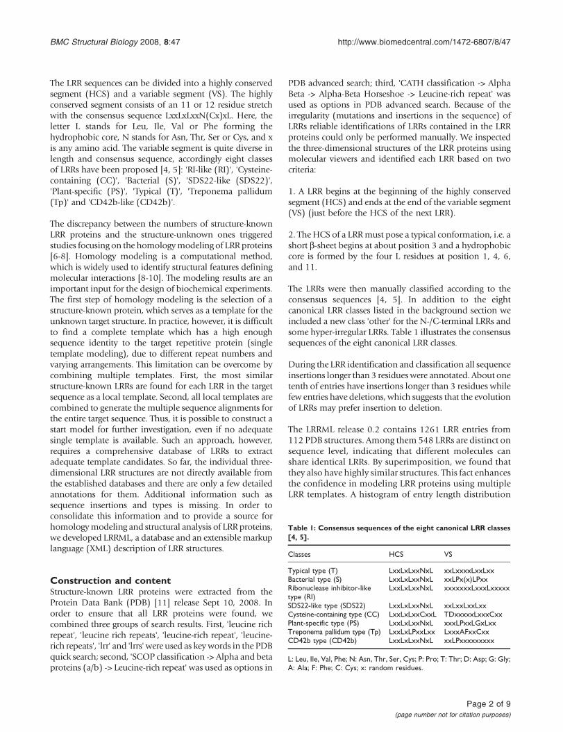
(Figure 1) shows that the LRR lengths are concentrated inthe interval from 20 to 29 which covers the characteristiclengths of consensus sequences of the eight canonicalLRR classes Some entries have a sequence longer than 30because they contain large insertions Table 2 presents thedistribution of LRR entries and PDB entries over the nineclasses respectively The classification results are consis-tent with a previous report which showed that LRRs fromdifferent classes never occur simultaneously in the sameprotein and have most probably evolved independently[4] Exceptions to this rule are the T and S types whichoften exist in the same protein forming the super motifSTT [12] It is assumed that both evolved from acommon precursor [1]
Currently there are several protein databases containinginformation on LRRs such as Pfam [13] InterPro [14]SMART [15] and Swiss-Prot [16] These databases predictthe LRR numbers and boundaries for their LRR proteinentries by various computational methods no matterwhether the entries have known three-dimensional
structures or not thereby false negative occurs frequentlyTable 3 lists the numbers of structure-known LRR proteinsand their LRRs covered by these databases As moredetailed examples LRR numbers of LRR proteins fromdifferent classes reported by the established databases arecompared in Table 4 Additionally the individual three-dimensional LRR structures are not directly available fromthese databases In order to combine the informationrequired for homology modeling and structural analysisLRRML is provided with three prominent characteristics
1 Each database entry is an individual three-dimen-sional LRR structure which was identified with highaccuracy
2 Extensive annotations such as systematic classifica-tion secondary structures HCSVS partitions andsequence insertion are provided
3 LRRs were extracted from all structure-known LRRprotein structures from PDB
Figure 1LRR entry length distribution The most common entry lengths vary from 20 to 29 Each LRR class has a characteristiclength distribution Some entries have a sequence length larger than 29 due to insertions
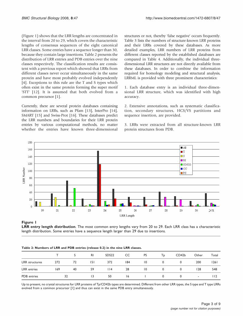
Table 2 Numbers of LRR and PDB entries (release 02) in the nine LRR classes
T S RI SDS22 CC PS Tp CD42b Other Total
LRR structures 272 72 151 372 184 10 0 0 200 1261
LRR entries 169 40 59 114 28 10 0 0 128 548
PDB entries 32 13 50 16 1 0 0 - 112
Up to present no crystal structures for LRR proteins of TpCD42b types are determined Different from other LRR types the S type and T type LRRsevolved from a common precursor [1] and thus can exist in the same PDB entry simultaneously
BMC Structural Biology 2008 847 httpwwwbiomedcentralcom1472-6807847
Page 3 of 9(page number not for citation purposes)
XML descriptionThe extensible markup language (XML) was standardizedin the 90s and is well established as a format forhierarchical data It can be queried and parsed moreeasily by application programs Therefore more andmore biological databases use the XML as data savingformat and database management system (DBMS)[17-19] LRRML was designed by using eXist [20] anXML DBMS and using XPathXQuery [21] for processingqueries and web forms We developed a LRR markuplanguage (LRRML) for exchanging and storing LRRstructures It consists of four blocks of information
1 The sequence information (XML tag ltlSequencegt)amino acid sequence and sequence length
2 The classification information (XML tag ltlTypegt)class name and consensus sequences
3 The sequence partitions (XML tag ltlRegionsgt) aminoacid sequence position length and insertion of HCSand VS
4 The corresponding PDB sources (XML tagltlSourcesgt) ID chain LRR number and classificationof the source PDB entries serial number positionDSSP [22] secondary structure and three-dimensional
coordinates of the current LRR in these source PDBentries
An example describing the LRR3 from PDB entry 2O6S isshown in Figure 2 The document type definition (DTD)file of LRRML is provided asAdditional file 1
UtilityWeb applicationThe entire database can be browsed by LRR IDs or byPDB IDs When browsing the entries appear in asummary table containing at first ID type and sequenceClicking on an ID opens an XML Stylesheet (XSLT) [21]converted HTML web page that presents the entry indetail The original XML file and the coordinates file inPDB format can also be downloaded The XSLT file usedis provided as Additional file 2 Aside from the textualview a LRR structure can be visualized by the onlinemolecular viewer Jmol [23] After loading users canchange the view settings flexibly by themselves LRRMLis provided with various search functions including PDBID search which returns all LRRs contained in this PDBstructure class search which returns all LRRs of this classor length search which returns all LRRs with thissequence length To simplify the homology modelingthe similarity search was implemented It returns thestructures of the most similar LRRs for a structure-unknown LRR The target LRR sequence can be searchedagainst the entire database a certain LRR class or LRRswith a certain length At first a global pair wise sequencealignment with sequence identity will be generated forthe target LRR and each of the LRRs in the user selectedset Then the most similar LRRs will be returned astemplate candidates ranked by sequence identity
The DBMS provides a REST-style application programminginterface (API) through HTTP which supports GET andPOST requests A unique resource identifier (URI) httpzeuskristgeouni-muenchende8081existrest is trea-ted by the server as path to a database collection Alsorequest parameters can help select any required elements
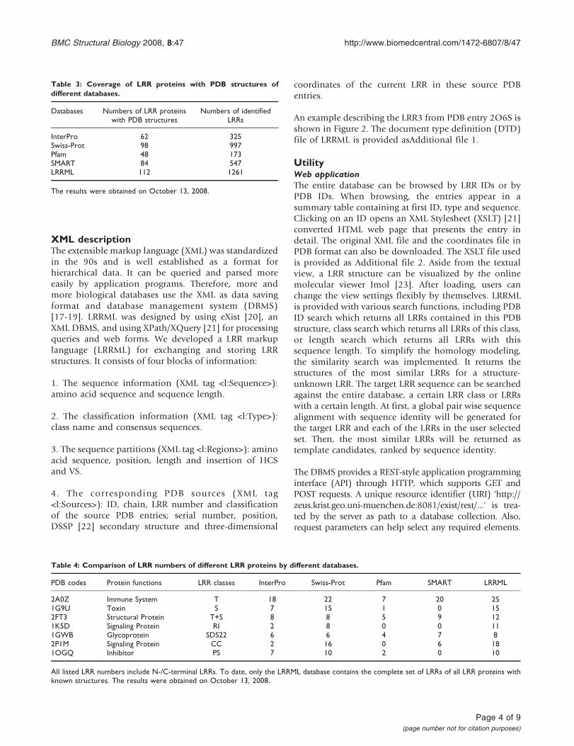
Table 3 Coverage of LRR proteins with PDB structures ofdifferent databases
Databases Numbers of LRR proteinswith PDB structures
Numbers of identifiedLRRs
InterPro 62 325Swiss-Prot 98 997Pfam 48 173SMART 84 547LRRML 112 1261
The results were obtained on October 13 2008
Table 4 Comparison of LRR numbers of different LRR proteins by different databases
PDB codes Protein functions LRR classes InterPro Swiss-Prot Pfam SMART LRRML
2A0Z Immune System T 18 22 7 20 251G9U Toxin S 7 15 1 0 152FT3 Structural Protein T+S 8 8 5 9 121K5D Signaling Protein RI 2 8 0 0 111GWB Glycoprotein SDS22 6 6 4 7 82P1M Signaling Protein CC 2 16 0 6 181OGQ Inhibitor PS 7 10 2 0 10
All listed LRR numbers include N-C-terminal LRRs To date only the LRRML database contains the complete set of LRRs of all LRR proteins withknown structures The results were obtained on October 13 2008
BMC Structural Biology 2008 847 httpwwwbiomedcentralcom1472-6807847
Page 4 of 9(page number not for citation purposes)
Figure 2The LRRML description of a LRR structure This entry is a 24 residue long typical LRR The first 11 residues composeits HCS and the last 13 residues compose its VS (no insertions) It is contained only in the chain A of PDB structure2O6S (a protein involved in the immune system) It is the third one of the 7 LRRs of 2O6S from position 77 to 100Its secondary structure was extracted from DSSP and its three-dimensional coordinate file is available though the hyperlink onthe corresponding web page
BMC Structural Biology 2008 847 httpwwwbiomedcentralcom1472-6807847
Page 5 of 9(page number not for citation purposes)
Table 5 Sequence identities () of target-template LRR pairs
NT 1 2 3 4 5 6 7 8 9 10 11 12 13 14 15 16 17 18 19 20 21 22 23 CT Avg
LRRML ID 406 65 212 465 151 177 110 259 8 203 64 293 270 357 65 152 259 316 152 239 239 92 80 101 173 mdash
PDB source 1XWD 2O6S 1G9U 1OZN 1XKU 1SQ0 2FT3 2V9S 1IO0 1H6U 2O6S 2Z64 2Z62 2Z81 2O6S 1XKU 2V9S 2Z7X 1XKU 1P8V 1P8V 2ID5 2O6Q 2ID5 1W8A mdash
Identity () 4760 4583 4583 4167 5000 5000 4615 4167 4138 4231 5000 5000 3333 4167 4286 5000 4000 3750 3846 5000 4583 5000 4167 4000 3929 4412
In the header line 1ndash25 denote canonical LRRs NT and CT denote N-C-terminal LRRs
BMC
StructuralB
iology2008
847httpw
wwbiom
edcentralcom1472-6807847
Page
6of
9(page
number
notfor
citationpurposes)
Figure 4Ramachandran plot of model and crystal structure of mouse TLR3 ectodomain (A) Predicted model of mouseTLR3 ectodomain (B) Crystal structure of mouse TLR3 ectodomain The different colored areas indicate disallowed (white)generously allowed (light yellow) additional allowed (yellow) and most favored (red) regions
Figure 3Comparison of model and crystal structure of mouse TLR3 ectodomain at the two ligand interaction regions Bluestructure obtained by homology modeling orange crystal structure (PDB code 3CIG) (A) The modeled backbone structure ofmouse TLR3 ectodomain (B) Model and crystal structure superimposed at the N-terminal interaction region The root mean squaredeviation is 196 Aring (C) Superimposition at the C-terminal interaction region The root mean square deviation is 19 Aring The reportedinteracting residues are presented with side chain and labelled with residue name and position in (B) and (C)
BMC Structural Biology 2008 847 httpwwwbiomedcentralcom1472-6807847
Page 7 of 9(page number not for citation purposes)
For example _query executes a specified XPathXQuerythe URL httpzeuskristgeouni-muenchende8081existrestdblrrml_query=LRR [TAbbr=S] returnsall the S type LRRs
Application in homology modelingLRRML was designed as a tool for template selection inhomology modeling of LRR proteins Traditionally thetemplate used in homology modeling is one or more fulllength protein structures obtained via similarity searchNevertheless due to the different repeat numbers andarrangements of LRRs the sequence identity between thetarget and the full length template is usually not highenough for homology modeling With LRRML the mostsimilar structure-known LRR can be found for each LRRin the target sequence as a local template The combina-tion of all local templates through multiple alignmentshelps to achieve a high sequence identity to the target
As test case we modeled the structure of mouse Toll-likereceptor 3 (TLR3) ectodomain We assumed that thestructure of mouse TLR3 ectodomain were unknown andexcluded the LRRs of mousehuman TLR3 ectodomainfrom LRRML Through similarity search the optimaltemplate for each of the 25 LRRs in mouse TLR3 wasfound The sequence identity between each LRR pair(targettemplate LRR) is listed in Table 5 Then a 26-linemultiple alignment was generated by the 25 templatesequences and the target sequence as the input ofMODELLER 9v3 [24] The resulting three-dimensionalmodel (Figure 3A) was evaluated by PROCHECK [25]with 982 residues falling into the most favored orallowed regions of the main chain torsion anglesdistribution whereas the result of the TLR3 crystalstructure (PDB code 3CIG) was 986 (Figure 4) Themouse TLR3 has been shown to bind double-strandedRNA ligand with both N-terminal and C-terminal siteson the lateral side of the convex surface of TLR3 [26] TheN-terminal interaction site is composed of LRRNT andLRR1-3 and the C-terminal site is composed of LRR19-21 We superimposed the resulting model onto thecrystal structure of mouse TLR3 ectodomain at the twointeraction sites by using SuperPose v10 [27] The rootmean square deviations of the structures are 196 Aring and19 Aring respectively (Figure 3BC) indicating that thepredicted model sufficiently well matched the crystalstructure and was useful for prediction of ligandinteraction sites These results demonstrate that homol-ogy modeling using combined multiple templatesobtained from LRRML can create valuable informationto trigger further biochemical research Interpretation ofstructural details however should be done exercisingdue care
ConclusionA specialised conformational leucine-rich repeats data-base called LRRML has been developed It is supportedby an XML database management system and can besearched and browsed with either an easy-to-use webinterface or REST like interface The interface is suitablefor most graphical web browsers and has been tested onthe Windows Mac and Linux operating systems LRRMLcontains individual three-dimensional LRR structureswith manual structural annotations It presents usefulsources for homology modeling and structural analysisof LRR proteins Since the amount of structure-deter-mined LRR proteins constantly increases we plan toupdate LRRML every 2 to 3 months
Availability and requirementsThis database is freely available at httpzeuskristgeouni-muenchende~lrrml
Authors contributionsTW and JG drafted the manuscript extracted the datacompiled the database wrote the code for the webinterface and performed the statistical analysis FJ WMHRWS and SCR conceived of the study built the databaseserver participated in the database design and coordina-tion and helped to draft the manuscript TW and JGshould be regarded as joint first authors All authors readand approved the final manuscript
Additional material
Additional file 1The document type definition (DTD) file of LRRMLClick here for file[httpwwwbiomedcentralcomcontentsupplementary1472-6807-8-47-S1dtd]
Additional file 2The XML Stylesheet (XSLT) of LRRMLClick here for file[httpwwwbiomedcentralcomcontentsupplementary1472-6807-8-47-S2xsl]
AcknowledgementsThis work was supported by Graduiertenkolleg 1202 of the DeutscheForschungsgemeinschaft
References1 Matsushima N Tanaka T Enkhbayar P Mikami T Taga M Yamada K
and Kuroki Y Comparative sequence analysis of leucine-richrepeats (LRRs) within vertebrate toll-like receptors BMCGenomics 2007 8124ndash143
2 Dolan J Walshe K Alsbury S Hokamp K OKeeffe S Okafuji TMiller SFC Guy Tear G and Mitchell KJ The extracellularLeucine-Rich Repeat superfamily a comparative survey andanalysis of evolutionary relationships and expression pat-terns BMC Genomics 2007 8320ndash343
BMC Structural Biology 2008 847 httpwwwbiomedcentralcom1472-6807847
Page 8 of 9(page number not for citation purposes)
3 Kobe B and Deisenhofer J Crystal structure of porcineribonuclease inhibitor a protein with leucine-rich repeatsNature 1993 366751ndash756
4 Kobe B and Kajava AV The leucine-rich repeat as a proteinrecognition motif Curr Opin Struct Biol 2001 11725ndash732
5 Bell JK Mullen GE Leifer CA Mazzoni A Davies DR and Segal DMLeucine-rich repeats and pathogen recognition in Toll-likereceptors Trends Immunol 2003 24528ndash533
6 Kajava AV Structural Diversity of Leucine-rich RepeatProteins J Mol Biol 1998 277519ndash527
7 Stumpp MT Forrer P Binz HK and Plckthun A Designing RepeatProteins Modular Leucine-richRepeat Protein LibrariesBased on the Mammalian Ribonuclease Inhibitor Family JMol Biol 2003 332471ndash487
8 Kubarenko A Frank M and Weber AN Structure-functionrelationships of Toll-like receptor domains through homol-ogy modelling and molecular dynamics Biochem Soc Trans2007 351515ndash1518
9 Roumlssle SC Bisch PM Lone YC Abastado JP Kourilsky P and Bellio MMutational analysis and molecular modeling of the bindingof Staphylococcus aureus enterotoxin C2 to a murine T cellreceptor Vbeta10 chain Eur J Immunol 2002 322172ndash2178
10 Hazai E and Bikaacutedi Z Homology modeling of breast cancerresistance protein (ABCG2) J Struct Biol 2008 16263ndash74
11 Berman HM Westbrook J Feng Z Gilliland G Bhat TN Weissig HShindyalov IN and Bourne PE The Protein Data Bank NucleicAcids Res 2000 28235ndash242
12 Matsushima N Kamiya M Suzuki N and Tanaka T Super-Motifs ofLeucine-Rich Repeats (LRRs) Proteins Genome Inform 200011343ndash345
13 Finn RD Tate J Mistry J Coggill PC Sammut JS Hotz HR Ceric GForslund K Eddy SR Sonnhammer EL and Bateman A The Pfamprotein families database Nucleic Acids Res 2008 36D281ndash288
14 Mulder NJ and Apweiler R InterPro and InterProScan tools forprotein sequence classification and comparison Methods MolBiol 2007 39659ndash70
15 Schultz J Copley RR Doerks T Ponting CP and Bork P SMART aweb-based tool for the study of genetically mobile domainsNucleic Acids Res 2000 28231ndash234
16 Wu CH Apweiler R Bairoch A Natale DA Barker WCBoeckmann B Ferro S Gasteiger E Huang H Lopez RMagrane M Martin MJ Mazumder R ODonovan C Redaschi Nand Suzek B The Universal Protein Resource (UniProt) anexpanding universe of protein information Nucleic Acids Res2006 34D187ndash191
17 Heida N Hasegawa Y Mochizuki Y Hirosawa K Konagaya A andToyoda T TraitMap an XML-based genetic-map databasecombining multigenic loci and biomolecular networksBioinformatics 2004 20 Suppl 1i152ndashi160
18 Kunz H Derz C Tolxdorff T and Bernarding J XML knowledgedatabase of MRI-derived eye models Comput Methods ProgramsBiomed 2004 73203ndash208
19 Jiang K and Nash C Application of XML database technologyto biological pathway datasets Conf Proc IEEE Eng Med Biol Soc2006 14217ndash4220
20 eXist-db an open source database management system httpexist-dborg
21 The World Wide Web Consortium httpwwww3org22 Kabsch W and Sander C Dictionary of protein secondary
structure pattern recognition of hydrogen-bonded andgeometrical features Biopolymers 1983 222577ndash2637
23 Jmol an open-source Java viewer for chemical structures in3D httpwwwjmolorg
24 Fiser A Do RK and Sali A Modeling of loops in proteinstructures Protein Sci 2000 91753ndash1773
25 Laskowski RA MacArthur MW Moss DS and Thornton JMPROCHECK a program to check the stereochemicalquality of protein structures J Appl Cryst 1993 26283ndash291
26 Liu L Botos I Wang Y Leonard JN Shiloach J Segal DM andDavies DR Structral basis of Toll-like receptor 3 signalingwith double-stranded RNA Science 2008 320379ndash381
27 Maiti R Van Domselaar GH Zhang H and Wishart DS SuperPosea simple server for sophisticated structural superpositionNucleic Acids Res 2004 32W590ndash594
Publish with BioMed Central and every scientist can read your work free of charge
BioMed Central will be the most significant development for disseminating the results of biomedical research in our lifetime
Sir Paul Nurse Cancer Research UK
Your research papers will be
available free of charge to the entire biomedical community
peer reviewed and published immediately upon acceptance
cited in PubMed and archived on PubMed Central
yours mdash you keep the copyright
Submit your manuscript herehttpwwwbiomedcentralcominfopublishing_advasp
BioMedcentral
BMC Structural Biology 2008 847 httpwwwbiomedcentralcom1472-6807847
Page 9 of 9(page number not for citation purposes)
- Abstract
-
- Background
- Description
- Conclusion
-
- Background
- Construction and content
- XML description
- Utility
-
- Web application
- Application in homology modeling
-
- Conclusion
- Availability and requirements
- Authors contributions
- Additional material
- Acknowledgements
- References
-
The LRR sequences can be divided into a highly conservedsegment (HCS) and a variable segment (VS) The highlyconserved segment consists of an 11 or 12 residue stretchwith the consensus sequence LxxLxLxxN(Cx)xL Here theletter L stands for Leu Ile Val or Phe forming thehydrophobic core N stands for Asn Thr Ser or Cys and xis any amino acid The variable segment is quite diverse inlength and consensus sequence accordingly eight classesof LRRs have been proposed [4 5] RI-like (RI) Cysteine-containing (CC) Bacterial (S) SDS22-like (SDS22)Plant-specific (PS) Typical (T) Treponema pallidum(Tp) and CD42b-like (CD42b)
The discrepancy between the numbers of structure-knownLRR proteins and the structure-unknown ones triggeredstudies focusing on the homologymodeling of LRR proteins[6-8] Homology modeling is a computational methodwhich is widely used to identify structural features definingmolecular interactions [8-10] The modeling results are animportant input for the design of biochemical experimentsThe first step of homology modeling is the selection of astructure-known protein which serves as a template for theunknown target structure In practice however it is difficultto find a complete template which has a high enoughsequence identity to the target repetitive protein (singletemplate modeling) due to different repeat numbers andvarying arrangements This limitation can be overcome bycombining multiple templates First the most similarstructure-known LRRs are found for each LRR in the targetsequence as a local template Second all local templates arecombined to generate the multiple sequence alignments forthe entire target sequence Thus it is possible to construct astart model for further investigation even if no adequatesingle template is available Such an approach howeverrequires a comprehensive database of LRRs to extractadequate template candidates So far the individual three-dimensional LRR structures are not directly available fromthe established databases and there are only a few detailedannotations for them Additional information such assequence insertions and types is missing In order toconsolidate this information and to provide a source forhomologymodeling and structural analysis of LRR proteinswe developed LRRML a database and an extensible markuplanguage (XML) description of LRR structures
Construction and contentStructure-known LRR proteins were extracted from theProtein Data Bank (PDB) [11] release Sept 10 2008 Inorder to ensure that all LRR proteins were found wecombined three groups of search results First leucine richrepeat leucine rich repeats leucine-rich repeat leucine-rich repeats lrr and lrrs were used as keywords in the PDBquick search second SCOP classification -gt Alpha and betaproteins (ab) -gt Leucine-rich repeat was used as options in
PDB advanced search third CATH classification -gt AlphaBeta -gt Alpha-Beta Horseshoe -gt Leucine-rich repeat wasused as options in PDB advanced search Because of theirregularity (mutations and insertions in the sequence) ofLRRs reliable identifications of LRRs contained in the LRRproteins could only be performed manually We inspectedthe three-dimensional structures of the LRR proteins usingmolecular viewers and identified each LRR based on twocriteria
1 A LRR begins at the beginning of the highly conservedsegment (HCS) and ends at the end of the variable segment(VS) (just before the HCS of the next LRR)
2 TheHCS of a LRRmust pose a typical conformation ie ashort b-sheet begins at about position 3 and a hydrophobiccore is formed by the four L residues at position 1 4 6and 11
The LRRs were then manually classified according to theconsensus sequences [4 5] In addition to the eightcanonical LRR classes listed in the background section weincluded a new class other for the N-C-terminal LRRs andsome hyper-irregular LRRs Table 1 illustrates the consensussequences of the eight canonical LRR classes
During the LRR identification and classification all sequenceinsertions longer than 3 residueswere annotated About onetenth of entries have insertions longer than 3 residues whilefew entries have deletions which suggests that the evolutionof LRRs may prefer insertion to deletion
The LRRML release 02 contains 1261 LRR entries from112 PDB structures Among them 548 LRRs are distinct onsequence level indicating that different molecules canshare identical LRRs By superimposition we found thatthey also have highly similar structures This fact enhancesthe confidence in modeling LRR proteins using multipleLRR templates A histogram of entry length distribution
Table 1 Consensus sequences of the eight canonical LRR classes[4 5]
Classes HCS VS
Typical type (T) LxxLxLxxNxL xxLxxxxLxxLxxBacterial type (S) LxxLxLxxNxL xxLPx(x)LPxxRibonuclease inhibitor-liketype (RI)
LxxLxLxxNxL xxxxxxxLxxxLxxxxx
SDS22-like type (SDS22) LxxLxLxxNxL xxLxxLxxLxxCysteine-containing type (CC) LxxLxLxxCxxL TDxxxxxLxxxCxxPlant-specific type (PS) LxxLxLxxNxL xxxLPxxLGxLxxTreponema pallidum type (Tp) LxxLxLPxxLxx LxxxAFxxCxxCD42b type (CD42b) LxxLxLxxNxL xxLPxxxxxxxxx
L Leu Ile Val Phe N Asn Thr Ser Cys P Pro T Thr D Asp G GlyA Ala F Phe C Cys x random residues
BMC Structural Biology 2008 847 httpwwwbiomedcentralcom1472-6807847
Page 2 of 9(page number not for citation purposes)
(Figure 1) shows that the LRR lengths are concentrated inthe interval from 20 to 29 which covers the characteristiclengths of consensus sequences of the eight canonicalLRR classes Some entries have a sequence longer than 30because they contain large insertions Table 2 presents thedistribution of LRR entries and PDB entries over the nineclasses respectively The classification results are consis-tent with a previous report which showed that LRRs fromdifferent classes never occur simultaneously in the sameprotein and have most probably evolved independently[4] Exceptions to this rule are the T and S types whichoften exist in the same protein forming the super motifSTT [12] It is assumed that both evolved from acommon precursor [1]
Currently there are several protein databases containinginformation on LRRs such as Pfam [13] InterPro [14]SMART [15] and Swiss-Prot [16] These databases predictthe LRR numbers and boundaries for their LRR proteinentries by various computational methods no matterwhether the entries have known three-dimensional
structures or not thereby false negative occurs frequentlyTable 3 lists the numbers of structure-known LRR proteinsand their LRRs covered by these databases As moredetailed examples LRR numbers of LRR proteins fromdifferent classes reported by the established databases arecompared in Table 4 Additionally the individual three-dimensional LRR structures are not directly available fromthese databases In order to combine the informationrequired for homology modeling and structural analysisLRRML is provided with three prominent characteristics
1 Each database entry is an individual three-dimen-sional LRR structure which was identified with highaccuracy
2 Extensive annotations such as systematic classifica-tion secondary structures HCSVS partitions andsequence insertion are provided
3 LRRs were extracted from all structure-known LRRprotein structures from PDB
Figure 1LRR entry length distribution The most common entry lengths vary from 20 to 29 Each LRR class has a characteristiclength distribution Some entries have a sequence length larger than 29 due to insertions
Table 2 Numbers of LRR and PDB entries (release 02) in the nine LRR classes
T S RI SDS22 CC PS Tp CD42b Other Total
LRR structures 272 72 151 372 184 10 0 0 200 1261
LRR entries 169 40 59 114 28 10 0 0 128 548
PDB entries 32 13 50 16 1 0 0 - 112
Up to present no crystal structures for LRR proteins of TpCD42b types are determined Different from other LRR types the S type and T type LRRsevolved from a common precursor [1] and thus can exist in the same PDB entry simultaneously
BMC Structural Biology 2008 847 httpwwwbiomedcentralcom1472-6807847
Page 3 of 9(page number not for citation purposes)
XML descriptionThe extensible markup language (XML) was standardizedin the 90s and is well established as a format forhierarchical data It can be queried and parsed moreeasily by application programs Therefore more andmore biological databases use the XML as data savingformat and database management system (DBMS)[17-19] LRRML was designed by using eXist [20] anXML DBMS and using XPathXQuery [21] for processingqueries and web forms We developed a LRR markuplanguage (LRRML) for exchanging and storing LRRstructures It consists of four blocks of information
1 The sequence information (XML tag ltlSequencegt)amino acid sequence and sequence length
2 The classification information (XML tag ltlTypegt)class name and consensus sequences
3 The sequence partitions (XML tag ltlRegionsgt) aminoacid sequence position length and insertion of HCSand VS
4 The corresponding PDB sources (XML tagltlSourcesgt) ID chain LRR number and classificationof the source PDB entries serial number positionDSSP [22] secondary structure and three-dimensional
coordinates of the current LRR in these source PDBentries
An example describing the LRR3 from PDB entry 2O6S isshown in Figure 2 The document type definition (DTD)file of LRRML is provided asAdditional file 1
UtilityWeb applicationThe entire database can be browsed by LRR IDs or byPDB IDs When browsing the entries appear in asummary table containing at first ID type and sequenceClicking on an ID opens an XML Stylesheet (XSLT) [21]converted HTML web page that presents the entry indetail The original XML file and the coordinates file inPDB format can also be downloaded The XSLT file usedis provided as Additional file 2 Aside from the textualview a LRR structure can be visualized by the onlinemolecular viewer Jmol [23] After loading users canchange the view settings flexibly by themselves LRRMLis provided with various search functions including PDBID search which returns all LRRs contained in this PDBstructure class search which returns all LRRs of this classor length search which returns all LRRs with thissequence length To simplify the homology modelingthe similarity search was implemented It returns thestructures of the most similar LRRs for a structure-unknown LRR The target LRR sequence can be searchedagainst the entire database a certain LRR class or LRRswith a certain length At first a global pair wise sequencealignment with sequence identity will be generated forthe target LRR and each of the LRRs in the user selectedset Then the most similar LRRs will be returned astemplate candidates ranked by sequence identity
The DBMS provides a REST-style application programminginterface (API) through HTTP which supports GET andPOST requests A unique resource identifier (URI) httpzeuskristgeouni-muenchende8081existrest is trea-ted by the server as path to a database collection Alsorequest parameters can help select any required elements
Table 3 Coverage of LRR proteins with PDB structures ofdifferent databases
Databases Numbers of LRR proteinswith PDB structures
Numbers of identifiedLRRs
InterPro 62 325Swiss-Prot 98 997Pfam 48 173SMART 84 547LRRML 112 1261
The results were obtained on October 13 2008
Table 4 Comparison of LRR numbers of different LRR proteins by different databases
PDB codes Protein functions LRR classes InterPro Swiss-Prot Pfam SMART LRRML
2A0Z Immune System T 18 22 7 20 251G9U Toxin S 7 15 1 0 152FT3 Structural Protein T+S 8 8 5 9 121K5D Signaling Protein RI 2 8 0 0 111GWB Glycoprotein SDS22 6 6 4 7 82P1M Signaling Protein CC 2 16 0 6 181OGQ Inhibitor PS 7 10 2 0 10
All listed LRR numbers include N-C-terminal LRRs To date only the LRRML database contains the complete set of LRRs of all LRR proteins withknown structures The results were obtained on October 13 2008
BMC Structural Biology 2008 847 httpwwwbiomedcentralcom1472-6807847
Page 4 of 9(page number not for citation purposes)
Figure 2The LRRML description of a LRR structure This entry is a 24 residue long typical LRR The first 11 residues composeits HCS and the last 13 residues compose its VS (no insertions) It is contained only in the chain A of PDB structure2O6S (a protein involved in the immune system) It is the third one of the 7 LRRs of 2O6S from position 77 to 100Its secondary structure was extracted from DSSP and its three-dimensional coordinate file is available though the hyperlink onthe corresponding web page
BMC Structural Biology 2008 847 httpwwwbiomedcentralcom1472-6807847
Page 5 of 9(page number not for citation purposes)
Table 5 Sequence identities () of target-template LRR pairs
NT 1 2 3 4 5 6 7 8 9 10 11 12 13 14 15 16 17 18 19 20 21 22 23 CT Avg
LRRML ID 406 65 212 465 151 177 110 259 8 203 64 293 270 357 65 152 259 316 152 239 239 92 80 101 173 mdash
PDB source 1XWD 2O6S 1G9U 1OZN 1XKU 1SQ0 2FT3 2V9S 1IO0 1H6U 2O6S 2Z64 2Z62 2Z81 2O6S 1XKU 2V9S 2Z7X 1XKU 1P8V 1P8V 2ID5 2O6Q 2ID5 1W8A mdash
Identity () 4760 4583 4583 4167 5000 5000 4615 4167 4138 4231 5000 5000 3333 4167 4286 5000 4000 3750 3846 5000 4583 5000 4167 4000 3929 4412
In the header line 1ndash25 denote canonical LRRs NT and CT denote N-C-terminal LRRs
BMC
StructuralB
iology2008
847httpw
wwbiom
edcentralcom1472-6807847
Page
6of
9(page
number
notfor
citationpurposes)
Figure 4Ramachandran plot of model and crystal structure of mouse TLR3 ectodomain (A) Predicted model of mouseTLR3 ectodomain (B) Crystal structure of mouse TLR3 ectodomain The different colored areas indicate disallowed (white)generously allowed (light yellow) additional allowed (yellow) and most favored (red) regions
Figure 3Comparison of model and crystal structure of mouse TLR3 ectodomain at the two ligand interaction regions Bluestructure obtained by homology modeling orange crystal structure (PDB code 3CIG) (A) The modeled backbone structure ofmouse TLR3 ectodomain (B) Model and crystal structure superimposed at the N-terminal interaction region The root mean squaredeviation is 196 Aring (C) Superimposition at the C-terminal interaction region The root mean square deviation is 19 Aring The reportedinteracting residues are presented with side chain and labelled with residue name and position in (B) and (C)
BMC Structural Biology 2008 847 httpwwwbiomedcentralcom1472-6807847
Page 7 of 9(page number not for citation purposes)
For example _query executes a specified XPathXQuerythe URL httpzeuskristgeouni-muenchende8081existrestdblrrml_query=LRR [TAbbr=S] returnsall the S type LRRs
Application in homology modelingLRRML was designed as a tool for template selection inhomology modeling of LRR proteins Traditionally thetemplate used in homology modeling is one or more fulllength protein structures obtained via similarity searchNevertheless due to the different repeat numbers andarrangements of LRRs the sequence identity between thetarget and the full length template is usually not highenough for homology modeling With LRRML the mostsimilar structure-known LRR can be found for each LRRin the target sequence as a local template The combina-tion of all local templates through multiple alignmentshelps to achieve a high sequence identity to the target
As test case we modeled the structure of mouse Toll-likereceptor 3 (TLR3) ectodomain We assumed that thestructure of mouse TLR3 ectodomain were unknown andexcluded the LRRs of mousehuman TLR3 ectodomainfrom LRRML Through similarity search the optimaltemplate for each of the 25 LRRs in mouse TLR3 wasfound The sequence identity between each LRR pair(targettemplate LRR) is listed in Table 5 Then a 26-linemultiple alignment was generated by the 25 templatesequences and the target sequence as the input ofMODELLER 9v3 [24] The resulting three-dimensionalmodel (Figure 3A) was evaluated by PROCHECK [25]with 982 residues falling into the most favored orallowed regions of the main chain torsion anglesdistribution whereas the result of the TLR3 crystalstructure (PDB code 3CIG) was 986 (Figure 4) Themouse TLR3 has been shown to bind double-strandedRNA ligand with both N-terminal and C-terminal siteson the lateral side of the convex surface of TLR3 [26] TheN-terminal interaction site is composed of LRRNT andLRR1-3 and the C-terminal site is composed of LRR19-21 We superimposed the resulting model onto thecrystal structure of mouse TLR3 ectodomain at the twointeraction sites by using SuperPose v10 [27] The rootmean square deviations of the structures are 196 Aring and19 Aring respectively (Figure 3BC) indicating that thepredicted model sufficiently well matched the crystalstructure and was useful for prediction of ligandinteraction sites These results demonstrate that homol-ogy modeling using combined multiple templatesobtained from LRRML can create valuable informationto trigger further biochemical research Interpretation ofstructural details however should be done exercisingdue care
ConclusionA specialised conformational leucine-rich repeats data-base called LRRML has been developed It is supportedby an XML database management system and can besearched and browsed with either an easy-to-use webinterface or REST like interface The interface is suitablefor most graphical web browsers and has been tested onthe Windows Mac and Linux operating systems LRRMLcontains individual three-dimensional LRR structureswith manual structural annotations It presents usefulsources for homology modeling and structural analysisof LRR proteins Since the amount of structure-deter-mined LRR proteins constantly increases we plan toupdate LRRML every 2 to 3 months
Availability and requirementsThis database is freely available at httpzeuskristgeouni-muenchende~lrrml
Authors contributionsTW and JG drafted the manuscript extracted the datacompiled the database wrote the code for the webinterface and performed the statistical analysis FJ WMHRWS and SCR conceived of the study built the databaseserver participated in the database design and coordina-tion and helped to draft the manuscript TW and JGshould be regarded as joint first authors All authors readand approved the final manuscript
Additional material
Additional file 1The document type definition (DTD) file of LRRMLClick here for file[httpwwwbiomedcentralcomcontentsupplementary1472-6807-8-47-S1dtd]
Additional file 2The XML Stylesheet (XSLT) of LRRMLClick here for file[httpwwwbiomedcentralcomcontentsupplementary1472-6807-8-47-S2xsl]
AcknowledgementsThis work was supported by Graduiertenkolleg 1202 of the DeutscheForschungsgemeinschaft
References1 Matsushima N Tanaka T Enkhbayar P Mikami T Taga M Yamada K
and Kuroki Y Comparative sequence analysis of leucine-richrepeats (LRRs) within vertebrate toll-like receptors BMCGenomics 2007 8124ndash143
2 Dolan J Walshe K Alsbury S Hokamp K OKeeffe S Okafuji TMiller SFC Guy Tear G and Mitchell KJ The extracellularLeucine-Rich Repeat superfamily a comparative survey andanalysis of evolutionary relationships and expression pat-terns BMC Genomics 2007 8320ndash343
BMC Structural Biology 2008 847 httpwwwbiomedcentralcom1472-6807847
Page 8 of 9(page number not for citation purposes)
3 Kobe B and Deisenhofer J Crystal structure of porcineribonuclease inhibitor a protein with leucine-rich repeatsNature 1993 366751ndash756
4 Kobe B and Kajava AV The leucine-rich repeat as a proteinrecognition motif Curr Opin Struct Biol 2001 11725ndash732
5 Bell JK Mullen GE Leifer CA Mazzoni A Davies DR and Segal DMLeucine-rich repeats and pathogen recognition in Toll-likereceptors Trends Immunol 2003 24528ndash533
6 Kajava AV Structural Diversity of Leucine-rich RepeatProteins J Mol Biol 1998 277519ndash527
7 Stumpp MT Forrer P Binz HK and Plckthun A Designing RepeatProteins Modular Leucine-richRepeat Protein LibrariesBased on the Mammalian Ribonuclease Inhibitor Family JMol Biol 2003 332471ndash487
8 Kubarenko A Frank M and Weber AN Structure-functionrelationships of Toll-like receptor domains through homol-ogy modelling and molecular dynamics Biochem Soc Trans2007 351515ndash1518
9 Roumlssle SC Bisch PM Lone YC Abastado JP Kourilsky P and Bellio MMutational analysis and molecular modeling of the bindingof Staphylococcus aureus enterotoxin C2 to a murine T cellreceptor Vbeta10 chain Eur J Immunol 2002 322172ndash2178
10 Hazai E and Bikaacutedi Z Homology modeling of breast cancerresistance protein (ABCG2) J Struct Biol 2008 16263ndash74
11 Berman HM Westbrook J Feng Z Gilliland G Bhat TN Weissig HShindyalov IN and Bourne PE The Protein Data Bank NucleicAcids Res 2000 28235ndash242
12 Matsushima N Kamiya M Suzuki N and Tanaka T Super-Motifs ofLeucine-Rich Repeats (LRRs) Proteins Genome Inform 200011343ndash345
13 Finn RD Tate J Mistry J Coggill PC Sammut JS Hotz HR Ceric GForslund K Eddy SR Sonnhammer EL and Bateman A The Pfamprotein families database Nucleic Acids Res 2008 36D281ndash288
14 Mulder NJ and Apweiler R InterPro and InterProScan tools forprotein sequence classification and comparison Methods MolBiol 2007 39659ndash70
15 Schultz J Copley RR Doerks T Ponting CP and Bork P SMART aweb-based tool for the study of genetically mobile domainsNucleic Acids Res 2000 28231ndash234
16 Wu CH Apweiler R Bairoch A Natale DA Barker WCBoeckmann B Ferro S Gasteiger E Huang H Lopez RMagrane M Martin MJ Mazumder R ODonovan C Redaschi Nand Suzek B The Universal Protein Resource (UniProt) anexpanding universe of protein information Nucleic Acids Res2006 34D187ndash191
17 Heida N Hasegawa Y Mochizuki Y Hirosawa K Konagaya A andToyoda T TraitMap an XML-based genetic-map databasecombining multigenic loci and biomolecular networksBioinformatics 2004 20 Suppl 1i152ndashi160
18 Kunz H Derz C Tolxdorff T and Bernarding J XML knowledgedatabase of MRI-derived eye models Comput Methods ProgramsBiomed 2004 73203ndash208
19 Jiang K and Nash C Application of XML database technologyto biological pathway datasets Conf Proc IEEE Eng Med Biol Soc2006 14217ndash4220
20 eXist-db an open source database management system httpexist-dborg
21 The World Wide Web Consortium httpwwww3org22 Kabsch W and Sander C Dictionary of protein secondary
structure pattern recognition of hydrogen-bonded andgeometrical features Biopolymers 1983 222577ndash2637
23 Jmol an open-source Java viewer for chemical structures in3D httpwwwjmolorg
24 Fiser A Do RK and Sali A Modeling of loops in proteinstructures Protein Sci 2000 91753ndash1773
25 Laskowski RA MacArthur MW Moss DS and Thornton JMPROCHECK a program to check the stereochemicalquality of protein structures J Appl Cryst 1993 26283ndash291
26 Liu L Botos I Wang Y Leonard JN Shiloach J Segal DM andDavies DR Structral basis of Toll-like receptor 3 signalingwith double-stranded RNA Science 2008 320379ndash381
27 Maiti R Van Domselaar GH Zhang H and Wishart DS SuperPosea simple server for sophisticated structural superpositionNucleic Acids Res 2004 32W590ndash594
Publish with BioMed Central and every scientist can read your work free of charge
BioMed Central will be the most significant development for disseminating the results of biomedical research in our lifetime
Sir Paul Nurse Cancer Research UK
Your research papers will be
available free of charge to the entire biomedical community
peer reviewed and published immediately upon acceptance
cited in PubMed and archived on PubMed Central
yours mdash you keep the copyright
Submit your manuscript herehttpwwwbiomedcentralcominfopublishing_advasp
BioMedcentral
BMC Structural Biology 2008 847 httpwwwbiomedcentralcom1472-6807847
Page 9 of 9(page number not for citation purposes)
- Abstract
-
- Background
- Description
- Conclusion
-
- Background
- Construction and content
- XML description
- Utility
-
- Web application
- Application in homology modeling
-
- Conclusion
- Availability and requirements
- Authors contributions
- Additional material
- Acknowledgements
- References
-
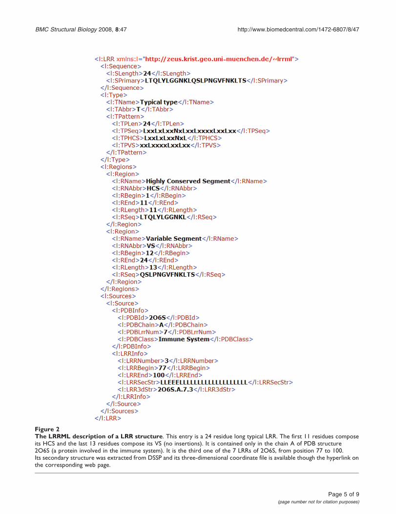
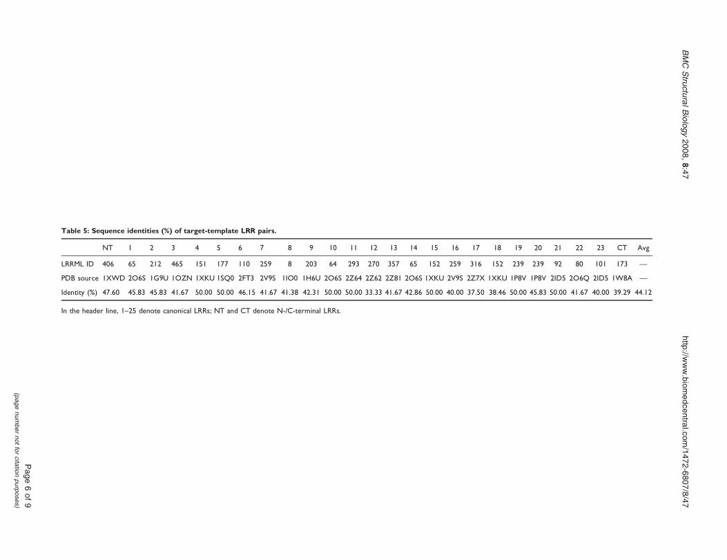
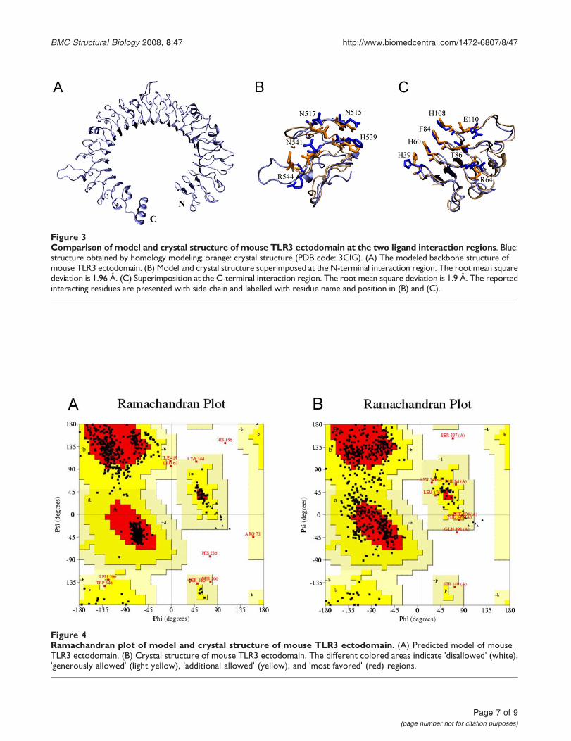
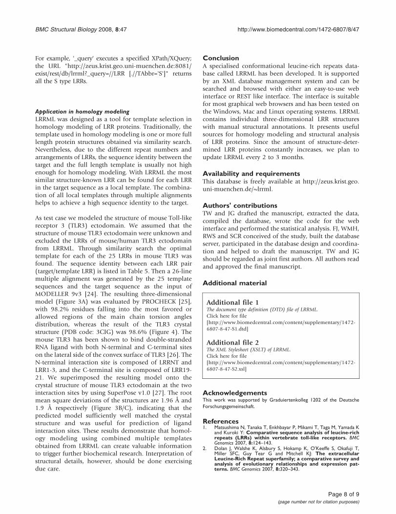