bme 560 medical imaging: x-ray, ct, and nuclear methods radiation physics part 1
TRANSCRIPT
BME 560Medical Imaging: X-ray, CT, and
Nuclear Methods
Radiation Physics Part 1

Today
• Forms of Radiation
• Particulate Radiation– Electron interactions with matter
• EM Radiation
• Nuclear Transitions
• Decay

What is Radiation?
• Radiation: Energy in the form of traveling waves or subatomic particles moving through space– May or may not have mass– May or may not have charge

Types of Radiation
• Ionizing: capable of producing an ion pair from interaction with an atom– Usually with energy > 13.6 eV
• Non-ionizing: incapable of producing an ion pair
• Ion pairs are reactive and may cause biological damage
Radiation
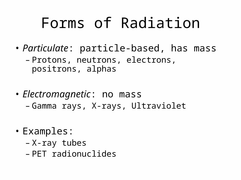
Forms of Radiation
• Particulate: particle-based, has mass– Protons, neutrons, electrons, positrons, alphas
• Electromagnetic: no mass– Gamma rays, X-rays, Ultraviolet
• Examples:– X-ray tubes– PET radionuclides

Ionization and Excitation
• Both transfer energy to an orbital electron– Let Eb be the electron binding energy
– Ionization: Energy is enough to remove electron from orbit
– Excitation: Energy is enough to transition electron to higher orbital
– In both cases, the hole will be filled and energy released.
Radiation (E)
K L M
N
Ejected electron (Ee)

Particulate Radiation
• Alpha particle: He nucleus (2 protons, 2 neutrons)
• Beta particle: Electron (-) or positron (+)
• Proton
• Neutron

Particulate Radiation
• Properties:– Alpha: very high mass, highly charged, very
highly ionizing, very low penetration– Beta: low mass, charged, highly ionizing, low
penetration– Proton: high mass, charged, highly ionizing, low
penetration– Neutron: high mass, no charge, less ionizing,
unusual penetration properties
• In all cases, penetration depends on energy.

Particulate Radiation
• Medical uses:– Alpha: Radiotherapy– Beta: X-ray production, PET imaging,
Radiotherapy– Proton: Radiotherapy– Neutron: Radiotherapy
• So we are primarily interested in electrons and positrons for imaging.

Energetic Electron Energy Transfer
• How electrons lose energy to a medium:– Collisional transfer: Interaction between particle
and orbital electrons• High-energy: Ionization• Low-energy: Heat• Energetic electron loses kinetic energy
– Radiative Transfer: Energy transfer results in production of X-ray
• Characteristic X-rays• Bremsstrahlung radiation
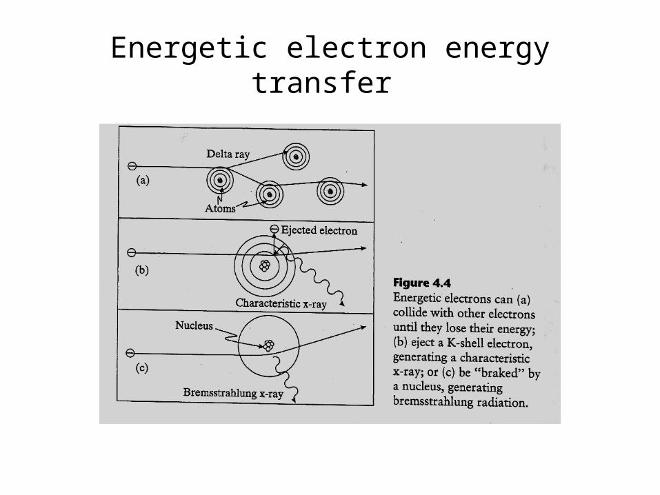
Energetic electron energy transfer

Energy loss rate vs. electron energy in water and lead
therapydiagnostic
Pb207
82

Characteristic X-rays
• Electron moves from higher energy level to a lower energy level.
• Since the orbital energy levels are well defined for individual elements, each element has its own characteristic transitions.– These are very specific energy levels.
• Usually, it is the transitions into the K shell that are important.

Characteristic X-rays

Bremsstrahlung• “Braking radiation”• The effect of charge-charge interaction between an
energetic electron and an atomic nucleus• The electron slows and releases energy in the form of
an X-ray photon.• Bremsstrahlung X-rays may have any energy up to
the energy of the incident electrons.

“White” bremsstrahlung x-rays
Filtered x-rays
Bremsstrahlung production
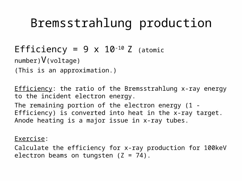
Efficiency = 9 x 10⁻¹⁰ Z (atomic number)V(voltage)
(This is an approximation.)
Efficiency: the ratio of the Bremsstrahlung x-ray energy to the incident electron energy.
The remaining portion of the electron energy (1 - Efficiency) is converted into heat in the x-ray target. Anode heating is a major issue in x-ray tubes.
Exercise:
Calculate the efficiency for x-ray production for 100keV electron beams on tungsten (Z = 74).

Exercises:
What is the rest energy of an electron? (mass = 9.11 x 10⁻²⁸ g)
What is the rest energy of a proton? (mass = 1.67 x 10⁻²⁴ g)
Electron energy, mass and velocity
m m0
1 2 /c 2(c : speed of light)
E mc 2
KE E E0Kinetic energy (KE)

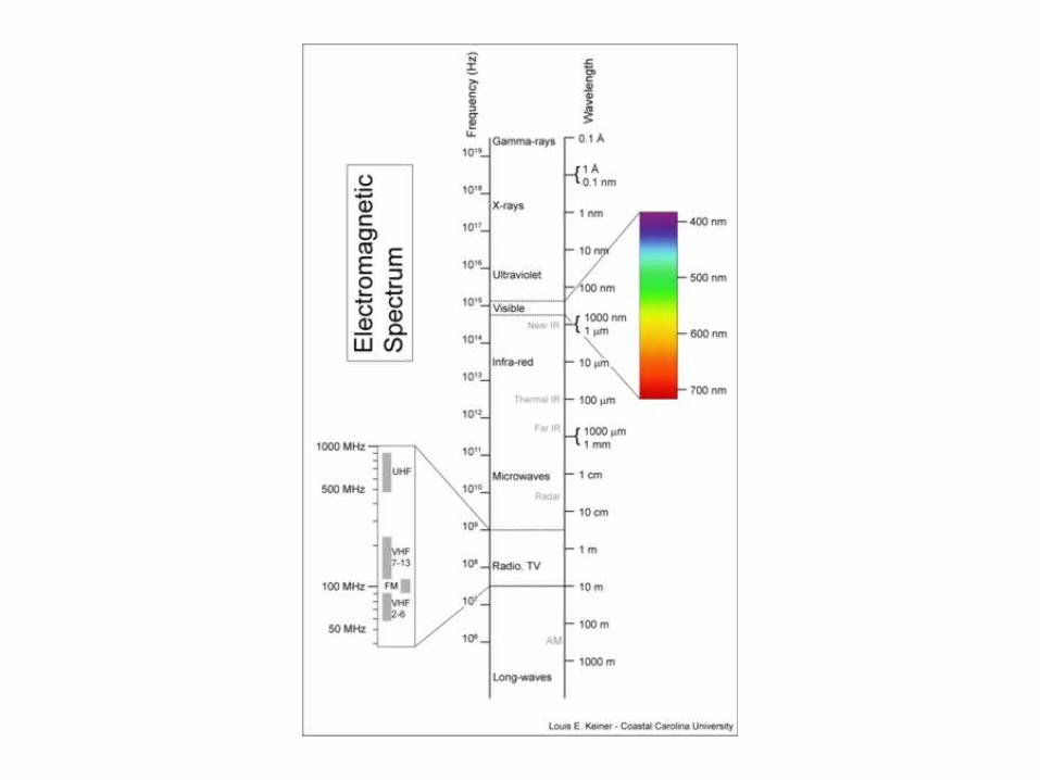
EM Radiation
• Electromagnetic radiation is considered in the form of photons.– No charge, no mass– The wavelengths of interest are much smaller than
the scales of typical interactions, so we are safe to treat this radiation as “particles” rather than “waves”.

EM Radiation
• X-rays versus gamma-rays (or gamma particles)– Physically, they are the same thing.– X-rays: Produced by energetic electrons striking a
material• Man-made
• Secondary effects
– Gamma-rays: Produced by radioactive decay of materials

Radiation

Electromagnetic (EM) radiation • Energy (keV): 1 eV = 1.602 x 10⁻¹⁹ J
• Frequency (Hz): E = hf (Hz)
Planck Constant: h = 6.625 x 10 -27 erg-second = 4.136 x 10⁻¹⁵ eV-s
• Wavelength (): E (keV)= 1.24 /(nm)

(Physical Principles of Medical Imaging by Perry Sprawls)

X-ray production process in imaging and therapy systems involves both particulate and EM types of
radiation
1) Free electron production (temporal res. control)
Electrons are “pulled” out of filament (thermionic or field emission).
1) Electron acceleration
Electron energy under voltage (E = eV)
3) Electron bombards anode to produce
Bremsstrahlung x-rays
(spatial res. Control- focal spot size)
Once x-rays are generated, they are
shaped and controlled to suit the need
of the specific application.
EM Radiation
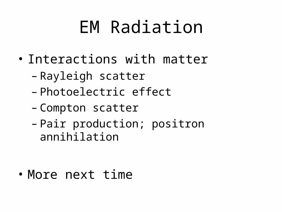
• Interactions with matter– Rayleigh scatter– Photoelectric effect– Compton scatter– Pair production; positron annihilation
• More next time

Radioisotopes
• Gamma photons are given off as unstable or metastable isotopes try to go to a more stable atomic state.
• The nuclear transitions are well defined for a given isotope.– Specific energies are emitted– Sometimes, there are multiple transition paths;
thus, multiple energies.

Atomic Structure
• Atomic structure– Nucleus (Z protons and N neutrons)
– Z orbital electrons
• Atomic number: Z (element)
• Mass number: A = Z+ N(total number of nucleons)
• Symbols
ZA X X A
element
Atomic number
mass number
Example : 612C C 12

Radionuclide
Definition: Certain natural and man-made atoms with unstable nucleus that can undergo spontaneous breakup or decay and, in the process, emit alpha, beta, or gamma radiation.
Naturally occurring radionuclides (U-238, Ra-226, Rn-222)
Man-made radionuclides (isotope)– All radionuclides for diagnostics and therapy are man-made.
Therapy: I-125, Ir-192, Cs-137, Co-60, etc.Diagnostic: Ga-67, Tc-99, I-131, C-11, O-15, etc.

Nucleus instability and decay
• A nucleus with excess energy is at an excited state. It will release the energy and go to the ground (energy) stable state eventually.
• N/P (number of neutrons over protons) ratio is a good indicator of nucleus stability. For low (<15) Z atoms, the stable N/P ratio is 1.0 and it increases to ~1.5 for high Z atoms.
N/P =1
H He Li Be B C N O F Ne Na Mg Al Si P S

Nuclear transitions
There are several ways an unstable nucleus can decay:
• Isotopic transition (mother and daughter nuclei have the same
number of protons) – Lose a neutron
• Isobaric transition (different number of protons but same
number of nucleons (N+P)) – Exchange proton and neutron
• Isomeric transition (different energy levels) – Lose energy from
nucleus
• Isotonic transition (have the same number of neutrons) – Lose a
proton
Isotopes, isobars, isomers, and isotones.

Examples of isobars, isotones, and isotopes
Products of radioactive decay:
• Beta particle (electron)Isobaric transition
• PositronIsobaric transition
• Electron capture Isobaric transition
• Gamma rayIsomeric transition
• Alpha particle
Secondary decay products:
• Characteristic x-rays
• Auger electrons
Isotones

Examples
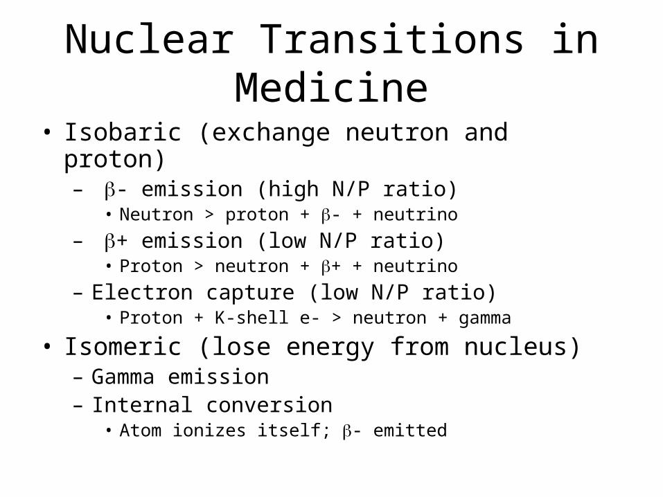
Nuclear Transitions in Medicine
• Isobaric (exchange neutron and proton)– - emission (high N/P ratio)
• Neutron > proton + - + neutrino
– + emission (low N/P ratio)• Proton > neutron + + + neutrino
– Electron capture (low N/P ratio)• Proton + K-shell e- > neutron + gamma
• Isomeric (lose energy from nucleus)– Gamma emission– Internal conversion
• Atom ionizes itself; - emitted

Composite diagram of nuclear transitions
Stable N/P
K shell
High N/PLow N/P
positronbeta
Electron capture
Gamma
Internal conversion
electron
Auger
X-rays
Z, atomic number
ener
gy
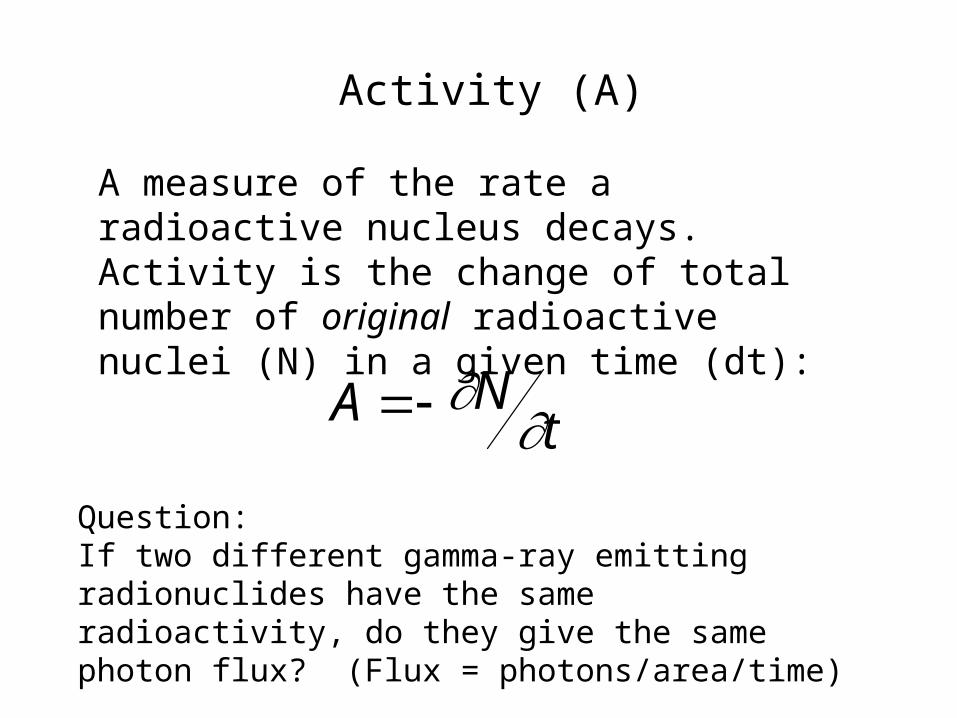
Activity (A)
A measure of the rate a radioactive nucleus decays. Activity is the change of total number of original radioactive nuclei (N) in a given time (dt):
Question: If two different gamma-ray emitting radionuclides have the same radioactivity, do they give the same photon flux? (Flux = photons/area/time)
A Nt

Activity Units: curie (Ci) and becquerel (Bq)
Ci = 3.7 x 10¹⁰ dps
Bq = 1 dps
dps: disintegrations per second
Curie is a very large quantity of radioactivity.
Commonly used activity level is ~ 100 mCi for
therapy and ~ mCi for diagnostics.
The SI unit is becquerel and 1mCi = 37M Bq.
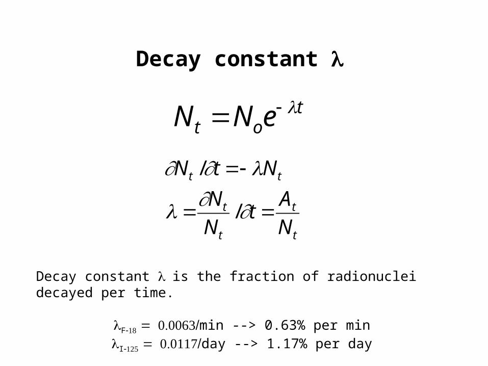
Decay constant
N t Noe t
N t /t N t
N t
N t
/t At
N t
Decay constant is the fraction of radionuclei decayed per time.
Fmin --> 0.63% per minIday --> 1.17% per day

Half life T1/2
• T1/2: time for 50% of the original radionuclides to decay.
N tT1/2N0
2N0
2N0e
T1/2
ln 2 T1/2 T1/2 ln 2
Nremaining = Noriginal/2n (n: number of half lives elapsed)
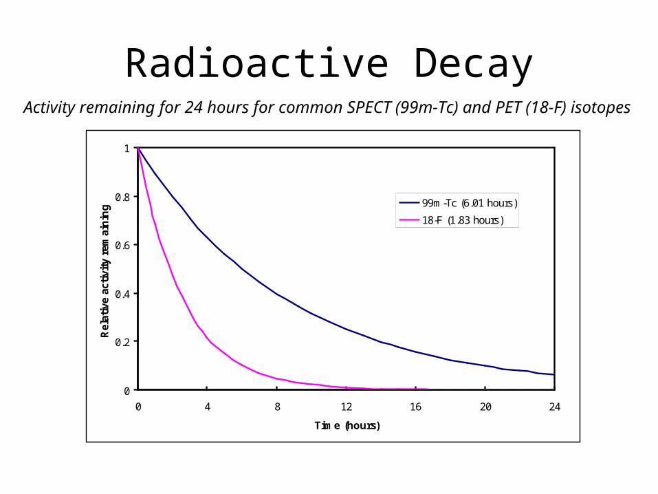
Radioactive Decay
0
0.2
0.4
0.6
0.8
1
0 4 8 12 16 20 24
Time (hours)
Rel
ativ
e ac
tivi
ty r
emai
nin
g 99m-Tc (6.01 hours)
18-F (1.83 hours)
Activity remaining for 24 hours for common SPECT (99m-Tc) and PET (18-F) isotopes
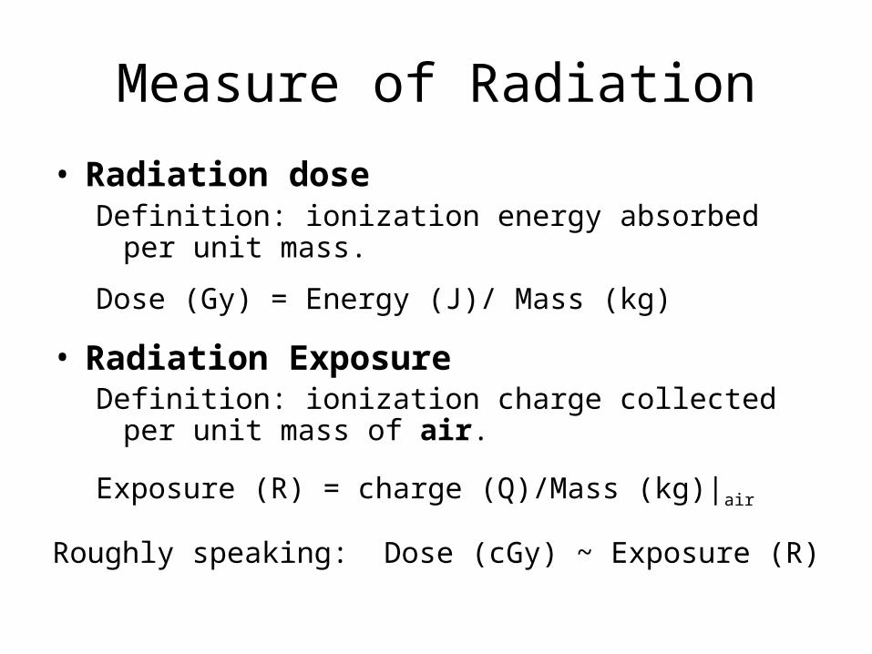
Measure of Radiation
• Radiation doseDefinition: ionization energy absorbed per unit mass.
Dose (Gy) = Energy (J)/ Mass (kg)
• Radiation ExposureDefinition: ionization charge collected per unit mass of air.
Exposure (R) = charge (Q)/Mass (kg)|air
Roughly speaking: Dose (cGy) ~ Exposure (R)