bobic - lamina splendens - chicago may 2015.pptx
TRANSCRIPT

Disclosure:

12th ICRS World Congress Chicago May 9, 2015
What is Lamina Splendens and What Does it Do?
Prof. Vladimir Bobic, MD, FRCSEd Consultant Orthopaedic Knee Surgeon
Chester Knee Clinic @ Nuffield Health, The Grosvenor Hospital Chester, UK

Structure of Articular Cartilage: Superficial Zone

Articular cartilage + Subchondral plate + Trabecular bone = biologically and functionally inseparable OsteoChondral unit
which absorbs and distributes loads across the joint.
But, what about the articular surface? The surface which undergoes a lot of friction and does the actual hard work of endless articulation with other surfaces!


What is Lamina Splendens and What Does it Do?
What’s in a Name? Lamina Splendens or Superficial Zone or Gliding Zone or …? "A rose by any other name would smell as sweet”. William Shakespeare
How Does Lamina Splendens Look Like?

Arthroscopic Appearance:
Is this the lamina splendens? Or just a chunk of semitransparent “surface layer”? CKC UK

Arthroscopic Appearance:
Is this the lamina splendens? Or just a small flap of semitransparent “surface layer”? CKC UK

Arthroscopic Appearance:
Is this the lamina splendens?

Is this Lamina Splendens? Looks like a cling-film …
CC UK

Like a cling-film? On top of tough articulating surface? Not really!
The structure of the LS must be very different to withstand a lifetime of weight-bearing, movement and friction

What’s in a Name: Lamina Spenders or Surface Amorphous Layer?
• MacConaill (1951) observed a bright line at the surface of the articular cartilage using a phase contrast microscope and called it the 'lamina splendens’.
• Sokoloff (1969) and Aspden & Hukins (1979) have disputed the existence of the ‘lamina splendens' concluding that it was nothing but an optical effect produced by phase contrast microscopy.
• However, some researchers using conventional SEM or TEM (Weissetal.1968; Weiss,1982; Jeffery et al.1991) considered that the observed surface layer might correspond to the ‘lamina splendens’ of MacConaill.
• Setting aside the question of the ‘lamina splendens', the surface amorphous layer observed using the cryo-SEM is “believed to correspond to the non collagenous layer on the articular surface”
• Basically, very confusing …

Does it really exist?

“Lamina Splendens: a layer of fine filaments at the very surface of the gliding zone
Henry J Mankin: The Articular Cartilage. Chapter in: "Textbook of Small Animal Orthopaedics” by Charles D. Newton and David M. Nunamaker, J.B. Lippincott, 1985

EM of Lamina Splendens or “a skin”
Henry J Mankin: The Articular Cartilage. Chapter in: "Textbook of Small Animal Orthopaedics” by Charles D. Newton and David M. Nunamaker, J.B. Lippincott, 1985


SEM of Lamina Splendens: “distinctly different layer”
Teshima R et al: Structure of the most superficial layer of articular cartilage. JBJS Br 1995:77-B: 460-4


(A). A semitransparent membrane corresponding to the lamina splendens (LS) was physically peeled off from normal articular cartilage (N) of a cow femoral head (unloading region).(B). A 3D image of the lamina splendens shows the collagen network within it is compromised of unique interwoven collagen bundles (ICB). (C). The corresponding MBI of the collagen network in LS in Fig (B). (D). Traditional histology shows the site where the lamina splendens was separated from the normal (cow) cartilage. (E). Traditional histology of early arthritic cartilage from a human femoral head shows disrupting the articular surface in early OA is a process similar to physically peeling off the lamina splendens. (F). Traditional histology of normal cartilage physically peeled the lamina splendens (indicated as CP (cartilage peeled lamina splendens) in Fig 2(A)) shows loss of the most superficial layer of articular cartilage can expose some chondrocytes near the surface to the joint cavity.
Wu et al. Journal of Orthopaedic Surgery and Research 2008 3:29
What is Lamina Splendens?

What is the Purpose of Lamina Splendens?
A schematic structure of the collagen network in AC shows that the interwoven collagen bundles in the lamina splendens integrate the obliquely oriented collagen fibres and those in the deeper region to form a 3D collagen scaffold, which anchors to the subchondral bone.
Wu et al. Journal of Orthopaedic Surgery and Research 2008 3:29
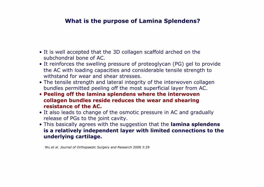
What is the purpose of Lamina Splendens?
• It is well accepted that the 3D collagen scaffold arched on the subchondral bone of AC.
• It reinforces the swelling pressure of proteoglycan (PG) gel to provide the AC with loading capacities and considerable tensile strength to withstand for wear and shear stresses.
• The tensile strength and lateral integrity of the interwoven collagen bundles permitted peeling off the most superficial layer from AC.
• Peeling off the lamina splendens where the interwoven collagen bundles reside reduces the wear and shearing resistance of the AC.
• It also leads to change of the osmotic pressure in AC and gradually release of PGs to the joint cavity.
• This basically agrees with the suggestion that the lamina splendens is a relatively independent layer with limited connections to the underlying cartilage.
Wu et al. Journal of Orthopaedic Surgery and Research 2008 3:29

• Furthermore, the collagen fibres changed from oblique orientation to perpendicular orientation after peeling off the most superficial layer of AC could be associated to the remodelling of the osmotic pressure and subsequent expansion of the proteoglycans in the AC.
• Previously, oblique collagen fibres have been reported to run between the articular surface and subchondral bone and they have further been suggested to be compatible to the requirement of entrapment of proteoglycans and strengthen the tensile properties.
• Therefore, the oblique collagen fibres contained by normal AC may also have contributed to the normal mechanical function of AC.
• Conversely, the perpendicular collagen orientation found in majority of early OA cartilage may contribute little to retain proteoglycans and enhance the tensile property of the cartilage to wear and sharing stresses.
Wu et al. Journal of Orthopaedic Surgery and Research 2008 3:29
What is the purpose of Lamina Splendens?

Superficial Zone and Lubricin:
CKC UK

Superficial Zone and Lubricin:
Roberts S et al. Cartilage 2010


Is Lamina Splendens Important?
CKC UK

Is Lamina Splendens Important?


Light-sheet fluorescence microscopy Light Sheet Microscopy is one of the greatest recent breakthrough technologies
Main advantages: light-sheet microscopy is faster and less phototoxic than other fluorescence microscopy techniques, making it ideal for studying living organisms and the biological processes that take place within them.


Lamina Splendens mounted on a Light-sheet setup
Lamina Splendens embedded in a cylinder of agarose
Source: Marco Marcello, Liverpool IIB/CCI
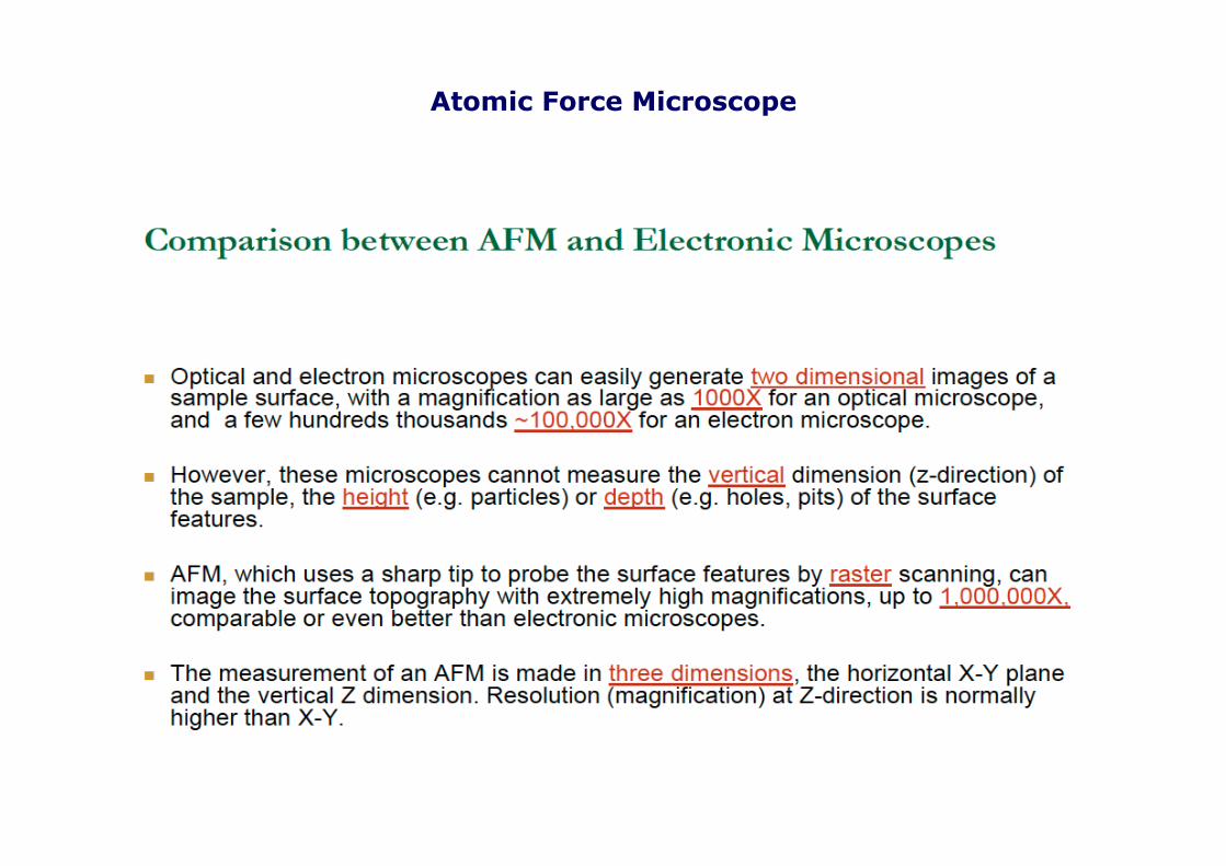
Atomic Force Microscope

Atomic Force Microscope

Atomic Force Microscope

• The surface zone of articular cartilage is a critical component of the mature tissue because its collagen fibrils are oriented parallel to the plane of the tissue surface and so endow it with resistance to shear forces in the joint.
• It is also a critical component of the immature tissue because it drives appositional growth and may allow spontaneous healing when there is fibrillation at the surface.
• It follows that tissue-engineered cartilage implants that do not have a lamina splendens will not function mechanically in the same way as the natural tissue because they will lack resistance to shear forces.
• Loss of the surface zone early in OA may be devastating because it will remove the main driver of the appositional growth of cartilage.
• Failure of repair of the lamina splendens may reflect a failure of this niche to function normally in the injured adult joint, leading ultimately to cartilage erosion and loss of joint function.
• Enriching the surface of engineered implants with mesenchymal cells derived from the synovial membrane may be a way of reconstructing the lamina splendens.
Hollander A, at al. Stem Cells and Cartilage Development: Complexities of a Simple Tissue. STEM CELLS 2010;28:1992–1996
What is Lamina Splendens and What Does it Do?

In conclusion, both synovium and infra patellar fat pad are promising cell sources for tissue engineering of superficial cartilage zone
Tissue Engineering of Gliding Zone and Lamina Splendens?

Summary
SEM of normal articular cartilage. Source: Norimasa Nakamura, Osaka, Japan
Lamina Splendens
• Lamina splendens definitely exists as a separate layer and has a huge mechanical (tribological) role,
• SZP (Lubricin) plays an intrinsic and critical tribological role in boundary lubrication at the articular surface of cartilage,
• Interwoven collagen bundles in the lamina splendens integrate the obliquely oriented collagen fibres and those in the deeper region to form a 3D collagen scaffold, which anchors to the subchondral bone
• Provides resistance to shear forces • Provides low-friction surface
• Drives appositional growth

Linkedin Lamina Splendens Interest Group (dormant since 2011, until a few weeks ago)

Linkedin Lamina Splendens Interest Group (dormant since 2011, until a few weeks ago)


Thank You