both tnf receptors are required for direct tnf-mediated cytotoxicity in microvascular endothelial...
TRANSCRIPT

0014-2980/98/1111-3577$17.50+.50/0© WILEY-VCH Verlag GmbH, D-69451 Weinheim, 1998
Both TNF receptors are required for direct TNF-mediated cytotoxicity in microvascular endothelialcells
Rudolf Lucas1, 2, Irene Garcia3, Yves R. A. Donati1, Marusa Hribar2, Stefano J.Mandriota4, Christine Giroud1, Wim A. Buurman5, Lucie Fransen6, Peter M. Suter1,Gabriel Nunez7, Michael S. Pepper4, and Georges E. Grau1, 8
1 Laboratory of Immunopathology, Department of Anaesthesiology, Pharmacology and SurgicalIntensive Care, University Medical Center, University of Geneva, Geneva, Switzerland
2 Laboratory of Intensive Care, Department of Internal Medicine, University Medical Center,University of Geneva, Geneva, Switzerland
3 Department of Pathology, University Medical Center, University of Geneva, Geneva,Switzerland
4 Department of Morphology, University Medical Center, University of Geneva, Geneva,Switzerland
5 Department of Surgery, University of Limburg, Maastricht, The Netherlands6 Innogenetics, Ghent, Belgium7 University of Michigan Medical School, Ann Arbor, USA8 CNRS – UPRES A6020, Faculty of Medicine, Universite de la Mediterranee, Marseille, France
The conditions under which tumor necrosis factor- § (TNF) induces apoptosis in primarymicrovascular endothelial cells (MVEC) were investigated. In the absence of sensitizingagents, TNF induced apoptosis after 3 days of incubation in confluent MVEC. In contrast,upon addition of the transcriptional inhibitor actinomycin D (Act. D), confluence was no long-er required and apoptosis occurred already after 16 h. To assess the role of either TNFreceptor (TNFR) type in apoptosis, MVEC isolated from mice genetically deficient in TNFR1(Tnfr1° mice) or TNFR2 (Tnfr2° mice) were incubated with TNF in the presence or absence ofAct. D. Under sensitized conditions, Tnfr2° MVEC were lysed like controls, whereas Tnfr1°MVEC were completely resistant, indicating an exclusive role for TNFR1. In contrast, in theabsence of Act. D, confluent monolayers of wild-type cells were lysed by TNF, but bothTnfr1° and Tnfr2° MVEC were resistant to TNF-mediated toxicity, indicating a requirementfor both TNFR types. Overexpression of the anti-apoptotic protein bcl-xL in MVEC led to aprotection against the direct, but not the sensitized cytotoxicity of TNF. In conclusion, inpathophysiologically relevant conditions, both TNFR appear to be required for TNF-inducedapoptosis in MVEC.
Key words: TNF / Endothelial cell / Apoptosis / bcl-xL
Received 8/7/98Revised 5/8/98Accepted 6/8/98
[I 18599]
Abbreviations: MVEC: Microvascular endothelial cellsLVEC: Large vessel endothelial cells Act. D: ActinomycinD CHX: Cycloheximide ECGS: Endothelial cell growthsupplement m: Mouse h: Human wt: Wild-type
1 Introduction
The capacity of TNF to induce hemorrhagic necrosis ofcertain tumors in vivo [1] has been shown to be partlymediated by endothelial injury of the tumor microvascu-lature [2, 3]. Thus, microvascular endothelial cells(MVEC) are important target cells for the tumor necroticactivity of TNF. Moreover, MVEC are critical in the devel-
opment of tissue lesions associated with an overproduc-tion of TNF, as can be found in septic shock (lung MVEC)[4] or cerebral malaria (brain MVEC) [5]. In vivo observa-tions have revealed that MVEC are more sensitive to TNFthan large vessel endothelial cells (LVEC) following sys-temic infusion [6]. On the other hand, in vitro, lung MVEChave been reported to be resistant towards the directlytic activity of TNF [7], in contrast to LVEC, which vari-ous groups found to undergo apoptosis in response toTNF [7–10].
To investigate whether TNF can lyse MVEC under spe-cific conditions, we isolated brain MVEC and testedthem for their sensitivity to TNF, using confluent versussub-confluent conditions. Moreover, we wished to
Eur. J. Immunol. 1998. 28: 3577–3586 TNF cytotoxicity in microvascular endothelial cells 3577

Figure 1. Conditions under which TNF induces toxicity inMVEC. (A) Cytotoxic activity of mTNF on CBA/J brain MVECunder confluent (5 × 10⁴ cells/well) vs. sub-confluent(3 × 10⁴ cells/well) conditions after 72 h of incubation. (B)Cytolytic activity of mTNF on CBA/J brain MVEC in the pres-ence or absence of 1 ? g/ml Act. D and/or 3 ? g/ml CHX after24 h of incubation. Cells were pretreated for 15 min withAct. D and for 4 h with CHX before the addition of mTNF. Allexperiments represent the means of triplicates of threeexperiments ± SD.
Figure 2. mTNF (10 ng/ml) induces apoptosis in brainMVEC. The number of apoptotic cells was assessed by acri-dine orange staining after 48 h of incubation with 10 ng/mlmTNF or after 16 h of incubation with 10 ng/ml mTNF and1 ? g/ml Act. D. The data represent the means of quadrupli-cates ± SD.
assess the role of both TNFR in TNF-induced cytotoxic-ity. Since MVEC express both the 55-kDa (TNFR1) andthe 75-kDa (TNFR2) TNFR, we assessed the importanceof each of these receptors by using MVEC isolated fromTNFR gene-deficient mice [11, 12]. In this respect, itshould be noted that TNFR1 contains a death domain[13, 14], whereas TNFR2 does not. However, the latterTNFR was recently shown to be implicated in apoptosisinduction in activated T cells, and this occurred throughinhibition of the expression of the anti-apoptotic proteinbcl-xL [15].
2 Results
2.1 Either confluence or transcriptional inhibitionare required for cytolytic activity of TNF onMVEC
Under sub-confluent conditions (3 × 10⁴ cells/well at thestart of the assay), no lysis of CBA/J brain MVEC wasdetectable after 24, 48, 72 or 96 h of incubation withmouse (m)TNF (up to 1000 IU/ml), as measured by ethi-dium homodimer incorporation (data for 72 h incubationare shown in Fig. 1A). In contrast, under initial confluentconditions (5 × 10⁴ cells/well), these cells were lysedafter 72 h of incubation with mTNF in a dose-dependentmanner (Fig. 1A). In the presence of the transcriptionalinhibitor actinomycin D (Act. D) (1 ? g/ml) however, brainMVEC became sensitive to TNF-induced cytolysis, evenunder sub-confluent conditions, whereas Act. D alonehad no effect (Fig. 1B). This cytolysis was already detect-able after 24 h of incubation (Fig. 1B). When the proteinsynthesis inhibitor cycloheximide (CHX; 3 ? g/ml) wasadded 4 h prior to TNF and Act. D, i.e. under conditionsthat are optimal for protein synthesis inhibition [16], thelevel of TNF and Act. D-induced MVEC cytolysis wasreduced to that observed with CHX alone, i.e. ˚ 15 %(Fig. 1B). All these results were confirmed using murineCBA/J lung MVEC and bovine adrenocortical MVEC(data not shown).
2.2 TNF kills MVEC by means of apoptosis
Apoptosis was assessed by means of fluorescencemicroscopy by counting cells stained with acridineorange which displayed the typical characteristics ofapoptosis, such as chromatin condensation (seen asintensified fluorescence), contraction of the cytoplasmand the formation of projections or blebs. Apoptosis wasinduced in confluent brain MVEC treated for 48 h with10 ng/ml mTNF. Under these conditions, 37 ± 5 % of theTNF-treated cells were apoptotic, compared to 5 ± 2 %in the control group (Fig. 2). In the presence of Act. D,
3578 R. Lucas et al. Eur. J. Immunol. 1998. 28: 3577–3586
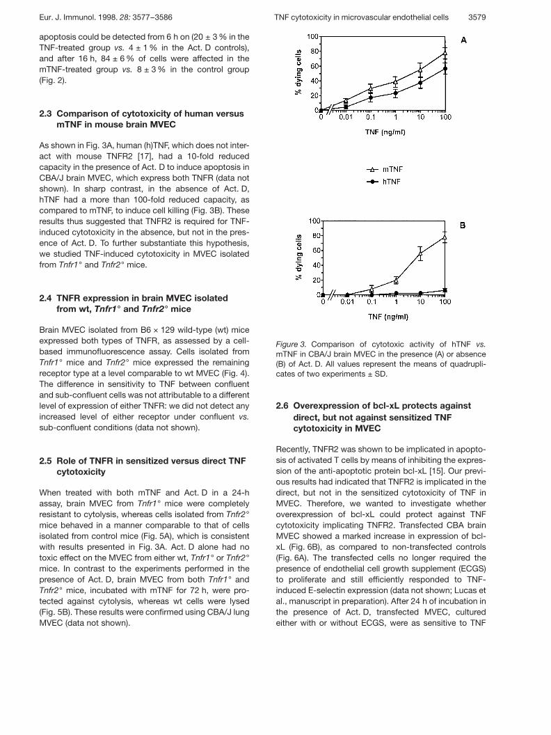
Figure 3. Comparison of cytotoxic activity of hTNF vs.mTNF in CBA/J brain MVEC in the presence (A) or absence(B) of Act. D. All values represent the means of quadrupli-cates of two experiments ± SD.
apoptosis could be detected from 6 h on (20 ± 3 % in theTNF-treated group vs. 4 ± 1 % in the Act. D controls),and after 16 h, 84 ± 6 % of cells were affected in themTNF-treated group vs. 8 ± 3 % in the control group(Fig. 2).
2.3 Comparison of cytotoxicity of human versusmTNF in mouse brain MVEC
As shown in Fig. 3A, human (h)TNF, which does not inter-act with mouse TNFR2 [17], had a 10-fold reducedcapacity in the presence of Act. D to induce apoptosis inCBA/J brain MVEC, which express both TNFR (data notshown). In sharp contrast, in the absence of Act. D,hTNF had a more than 100-fold reduced capacity, ascompared to mTNF, to induce cell killing (Fig. 3B). Theseresults thus suggested that TNFR2 is required for TNF-induced cytotoxicity in the absence, but not in the pres-ence of Act. D. To further substantiate this hypothesis,we studied TNF-induced cytotoxicity in MVEC isolatedfrom Tnfr1° and Tnfr2° mice.
2.4 TNFR expression in brain MVEC isolatedfrom wt, Tnfr1° and Tnfr2° mice
Brain MVEC isolated from B6 × 129 wild-type (wt) miceexpressed both types of TNFR, as assessed by a cell-based immunofluorescence assay. Cells isolated fromTnfr1° mice and Tnfr2° mice expressed the remainingreceptor type at a level comparable to wt MVEC (Fig. 4).The difference in sensitivity to TNF between confluentand sub-confluent cells was not attributable to a differentlevel of expression of either TNFR: we did not detect anyincreased level of either receptor under confluent vs.sub-confluent conditions (data not shown).
2.5 Role of TNFR in sensitized versus direct TNFcytotoxicity
When treated with both mTNF and Act. D in a 24-hassay, brain MVEC from Tnfr1° mice were completelyresistant to cytolysis, whereas cells isolated from Tnfr2°mice behaved in a manner comparable to that of cellsisolated from control mice (Fig. 5A), which is consistentwith results presented in Fig. 3A. Act. D alone had notoxic effect on the MVEC from either wt, Tnfr1° or Tnfr2°mice. In contrast to the experiments performed in thepresence of Act. D, brain MVEC from both Tnfr1° andTnfr2° mice, incubated with mTNF for 72 h, were pro-tected against cytolysis, whereas wt cells were lysed(Fig. 5B). These results were confirmed using CBA/J lungMVEC (data not shown).
2.6 Overexpression of bcl-xL protects againstdirect, but not against sensitized TNFcytotoxicity in MVEC
Recently, TNFR2 was shown to be implicated in apopto-sis of activated T cells by means of inhibiting the expres-sion of the anti-apoptotic protein bcl-xL [15]. Our previ-ous results had indicated that TNFR2 is implicated in thedirect, but not in the sensitized cytotoxicity of TNF inMVEC. Therefore, we wanted to investigate whetheroverexpression of bcl-xL could protect against TNFcytotoxicity implicating TNFR2. Transfected CBA brainMVEC showed a marked increase in expression of bcl-xL (Fig. 6B), as compared to non-transfected controls(Fig. 6A). The transfected cells no longer required thepresence of endothelial cell growth supplement (ECGS)to proliferate and still efficiently responded to TNF-induced E-selectin expression (data not shown; Lucas etal., manuscript in preparation). After 24 h of incubation inthe presence of Act. D, transfected MVEC, culturedeither with or without ECGS, were as sensitive to TNF
Eur. J. Immunol. 1998. 28: 3577–3586 TNF cytotoxicity in microvascular endothelial cells 3579

Figure 4. Expression levels of TNFR1 and TNFR2 in MVECisolated from wt B6 × 129, Tnfr1° and Tnfr2° mice. The val-ues represent the means of quadruplicates ± SD.
Figure 5. Comparison of sensitivity of brain MVEC isolatedfrom wt B6 × 129 mice vs. Tnfr1° and Tnfr2° mice towards:(A) the combined lytic effect of mTNF and Act. D after 24 hof incubation (3 × 10⁴ cells/well), and (B) the direct lyticeffect of mTNF after 72 h of incubation (5 × 10⁴ cells/well).All values represent the means of quadruplicates of threeexperiments ± SD.
Figure 6. Expression levels of bcl-xL in (A) wt brain CBA/JMVEC and (B) pSFFV bcl-xL-transfected CBA/J brainMVEC.
cytotoxicity as the non-transfected cells (Fig. 7A). Insharp contrast, after 72 h of incubation, confluent mono-layers of bcl-xL-transfected MVEC were resistant todirect TNF cytotoxicity, whereas the control cells or thecells microinjected with the control vector were effi-ciently lysed (Fig. 7B).
3 Discussion
In vivo, both TNFR appear to be involved in the systemictoxicity of TNF. Both Tnfr1° [11, 18, 19] and Tnfr2° mice[12] were shown to be resistant to lethal doses of mTNF,with resistance being more marked in the former [18].Apart from being directly cytotoxic, TNF can indirectlycause tissue damage, e.g. by means of inducing adhe-sion molecules, such as ICAM-1, that increases the inter-action between the endothelium and leukocytes [20, 21]or platelets [22]. Moreover, the decreased activation of
3580 R. Lucas et al. Eur. J. Immunol. 1998. 28: 3577–3586

Figure 7. Comparison of sensitivity to (A) Act. D-sensitizedand (B) direct mTNF cytotoxicity between wt, control vector-microinjected and bcl-xL-overexpressing CBA/J brainMVEC. All values represent the means of triplicates of threeexperiments ± SD.
the integrin § v g 3, an adhesion molecule which plays akey role in tumor angiogenesis, by the combined treat-ment of human endothelial cells with TNF and IFN- + , wasrecently proposed to be one of the mechanisms impli-cated in the anti-vascular activity of these cytokines [23].
Here, we have addressed the direct in vitro toxicity ofTNF on primary endothelial cells, which represent one ofthe major target cells relevant for TNF pathology in vivo.Most published studies have made use of LVEC that areboth morphologically and functionally different fromMVEC [24–26]. In this report, we have shown that mouseMVEC, which are more affected than LVEC during TNFinfusion in vivo [6], can also be killed by mTNF in vitro,provided they are at confluence; following a 72-h incuba-tion with mTNF under these conditions, the cellsundergo apoptosis. However, under sub-confluent con-ditions, MVEC are not lysed directly by TNF. The latter
result is in accordance with earlier findings of experi-ments performed with bovine lung MVEC [7]. The reasonfor this confluence-dependent sensitivity of MVECtowards TNF remains unclear. We could not find signifi-cant changes in TNFR expression between confluentand sub-confluent cultures (data not shown), as wasreported for epithelial and myeloid cells [27]. Neithercould we find a role for membrane-bound TNF, whichshould only be active during cell-cell contact and wasshown to be induced by exogenously added TNF inHeLa cells [28]. In this respect, we could not inhibit cyto-toxicity induced by hTNF in murine MVEC with neutraliz-ing antibodies towards membrane-bound mTNF (kindgift from M. Grell and K. Pfizenmaier, University of Stut-tgart; data not shown), arguing against a role for endoge-nously produced membrane-bound mTNF in cytotoxicityin these cells.
Since the pretreatment of various cell types with Act. Dcan either induce or increase sensitivity to TNF, resultsobtained in the presence of transcriptional inhibitors donot necessarily reflect the in vivo effect of TNF on thesecells. We found that in the presence of Act. D, TNFR1was necessary and sufficient to mediate TNF-inducedapoptosis in MVEC. This result is in agreement with thereported resistance of hepatocytes isolated from Tnfr1°mice against cytotoxicity induced by the combination ofTNF and Act. D [16]. However, in contrast to confluentMVEC, TNF had no direct cytotoxic effect on hepato-cytes. Moreover, the combined effect of TNF and Act. Ddisappeared after several in vitro passages of hepato-cytes [16]. These results differ from our observationswith MVEC, which are directly killed by TNF at conflu-ence and the sensitivity of which to TNF and Act. D doesnot decrease upon serial passaging (tested for up to fourpassages). The observation that MVEC isolated fromTnfr2° mice are still efficiently lysed by a combined TNFand Act. D treatment also indicates that TNFR1, stillexpressed in these cells, is biologically active. The sameholds true for TNFR2 in Tnfr1° MVEC, which still medi-ates up-regulation of ICAM-1 induced by membrane-bound TNF [29].
In contrast to the results obtained under conditions oftranscriptional inhibition, both TNFR1 and TNFR2 arerequired for direct TNF-induced MVEC apoptosis after72 h. This result is in agreement with a previous reportthat showed that both TNFR types are necessary toinduce DNA fragmentation in U-937 cells and cytotoxic-ity in PC 60 cells [30, 31]. Thus, depending on the pres-ence or absence of transcriptional inhibitors, differentpathways could be implicated in the TNF-induced killingof MVEC. Likewise, others have provided evidence thatdirect TNF-mediated cytotoxicity is genetically, pharma-cologically, and temporally distinct from the cytotoxicity
Eur. J. Immunol. 1998. 28: 3577–3586 TNF cytotoxicity in microvascular endothelial cells 3581

mediated by TNF during protein synthesis inhibition [32].Recently, TNF was shown to trigger apoptosis in CHX-treated human endothelial cells by means of anIL-1-converting enzyme (ICE)-like protease-dependentpathway, since this activity could be blocked by trans-fected CrmA protein or Z-VAD.fmk peptide, but not bytransfected anti-apoptotic proteins such as bcl-2, bcl-xLor A1 [33]. In contrast, the co-treatment of these cellswith TNF and C6-ceramide resulted in apoptosis inhibit-able by transfected anti-apoptotic proteins but not byICE-like protease inhibitors [33]. Since most studiesassessing the cytotoxicity of TNF in endothelial cellshave been performed in the presence of transcriptionalor translational inhibitors, results from these experimentsshould be interpreted with caution, since a role forTNFR2 in cytotoxicity of endothelial cells would not bedetected under these conditions.
Since TNFR2, in contrast to TNFR1, does not have adeath domain [13, 14], its implication in apoptosis israther surprising. However, a recent report showed thatTNFR2 activation in activated T cells leads to a dimin-ished expression of the anti-apoptotic protein bcl-xL inthese cells [15]. Bcl-xL was recently shown to inhibit theassociation of the mammalian homologue of CED-4(Apaf-1) and caspase 9, thus preventing the Apaf-1-mediated maturation of caspase-9 [34]. Our experimentswith bcl-xL-overexpressing brain MVEC have indicatedthat these cells are still sensitive to TNF-mediated effectswhich are exclusively mediated by TNFR1, such as E-selectin induction (completely absent in MVEC fromTnfr1° mice) and Act. D combined cytotoxicity. The latterresult is in contrast to recent findings that MVEC overex-pressing the anti-apoptotic protein A1, of the bcl-2 pro-tein family, are resistant to the combined mTNF andAct. D cytotoxicity [35]. Interestingly, bcl-xL-overexpressing MVEC were resistant to effects involvingTNFR2, such as direct TNF cytotoxicity. Therefore, theseresults suggest the existence of a cross-talk betweenTNFR2 and bcl-xL in the absence of transcriptional inhi-bition: one factor, upon activation (TNFR2) or overex-pression (bcl-xL), inhibits the effect of the other. Thus,the interaction between TNFR2 and bcl-xL might be oneof the mechanisms by which this TNFR type is impli-cated in TNF cytotoxicity in MVEC. Although the basalexpression levels of anti-apoptotic proteins such as bcl-2 and bcl-xL are consistently low or undetectable inendothelial cells, these levels can be significantlyincreased during viral infection of these cells. Indeed, theexpression of bcl-2 has recently been shown to be acti-vated in endothelial cells chronically expressing thehuman T cell lymphotropic virus type I, and this might bepathophysiologically relevant [36]. Alternatively, TNFR2was proposed to increase the TNFR1-mediated apopto-sis of HeLa cells by up-regulating cytosolic phospholi-
pase A2 levels [37]. Moreover, a recent study has shownthat the absence of a TNFR2-associated protein TRAF2can decrease the sensitivity of HeLa cells for TNF-induced killing by a factor of 1000. Since the TNFR2-associated signal transducer TRAF2 is a component ofthe TNFR1 signaling complex [38], these results indicatecross-talk between both TNFR for TNF-mediated cyto-toxicity in these cells [28]. Since TNFR2, but not TNFR1expression, is increased in lung MVEC in patients withacute respiratory distress syndrome [39], in hepatocytesfrom patients with chronic hepatitis B [40] and in brainMVEC from mice with cerebral malaria [20, 29], this indi-cates that this TNFR type, in addition to TNFR1, mightalso play an important role in TNF-mediated pathology.
In conclusion, our results show that proliferating MVECare not killed by TNF alone, unless they are treated withtranscriptional inhibitors. This combined TNF and Act. Dtoxicity is mediated by TNFR1. In contrast, in confluentmonolayers of MVEC, TNF directly induces apoptosis,which is inhibited by either TNFR1 or TNFR2 deficiency.These results suggest that in physiologically relevantconditions, both TNFR are important for the direct cyto-toxic effect of TNF on MVEC.
4 Materials and methods
4.1 Mice and cells
Male wt B6 × 129, as well as Tnfr1° [11] and Tnfr2° mice [12]of an age of 8–10 weeks were provided by F. Hoffmann-LaRoche (Basel, Switzerland). Male CBA/J mice of the sameage were purchased from Iffa Credo, Les Oncins, France.
Mouse brain MVEC were isolated as described [41], usingmagnetic beads (Dynabeads M-450, Dynal, Oslo, Norway),covalently bound to a purified rat-anti-mouse platelet-endothelial cell adhesion molecule (PECAM-1) mAb (kindgift from Dr. B. Imhof, Geneva). The cells were characterizedfor the presence of tight junctions, von Willebrand Factorexpression, acetylated low density lipoprotein uptake andTNF-induced E-selectin expression (the latter being nega-tive in cells lacking TNFR1). The cells were plated onto T25flasks pre-coated with 2 % gelatin (Sigma, Buchs, Switzer-land) and cultured at 37 °C in a 5 % CO2 atmosphere. MVECwere passaged up to four times.
4.2 Buffers and reagents
MVEC were resuspended in DMEM containing 2 mM L-glutamine, 100 U/ml penicillin, 10 ? g/ml streptomycin, 20 %FCS, 40 U/ml heparin and 100 ? g/ml ECGS (Sigma).Recombinant mTNF and hTNF (Innogenetics, Ghent, Bel-gium) had a specific activity of 2 × 10⁸ and 2 × 10⁷ IU/mg,
3582 R. Lucas et al. Eur. J. Immunol. 1998. 28: 3577–3586

respectively. Act. D, CHX and acridine orange were obtainedfrom Sigma, Buchs, Switzerland.
4.3 TNF cytotoxicity assay
An ethidium homodimer-1 incorporation assay was used toscreen for lysis of endothelial cells. Ethidium homodimer-1(Molecular Probes, Leiden, The Netherlands) is a high-affinity red fluorescent DNA dye that is only internalizedthrough altered cell membranes. This assay has recentlybeen shown to be more sensitive than the MTT incorpora-tion assay when screening for TNF cytotoxicity [42] and haspreviously been used to screen for TNF-induced killing ofMVEC [43]. Briefly, MVEC (3 × 10⁵ cells/ml) kept in DMEMwithout phenol red (phenol red is a quencher of the ethidiumhomodimer signal) were incubated for 24 h in the presenceof various concentrations of mTNF or hTNF and 1 ? g/mlAct. D. Alternatively, confluent monolayers of MVEC (5 × 10⁵
cells/ml) were treated for 72 h with various concentrations ofmTNF or hTNF. Subsequently, the MVEC were treated for30 min at 37 °C with 8 mM ethidium homodimer solution. Ascontrols, cells pretreated for 30 min at room temperaturewith 33 % ethanol (positive control) as well as cells incu-bated in medium (negative control) were included. The fluo-rescence signal was then read using 530 nm as excitationwavelength and 620 nm as emission wavelength in aCytoFluor® II fluorescence Multi-Well Plate Reader (PerSept-ive Biosystems, MA), after which the percentage of dyingcells was calculated, using the following formula: [(experi-mental signal) − (medium signal)]/[(ethanol signal) − (mediumsignal)] × 100 %.
4.4 Apoptosis detection
Quantification of apoptotic cells was accomplished by acri-dine orange uptake as described [44, 45]. Cells were incu-bated with 1 ? g/ml acridine orange for 5 min and fixed in4 % paraformaldehyde. Cellular morphology was subse-quently assessed by fluorescence microscopy [44, 45].Apoptosis was identified by the findings of condensation ofchromatin and blebbing of the cytoplasm [45]. A total of fiverandom microscope fields, each containing about 100 cells,were examined under each experimental condition. The totalnumber of cells with histomorphological evidence of apop-tosis was counted in each field.
4.5 Microinjection of DNA into MVEC
Construction of plasmids to express a FLAG tag attached tobcl-xL protein has been described [46]. MVEC were culturedin gelatin-precoated 3.5-cm diameter dishes for 2 days.Rectangles (10 by 3 mm) containing 150 to 200 mouse brainMVEC were drawn on the bottom of each dish to allow local-ization of injected cells. pSFFV-bcl-xL-plasmid or the emptypSFFV-plasmid solution at 100 ng/ml were injected into
MVEC nuclei with a mechanical Leitz manipulator and anINJECT + MATIC microinjector [46]. After 10 days in culturein the presence of medium with ECGS and 100 ? g/ml ge-neticin, the percentage of microinjected MVEC expressingFLAG-bcl-xL was determined by immunohistochemistry,using M2, an mAb specific for FLAG (International Biotech-nologies), followed by FITC-conjugated rat-anti-mouse IgG.Greater than 80 % of the MVEC microinjected were posi-tively stained. Cells microinjected with the control vectorsurvived the antibiotic treatment, while control cells were allkilled after 10 days in the presence of 100 ? g/ml geneticin.
4.6 Cell-based ELISA
MVEC cells (3 × 10⁵ cells/ml) were left to adhere for 12 h in96-well plates (Falcon) in DMEM + 20 % FCS. The plate wasthen washed once with HBSS, 20 mM Hepes ( j washingbuffer), after which the cells were fixed for 45 min at roomtemperature with DMEM + 1 % paraformaldehyde. After onewashing step, the cells were incubated for 45 min at roomtemperature in blocking solution (PBS + 5 % BSA + 0.02 %NaN3).
Subsequently, the wells were aspirated and 30 ? l of either apolyclonal rabbit-anti-mTNFR1 or a rabbit-anti-mTNFR2[47] was added for 45 min at room temperature. To assessnon-specific binding, the reactivity of rabbit-anti-mTNFR1with MVEC from Tnfr1° and of rabbit-anti-mTNFR2 withMVEC from Tnfr2° was measured. After two washing steps,50 ? l/well of a goat anti-rabbit-alkaline phosphatase com-plex (Biogenex, 1 ? g/ml) was added for 75 min undersmooth agitation at room temperature. Finally, after twowashing steps with washing buffer and one washing stepwith 2.5 M diethanolamine buffer, pH 9.5, 100 ? l/well of thesubstrate solution, consisting of the substrate AttophosTM
(Europa Research Products, Ely, GB) (final concentration1 mM) was diluted in 2.5 M diethanolamine buffer, pH 9.5, towhich levamisole (an inhibitor of endogenous alkaline phos-phatase, final concentration 10 mM) was added. After 2 minof incubation, the plate was read at 485 nm excitation wave-length and 530 nm emission wavelength in a Cytofluor® IIfluorescence multi-well plate reader (PerSeptive Biosys-tems).
Acknowledgments: We thank Dr. H. Bluethmann (F.Hoffmann-La Roche, Switzerland) for the Tnfr1° and theTnfr2° mice and Dr. K. Pfizenmaier and Dr. M. Grell (Univer-sity of Stuttgart, Germany) for their generous gift of anti-bodies towards membrane-bound mTNF. We also thankMrs. Laperrousaz for the photograph mounting. This workwas supported by grants from the Swiss National ScienceFoundation (grant 3200-043583.95 to G. E. G., grant 0031-43364.95 to M. S. P., and grant 3100-042275.94 and 3234-041729.94 to I. G.) and by the Ligue Genevoise contre leCancer (to G. E. G.).
Eur. J. Immunol. 1998. 28: 3577–3586 TNF cytotoxicity in microvascular endothelial cells 3583
5 References
1 Carswell, E. A., Old, L. J., Kassel, R. L., Green, S.,Fiore, N. and Williamson, B., An endotoxin-inducedserum factor that causes necrosis of tumors. Proc. Natl.Acad. Sci. USA 1975. 72: 3666–3670.
2 Renard, N., Lienard, D., Lespagnard, L., Eggermont,A., Heimann, R. and Lejeune, F., Early endotheliumactivation and polymorphonuclear cell invasion precedespecific necrosis of human melanoma and sarcomatreated by intravascular high-dose tumour necrosis fac-tor alpha. Int. J. Cancer 1994. 57: 656–663.
3 Eggermont, A. M. M., Koops, H. S., Klausner, J. M.,Kroon, B. B. R., Schlag, P. M., Lienard, D., Vangeel, A.N., Hoekstra, H. J., Meller, I., Nieweg, O. E., Kettel-hack, C., Benari, G., Pector, J. C. ad Lejeune, F. J.,Isolated limb perfusion with tumor necrosis factor andmelphalan for limb salvage in 186 patients with locallyadvanced soft tissue extremity sarcomas: The cumula-tive multicenter European experience. Ann. Surg. 1996.224: 756–764.
4 Tracey, K. J., The acute and chronic pathophysiologiceffects of TNF-mediation of septic shock and wasting. InBeutler, B. (Ed.) Tumor Necrosis Factors; the moleculesand their emerging role in medicine. Raven Press,New York 1992, pp 255–283.
5 Grau, G. E., Fajardo, L. F., Piguet, P. F., Allet, B., Lam-bert, P. H. and Vassalli, P., Tumor necrosis factor as anessential mediator in murine cerebral malaria. Science1987. 237: 1210–1212.
6 Talmadge, J. E., Bowersox, O., Tribble, H., Lee, S. H.,Shepard, H. M. and Liggit, D., Toxicity of tumor necro-sis factor is synergistic with gamma-interferon and canbe reduced with cyclooxygenase inhibitors. Am. J.Pathol. 1987. 128: 410–425.
7 Meyrick, B., Christman, B. and Jesmok, G., Effects oftumor necrosis factor alpha on cultured pulmonaryartery and lung microvascular endothelial monolayers.Am. J. Pathol. 1991. 138: 93–101.
8 Robaye, B., Mosselmans, R., Fiers, W., Dumont, J. E.and Galand, P., Tumor necrosis factor induces apopto-sis in normal endothelial cells in vitro. Am. J. Pathol.1991. 138: 447–453.
9 Polunovsky, V. A., Wendt, C. H., Ingbar, D. H.,Peterson, M. S. and Bitterman, P. B., Induction ofendothelial cell apoptosis by TNF alpha: modulation byinhibitors of protein synthesis. Exp. Cell Res. 1994. 214:584–594.
10 Spyridopoulos, I., Brogi, E., Kearny, M., Sullivan, A.B., Cetrulo, C., Isner, J. M. and Losordo, D. W., Vascu-lar endothelial growth factor inhibits endothelial cellapoptosis induced by tumor necrosis factor- § : balance
between growth and death signals. J. Mol. Cell Cardiol.1997. 29: 1321–1330.
11 Rothe, J., Lesslauer, W., Loetscher, H., Lang, Y., Koe-bel,, P., Kontgen, F., Althage, A., Zinkernagel, R.,Steinmetz, M. and Bluethmann, H., Mice lacking theTumour Necrosis Factor Receptor-1 are resistant toTNF-mediated toxicity, but highly susceptible to infec-tion by Listeria monocytogenes. Nature 1993. 364:798–802.
12 Erickson, S. L., Desauvage, F. J., Kikly, K., Carver-moore, K., Pittsmeek, S., Gillett, N., Sheehan, K. C. F.,Schreiber, R. D., Goeddel, D. V. and Moore, M. W.,Decreased sensitivity to tumour necrosis factor but nor-mal T-cell development in TNF receptor-2-deficientmice. Nature 1994. 372: 560–563.
13 Tartaglia, L. A., Ayres, T. M., Wong, G. H. W. andGoeddel, D. V., A novel death domain within the 55 kDaTNF receptor signals cell death. Cell 1993. 74: 845–853.
14 Varfolomeev, E. E., Boldin, M. P., Goncharov, T. M.and Wallach, D., A potential mechanism of cross-talkbetween the p55 tumor necrosis factor receptor andFas/APO1: Proteins binding to the death domains of thetwo receptors also bind to each other. J. Exp. Med.1996. 183: 1271–1275.
15 Lin, R. H., Hwang, Y. W., Yang, B. C. and Lin, C. S.,TNF receptor-2-triggered apoptosis is associated withthe down-regulation of bcl-xL on activated T cells andcan be prevented by CD28 costimulation. J. Immunol.1997. 158: 598–603.
16 Leist, M., Gantner, F., Jilg, S. and Wendel, A., Activa-tion of the 55 kDa TNF receptor is necessary and suffi-cient for TNF-induced liver failure, hepatocyte apoptosisand nitrite release. J. Immunol. 1995. 154: 1307–1316.
17 Lewis, M., Tartaglia, L. A., Lee, A., Bennett, G. L.,Rice, G. C., Wong, G. H. W., Chen, E. Y. and Goedell,D. V., Cloning and expression of cDNA for 2 distinctmurine Tumor Necrosis factors demonstrate one recep-tor is species-specific. Proc. Natl. Acad. Sci. USA 1991.88: 2830–2834.
18 Vandenabeele, P., Declercq, W., Beyaert, R. and Fiers,W., Two tumour necrosis factor receptors: Structure andfunction. Trends Cell Biol. 1995. 5: 392–399.
19 Pfeffer, K., Matsuyama, T., Kundig, T. M., Wakeham,A., Kishihara, K., Shahinian, A., Wiegmann, K., Oha-shi, P. S., Kronke, M. and Mak, T. W., Mice deficient forthe 55 kDa TNF receptor are resistant to endotoxicshock, yet succumb to L. monocytogenes infection. Cell1993. 73: 457–467.
20 Lucas, R., Lou, J. N., Juillard, P., Moore, M., Blueth-mann, H. and Grau, G. E., Respective role of TNFreceptors in the development of experimental cerebralmalaria. J. Neuroimmunol. 1997. 72: 143–148.
3584 R. Lucas et al. Eur. J. Immunol. 1998. 28: 3577–3586
21 Barbara, J. A., Smith, W. B., Gamble, J. R., VanOstade, X., Vandenabeele,, P., Tavernier, J., Fiers, W.,Vadas, M. A. and Lopez, A. F., Dissociation of TNFalpha cytotoxic and proinflammatory activities by p55receptor- and p75 receptor-selective TNF-alphamutants. EMBO J. 1994. 13: 843–850.
22 Lou, J. N., Donati, Y. R. A., Juillard, P., Giroud, C.,Vesin, C., Mili, N. and Grau, G. E., Platelets play animportant role in TNF-induced microvascular endothelialcell pathology. Am. J. Pathol. 1997. 151: 1397–1405.
23 Ruegg, C., Yilmaz, A., Bieler, G., Bamat, J., Chaubert,P. and Lejeune, F. J., Evidence for the involvement ofendothelial cell integrin alphaVbeta3 in the disruption ofthe tumor vasculature induced by TNF and IFN-gamma.Nature Med. 1989. 4: 408–414.
24 Grau, G. E. and Lou, J., TNF in vascular pathology:importance of platelet-endothelium interactions. Res.Immunol. 1993. 144: 355–363.
25 Scott, P. A. E. and Bicknell, R., The isolation and cul-ture of microvascular endothelium. J. Cell Sci. 1993.105: 269–273.
26 Swerlick, R. A., Lee, K. H., Wick, T. M. and Lawley, T.J., Human dermal microvascular endothelial but nothuman umbilical vein endothelial cells express CD36 invivo and in vitro. J. Immunol. 1992. 148: 78–83.
27 Pocsik, E., Mihalik, K., Aliosman, F. and Aggarwal, B.B., Cell density-dependent regulation of cell surfaceexpression of two types of human tumor necrosis factorreceptors and its effect on cellular response. J. Cell. Bio-chem. 1994. 54: 453–464.
28 Weiss, T., Grell, M., Hessabi, B., Bourteele, S., Müller,G., Scheurich, P. and Wajant, H., Enhancement of TNFreceptor p60-mediated cytotoxicity by TNF receptorp80. Requirement of the TNF Receptor-AssociatedFactor-2 binding site. J. Immunol. 1997. 158:2398–2404.
29 Lucas, R., Juillard, P., Decoster, E., Redard, M., Bur-ger, D., Donati, Y., Giroud, C., Monso-Hinard, C.,De Kesel, T., Buurman, W. A., Moore, M. W., Dayer, J.M., Fiers, W., Bluethmann, H. and Grau, G. E., Crucialrole of TNF receptor 2 and membrane-bound TNF inexperimental cerebral malaria. Eur. J. Immunol. 1997.27: 1719–1725.
30 Higuchi, M. and Aggarwal, B. B., Differential roles oftwo types of the TNF receptor in TNF-induced cytotoxic-ity, DNA fragmentation and differentiation. J. Immunol.1994. 152: 4017–4025.
31 Vandenabeele, P., Declercq, W., Vanhaesebroeck, B.,Grooten, J. and Fiers, W., Both TNF receptors arerequired for TNF-mediated induction of apoptosis inPC60 cells. J. Immunol. 1995. 154: 2904–2913.
32 Reid, T., Louie, P. and Heller, R. A., Mechanisms oftumor necrosis factor cytotoxicity and the cytotoxic sig-nals transduced by the p75 tumor necrosis factor recep-tor. Circ. Shock 1994. 44: 84–90.
33 Slowik, M. R., Min, W., Ardito, T., Karsan, A., Kashga-rian, M. and Pober, J. S., Evidence that TNF triggersapoptosis in human endothelial cells by interleukin-1-converting enzyme-like protease-dependent and -inde-pendent pathways. Lab. Invest. 1997. 77: 257–267.
34 Hu, Y., Benedict, M. A., Wu, D., Inohara, N. andNunez, G., Bcl-xL interacts with Apaf-1 and inhibitsApaf-1-dependent caspase-9 activation. Proc. Natl.Acad. Sci. USA 1998. 95: 4386–4391.
35 Karsan, A., Yee, E. and Harlan, J. M., Endothelial celldeath induced by tumor necrosis factor- § is inhibited bythe bcl-2 family member A1. J. Biol. Chem. 1996. 271:27201–27204.
36 Nicot, C., Astier-Gin, T. and Guillemain, B., Activationof Bcl-2 expression in human endothelial cells chroni-cally expressing the human T-cell lymphotropic virustype I. Virology 1997. 236: 47–53.
37 MacEwan, D. J., Elevated cPLA2 levels as a mechanismby which the p70 TNF and p75 NGF receptors enhanceapoptosis. FEBS Lett. 1996. 379: 77–81.
38 Shu, H. B., Takeuchi, M. and Goeddel, D. V., The tumornecrosis factor receptor 2 signal transducers TRAF2 andc-IAP1 are components of the tumor necrosis factorreceptor 1 signalling complex. Proc. Natl. Acad. Sci.USA 1996. 93: 13973–13978.
39 Grau, G. E., Mili, N., Lou, J. N., Morel, D. R., Ricou, B.,Lucas, R. and Suter, P. M., Phenotypic analysis of pul-monary microvascular endothelial cells from patientswith acute respiratory distress syndrome. Lab. Invest.1996. 74: 761–770.
40 Marinos, G., Naoumov, N. V., Rossol, S., Torrew, F.,Wong, P. Y., Gallati, H., Portmann, B. and Williams, R.,Tumor necrosis factor receptors in patients with chronichepatitis B virus infection. Gastroenterology 1995. 108:1453–1463.
41 Jackson, C. J., Garbett, P. K., Nissen, B. and Schrie-ber, L., Binding of human endothelium to Ulexeuropeaus-l-coated Dynabeads: application to the isola-tion of microvascular endothelium. J. Cell Science 1990.96: 257–262.
42 Levesque, M. C., Paquet, A. and Page, M., Improvedfluorescent bioassay for the detection of tumor necrosisfactor activity. J. Immunol. Methods 1995. 178: 71–76.
43 Lucas, R., Echtenacher, B., Sablon, E., Juillard, P.,Magez, S., Lou, J. N., Donati, Y. R. A., Bosman, F., VanDe Voorde, A., Fransen, L., Männel, D. N., Grau, G. E.and De Baetselier, P., Generation and characterizationof a mouse tumor necrosis factor mutant with antiperito-
Eur. J. Immunol. 1998. 28: 3577–3586 TNF cytotoxicity in microvascular endothelial cells 3585

nitis and desensitization activities comparable to thoseof the wild type but with reduced systemic toxicity.Infect. Immun. 1997. 65: 2006–2010.
44 Gregory, C. D., Div, C., Henderson, S., Smith, C. A.,Williams, G. T., Gordon, J. and Rickinson, S. B., Acti-vation of Epstein-Barr virus latent genes protect humang cells from death by apoptosis. Nature 1991. 349:
612–614.
45 Garcia, I., Martinou, I., Tsujimoto, Y. and Martinou, J.C., Prevention of programmed cell death of sympathicneurons by the bcl-2 proto-oncogene. Science 1992.258: 302–304.
46 Gonzalez-Garcia, M., Perez-Ballestero, R., Ding, L.,Duan, L., Boise, L. H., Thompson, C. B. and Nunez,G., Bcl-xL is the major bcl-x mRNA form expressed dur-ing murine development and its product localizes tomitochondria. Development 1994. 120: 3033–3042.
47 Bemelmans, M. H. A., Abramowicz, D., Douma, D. J.,Goldman, M. and Buurman, W. A., In vivo T cell activa-tion by anti-CD3 monoclonal antibody induces solubleTNF receptor release in mice – Effects of pentoxifylline,methylprednisolone, anti-TNF, and anti-IFN-gamma anti-bodies. J. Immunol. 1994. 153: 499–506.
Correspondence: Rudolf Lucas, Laboratory of IntensiveCare Research, Dept. of Internal Medicine, University Medi-cal Center, Rue Michel-Servet 1, CH-1211 Geneva 4, Swit-zerlandFax: +41 22 702 54 52e-mail: lucas — cmu.unige.ch
3586 R. Lucas et al. Eur. J. Immunol. 1998. 28: 3577–3586