brian s. hercyk and maitreyi e. das department of
TRANSCRIPT
F-BAR Cdc15 Promotes Cdc42 Activation During Cytokinesis and Cell 1
Polarization in S. pombe 2
3
Brian S. Hercyk and Maitreyi E. Das 4
Department of Biochemistry & Cellular and Molecular Biology, University of Tennessee, 5
Knoxville, TN, USA, 37996. 6
7
Genetics: Early Online, published on October 7, 2019 as 10.1534/genetics.119.302649
Copyright 2019.

Running title: Cdc15 promotes Gef1 localization 8
9
Key words: Cdc15, Cdc42, Gef1, cytokinesis, polarity 10
11
Corresponding Author: 12
Maitreyi Das 13
University of Tennessee, Knoxville 14
415, Ken and Blaire Mossman Building 15
Knoxville, TN, 37996 16
ABSTRACT 18
Cdc42, a Rho-family GTPase, is a master regulator of cell polarity. Recently, it has been 19
shown that Cdc42 also facilitates proper cytokinesis in the fission yeast 20
Schizosaccharomyces pombe. Cdc42 is activated by two partially redundant GEFs, 21
Gef1 and Scd1. Although both GEFs activate Cdc42, their deletion mutants display 22
distinct phenotypes, indicating that they are differentially regulated, by an unknown 23
mechanism. During cytokinesis, Gef1 localizes to the division site and activates Cdc42 24
to initiate ring constriction and septum ingression. Here we report that the F-BAR 25
protein Cdc15 promotes Gef1 localization to its functional sites. We show that cdc15 26
promotes Gef1 association with cortical puncta at the incipient division site to activate 27
Cdc42 during ring assembly. Moreover, cdc15 phospho-mutants phenocopy the polarity 28
phenotypes of gef1 mutants. In a hypermorphic cdc15 mutant, Gef1 localizes 29
precociously to the division site and is readily detected at the cortical patches and the 30
cell cortex. Correspondingly, the hypermorphic cdc15 mutant shows increased bipolarity 31
during interphase and precocious Cdc42 activation at the division site during 32
cytokinesis. Finally, loss of gef1 in hypermorphic cdc15 mutants abrogates the 33
increased bipolarity and precocious Cdc42 activation phenotype. We did not see any 34
change in the localization of the other GEF Scd1 in a Cdc15-dependent manner. Our 35
data indicate that Cdc15 facilitates Cdc42 activation at the division site during 36
cytokinesis at the cell cortex to promote bipolarity and this is mediated by promoting 37
Gef1 localization to these sites. 38
39

INTRODUCTION 40
The conserved Cdc42 is a master regulator of polarized cell growth in fission yeast 41
(MILLER AND JOHNSON 1994; JOHNSON 1999; ESTRAVIS et al. 2012; DAS AND VERDE 2013). 42
Recently, it has also been shown that Cdc42 has a role in cytokinesis, the final step in 43
cell division (WEI et al. 2016). Through the regulation of actin and membrane trafficking, 44
Cdc42 controls cellular processes such as growth, cell polarity, and cytokinesis (MARTIN 45
et al. 2007; HARRIS AND TEPASS 2010; ESTRAVIS et al. 2011; ESTRAVIS et al. 2012). Given 46
the complexities of these cellular processes, Cdc42 activation needs to be precisely 47
regulated in a spatiotemporal manner. A prime example of this precise regulation is the 48
oscillation of Cdc42 activation between the two cell ends during bipolar growth (DAS et 49
al. 2012; DAS AND VERDE 2013). Disrupting Cdc42 activation patterns lead to defects in 50
cell shape and cytokinesis (DAS et al. 2012; WEI et al. 2016; ONWUBIKO et al. 2019). 51
While much is known about how Cdc42 promotes actin organization and polarization, 52
the spatiotemporal manner in which regulation of Cdc42 is fine-tuned is not well 53
understood. 54
Cdc42 is activated by GEFs (guanine nucleotide exchange factors) which exchange 55
GDP for GTP, and inactivated by GAPs (GTPase activating proteins) which enhance 56
the intrinsic rate of GTP hydrolysis (BOS et al. 2007). Fission yeasts have two GEFs, 57
Scd1 and Gef1 that control polarization and cytokinesis (CHANG et al. 1994; COLL et al. 58
2003). While the double deletion of the two GEFs is not viable (COLL et al. 2003; HIROTA 59
et al. 2003), scd1Δ and gef1Δ mutants exhibit distinct phenotypes, indicating that they 60
differentially activate Cdc42. scd1Δ cells are depolarized and exhibit defects in septum 61
morphology (CHANG et al. 1994; WEI et al. 2016). In contrast, gef1Δ mutants exhibit 62
monopolar growth and a delayed onset of ring constriction (COLL et al. 2003; DAS et al. 63
2015; WEI et al. 2016; ONWUBIKO et al. 2019). This suggests that the two GEFs allow for 64
distinct Cdc42 activation patterns, which regulate different aspects of cell polarity 65
establishment and cytokinesis. It is unclear how the two Cdc42 GEFs result in distinct 66
phenotypes given they both activate the same GTPase. One potential explanation could 67
be differential regulation of these GEFs. Indeed, during cytokinesis, first Gef1 localizes 68
to the membrane proximal to the actomyosin ring where it activates Cdc42 to promote 69
timely onset of ring constriction and septum initiation (WEI et al. 2016). Next, Scd1 70
localizes to the ingressing membrane to promote proper septum maturation (WEI et al. 71
2016). 72
It is unknown what gives rise to the temporal localization pattern of the GEFs. Gef1 73
contains a BAR domain that is required for its function but not for its localization (DAS et 74
al. 2015). The N-terminal region of Gef1 is necessary and sufficient for its localization 75
(DAS et al. 2015). Phosphorylation of the N-terminal region by Orb6 kinase generates a 76
14-3-3 binding site that results in the sequestration of Gef1 in the cytoplasm (DAS et al. 77

2009; DAS et al. 2015). While it is known how Gef1 is removed from its site of action, it 78
is unclear what localizes Gef1 to these sites. 79
Here we show that Gef1 localization to its site of action is aided by the F-BAR protein 80
Cdc15. Cdc15 localizes to endocytic patches during interphase and to the division site, 81
where it scaffolds the actomyosin ring (WU et al. 2003; ARASADA AND POLLARD 2011; 82
MCDONALD et al. 2017). We report that Gef1 localizes to cortical patches at the division 83
site during ring assembly in a cdc15-dependent manner. Similarly, we find that cdc15 84
promotes Gef1 localization to the cortical patches and cell tips. We show that cdc15 85
phospho-mutants phenocopy gef1 polarity phenotypes. A hypermorphic cdc15 allele 86
shows precocious Cdc42 activation at the division site during cytokinesis and increased 87
bipolarity during interphase. Finally, we show that enhanced bipolarity and premature 88
Cdc42 activation is abrogated upon deletion of gef1 in the hypermorphic cdc15 mutant. 89
Here we show that Cdc15 regulates cell polarization by promoting Cdc42 activation 90
through the regulation of Gef1. We did not see any change in the localization of the 91
other GEF, Scd1, in a cdc15-dependent manner. Taken together our data indicates that 92
Cdc15 specifically promotes Gef1 localization to the division site and the cell cortex to 93
promote Cdc42 activation. 94
95
MATERIALS AND METHODS 96
Strains and cell culture 97
The S. pombe strains used in this study are listed in Supplemental Table S1. All strains 98
are isogenic to the original strain PN567. Cells were cultured in yeast extract (YE) 99
medium and grown exponentially at 25°C, unless specified otherwise. Standard 100
techniques were used for genetic manipulation and analysis (MORENO et al. 1991). Cells 101
were grown exponentially for at least 3 rounds of eight generations before imaging. 102
103
Microscopy 104
Cells were imaged at room temperature (23–25°C) with an Olympus IX83 microscope 105
equipped with a VTHawk two-dimensional array laser scanning confocal microscopy 106
system (Visitech International, Sunderland, UK), electron-multiplying charge-coupled 107
device digital camera (Hamamatsu, Hamamatsu City, Japan), and 100×/numerical 108
aperture 1.49 UAPO lens (Olympus, Tokyo, Japan). Images were acquired with 109
MetaMorph (Molecular Devices, Sunnyvale, CA) and analyzed by ImageJ (National 110
Institutes of Health, Bethesda, MD (SCHNEIDER et al. 2012)). For still and z-series 111
imaging, the cells were mounted directly on glass slides with a #1.5 coverslip (Fisher 112
Scientific, Waltham, MA) and imaged immediately; fresh slides were prepared every 10 113
minutes. Z-series images were acquired with a depth interval of 0.4 μm. For time-lapse 114
images, the cells were placed in 3.5-mm glass-bottom culture dishes (MatTek, Ashland, 115
MA) and overlaid with YE medium plus 0.6% agarose with 100μM ascorbic acid as an 116
antioxidant to minimize toxicity to the cell, as reported previously (FRIGAULT et al. 2009; 117
WEI et al. 2017). 118
119
Analysis of fluorescent intensity 120
Mutants expressing fluorescent proteins were harvested from mid-log phase cultures at 121
OD(595) 0.5 and imaged on slides. Depending on the mutant and the fluorophore, 16-18 122
z-planes were collected at a z-interval of 0.4µm for either or both the 488nm and 561nm 123
channels. The respective controls were grown and imaged in an identical manner. 124
ImageJ was used to generate sum projections from the z-series, and to measure the 125
fluorescence intensity of a selected region. The cytoplasmic fluorescence of the same 126
cell was subtracted to generate the normalized intensity. Mean normalized intensity was 127
calculated for each image from all measurable cells (n>5) within each field. 128
129
Statistical tests 130
Statistical tests were performed using GraphPad Prism software. When comparing two 131
samples, a student’s t-test (two-tailed, unequal variance) was used to determine 132
significance. When comparing three or more samples, one-way ANOVA was used, 133
followed by a Tukey’s multiple comparisons post-hoc test to determine individual p-134
values. 135
136
Cell staining 137
To stain the septum and cell wall, cells were stained in YE liquid with 50 μg/ml 138
Calcofluor White M2R (Sigma-Aldrich, St. Louis, MO) at room temperature. 139
140
Latrunculin A treatment 141
Cells were treated with 10 μM latrunculin A in dimethyl sulfoxide (DMSO) in YE medium 142
for 30 min before imaging. Control cells were treated with only 0.1% DMSO in YE 143
medium. 144
145
Analysis of sin and cdc12 mutants 146
plo1-25, sid2-250, and control cells were grown in YE at 25°C to OD 0.2, then shifted to 147
the restrictive temperature at 35.5°C. Slides were then prepared and imaged from the 148
cultures at 0, 1, 2, and 4 hour time points. Cells expressing cdc12ΔC-GFP were initially 149
grown in EMM (Edinburgh minimal medium) with 150µM Thiamine. Induction of 150
cdc12ΔC-GFP expression was performed as described previously (YONETANI AND 151
CHANG 2010). Briefly, cultures were harvested by low speed centrifugation, rinsed, and 152
then grown in EMM without thiamine for 18 hours prior to imaging. 153
154
Cell synchronization 155
Cells expressing Gef1-tdTomato and Cdc15-GFP or Cdc15-27A-GFP were grown in YE 156
at 25°C to OD 0.2, then treated with 10mM hydroxyurea (HU) for 4 hours. Cells were 157
harvested by low speed centrifugation and washed 3 times in fresh YE to release them 158
from S-phase arrest. Fresh slides were prepared and imaged in 30 minute intervals until 159
they entered M-phase. 160
161
Data availability statement 162
Strains are available upon request. The authors affirm that all data necessary for 163
confirming the conclusions of the article are present within the article, figures, and 164
tables. 165

RESULTS 166
Gef1 localizes to cortical puncta 167
While we have previously characterized the distinct localization pattern and phenotypes 168
of the Cdc42 GEFs Gef1 and Scd1 during cytokinesis (WEI et al. 2016), what facilitates 169
their localization to the division site at the appropriate time is unknown. Since Gef1 is 170
detectable at the membrane proximal to the assembled actomyosin ring, we posited that 171
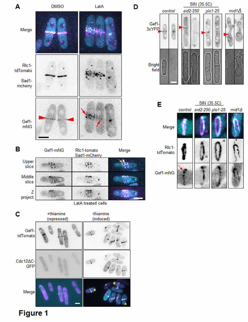
the ring is required for Gef1 localization. To test this, we treated cells with 10µM 172
Latrunculin A (LatA) for 30 min to depolymerize actin structures, then observed the 173
localization of Gef1-mNeonGreen (Gef1-mNG). Gef1-mNG localizes to the membrane 174
proximal to the actomyosin ring, marked by Rlc1-tdTomato, in mock DMSO treated cells 175
(Fig. 1A). Rlc1-tdTomato rings fragment upon treatment with LatA, as does Gef1-mNG, 176
indicating that an intact ring is necessary for proper Gef1 localization. We observe that 177
upon LatA treatment, Gef1-mNG does not diffuse away into the cytosol, but instead 178
localizes to cortical nodes about the cortex with Rlc1-tdTomato. Upon closer 179
examination of these nodes, one population of Gef1 can be seen to partially colocalize 180
with Rlc1, while the other population of Gef1 puncta do not overlap with Rlc1 (Fig. B). 181
These findings indicate that Gef1 localizes to cortical puncta near or overlapping with 182
Rlc1 containing puncta. 183
Since Gef1 promotes timely onset of ring constriction (WEI et al. 2016), we asked if Gef1 184
localization itself was under a temporal control. Given that Gef1 arrives at the division 185
site during anaphase as the actomyosin ring assembles (WEI et al. 2016), we asked 186
whether Gef1 localization is cell cycle-dependent. To test this, we induced ectopic ring 187
formation in interphase cells using the constitutively active formin mutant, cdc12ΔC-188
GFP (YONETANI AND CHANG 2010). In the presence of thiamine, cdc12ΔC-GFP 189
expression is repressed. In these conditions, Gef1-tdTomato localizes to the division 190
site of mitotic cells, which are approximately 14µm in length (Fig. 1C). However, Gef1-191
tdTomato also localizes to ectopic rings that form in cdc12ΔC-GFP expressing mono-192
nucleate interphase cells less than 10µm long (Fig. 1C). This indicates that Gef1 193
localization to the ring is not cell cycle-dependent, but rather that formation of the 194
actomyosin ring is sufficient for Gef1 localization. 195
Next, we asked what pathway localizes Gef1 to the division site. The Septation Initiation 196
Network (SIN) is a protein signaling network that coordinates the timing of cytokinesis 197
with chromosome segregation (ROBERTS-GALBRAITH AND GOULD 2008; JOHNSON et al. 198
2012; SIMANIS 2015). The SIN pathway promotes the localization and activity of proteins 199
involved in ring constriction and the coordinated process of septum formation (JIN et al. 200
2006; ROBERTS-GALBRAITH et al. 2010; BOHNERT et al. 2013). To determine whether the 201
SIN is required for Gef1 localization to the division site, we examined the localization of 202
Gef1-3YFP in two SIN protein kinase ts mutants, plo1-25 and sid2-250 (BAHLER et al. 203
1998; JIN et al. 2006; HACHET AND SIMANIS 2008). In plo1-25 and sid2-250, Gef1-3xYFP 204
localizes normally to the division site at the permissive temperature of 25°C. 205
Surprisingly, Gef1-3YFP still localizes to ring like structures in plo1-25 and sid2-250 206
cells shifted to the restrictive temperature of 35.5°C for 1, 2, or 4 hours (Fig. 1D). We 207
imaged plo1-25 and sid2-250 cells expressing Gef1-mNG and the ring marker Rlc1-208
tdTomato to better visualize the ring-like Gef1 structures and to determine whether 209
these structures represented components of the actomyosin ring. Indeed, Gef1-mNG 210
colocalizes with Rlc1-tdTomato in cells shifted to 35.5°C for 1, 2, or 4 hours, 211
demonstrating that Gef1 localization to the actomyosin ring is not dependent upon the 212
SIN pathway (Fig. 1E). Since we had observed Gef1 localization to cortical nodes, we 213
examined whether Gef1 localization was Mid1-dependent. Mid1 is an anillin-like protein 214
that is exported from the nucleus to form cortical nodes that define the division plane 215
(BAHLER et al. 1998; PAOLETTI AND CHANG 2000). It is to these nodes that various 216
contractile ring components are recruited, before coalescing to form the actomyosin ring 217
(COFFMAN et al. 2009; LAPORTE et al. 2011). In mid1Δ cells, Gef1-3xYFP localizes to 218
misplaced, extended ring-like structures (Fig.1D). Gef1-mNG and Rlc1-tdTomato 219
colocalize at these extended ring-like structures, similarly to the sin mutants (Fig.1E). 220
This demonstrates that the early node protein Mid1 is not required for the localization of 221
Gef1 to the actomyosin ring. 222
223
224
Gef1-dependent Cdc42 activation appears at the division site prior to ring 225
assembly 226
Gef1 is localized mainly in the cytoplasm and is not easily detected when present in 227
small quantities at the membrane. Gef1 is the first GEF to localize to the division site 228
and activate Cdc42 (WEI et al. 2016). To determine precisely when Gef1 localizes to the 229
division site, we carefully examined Gef1-mediated Cdc42 activity during ring assembly. 230
We monitored Cdc42 activity with the CRIB-3xGFP bio-probe that specifically binds to 231
active GTP-bound Cdc42 (TATEBE et al. 2008). In gef1+ cells, CRIB-3xGFP first appears 232
as a broad band at the division site, as it is lost from the cell tips, 8 minutes after the 233
Sad1-mCherry labelled spindle pole bodies (SBP) separate (Fig. 2A, red arrowhead, 234
2D). However, the actomyosin ring, visualized by Rlc1-tdTomato, does not fully 235
assemble for another 4 minutes (Fig. 2A, blue arrowhead). This suggests that Cdc42 is 236
activated at the membrane at the division site before the cortical nodes completely 237
condense to form the cytokinetic ring. In contrast, CRIB-3xGFP does not become active 238
at the division site until ~44 minutes after SPB separation in gef1Δ mutants (Fig. 2B, red 239
arrowhead, 2D). Thus, although Gef1 cannot be directly detected at the division site 240
during this period, our findings suggest that Gef1 specifically activates Cdc42 as the 241
ring assembles (Fig. 2B). 242
Since Gef1-mediated Cdc42 activation initiates during ring formation, we asked if a 243
protein involved in ring assembly regulates Gef1 localization to the division site. The F-244
BAR protein Cdc15 is recruited to the cortical nodes, involved in ring formation and acts 245
as a scaffold for multiple proteins involved in cytokinesis (FANKHAUSER et al. 1995; 246
CARNAHAN AND GOULD 2003; WACHTLER et al. 2006; ROBERTS-GALBRAITH et al. 2009; 247
REN et al. 2015). During cytokinesis Cdc15 is redistributed from the cell tips to the 248
division site. We find that while Cdc15 localizes to the division site ~ 4 minutes after 249
SPB separation, patches of Cdc15 remain at the polarized growth regions until ~10 250
minutes after SPB separation (Fig. 2C,E). This suggests that Cdc42 is activated at the 251
division site as Cdc15 is redistributed within the cell. Since Cdc42 activation is solely 252
Gef1-mediated during this period, we asked whether Cdc15 may promote Gef1 253
localization to the division site to activate Cdc42. A recent report indicates that Gef1 254
regulates Cdc15 distribution along the actomyosin ring (ONWUBIKO et al. 2019). It is 255
possible that Gef1 in turn depends on Cdc15 for its localization. 256
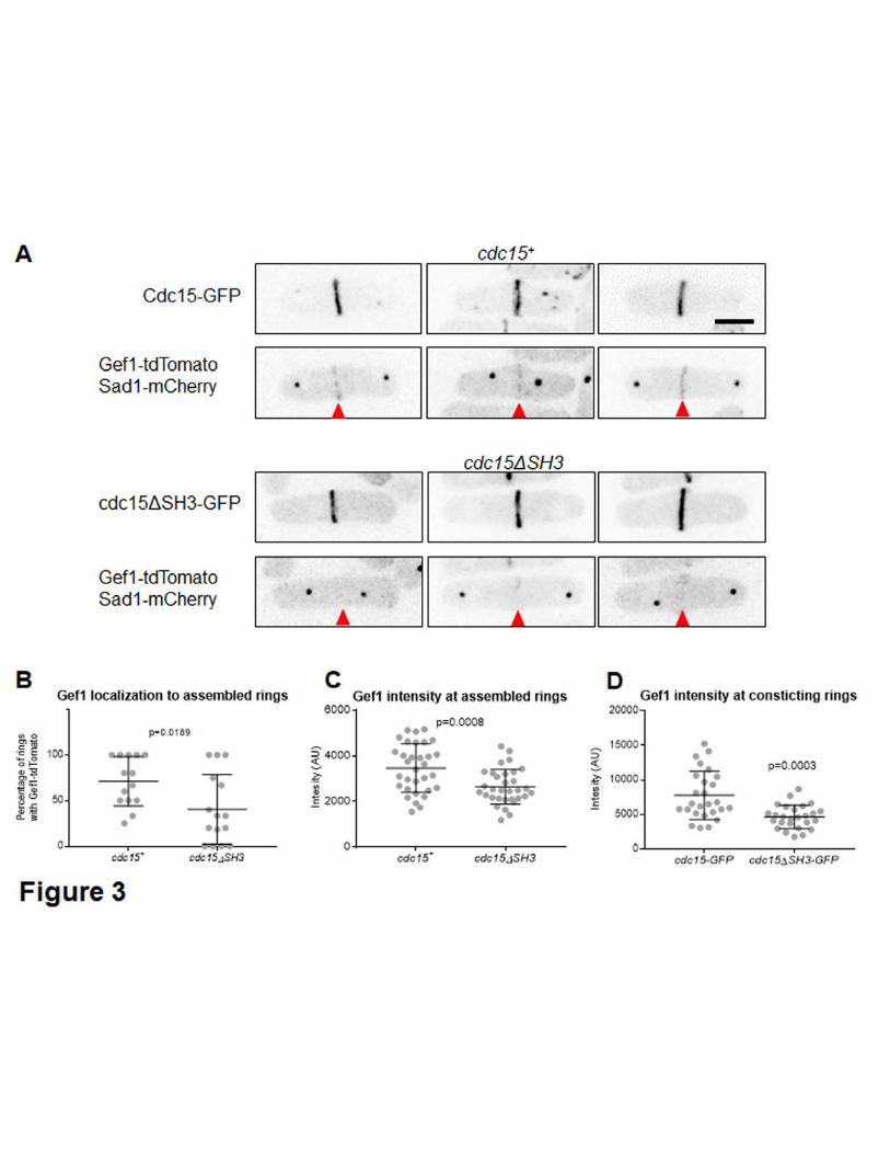
257
258

Cdc15 promotes Gef1 localization to the division site 259
Cdc15 associates with the membrane via its F-BAR domain and acts as a scaffold that 260
associates with proteins at the actomyosin ring (MCDONALD et al. 2015; REN et al. 2015; 261
MCDONALD et al. 2017). The scaffolding ability of Cdc15 is primarily conferred through 262
its C-terminal SH3 domain, through which it interacts with other proteins (ROBERTS-263
GALBRAITH et al. 2009; REN et al. 2015). While cdc15 is essential for fission yeast, a 264
cdc15ΔSH3 mutant is viable but displays defects in septum ingression and ring 265
constriction (ROBERTS-GALBRAITH et al. 2009). Similar to gef1Δ mutants, onset of ring 266
constriction and Bgs1 localization to the division site is delayed in cdc15ΔSH3 mutants 267
(ROBERTS-GALBRAITH et al. 2009; ARASADA AND POLLARD 2014; CORTES et al. 2015; WEI 268
et al. 2016). Since cdc15ΔSH3 and gef1Δ displayed similar cytokinetic defects, we 269
asked if Cdc15 promotes Gef1 localization to the division site. To test this, we examined 270
Gef1-tdTomato localization to assembled but not constricting rings , , in cells expressing 271
either Cdc15-GFP or cdc15ΔSH3-GFP. Gef1-tdTomato is present in ~70% of Cdc15-272
GFP rings, while Gef1-tdTomato is present in only ~40% of cdc15ΔSH3-GFP rings (Fig. 273
3A and Fig. 3B). Furthermore, Gef1-tdTomato fluorescent intensity is also reduced at 274
the assembled rings of the cdc15ΔSH3 mutant, with a relative intensity of only 76% that 275
of cdc15+ cells (Fig. 3A, 3C, Table 1). We find that Gef1-tdTomato localizes to the 276
division site in cells with a minimum SPB distance of 3µm in cdc15+ cells. In contrast, in 277
cdc15ΔSH3 mutants Gef1-tdTomato appears at the division site with a minimum SPB 278
distance of 7µm (Table 1). Moreover, in cdc15+ cells 61% of cells in anaphase B 279
displayed Gef1-tdTomato at the division site, while in cdc15ΔSH3 mutants only 12% of 280
anaphase B cells showed Gef1 localization (Table 1). In fission yeast, ring assembly 281
completes during anaphase B. While Gef1 localization to assembled rings is initially 282
impaired in cells expressing cdc15ΔSH3-GFP, all constricting rings have Gef1-283
tdTomato (Table 1). If Gef1 localization to the ring was solely impaired due to the 284
delayed onset of ring constriction defect exhibited by cdc15ΔSH3 mutants, Gef1 285
intensity should increase as soon as the actomyosin ring initiates constriction. However, 286
even in the constricting rings, Gef1-tdTomato levels in cdc15ΔSH3 mutants are only 287
60% that of cdc15+ cells (Fig.3D, p=0.003, Table 1). This suggests that Cdc15 likely 288
promotes Gef1 localization to the division site. 289
290

cdc15 phenocopies gef1 polarity phenotypes 291
Our data indicates a functional relationship between Gef1 and Cdc15 during 292
cytokinesis. This is further supported by the fact that cdc15∆SH3 and gef1 share a 293
common phenotype: a delay in the onset of ring constriction and Bgs1 localization at the 294
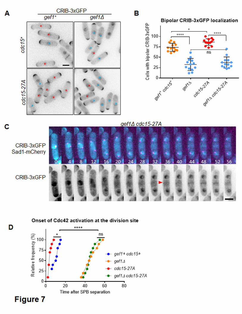
division site (ARASADA AND POLLARD 2014; CORTES et al. 2015; WEI et al. 2016). It is 295
possible that during cytokinesis Cdc15 recruits Bgs1 to the division site through Gef1. 296
gef1Δ cells are primarily monopolar, growing only from the old end (COLL et al. 2003; 297
DAS et al. 2015). In contrast, the hypermorphic allele gef1S112A exhibits precocious 298
new end growth, producing primarily bipolar cells (DAS et al. 2015). We asked if this 299
functional relationship between Gef1 and Cdc15 is specific to cytokinesis, or whether it 300
is also observed during polarized growth. Indeed, as compared to control cells, 301
cdc15∆SH3 mutants show decreased bipolarity in interphase cells, similar to gef1∆ cells 302
(Fig. 4A, 4B). Next, we asked if an increase in bipolarity was also observed in cdc15 303
mutants with increased cortical localization. When oligomerized, the F-BAR domain 304
enables Cdc15 to properly interact with the membrane (ROBERTS-GALBRAITH et al. 2010; 305
MCDONALD et al. 2015). Cdc15 is a phospho-protein where hyper-phosphorylation 306
disrupts proper oligomerization and at least in part impairs function (ROBERTS-307
GALBRAITH et al. 2010). In contrast, the de-phosphorylated form of Cdc15 shows 308
increased oligomerization and increased localization at cortical patches (ROBERTS-309
GALBRAITH et al. 2010). We find that similar to gef1∆ and cdc15∆SH3 mutants, the 310
phosphomimetic cdc15-27D allele exhibits decreased bipolarity (Fig. 4A, 4B). Further, 311
the non-phoshorylatable cdc15-27A allele is primarily bipolar, similar to gef1S112A 312
mutants (Fig. 4A, 4B). 313
To determine whether gef1 is epistatic to cdc15 we generated double mutants of gef1Δ 314
with different hypomorphic cdc15 alleles (Fig. 4C, Supplementary Fig. S1). We found 315
that gef1Δ cdc15-27D and gef1Δ cdc15ΔSH3 double mutants show a decrease in 316
bipolarity similar to that observed in gef1Δ, cdc15-27D, and cdc15ΔSH3 single mutants. 317
Moreover, the increased bipolarity in cdc15-27A mutant was reversed when combined 318
with a gef1Δ mutant (Fig. 4C, Supplementary Fig. S1). Together, this suggests that 319
these proteins functionally interact and that gef1 is epistatic to cdc15. In contrast, when 320
we analyzed the relationship between gef1S112A and cdc15 mutant alleles the 321
morphological defects were further enhanced (Supplementary Fig. S2). The gef1S112A 322
cdc15-27A double mutant, displayed an increase in aberrant morphology and 323
depolarized cells (Supplementary Fig. S2 A, B) while single gef1S112A and cdc15-27A 324
mutants displayed normal cell morphology with enhanced bipolarity (Fig. 4A-C). 325
Similarly, gef1S112A cdc15-27D and gef1S112A cdc15ΔSH3 double mutants showed 326
aberrant cell morphology defects with multiple cell poles, and large cell size 327
(Supplementary Fig. S2 A, B). The gef1S112A mutant has a mutation in the Orb6 328
kinase phosphorylation site and in the dephosphorylated form this allele does not 329

interact with the 14-3-3 binding protein Rad24 and remains localized to the plasma 330
membrane (DAS et al. 2009; DAS et al. 2015). Our observations indicate that Cdc15 331
functionally interacts with Gef1 via a mechanism distinct from the Orb6 kinase-332
gef1S112A allele mechanism. 333
334
335
cdc15-27A enhances Gef1 localization at cortical patches and division site 336
Gef1 is predominantly a cytosolic GEF during interphase, and only transiently localizes 337
to sites of polarized growth (DAS et al. 2015). Given that Gef1 localization at the division 338
site is dependent on Cdc15, we asked whether such a relationship also occurs at the 339
sites of polarized growth, as suggested by the polarity phenotypes exhibited by cdc15 340
mutants. While Cdc15-GFP is clearly visible at endocytic patches at the cell tips, Gef1-341
tdTomato is seldom observed (Fig. 5A, i). The non-phoshorylatable cdc15-27A mutants 342
tagged to GFP show increased localization at cortical patches during interphase. 343
Correspondingly, in cells expressing cdc15-27A-GFP, Gef1-tdTomato is readily 344
observed at the cell cortex (Fig. 5A, ii, iii). Moreover, we also observed that Gef1-345
tdTomato and cdc15-27A-GFP localize to the same cortical patches (Fig. 5A, iv, v, red 346
arrow). In addition to these patches, some regions of the cortex contain only Gef1-347
tdTomato or Cdc15-GFP (Fig. 5A, iv, v, green and orange arrowheads respectively). 348
Next, we asked if Cdc15 also promoted Gef1 localization to the division site. We 349
observe Gef1-mediated Cdc42 activation at the division site well before Gef1 itself is 350
detectable (Fig. 1A). Similar to a previous report, Gef1-tdTomato can be detected at the 351
division site only in rings that have completed assembly (WEI et al. 2016). We find that 352
in cdc15-27A mutants, Gef1-tdTomato localizes to the division site before the ring 353
completes assembly. Gef1-tdTomato colocalizes with cdc15-27A-GFP as the latter 354
condenses into a ring, while it is not yet detectable at this stage in cdc15+ cells (Fig. 355
5B). Since it is hard to distinguish Gef1 signal at the division site from the cytoplasmic 356
signal, it is not possible to precisely determine when Gef1 localizes to the division site 357
by time lapse microscopy. We therefore synchronized the cells in the S phase using 358
hydroxyurea treatment and then washed out the drug to allow for cell cycle progression. 359
We used Cdc15-GFP or cdc15-27A-GFP labeled actomyosin rings to determine cell 360
cycle stage. During early cytokinesis Cdc15 appears at the ring or at the precursor 361
cytokinetic nodes around the nucleus. We find that as the percentage of cells in 362
cytokinesis increases, the fraction of cells with cdc15-27A-GFP patches around the 363
nucleus containing Gef1-tdTomato also increased (Fig. 5C, D, green arrows). Thus, it is 364
possible that Gef1 localizes earlier to the division site in cdc15-27A mutants, most likely 365
to the cytokinetic nodes. 366
367
368

Cdc15 promotes Gef1-mediated Cdc42 activation 369
Given that Gef1 appears to precociously localize to nodes at the division site of cells 370
expressing cdc15-27A, we asked whether this was concomitant with precocious Cdc42 371
activation. Normally Cdc42 activity, visualized by CRIB-3xGFP, first appears at the 372
division site only after the cell initiates anaphase A (WEI et al. 2016). However, we find 373
that in cdc15-27A mutants, CRIB-3xGFP signal was visible at the medial region in 33% 374
of late G2-phase cells, prior to the separation of the Sad1-mCherry labelled SPB 375
separated (Fig. 6A, 6B). In these cells CRIB-3xGFP signal appeared as a broad band 376
that overlapped with the nucleus (Fig. 6A, red arrows). Next, we performed time lapse 377
microscopy to determine when Cdc42 was activated at the division site in cdc15-27A 378
mutants. Cdc42 is first activated ~10 minutes after SBP separation in cdc15+ cells. We 379
find that in cdc15-27A mutants, Cdc42 is activated earlier at ~4 minutes after SPB 380
separation, as determined by CRIB-3xGFP localization (Fig. 6C, red arrowhead, Fig. 381
7D). Further, similar to previous reports, in cdc15+ cells CRIB-3xGFP signal at the 382
division site appears concurrent with the loss of signal from the cell tips. We find that in 383
cdc15-27A mutants, CRIB-3xGFP signal appears at the division site well before the 384
signal is lost from the cell tips (Fig. 6C, yellow asterisk). While CRIB-3xGFP signal at 385
the cell medial region is clearly detected in cells with a single SPB by still imaging, we 386
did not detect CRIB-3xGFP signal at the division site prior to SPB separation by time 387
lapse imaging. This could be due to low abundance or photobleaching of the signal, or it 388
is possible that Cdc42 is only transiently activated at the medial region during 389
interphase in cdc15-27A cells. 390
Finally, we asked if the premature CRIB-3xGFP signal at the division site and the 391
increased bipolarity observed in cdc15-27A mutants was due to Gef1-mediated Cdc42 392
activation. To test this, we deleted gef1 in cdc15-27A mutants. We find that cdc15-27A 393
cells display an increase in bipolar CRIB-3xGFP localization at the cell tips, relative to 394
cdc15+ cells (Fig. 7A, 7B, p=0.039). This is consistent with our data indicating that 395
bipolar growth is enhanced by cdc15-27A (Fig. 4). Deletion of gef1 in cdc15-27A 396
mutants reduces bipolar CRIB-3xGFP localization, similar to that observed in gef1Δ 397
cells (Fig. 7A, B, p<0.0001). Likewise, premature Cdc42 activation at the division site in 398
cdc15-27A mutants is also abrogated in gef1Δ cdc15-27A cells. In gef1Δ cdc15-27A 399
mutants, CRIB-3xGFP did not appear at the division site until ~45minutes after SPB 400
separation, as was also observed in gef1Δ (Fig. 7C, 7D). Together, these results 401
indicate that Cdc15 promotes Gef1-mediated Cdc42 activation during cytokinesis and 402
cell polarization. 403
404

Cdc15 regulates Cdc42 activation likely independent of the other GEF Scd1 405
Next, we asked if Cdc15 also promoted Scd1-dependent Cdc42 activation. To address 406
this, we first examined the localization of the Cdc42 GEF Scd1 in cdc15-27A mutants. 407
Under normal conditions, Scd1-tdTomato appears as a cap at the cell tips during 408
interphase (KELLY AND NURSE 2011; DAS et al. 2012). We find that Scd1-tdTomato 409
localization in cdc15-27A cells does not differ from cdc15+ cells and does not localize to 410
interphase cortical patches and nodes (Fig. 8A). We did not observe any change in the 411
number of Cdc15 labelled actomyosin rings with Scd1-tdTomato (Fig. 8B). Moreover, 412
Scd1-td-tomato levels at the cell poles are similar for cdc15+ and cdc15-27A mutants 413
(Fig. 8C). This indicates that Cdc15 specifically regulates Gef1 localization during 414
cytokinesis and cell polarization but not that of Scd1. 415
Next we combined scd1Δ with cdc15 mutant alleles. Scd1 is essential for mating and 416
hence scd1Δ cells are sterile (CHANG et al. 1994; BENDEZU AND MARTIN 2013). Thus, in 417
order to generate double mutants, we transformed scd1Δ strains with a plasmid bearing 418
scd1 and mated this to cdc15 mutants. Once scd1Δ cdc15 double mutants were 419
selected we tried to remove the scd1-bearing plasmid. In scd1Δ cdc15-27A double 420
mutant cells that lost the plasmid were not viable (Fig. 8E) and appeared more 421
depolarized compared to scd1Δ mutants (Fig. 8D). A recent report indicates that in 422
scd1Δ mutants Gef1 cortical localization is enhanced and is ectopic (HERCYK et al. 423
2018). In the absence of scd1, gef1 is the only remaining GEF and scd1Δ gef1Δ double 424
mutants are inviable. It is possible that in the scd1Δ cdc15-27A mutant Gef1 localization 425
is severely impaired due to the combined effect of the two mutations thus leading to loss 426
of viability. In agreement with this, we find that cdc15-27D and cdc15ΔSH3 mutants that 427
phenocopy gef1Δ are also synthetically lethal with scd1Δ. Terminal colonies of scd1Δ 428
cdc15-27D and scd1Δ cdc15ΔSH3 without the scd1-bearing plasmid were observed 429
under the microscope. These mutants display severe morphological defects (Fig. 8E). 430
Thus, these observations indicate that Scd1 and Cdc15 function independently to 431
activate Cdc42. 432
DISCUSSION 433
The two Cdc42 GEFs, while partially redundant, show distinct phenotypes during cell 434
polarity and cytokinesis (CHANG et al. 1994; COLL et al. 2003; WEI et al. 2016). This 435
suggests that the GEFs may be regulated in different ways to precisely activate Cdc42 436
at its site of function. Although the role of the Cdc42 GEF, Gef1 in cytokinesis and cell 437
polarity is well established (DAS AND VERDE 2013; CHIOU et al. 2017), it is not clear how 438
Gef1 localizes to its site of action. Here we show that Gef1, but not the other GEF Scd1, 439
localizes to its site of action in a manner dependent on the F-BAR Cdc15. 440
While disintegration of the actomyosin ring by LatA treatment results in Gef1 localizing 441
to the cortical puncta, Gef1 does not become visible at the division site until the 442
cytokinetic nodes coalesce into the actomyosin ring (WEI et al. 2016). However, Gef1-443
dependent Cdc42 activity can be observed at the membrane overlapping the nodes a 444
few minutes before the nodes fully coalesce to form the ring. Given that Gef1 is a low 445
abundance protein, it is possible that Gef1 may be present at quantities beneath our 446
detection limit at the cortical nodes during the initial stages of ring assembly. Alternately, 447
it is possible that cytoplasmic Gef1 near the division site may activate Cdc42 at the 448
membrane overlapping the nucleus by an as yet unknown mechanism. 449
Given the timing of Cdc42 activation, Gef1 appears to be recruited late during the ring 450
assembly process. Thus, we looked at other proteins that are likewise recruited to the 451
division site during a similar time frame. It has previously been reported that the F-BAR 452
protein Cdc15 is one of the last proteins to be recruited to the cytokinetic nodes before 453
the ring assembles (WU et al. 2003). We find that Cdc15 localizes to the division site 454
shortly before Gef1-dependent Cdc42 activity initiates. Since Cdc15 serves as a 455
scaffold and promotes localization for many other proteins during cytokinesis we asked 456
if Cdc15 also promoted Gef1 localization, at the division site. We find that Gef1 457
localization is delayed in the hypomorphic cdc15ΔSH3 mutant, and Gef1 levels at the 458
division site remain low throughout constriction. Thus, our data suggests that Gef1 459
localization to the division site is cdc15 dependent. While our data indicates a 460
relationship between Gef1 and Cdc15, we did not observe any physical interaction 461
between these proteins. This suggests that Cdc15 does not promote Gef1 localization 462
by physically recruiting it to its site of action. It is possible that Cdc15 promotes Gef1 463
localization via indirect means. Cdc15 is required for endocytosis and also regulates the 464
organization of lipid-rich domains in the plasma membrane (WACHTLER et al. 2003; 465
TAKEDA et al. 2004; ARASADA AND POLLARD 2011). It is possible that Cdc15 may regulate 466
Gef1 localization via either of these processes. Indeed, in a recent report Gef1 has been 467
shown to be involved in endocytosis (ONWUBIKO et al. 2019). Further analysis will be 468
necessary to understand the molecular mechanism of Cdc15-dependent Gef1 469
localization. 470

We have previously reported that the β-1,3-glucan synthase Bgs1, the septum 471
synthesizing enzyme that drives membrane ingression, is delayed in gef1Δ cells (WEI et 472
al. 2016). A similar defect is observed in cdc15ΔSH3 mutants (ROBERTS-GALBRAITH et 473
al. 2009; CORTES et al. 2015). Given that Cdc15 promotes Gef1 localization to the 474
division site, and that cdc15ΔSH3 also exhibits the delayed onset of ring constriction, 475
characteristic of gef1Δ cells, we posited that Cdc15 acts upstream of Gef1 during 476
cytokinesis. Apart from its role in cytokinesis, Gef1 is also required for proper cell 477
polarity establishment (COLL et al. 2003). In fission yeast, immediately after division the 478
cells grow in a monopolar manner from the old end, and as the cells reach a certain 479
size, bipolarity ensues (DAS et al. 2012; DAS et al. 2015). Loss of gef1 leads to a delay 480
in initiation of bipolarity and as a result a large number of the cells in interphase are 481
monopolar (COLL et al. 2003; DAS et al. 2015). While Gef1 mainly localizes to the 482
cytoplasm, its cortical localization is enhanced in gef1S112A mutants rendering the cells 483
bipolar. We find that the gain of function cdc15-27A mutant resembles gef1S112A 484
mutants, in which the cells are predominantly bipolar. In contrast, cdc15-27D and 485
cdc15ΔSH3 mutants mimic gef1Δ mutants, in which cells are predominantly monopolar. 486
Moreover, both gain of function (cdc15-27A) and hypomorphic (cdc15ΔSH3 and cdc15-487
27D) cdc15 mutants displayed monopolarity similar to gef1Δ single mutants when 488
combined with gef1Δ. This provides further evidence that gef1 is epistatic over cdc15 489
and that the two proteins functionally interact. 490
A recent report suggests that Gef1 is primarily a cytosolic GEF, where it activates 491
Cdc42 (TAY et al. 2018). Rather our data suggest that Cdc15 recruits Gef1 to the 492
cortical patches to promote bipolar growth. During interphase Cdc15 is localized to the 493
endocytic patches where it promotes vesicle internalization (ARASADA AND POLLARD 494
2011). In the hypermorphic mutants, cdc15-27A-GFP levels are elevated at cortical 495
patches (ROBERTS-GALBRAITH et al. 2010). Correspondingly, these mutants also show 496
Gef1 localization to these patches. Moreover, Gef1 localization at the cortex is quite 497
prominent in these mutants. In agreement with increased Gef1 cortical localization, we 498
also observe increased Cdc42 activation at both cell poles resulting in increased 499
bipolarity. Gef1 cortical localization has been shown to increase under stress conditions 500
(DAS et al. 2015; TAY et al. 2018). It is possible that in cdc15-27A the cells undergo 501
stress resulting in enhanced cortical localization of Gef1. However, given that 502
hypomorphic cdc15 mutants impair Gef1 localization and Gef1-dependent Cdc42 503
activation, and that Gef1 localizes to cdc15-27A containing patches, we propose that 504
Cdc15 regulates Gef1-mediated Cdc42 activation. A recent paper demonstrates that 505
Gef1 regulates Cdc15 by controlling the size and lifetime of Cdc15 cortical patches 506
(ONWUBIKO et al. 2019). Above, we present data that demonstrate Cdc15 is upstream of 507
Gef1. These two observations are not contradictory, but rather reveal an elegant 508
regulatory pattern: Cdc15 promotes Gef1 localization to endocytic patches, where Gef1 509
in turn regulates the size of the Cdc15 patch via Cdc42 activation. Our observation that 510
Gef1-tdTomato and Cdc15-27A-GFP do not perfectly colocalize at the cortex can be 511
explained by the following model. Cdc15 initially recruits Gef1 to endocytic patches at 512
the cortex, resulting in colocalization. Once Gef1 facilitates patch internalization, Cdc15 513
is lost from the cortex while Gef1 remains for a short time. Further investigations will 514
determine how Gef1-mediated Cdc42 activity regulates Cdc15 cortical patch lifetime. 515
Given the abundance of Gef1 in the cytoplasm, small levels of Gef1 are not easily 516
detectable at cortical patches. Gef1 localization to the cortical patches and the cortex 517
may be enhanced by the increased abundance of cdc15-27A at cortical patches. Gef1 518
localization at the cell cortex is also regulated by the NDR kinase Orb6 (DAS et al. 519
2009). Orb6 kinase phosphorylates Gef1 at a serine at position 112 and this promotes 520
sequestration of Gef1 to the cytoplasm by the 14-3-3 protein Rad24 (DAS et al. 2015). 521
We find that gef1S112A mutants that constitutively localize to the membrane show 522
additive effects with cdc15 mutants. This suggests that Gef1 localization to the site of 523
action is regulated by multiple pathways. While Orb6 kinase is involved in preventing 524
Gef1 localization to the membrane, Cdc15 likely promotes its localization. The cell 525
shape defects with increased cell size observed in gef1S112A cdc15-27D and 526
gef1S112A cdc15ΔSH3 mutants suggests that in the absence of proper Cdc15 function, 527
constitutively localized gef1S112A can establish multiple growth poles. 528
Together these results indicate that Cdc15 promotes Gef1-mediated Cdc42 activity at 529
the cell poles and during cytokinesis. Cdc42 is activated by Gef1 and Scd1 and scd1Δ 530
gef1Δ double mutant is lethal (COLL et al. 2003; HIROTA et al. 2003). Our observation 531
that loss of function cdc15 mutants are synthetically lethal with deletion of the other 532
Cdc42 GEF scd1 provides further evidence that Cdc15 promotes Gef1 function. 533
The mechanistic understanding of factors that control Gef1 localization is sorely lacking. 534
Aside from the observation that the N-terminus of Gef1 is required for its localization to 535
the membrane, no other factors have been identified (DAS et al. 2015). It has also been 536
reported that Gef1 activates Cdc42 with the help of BAR Hob3 protein interaction (COLL 537
et al. 2007). Gef1 is a homolog of the mammalian GEF TUBA and contains a BAR 538
domain (DAS et al. 2015). However, previous reports show that the Gef1-BAR domain is 539
not required for its localization to the division site, nor is Hob3 required for Gef1 540
localization (Supplementary Fig. S3) (DAS et al. 2015). In contrast, the mechanism 541
removing Gef1 from the membrane has been elucidated. Gef1 is phosphorylated by 542
Orb6, generating a 14-3-3 binding site that results in Gef1 removal by Rad24 (DAS et al. 543
2009; DAS et al. 2015). Here, we identify Cdc15 as a factor that promotes Gef1 544
localization to both the cell tips and the division site. The role of Cdc15 in the processes 545
of cytokinesis and endocytosis is well established (FANKHAUSER et al. 1995; CARNAHAN 546
AND GOULD 2003; WACHTLER et al. 2006; ROBERTS-GALBRAITH et al. 2010; ARASADA AND 547
POLLARD 2011; ARASADA AND POLLARD 2014; MARTIN-GARCIA et al. 2014; REN et al. 548
2015; WILLET et al. 2015). Here we present data that reveals an additional role for 549
Cdc15 in the regulation of Cdc42 activation during cell polarization and cytokinesis. 550
Furthermore, this regulation is mediated by the specific regulation of Gef1 localization, 551
but not that of the other GEF Scd1. These studies begin to explain how, through 552
differential regulation and localization, two GEFs of the same GTPase can exhibit 553
distinct phenotypes. 554
555
556
557
ACKNOWLEDGEMENTS 558
We thank Kathleen Gould and Fred Chang for generously providing strains. This work 559
was funded by National Science Foundation, grant #1616495. 560

REFERENCES 561 Arasada, R., and T. D. Pollard, 2011 Distinct roles for F-BAR proteins Cdc15p and Bzz1p in actin 562
polymerization at sites of endocytosis in fission yeast. Curr Biol 21: 1450-1459. 563 Arasada, R., and T. D. Pollard, 2014 Contractile ring stability in S. pombe depends on F-BAR protein 564
Cdc15p and Bgs1p transport from the Golgi complex. Cell Rep 8: 1533-1544. 565 Bahler, J., A. B. Steever, S. Wheatley, Y. Wang, J. R. Pringle et al., 1998 Role of polo kinase and Mid1p in 566
determining the site of cell division in fission yeast. J Cell Biol 143: 1603-1616. 567 Bendezu, F. O., and S. G. Martin, 2013 Cdc42 explores the cell periphery for mate selection in fission 568
yeast. Curr Biol 23: 42-47. 569 Bohnert, K. A., A. P. Grzegorzewska, A. H. Willet, C. W. Vander Kooi, D. R. Kovar et al., 2013 SIN-570
dependent phosphoinhibition of formin multimerization controls fission yeast cytokinesis. 571 Genes Dev 27: 2164-2177. 572
Bos, J. L., H. Rehmann and A. Wittinghofer, 2007 GEFs and GAPs: critical elements in the control of small 573 G proteins. Cell 129: 865-877. 574
Carnahan, R. H., and K. L. Gould, 2003 The PCH family protein, Cdc15p, recruits two F-actin nucleation 575 pathways to coordinate cytokinetic actin ring formation in Schizosaccharomyces pombe. J Cell 576 Biol 162: 851-862. 577
Chang, E. C., M. Barr, Y. Wang, V. Jung, H. P. Xu et al., 1994 Cooperative interaction of S. pombe proteins 578 required for mating and morphogenesis. Cell 79: 131-141. 579
Chiou, J. G., M. K. Balasubramanian and D. J. Lew, 2017 Cell Polarity in Yeast. Annu Rev Cell Dev Biol 33: 580 77-101. 581
Coffman, V. C., A. H. Nile, I. J. Lee, H. Liu and J. Q. Wu, 2009 Roles of formin nodes and myosin motor 582 activity in Mid1p-dependent contractile-ring assembly during fission yeast cytokinesis. Mol Biol 583 Cell 20: 5195-5210. 584
Coll, P. M., S. A. Rincon, R. A. Izquierdo and P. Perez, 2007 Hob3p, the fission yeast ortholog of human 585 BIN3, localizes Cdc42p to the division site and regulates cytokinesis. The EMBO journal 26: 1865-586 1877. 587
Coll, P. M., Y. Trillo, A. Ametzazurra and P. Perez, 2003 Gef1p, a new guanine nucleotide exchange factor 588 for Cdc42p, regulates polarity in Schizosaccharomyces pombe. Molecular biology of the cell 14: 589 313-323. 590
Cortes, J. C., N. Pujol, M. Sato, M. Pinar, M. Ramos et al., 2015 Cooperation between Paxillin-like Protein 591 Pxl1 and Glucan Synthase Bgs1 Is Essential for Actomyosin Ring Stability and Septum Formation 592 in Fission Yeast. PLoS Genet 11: e1005358. 593
Das, M., T. Drake, D. J. Wiley, P. Buchwald, D. Vavylonis et al., 2012 Oscillatory Dynamics of Cdc42 594 GTPase in the Control of Polarized Growth. Science 337: 239-243. 595
Das, M., I. Nunez, M. Rodriguez, D. J. Wiley, J. Rodriguez et al., 2015 Phosphorylation-dependent 596 inhibition of Cdc42 GEF Gef1 by 14-3-3 protein Rad24 spatially regulates Cdc42 GTPase activity 597 and oscillatory dynamics during cell morphogenesis. Mol Biol Cell. 598
Das, M., and F. Verde, 2013 Role of Cdc42 dynamics in the control of fission yeast cell polarization. 599 Biochem Soc Trans 41: 1745-1749. 600
Das, M., D. J. Wiley, X. Chen, K. Shah and F. Verde, 2009 The conserved NDR kinase Orb6 controls 601 polarized cell growth by spatial regulation of the small GTPase Cdc42. Curr Biol 19: 1314-1319. 602
Estravis, M., S. Rincon and P. Perez, 2012 Cdc42 regulation of polarized traffic in fission yeast. Commun 603 Integr Biol 5: 370-373. 604
Estravis, M., S. A. Rincon, B. Santos and P. Perez, 2011 Cdc42 regulates multiple membrane traffic events 605 in fission yeast. Traffic 12: 1744-1758. 606
Fankhauser, C., A. Reymond, L. Cerutti, S. Utzig, K. Hofmann et al., 1995 The S. pombe cdc15 gene is a 607 key element in the reorganization of F-actin at mitosis. Cell 82: 435-444. 608

Frigault, M. M., J. Lacoste, J. L. Swift and C. M. Brown, 2009 Live-cell microscopy - tips and tools. J Cell Sci 609 122: 753-767. 610
Hachet, O., and V. Simanis, 2008 Mid1p/anillin and the septation initiation network orchestrate 611 contractile ring assembly for cytokinesis. Genes Dev 22: 3205-3216. 612
Harris, K. P., and U. Tepass, 2010 Cdc42 and vesicle trafficking in polarized cells. Traffic 11: 1272-1279. 613 Hercyk, B., J. Rich and M. E. Das, 2018 A novel interplay between GEFs orchestrates Cdc42 activation in 614
cell polarity and cytokinesis. bioRxiv. 615 Hirota, K., K. Tanaka, K. Ohta and M. Yamamoto, 2003 Gef1p and Scd1p, the Two GDP-GTP exchange 616
factors for Cdc42p, form a ring structure that shrinks during cytokinesis in Schizosaccharomyces 617 pombe. Mol Biol Cell 14: 3617-3627. 618
Jin, Q. W., M. Zhou, A. Bimbo, M. K. Balasubramanian and D. McCollum, 2006 A role for the septation 619 initiation network in septum assembly revealed by genetic analysis of sid2-250 suppressors. 620 Genetics 172: 2101-2112. 621
Johnson, A. E., D. McCollum and K. L. Gould, 2012 Polar opposites: Fine-tuning cytokinesis through SIN 622 asymmetry. Cytoskeleton (Hoboken) 69: 686-699. 623
Johnson, D. I., 1999 Cdc42: An essential Rho-type GTPase controlling eukaryotic cell polarity. 624 Microbiology and molecular biology reviews : MMBR 63: 54-105. 625
Kelly, F. D., and P. Nurse, 2011 Spatial control of Cdc42 activation determines cell width in fission yeast. 626 Mol Biol Cell 22: 3801-3811. 627
Laporte, D., V. C. Coffman, I. J. Lee and J. Q. Wu, 2011 Assembly and architecture of precursor nodes 628 during fission yeast cytokinesis. J Cell Biol 192: 1005-1021. 629
Martin-Garcia, R., P. M. Coll and P. Perez, 2014 F-BAR domain protein Rga7 collaborates with Cdc15 and 630 Imp2 to ensure proper cytokinesis in fission yeast. J Cell Sci 127: 4146-4158. 631
Martin, S. G., S. A. Rincon, R. Basu, P. Perez and F. Chang, 2007 Regulation of the formin for3p by cdc42p 632 and bud6p. Molecular biology of the cell 18: 4155-4167. 633
McDonald, N. A., A. L. Lind, S. E. Smith, R. Li and K. L. Gould, 2017 Nanoscale architecture of the 634 Schizosaccharomyces pombe contractile ring. Elife 6. 635
McDonald, N. A., C. W. Vander Kooi, M. D. Ohi and K. L. Gould, 2015 Oligomerization but Not Membrane 636 Bending Underlies the Function of Certain F-BAR Proteins in Cell Motility and Cytokinesis. Dev 637 Cell 35: 725-736. 638
Miller, P. J., and D. I. Johnson, 1994 Cdc42p GTPase is involved in controlling polarized cell growth in 639 Schizosaccharomyces pombe. Mol Cell Biol 14: 1075-1083. 640
Moreno, S., A. Klar and P. Nurse, 1991 Molecular genetic analysis of fission yeast Schizosaccharomyces 641 pombe. Methods in enzymology 194: 795-823. 642
Onwubiko, U. N., P. J. Mlynarczyk, B. Wei, J. Habiyaremye, A. Clack et al., 2019 A Cdc42 GEF, Gef1, 643 through endocytosis organizes F-BAR Cdc15 along the actomyosin ring and promotes concentric 644 furrowing. J Cell Sci. 645
Paoletti, A., and F. Chang, 2000 Analysis of mid1p, a protein required for placement of the cell division 646 site, reveals a link between the nucleus and the cell surface in fission yeast. Mol Biol Cell 11: 647 2757-2773. 648
Ren, L., A. H. Willet, R. H. Roberts-Galbraith, N. A. McDonald, A. Feoktistova et al., 2015 The Cdc15 and 649 Imp2 SH3 domains cooperatively scaffold a network of proteins that redundantly ensure 650 efficient cell division in fission yeast. Mol Biol Cell 26: 256-269. 651
Roberts-Galbraith, R. H., J. S. Chen, J. Wang and K. L. Gould, 2009 The SH3 domains of two PCH family 652 members cooperate in assembly of the Schizosaccharomyces pombe contractile ring. J Cell Biol 653 184: 113-127. 654
Roberts-Galbraith, R. H., and K. L. Gould, 2008 Stepping into the ring: the SIN takes on contractile ring 655 assembly. Genes Dev 22: 3082-3088. 656
Roberts-Galbraith, R. H., M. D. Ohi, B. A. Ballif, J. S. Chen, I. McLeod et al., 2010 Dephosphorylation of F-657 BAR protein Cdc15 modulates its conformation and stimulates its scaffolding activity at the cell 658 division site. Mol Cell 39: 86-99. 659
Schneider, C. A., W. S. Rasband and K. W. Eliceiri, 2012 NIH Image to ImageJ: 25 years of image analysis. 660 Nat Methods 9: 671-675. 661
Simanis, V., 2015 Pombe's thirteen - control of fission yeast cell division by the septation initiation 662 network. J Cell Sci 128: 1465-1474. 663
Takeda, T., T. Kawate and F. Chang, 2004 Organization of a sterol-rich membrane domain by cdc15p 664 during cytokinesis in fission yeast. Nat Cell Biol 6: 1142-1144. 665
Tatebe, H., K. Nakano, R. Maximo and K. Shiozaki, 2008 Pom1 DYRK regulates localization of the Rga4 666 GAP to ensure bipolar activation of Cdc42 in fission yeast. Current biology : CB 18: 322-330. 667
Tay, Y. D., M. Leda, A. B. Goryachev and K. E. Sawin, 2018 Local and global Cdc42 guanine nucleotide 668 exchange factors for fission yeast cell polarity are coordinated by microtubules and the Tea1-669 Tea4-Pom1 axis. J Cell Sci 131. 670
Wachtler, V., Y. Huang, J. Karagiannis and M. K. Balasubramanian, 2006 Cell cycle-dependent roles for 671 the FCH-domain protein Cdc15p in formation of the actomyosin ring in Schizosaccharomyces 672 pombe. Mol Biol Cell 17: 3254-3266. 673
Wachtler, V., S. Rajagopalan and M. K. Balasubramanian, 2003 Sterol-rich plasma membrane domains in 674 the fission yeast Schizosaccharomyces pombe. J Cell Sci 116: 867-874. 675
Wei, B., B. S. Hercyk, J. Habiyaremye and M. Das, 2017 Spatiotemporal Analysis of Cytokinetic Events in 676 Fission Yeast. J Vis Exp. 677
Wei, B., B. S. Hercyk, N. Mattson, A. Mohammadi, J. Rich et al., 2016 Unique Spatiotemporal Activation 678 Pattern of Cdc42 by Gef1 and Scd1 Promotes Different Events during Cytokinesis. Mol Biol Cell 679 27: 1235-1245. 680
Willet, A. H., N. A. McDonald, K. A. Bohnert, M. A. Baird, J. R. Allen et al., 2015 The F-BAR Cdc15 681 promotes contractile ring formation through the direct recruitment of the formin Cdc12. J Cell 682 Biol 208: 391-399. 683
Wu, J. Q., J. R. Kuhn, D. R. Kovar and T. D. Pollard, 2003 Spatial and temporal pathway for assembly and 684 constriction of the contractile ring in fission yeast cytokinesis. Dev Cell 5: 723-734. 685
Yonetani, A., and F. Chang, 2010 Regulation of cytokinesis by the formin cdc12p. Curr Biol 20: 561-566. 686
687

SPB distance at which Gef1 first appears at CDS
% of Anaphase B cells with Gef1 at CDS
*
% of cells with assembled rings with Gef1 at CDS
*
Relative Gef1-tdTomato intensity in assembled rings
% of cells with constricting rings with Gef1 at CDS
Relative Gef1-tdTomato intensity in constricting rings
cdc15+ 3µmN=26
61%N=26
78%N=56
1.0N=32
100%N=24
1.0N=26
cdc15ΔSH3 7µmn=26
12%n=26
48%n=37
0.76n=32
100%n=43
0.6N=26
Table 1. Characterization of Gef1 recruitment to the cell division site (CDS) in a cdc15-dependent manner
* The actomyosin ring assembles during anaphase B, hence while all cells with assembledrings are in anaphase B, not all cells in anaphase B have an assembled ring.