by lillian owen - ordiecole.com · by lillian owen fall 1998. introduction ... in 1663, robert...
TRANSCRIPT

By Lillian Owen
Fall 1998

Introduction
The microscope is a powerful tool used widely today by students, teachers,
scientists, doctors and researchers. It is used in many fields such as
medicine, physics, biology, engineering and countless others. Microscopes
are also used in industry and manufacturing. There are many types of
microscopes being used and the question arises; how did our high-powered
microscopes, such as the Scanning Probe Microscope come into being? The
history of the microscope is a fascinating story and this paper will explain
the progress and development of the microscope. Here is a list of the types
of microscopes that will be discussed.
Types of Microscopes
Optical Microscopes
• Simple Microscope
• Compound Microscope
• Ultraviolet Microscope
• Ultraviolet Fluorescent Microscope
TEM Transmission Electron Microscope
SEM Scanning Electron Microscope
STM Scanning Tunneling Microscope
AFM Atomic Force Microscope
SPM Scanning Probe Microscope

History of the Microscope 1
Terms
concave
convex
nanometer
nanolithography
nanotechnology
micron
micrometer
microstructure
photomicrograph
wavelength of light
Microscopes
Named by Giovanni Faber in 1625, the term 'microscope' originally
referred to what we know today as a telescope.2 Simple items such as a
magnifying glass or glasses are actually microscopes. A magnifying glass can
also be used as a telescope. The common feature of microscopes,
telescopes, eyeglasses and magnifying glasses is the lens. Lenses are made
of glass or plastic and can be shaped in various ways to make objects appear
closer or further away. In order to increase the size of an object, a lens
bends light rays away from each other.3
Microscopes today come in several forms. These instruments are
greatly valued for scientific investigation and research. The first
microscope invented was a light microscope, also called the optical
microscope, and was invented nearly 500 years ago. The microscope was not
immediately appreciated for the contribution it could and would eventually
make to science. The microscope has enabled us to learn more about our
world than any other instrument.4 It has enabled us to see what cannot be
History of the Microscope 2
Figure 2. 2700 year old Mesopotamian Clay Tablet with instructions for Glassmaking6
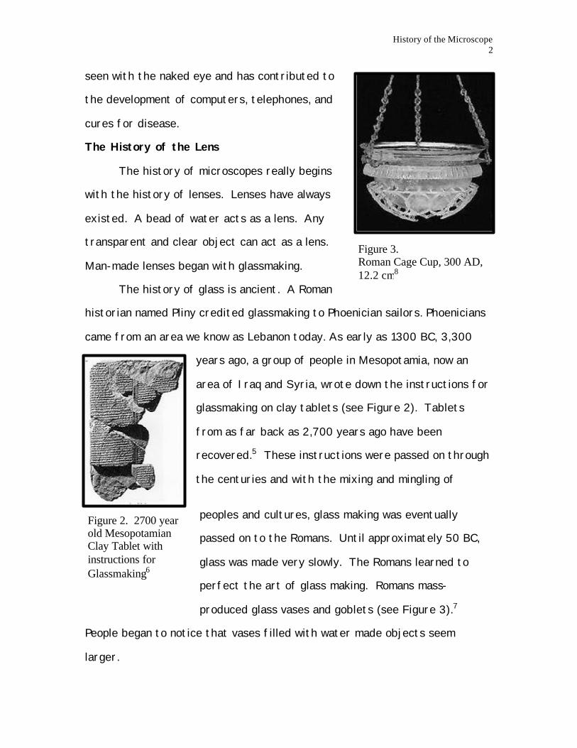
Figure 3. Roman Cage Cup, 300 AD, 12.2 cm8
seen with the naked eye and has contributed to
the development of computers, telephones, and
cures for disease.
The History of the Lens
The history of microscopes really begins
with the history of lenses. Lenses have always
existed. A bead of water acts as a lens. Any
transparent and clear object can act as a lens.
Man-made lenses began with glassmaking.
The history of glass is ancient. A Roman
historian named Pliny credited glassmaking to Phoenician sailors. Phoenicians
came from an area we know as Lebanon today. As early as 1300 BC, 3,300
years ago, a group of people in Mesopotamia, now an
area of Iraq and Syria, wrote down the instructions for
glassmaking on clay tablets (see Figure 2). Tablets
from as far back as 2,700 years ago have been
recovered.5 These instructions were passed on through
the centuries and with the mixing and mingling of
peoples and cultures, glass making was eventually
passed on to the Romans. Until approximately 50 BC,
glass was made very slowly. The Romans learned to
perfect the art of glass making. Romans mass-
produced glass vases and goblets (see Figure 3).7
People began to notice that vases filled with water made objects seem
larger.

History of the Microscope 3
Figure 4. Roger Bacon (1214-1292)11
In 1038 AD, an Arabian scholar, Alhazen, published a book called
"Opticae Thesaurus Alhazeni Arabius Basil." This book on optics described
optics of the eye, including how the eye focuses. In his book, Alhazen also
discussed ways to magnify objects by placing these objects in a dense,
spherical medium and turning the curved surface of these objects towards
the eye.9 This means that Alhazen had noticed, and had written down that
objects could be magnified.
Roger Bacon (Figure 4), a monk who lived in England
(1214-1292), studied various aspects of nature for most of
his life. He wrote a book called "Opus Majus." He wrote
about many things in his book including optics and lenses.
Bacon did experiments with lenses and found that putting
glass or crystal over letters with the convex side of the
crystal or glass towards the eye, made the letters appear
larger to him.10
By the late 13th century, spectacles, or glasses, were introduced in
Florence, Italy. Although it is not clear whom the inventor was, the use of
spectacles spread rapidly throughout Europe. The spread of glasses through
Europe undoubtedly led to the development of microscopes and telescopes.12
The Optical Microscope
Lenses are only one component of the optical microscope. Another
element is light. The most obvious source of light is the sun. Today we have
artificial light, but when microscopes were first invented, candles were used
as a light source; although, eventually sunlight was used as a light source as
well.13 Light normally travels in a straight line; although, when light

History of the Microscope 4
Figure 5. This is thought to be the first microscope, also called the Janssen Microscope17
encounters an object, it will bounce off or bend depending on the type of
object that it encounters. For example, if light hits a transparent object
such as glass, it will normally pass through the glass, bending a bit. If the
quality of the glass is poor, less light will go through, resulting in a distorted
view of the world beyond the glass.14 Optical microscopes capture this light
and use it to magnify objects.
There are really two main kinds of optical microscopes: simple and
compound. The simple microscope consists of one or two lenses. The lens is
placed close to the eye to yield a better view. To focus, the length between
the lens and the object is modified. The compound microscope uses more
than one lens. The first lens in a compound microscope is called an objective
lens. This lens magnifies the object first and then another lens called ocular
lens magnifies this already magnified image once again.15
By the 1500's, the art of grinding glass into lenses was highly
developed, especially in Holland. From the development of lenses, it became
apparent that there was more to be seen in the heavens and on earth and
someone realized that combining two or more lenses resulted in greater
magnification or greater "seeing power." These combinations of lenses were
the first microscopes and telescopes.
The original inventor of the first microscope is
debatable. From records that exist, it seems that the earliest
microscopes were made in the 1590`s. The first microscope is
thought to have been invented by Hans and Sacharias Janssen
(1588-1630),16 a father and son (respectively) who ran a lens-
grinding business in Holland. A friend of Sacharias Janssen

History of the Microscope 5
Figure 7. Antoni van Leeuwenhoek2
Figure 6. Compound Microscope used by Hooke20
named Cornelius Drebbel was able to document his observations of the Janssen
microscope. Drebbel described the microscope as having 3 brass sliding tubes that
stood vertically from 3 legs. When completely extended this microscope was 18
inches long with a diameter of 2 inches. This microscope invented by the
Jannsen's was a compound microscope (see Figure 5).18
Nearly half a century passed before the
microscope received any attention. In 1663, Robert
Hooke (1635-1703), curator of exhibits at the Royal
Society of London, began giving weekly lectures on his
microscope experiments.19 He looked at various
objects including cork, needles and compound eyes of
different bugs. Micrographia, a book written by
Hooke, was published in 1665. Micrographia contained
information and drawings about the various
experiments he did with the compound microscope (see Figure 6). Hooke took
common materials and put them under the microscope to look at the
microstructure. Although Hook used a two-lens microscope design, he felt a single
lens would provide greater magnification; however, Hooke believed that this type
of microscope would be very difficult to use. He was correct. The other
microscope that Hooke described in his book is called the simple
microscope. The distance between the object in the lens has to be
very short in order to gain a high magnification.21
Antoni van Leeuwenhoek (1632-1723), a Dutch cloth merchant
and a naturalist, was inspired by Micrographia and built over 500
microscopes during his life of the variety that Hooke theorized

History of the Microscope 6
Figure 9. George Adam's Universal Microscope28
Figure 8. Leeuwenhoek's Microscope24
about. Leeuwenhoek was able to overcome the obstacle of
needing a very short distance between the object and lens.
The Leeuwenhoek microscopes were very small, about 3 to
4 inches long or the size of the average human thumb (see
Figure 8), with a sharp point where the specimen would
attach. The single lens was mounted in a metal plate and
could be adjusted by the screws. This microscope enabled
Leeuwenhoek to see things that people did not even know
existed. For example, he saw bacteria.23 Like Hooke,
Leeuwenhoek wrote down what he saw and drew
descriptions. In addition to bacteria, he described
spermatozoa and red blood cells. The microscopes could
obtain a resolution of 1.4 micrometers or a magnification of about 300 times.25 A
human hair is approximately 50 micrometers wide. So Leeuwenhoek was able to
view samples that were about 1/35th the size of the width of a human hair!
The earliest microscopy devices were made from simple
materials such as cardboard tubing, which often held the lenses. In
1744, John Cuff made a brass microscope with a rotating nosepiece
that had 2 or more objective lenses of different magnifications.26
Many other microscopes were produced based on the Cuff design.
In 1746, George Adams came on the scene with a variation that also
had two rotating nosepieces and multiple lenses of differing
magnification. Though the microscope was sturdier than earlier
microscopes, it did not improve the poor quality of the magnified
images. The blurry and distorted images were the result of the
History of the Microscope 7
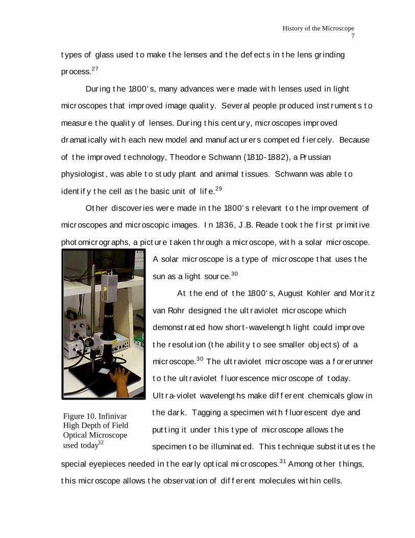
Figure 10. Infinivar High Depth of Field Optical Microscope used today32
types of glass used to make the lenses and the defects in the lens grinding
process.27
During the 1800's, many advances were made with lenses used in light
microscopes that improved image quality. Several people produced instruments to
measure the quality of lenses. During this century, microscopes improved
dramatically with each new model and manufacturers competed fiercely. Because
of the improved technology, Theodore Schwann (1810-1882), a Prussian
physiologist, was able to study plant and animal tissues. Schwann was able to
identify the cell as the basic unit of life.29
Other discoveries were made in the 1800's relevant to the improvement of
microscopes and microscopic images. In 1836, J.B. Reade took the first primitive
photomicrographs, a picture taken through a microscope, with a solar microscope.
A solar microscope is a type of microscope that uses the
sun as a light source.30
At the end of the 1800's, August Kohler and Moritz
van Rohr designed the ultraviolet microscope which
demonstrated how short-wavelength light could improve
the resolution (the ability to see smaller objects) of a
microscope.30 The ultraviolet microscope was a forerunner
to the ultraviolet fluorescence microscope of today.
Ultra-violet wavelengths make different chemicals glow in
the dark. Tagging a specimen with fluorescent dye and
putting it under this type of microscope allows the
specimen to be illuminated. This technique substitutes the
special eyepieces needed in the early optical microscopes.31 Among other things,
this microscope allows the observation of different molecules within cells.

History of the Microscope 8
Figure 11. JSM-840 Scanning Electron Microscope 36
Figure 12. Max Ruska34
The ultraviolet microscope was an improvement in microscopy but still did
not allow the observation of things smaller than 0.5 microns (1/100 the size of a
hair). This obstacle was overcome in the 20th century with the development of the
Transmission Electron Microscope (TEM) and the Scanning Probe Microscope
(SPM).
The Electron Microscope
In 1897, an English physicist, J.J. Thompson, discovered
the electron. In 1924, a French physicist, Louis de Broglie found
that electrons could behave in a wave-motion like light does. The
length of this electron wave was determined to be much smaller
than the wavelength of light. This means that by using an
electron beam, instead of a light beam, it might be possible to
view those objects smaller than the wavelength of light.33 These
important discoveries ushered in the next technological
breakthrough in microscopes: the Transmission Electron
Microscope or TEM and Scanning Electron Microscopes or SEM (see figure 11).
In 1931 a young Russian Ph.D. student, Max Ruska,
produced the first electron microscope that could see an
object that was 50 nanometers in size! If we return to
the hair example, this would be 1/100 the width of the
average human hair. This microscope was 4 times more
powerful than a light microscope. Ruska (see Figure 12)35
was awarded the Nobel Prize in Physics for this
achievement in 1986.
History of the Microscope 9
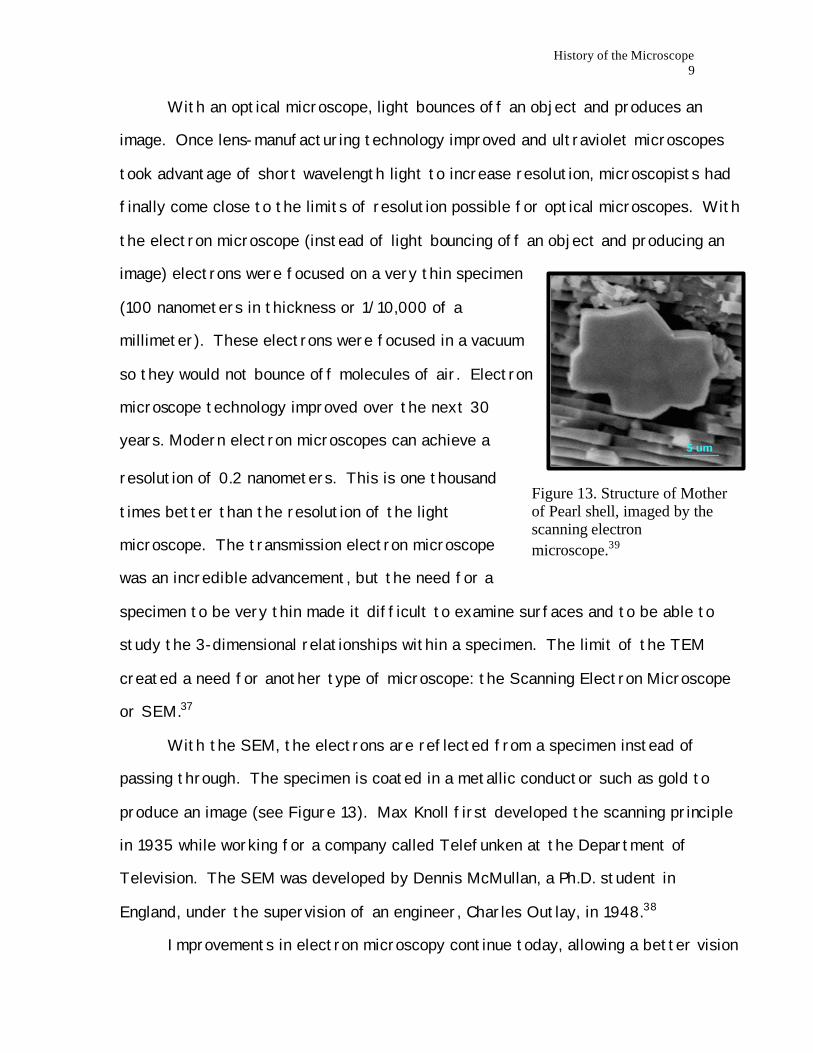
Figure 13. Structure of Mother of Pearl shell, imaged by the scanning electron microscope.39
With an optical microscope, light bounces off an object and produces an
image. Once lens-manufacturing technology improved and ultraviolet microscopes
took advantage of short wavelength light to increase resolution, microscopists had
finally come close to the limits of resolution possible for optical microscopes. With
the electron microscope (instead of light bouncing off an object and producing an
image) electrons were focused on a very thin specimen
(100 nanometers in thickness or 1/10,000 of a
millimeter). These electrons were focused in a vacuum
so they would not bounce off molecules of air. Electron
microscope technology improved over the next 30
years. Modern electron microscopes can achieve a
resolution of 0.2 nanometers. This is one thousand
times better than the resolution of the light
microscope. The transmission electron microscope
was an incredible advancement, but the need for a
specimen to be very thin made it difficult to examine surfaces and to be able to
study the 3-dimensional relationships within a specimen. The limit of the TEM
created a need for another type of microscope: the Scanning Electron Microscope
or SEM.37
With the SEM, the electrons are reflected from a specimen instead of
passing through. The specimen is coated in a metallic conductor such as gold to
produce an image (see Figure 13). Max Knoll first developed the scanning principle
in 1935 while working for a company called Telefunken at the Department of
Television. The SEM was developed by Dennis McMullan, a Ph.D. student in
England, under the supervision of an engineer, Charles Outlay, in 1948.38
Improvements in electron microscopy continue today, allowing a better vision

History of the Microscope 10
Figure 16. Atomic Force Microscope44
Figure 15. Gerd Binnig42
Figure 14. Heinrich Rohrer41
of the microstructure of biological and non-biological specimens and creating new
forms of the microscope. Even so, the electron microscope has its constraints and
this gave rise to a need for a new type of microscope technology: the Scanning
Probe Microscopes or SPM.
The Scanning Probe Microscopes
The next step in microscopy came in 1982 with the
development of Scanning Tunneling Microscopy
(STM) by two Swiss Scientists, Heinrich Rohrer
and Gerd Binnig. For this achievement they were
awarded the Nobel Prize in Physics in 1986 (see
Figures 14 and 15).40 They developed a
microscope that probes the surface of
specimens instead of looking through them.
The probe has a tip that is 1 atom in thickness and moves up and down over
the surface of an object. The probe scans the object producing image
information of a small area of the object. The STM
requires an electrically conductive sample because the
tip itself carries a small voltage. When it comes near
the surface of the sample, a small current will flow
between the sample and the tip. The current changes
with the height of the sample and an image is produced
based on this change. The limits in STM, mainly the
need for an electrically conductive sample, created a
need for another type of microscope: the Atomic
Force Microscope or the AFM.43

History of the Microscope 11
Figure 17. Probe tip of the AFM46
Image 18. Red Blood Cell image taken with the AFM.48
The AFM (see Figure 16) was developed in 1985 with components of the STM
by Binnig, Quate and Gerber and works in a similar fashion to the STM. Instead of
reading the change in current to produce an
image, the probe of the AFM moves over the
surface of the sample and reads the repulsive
force between the sample and the probe tip (see
Figure 17). As with the STM, there is not much
space between the probe tip and sample surface.
The AFM can be used to look at non-conductive
samples like bacteria, viruses, and cells (see image 18) as well as samples such as
CDs and computer chips.45
Since STM and AFM are still fairly young, the possibilities for the use of
these microscopes are still in development. Even so, STM and AFM technology have
opened the door to an entirely new area of science called nanotechnology. The
computer industry uses these microscopes to produce smaller and smaller electrical
circuits and to control the quality of products that they are making.47 Probes are
also used to write on circuit boards. This is called nanolithography. Scientists are
also using these microscopes to learn
more about DNA.
Conclusion The microscope evolved over the
past 500 years branching into different
directions, all with the same purpose, to
enable the exploration of the
microworld and nanoworld. From the
History of the Microscope 12
earliest Janssen microscope to the Electron and Scanning Probe microscopes of
today, microscopy has revolutionized the world we live in, allowing us to see a part
of the world that our eyes alone cannot. The possibilities for the use of
microscopes are beyond our wildest dreams and many more microscopes exist today
than could be discussed in this historical account. The Phoenicians making glass on
a beach 3000 years ago probably had no idea of what was to come!
Acknowledgements I would like to thank the following people for their assistance in preparing this history: Leanne Bosson, Laurie Luckenbill and Lorna Stulen-Glaunsinger. Footnotes 1. Ford, Brian J. (1992), p166. 2. Burgess, et al. (1990), p 186. 3. Klein. (1980), p24. 4. Klein. (1980), p14. 5. Corning Museum of Glass 6. Clay Tablet from the Corning Museum of Glass 7. Corning Museum of Glass 8. Roman Cage Cup from the Corning Museum of Glass 9. Clay and Court. (1975), p4. 10. Clay and Court. (1975), p5. 11. Picture of Roger Bacon from Compton's Encyclopedia On-line 12. Jones, Chapter 1. 13. Burgess, et al. (1990), p187. 14. Klein. (1980), p23-24. 15. Comptom's Encyclopedia On-Line 16. Jones, Chapter 2. 17. Picture of what is thought to be a Janssen microscope from Jones,
Chapter 2. 18. Ruestow. (1996), p7. 19. Burgess, et al. (1990), p186. 20.Picture of Hooke's microscope from Fournier. (1996), p17.

History of the Microscope 13
21. Burgess, et al. (19990), p186. 22.Picture of Antoni van Leeuwenhoek from Antoni van Leeuwenhoek, UC
Berkeley. 23.Antoni van Leeuwenhoek, UC Berkeley. 24.Picture of Antoni van Leeuwenhoek's microscope from Antoni van
Leeuwenhoek, UC Berkeley. 25.Burgess, et al. (1990), p186. 26.Clay and Court. (1975), p140. 27.Burgess, et al. (1990), p187. 28.Jones, Chapter 4. 29.Burgess, et al. (1990), p188. 30.Ford. (1973), p108. 31. Burgess, et al. (1990), p188. 32.WISE image taken by Lillian Owen at ASU Optical Microscopy Lab,
thanks Dr. Michael McKelvy. 33.Burgess, et al. (1990), p189. 34.Picture of Max Ruska from The Nobel Prize Internet Archive. 35.Press Release: 1986 Nobel Prize in Physics. 36.WISE image taken by Lillian Owen at ASU Electron Microscopy Lab,
thanks Dr. Michael McKelvy. 37.Burgess, et al. (1990), p189-190. 38.Burgess, et al. (1990), p189-190. 39.Mother of Pearl image taken by Velavan Varadarajan, courtesey of Dr.
B.L. Ramakrishna. 40.Press Release: 1986 Nobel Prize in Physics. 41. Picture of Rohrer from 1986 Nobel Prize in Physics web site. 42.Picture of Binnig from 1986 Nobel Prize in Physics web site. 43.Martin. (1996). 44.WISE picture taken by Lillian Owen at ASU IN-VSEE Lab, thanks Dr. Ed
Ong. 45.Martin. (1996). 46.AFM probe tip image taken by Dr. Ed Ong at ASU, courtesy of Dr. Ed
Ong. 47.Wickramasinghe. (1989). 48.Red Blood Cell Image courtesy of Dr. B.L. Ramakrishna. Bibliography
History of the Microscope 14
Bunch, Bryan and Alexander Hellemans. (1993). The timetables of technology. New York: Simon and Schuster. Burgess, Jeremy., Martin, Michael., and Rosemary Taylor. (1990). Under the microscope: a hidden world revealed. New York: Cambridge University Press. Clay, Reginald S., and Thomas H. Court. (1975). The history of the microscope. London: Holland Press. Ford, Brian J. (1992). Images of science: a history of scientific illustration. Oxford: Oxford University Press. Ford, Brian J. (1973). The revealing lens: mankind and the microscope. London: Harrap & Company. Fournier, Marian. (1996). The fabric of life: microscopy in the seventeenth century. Baltimore: Johns Hopkins University Press. Klein, Aaron K. (1980). The complete beginners guide to microscopes and telescopes. Garden City, New York: Doubleday & Company. Martin, Yves. (1996). An Introduction to Scanning Probe Microscopes. Paper. Ruestow, Edward. (1996). The microscope in the Dutch Republic: the shaping of discovery. New York: Cambridge University Press. Turner, Gerald. (1989). The great age of the microscope. New York: IOP Publishing. Wickramasinghe, H. Kumar. Scanned-Probe Microscopes. Scientific American pages 98-105; October 1989. WWW Sources: Antoni van Leeuwenhoek. Encarta On-line. May 28, 1998. <http://encarta.msn.com/find/concise/default.asp?vs=x97&la=na&ty=2&vo=PID&ti=11998>

History of the Microscope 15
Antony van Leeuwenhoek. University of California at Berkeley. June, 9 1998. <http://www.ucmp.berkeley.edu/history/leeuwenhoek.html> Roger Bacon. Comptom's Encyclopedia On-line. June 3, 1998. <http://comptons3.aol.com/encyclopedia/ARTICLES/00378_A.html#112d1c3> Jones, Thomas E. Home page. History of the Light Microscope. June 1998. <http://www.utmem.edu/personal/thjones/hist/hist_mic.htm> Max Ruska. The Nobel Prize Internet Archive. June 13, 1998. < http://www.th.physik.uni-frankfurt.de/~jr/gif/phys/ruska.jpg> Microscopes. Comptom's Encyclopedia On-line. June 3, 1998. <http://comptons2.aol.com/encyclopedia/ARTICLES/03151_A.html> A resource for glass. The Corning Museum of Glass. May 27, 1998. <http://www.pennynet.org/glmuseum> Press Release: The 1986 Nobel Prize in Physics. The Royal Swedish Academy of Sciences. June 13, 1998. < http://www.nobel.se/laureates/physics-1986-press.html> The 1986 Nobel Prize in Physics. The Royal Swedish Academy of Sciences. June 13, 1998. < http://nobel.sdsc.edu/laureates>