c regionale immunoematologia e tipizzazione … · accreditato dall’american society...
TRANSCRIPT
CENTRO REGIONALE IMMUNOEMATOLOGIA E TIPIZZAZIONE TISSUTALE Registro Regionale Donatori Midollo Osseo
Accreditato dall’American Society for Histocompatibility and Immunogenetics (A.S.H.I.) n° 11-9-IT-04-1 Accreditato dall’European Federation for Immunogenetics (E.F.I.) n° 07-IT-024.976
(P.O. “S.Salvatore” – via Lorenzo Natali 67100 - L’Aquila) Tel. +39 0862368236 ; Fax +39 0862 368603 Direttore: dott. Franco Papola

Fig. 3. Histology of the normal cornea. In this photomicrograph the structure of the normal cornea, consisting of an outer layer of epithelium, a middle layer of collagenous stroma with scattered keratocytes, and an inner layer of endothelial cells, is shown (×40 magnification).

Acta
Oph
thal
mol
ogic
a 20
09

Fig. 2. Histopathology of corneal allograft rejection. In this photomicrograph of a corneal allograft undergoing rejection, numerous white blood cells of different morphologies can be seen in the stroma and epithelium of the graft. In particular, cells having the morphology of lymphocytes, macrophages, and polymorphonuclear leukocytes can be seen. Blood vessels are present in the corneal stroma (×100 magnification).

Fig. 4. Neovascularization of the rabbit cornea with silk sutures. (A) Slit lamp photograph of a vascularized cornea shortly after removal of the silk sutures. The dense neovascularization of the cornea in the area where the sutures were placed can be seen (arrows). (B) Photomicrograph showing the histopathology of corneal neovascularization induced by a silk suture. The presence of blood vessels and inflammatory cells in the stroma and disruption of the stromal matrix induced by the suture can be seen (×100 magnification).


Acta
Oph
thal
mol
ogic
a 20
09

Meccanismi immunologici nel rigetto

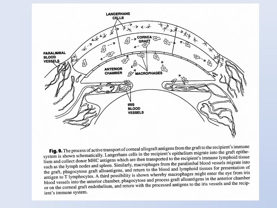
Donor Organ
Capillary Endothelial
cell
Formation of Antigen-Ab
complex
C1 complex
Pathophysiology of AMR
Damaged Cell
Releases platelet
aggregation factors,
cytokines
Endothelial cell
necrosis
Schwartz, NEJM 2010
C4
C4b C4d
C4d is by-product and marker of complement activation
4

Fig. 1. Combined stromal and endothelial graft rejection showing differential graft edema with stromal haze. (Note the prominent cut suture end with vascularization, which was responsible for the rejection.)
Surv Ophthalmol 52 (4) July--August 2007

Effects of corticosteroids

Cyclosporine A and FK506 inactivate calcineurin (a calcium binding protein), which is required for T, B and granulocyte activation

Overview of Immune System Responses

Per agire sulla presenza di anticorpi, abbassare il titolo anticorpale e/o frenare gli effetti degli Ab :
The boron atom in bortezomib binds the catalytic site of the 26S proteasome[4] with high affinity and specificity. In normal cells, the proteasome regulates protein expression and function by degradation of ubiquitylated proteins,
Il rituximab, è un farmaco appartenente alla classe degli anticorpi monoclonali; il suo bersaglio è la proteina CD20. Viene utilizzato nel trattamento del Linfoma non Hodgkin delle cellule B, nelle leucemie delle cellule B e in talune malattie autoimmuni.

Fig. 1. The effects of passively transfered donor-specific antiserum on surviving of corneal allografts. TheBALB/c recipients of B10 corneal allografts were untreated (9 mice) or were treated with anti-B10 antiserum administered at the period of grafting (10 mice) or at 2 weeks after transplantation (9 mice).
V. Hol´aˇn et al. / Immunology Letters 100 (2005) 211–213
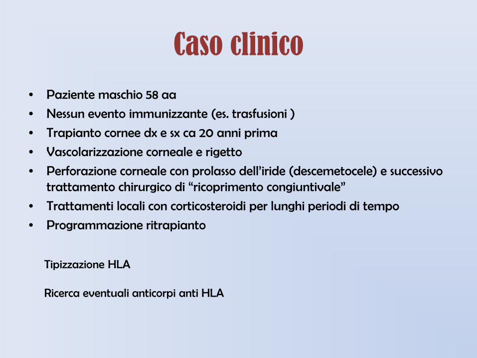
Caso clinico
• Paziente maschio 58 aa
• Nessun evento immunizzante (es. trasfusioni )
• Trapianto cornee dx e sx ca 20 anni prima
• Vascolarizzazione corneale e rigetto
• Perforazione corneale con prolasso dell’iride (descemetocele) e successivo trattamento chirurgico di “ricoprimento congiuntivale”
• Trattamenti locali con corticosteroidi per lunghi periodi di tempo
• Programmazione ritrapianto
Tipizzazione HLA Ricerca eventuali anticorpi anti HLA


Considerazioni immunologiche e rigetto del trapianto di cornea
RIGETTO: RISPOSTA
CELLULO-MEDIATA/ UMORALE
Incompatibilità HLA
Attivazione Linfociti T
Citotossici e T Helper
Anticorpi anti – HLA nel siero
In pazienti a rischio di rigetto immunologico 2 o più mismatch HLA classe I 1 o più mismatch HLA classe II Anticorpi HLA nel siero (paziente immunizzato)
Rischio ↑ 14 volte
Rischio ↑ 1000 volte
HLA

Utilizzare cornee che, oltre ad essere compatibili, non posseggono antigeni proibiti (es. DR4, DR16, DR1) in quanto gli anticorpi già presenti avrebbero un effetto sia diretto (rigetto acuto) che favorente la rivascolarizzazione e quindi la perdita precoce della cornea ritrapiantata.
Come comportarsi nel caso di pazienti ad alto rischio
Tipizzare il paziente per gli antigeni HLA di I° e II° classe,
Effettuare lo screening anticorpale per HLA di I° e II° classe,
Evidenziare gli eventuali anticorpi specifici HLA di I° o di II° classe,

GRAZIE per l’attenzione!