calcium-evoked dendritic exocytosis in cultured hippocampal
TRANSCRIPT
Calcium-Evoked Dendritic Exocytosis in Cultured HippocampalNeurons. Part I: Trans-Golgi Network-Derived Organelles UndergoRegulated Exocytosis
Mirjana Maletic-Savatic and Roberto Malinow
Cold Spring Harbor Laboratory, Cold Spring Harbor, New York 11724
Exocytosis is a widely observed cellular mechanism for deliv-ering transmembrane proteins to the cell surface and releasingsignaling molecules into the extracellular space. Calcium-evoked exocytosis, traditionally thought to be restricted topresynaptic specializations in neurons, has been describedrecently in many cells. Here, calcium-evoked dendritic exocy-tosis (CEDE) is visualized in living cultured hippocampal neu-rons. Organelles that undergo CEDE are in somata, dendrites,and perisynaptic regions, identified by using immunocyto-chemistry and correlative light and electron microscopy. CEDEis regulated developmentally: neurons ,9 d in vitro do not showCEDE. In addition, CEDE is blocked by tetanus toxin, an inhib-
itor of regulated exocytosis, and nocodazole, an inhibitor ofmicrotubule polymerization. Organelles that undergo CEDE of-ten are found on the base of spines, putative sites of synapticplasticity. CEDE therefore could be involved in structural andfunctional modification of spines and could play a role in syn-aptic plasticity, where it might involve changes in receptor/channel density, release of active compounds having effect onpre- and postsynaptic function, and/or growth of synapticstructures.
Key words: exocytosis; trans-Golgi network; dendrite; pyra-midal neurons; hippocampal culture; time-lapse imaging; FM1-43; immunocytochemistry; tetanus toxin; microtubules
Constitutive exocytosis is ubiquitous in eukaryotic cells. It isresponsible for the recycling and renewal of plasma membranecomponents and for the secretion of molecules into the extracel-lular space (Alberts et al., 1989). In neurons the exocytosis ofneurotransmitter molecules from presynaptic terminals repre-sents the fundamental mechanism for the transfer of information.Although presynaptic vesicle exocytosis has been examined ex-tensively, exocytosis has not yet been demonstrated directly in thedendritic regions of neurons, where it might provide a regulatedmeans for effecting synaptic plasticity and/or homeostasis. Evi-dence exists that nonfunctional pools of postsynaptic membraneproteins are present (Hampson et al., 1992; Baude et al., 1995;Rubio and Wenthold, 1997) and that postsynaptic exocytosis isrequired for some forms of synaptic plasticity (Lledo et al., 1998).Calcium-evoked exocytosis has been demonstrated in neuronalsomata (Huang and Neher, 1996) as well as in non-neuronal cells(Morimoto et al., 1995). In addition, lysosomes in fibroblasts,epithelial cells, and myoblasts undergo calcium-regulated exocy-tosis, which is temperature- and ATP-dependent (Rodriguez etal., 1997). In this study we demonstrate calcium-evoked exocyto-sis of dendritic compartments, consistent with the view that someforms of synaptic plasticity might occur via the regulated exocy-tosis of dendritic organelles.
In the past several years the mechanisms of presynaptic vesicleexocytosis have been examined in detail, including studies usingfluorescent membrane probes. The styryl dye FM1-43 has been
used effectively as a probe to monitor at the light microscope levelthe dynamics of presynaptic vesicles in living cells (Betz et al.,1992). A brief exposure to FM1-43, combined with the depolar-ization of the cell, induces selective internalization of the dye intothe presynaptic terminals (Ryan et al., 1993). With triggeredexocytosis the dye is released into the extracellular space, and theloss of fluorescence is used as a measure of exocytosis (Ryan etal., 1993).
FM1-43 nonspecifically attaches to the whole surface of a cell,and on endocytosis the dye becomes internalized. In fully polar-ized hippocampal neurons, endocytosis occurs over the entiresomatodendritic surface (Parton and Dotti, 1993). Using theseproperties, we developed a method to label dendritic membra-nous compartments with FM1-43. This method allowed us tovisualize the behavior of labeled compartments in living culturedhippocampal neurons and to determine the cellular mechanismscontrolling their exocytosis. Furthermore, because there is evi-dence for mixing between endocytic and biosynthetic pathways(Trowbridge et al., 1993), studies monitoring the fate of endocy-tosed FM1-43 may shed light on the mechanisms by which newlysynthesized membrane-bound proteins, like receptors, are trans-ported and inserted into synapses.
MATERIALS AND METHODSCell cultureCortical astrocytes were derived from 1-d-old rat pups and plated ontopoly-L-lysine-coated coverslips. Hippocampal neurons were generatedfrom 19-d-old rat embryos (Banker and Goslin, 1990) and plated onto aconfluent monolayer of astrocytes. Astrocytes and neurons were platedat 50,000 and 30,000 cells per 18-mm-round glass coverslip (FisherScientific, Pittsburgh, PA), respectively. For relocation experiments theastrocytes and neurons were plated at 100,000 and 85,000 cells per25-mm-square gridded glass coverslip (Bellco Glass, Vineland, NJ), re-spectively. Cultures were maintained in a serum-free medium (Bankerand Goslin, 1990).
Received March 23, 1998; revised June 18, 1998; accepted June 22, 1998.This study was supported by the Mathers Foundation and National Institutes of
Health. We are grateful to Nancy Dawkins for preparing the cultures, TamaraHoward for assistance with the electron microscopy, Jason Kass for technicalassistance with immunocytochemistry, and Irena Miloslavskaya for assistance inimage processing.
Correspondence should be addressed to Dr. Roberto Malinow, Cold SpringHarbor Laboratory, Cold Spring Harbor, NY 11724.Copyright © 1998 Society for Neuroscience 0270-6474/98/186803-11$05.00/0
The Journal of Neuroscience, September 1, 1998, 18(17):6803–6813

Labeling neurons with FM1-43Acute FM1-43 loading. Cultures were exposed to 15 mM FM1-43 com-bined with 90 mM KCl for 1 min while on the microscope stage andwashed for 10 min with constantly perfusing bath solution [containing (inmM) 119 NaCl, 2.5 KCl, 1.3 MgCl2 , 2.5 CaCl2 , 1 NaH2PO4 , 26.2NaHCO3 , and 11 glucose].
Overnight FM1-43 loading. Cultures were exposed 16–36 hr to 1.5 mMFM1-43 at 35.5°C before transfer to the microscope stage. Cultures wererinsed with 0.1% DMSO for 1 min and maintained in constantly perfus-ing bath solution for 1 hr. The bath solution (see above) contained 1 mMtetrodotoxin to abolish action potentials.
Time-dependent loading. Mature cultures (.9 d in vitro, DIV) wereloaded with 15 mM FM1-43 for 1–16 hr. After loading, they were trans-ferred to a microscope stage and washed with the bath solution for 30min. They were challenged with vehicle (0.1% DMSO) or 1 mM A23187for 1 min. Calcium-evoked dendritic exocytosis (CEDE) was monitoredfor at least 15 min.
Calcium-evoked dendritic exocytosisAfter overnight loading with 1.5 mM FM1-43, the cultures were trans-ferred to the microscope stage and maintained in constantly perfusingbath solution for 1 hr. Calcium ionophore A23187 (1 mM) was applieddirectly onto the coverslip for 1 min. Fluorescence was monitored for 30min before and 30 min after A23187 application, at 15 min intervals. Anadditional image was taken ;5 min after ionophore application. Allimages in a given experiment were taken with the same exposure time(100–500 msec, chosen so that the fluorescent signal did not saturate thecamera detection limit). A number of cells (.20) were tested for viabilitywith 0.4% trypan blue. More than 90% of the neurons excluded the dyeafter A23187 challenge (data not shown), indicating that ionophoreapplication did not have toxic effects. Viability also was confirmed in anumber of experiments (.5) in which the subsequent application ofA23187 combined with FM1-43 produced loading (data not shown).
EGTA treatment. Mature cultures (.9 DIV) were loaded with 1.5 mMFM1-43 overnight and washed as above. Cells were incubated in 5 mMEGTA, pH 7.0, in Ca 21-free artificial CSF for 10 min before A23187challenge and for 15 min thereafter.
Tetanus toxin treatment. Mature cultures (.9 DIV) were loaded with1.5 mM FM1-43 for 14 hr and then exposed for 5 hr to 10 nM tetanus toxin(generously provided by Dr. Giampetro Schiavo, Rockefeller University,New York, NY) and 1.5 mM FM1-43 at 35.5°C. Experiments wereperformed as above.
Nocodazole treatment. Mature cultures (.9 DIV), loaded overnightwith 1.5 mM FM1-43, were exposed for 45 min to 2.5 mg/ml nocodazoleand 1.5 mM FM1-43 at 35.5°C. Experiments were performed as above.
Field electrical stimulation. Mature cultures (.9 DIV), loaded over-night with 1.5 mM FM1-43, were stimulated with repetitive field stimuli.The current was delivered between the two silver wires placed 3 mmapart onto the coverslip with cultured neurons. Different frequencies (1,10, 50, and 100 Hz) and numbers of stimuli were applied.
Measurement of released FM1-43. Neurons loaded overnight with 1.5mM FM1-43 were washed in the same manner as for imaging experiments.They were incubated for 15 min in HEPES buffer, pH 7.2. An aliquot ofthis bathing solution (Apre ) was recovered. Then the neurons wereexposed to 1 mM A23187 for 1 min, followed by fresh HEPES buffer for15 min. An aliquot from this bathing solution (Apost ) was recovered. Apreand Apost solutions were pooled from 10 cultures. FM1-43 was extractedfrom these aqueous solutions with an equal volume of water-saturatedbutanol. The butanol fraction was recovered and evaporated. (3-[(3-Cholamidopropyl)dimethylammonio]-1-propane-sulfonate) (CHAPS)(2%) was added (FM1-43 binds to CHAPS and increases fluorescence;Henkel et al., 1996). This solution was put in a quartz rectangularcapillary, and FM1-43 fluorescence was measured (FITC filters; ZeissAxioskop, Oberkochen, Germany). We measured the extracted fluores-cence of HEPES buffer, Apre and Apost. The amount of released FM1-43was calculated as [Apost 2 buffer]/[ Apre 2 buffer]. Calibration curveswere generated from 1.5 nM to 1.5 mM FM1-43 before each experiment(n 5 7).
MicroscopyImages were acquired with a computer-controlled cooled CCD camera(Photometrics, Tucson, AZ), using FITC filters (peak at 450 nm) on aZeiss Axioskop epifluorescence microscope (50 W mercury lamp), andprocessed with Photometrics-supplied software (PMIS). Time-lapse im-
ages of mature neurons loaded with FM1-43 were acquired every 2 minover a 1 hr period, using FITC filters on a Zeiss Axiovert invertedmicroscope (50 W mercury lamp; 1003 lens with additional 2.53magnification).
ImmunocytochemistryMature neurons (.12 DIV) were loaded with 1.5 mM FM1-43 overnightand then washed for 1 hr with the bathing solution. Cultures were fixedin 4% paraformaldehyde in 0.1 M PBS containing 0.12 M sucrose for 20min at 14°C and immediately permeabilized with 0.3% Triton X-100 inPBS for 5 min at room temperature. FM1-43-labeled compartments wereimaged after fixation (1003 lens, FITC filter, Zeiss Axioskop, 75 Wxenon lamp) to avoid confounding results if the labeled organelles movedbefore fixation. Imaging with Texas Red filters (Omega Optical, Brattle-boro, VT) did not reveal any FM1-43 fluorescence. Cultures were incu-bated in blocking solution (10% horse serum, 1% goat-serum, and 0.1%Triton X-100 in PBS) for 1 hr at room temperature and then immuno-stained overnight with the primary antibody [polyclonal anti-synapsin Iantibody: 1:10, polyclonal rabbit sera number 6246, generously providedby Dr. Pietro De Camilli (Yale University, New Haven, CT); polyclonalanti-transferrin receptor antibody raised in sheep: 215 mg/ml, Sigma (St.Louis, MO); polyclonal anti-mannose-6-phosphate receptor antibody:1:50, generously provided by Dr. W. J. Brown (Cornell University,Ithaca, NY); polyclonal anti-cathepsin D antibody: 1:100, generouslyprovided by Dr. W. J. Brown (Cornell University); polyclonal anti-inositol triphosphate (IP3 ) receptor antibody: 1:50, generously providedby Dr. T. Jayaraman (Mount Sinai School of Medicine, New York, NY);monoclonal anti-BiP antibody: 16 mg/ml, Stress Gen (Sidney, Canada)].Primary antibody was washed out three times for 10 min with theblocking solution. Then the cells were incubated with the appropriatesecondary biotinylated antibody (2.5 mg/ml; Cappel, West Chester, PA)for 1 hr at room temperature. After the washout of the biotinylatedantibody, the cultures were incubated with Texas Red-conjugated avidin(25 mg/ml; Cappel) for 1 hr at room temperature. Immunoreactivity wasanalyzed immediately after the Texas Red–avidin was washed out withPBS (1003 lens, Texas Red filter). Neurons were relocalized by usinggridded coverslips (Bellco).
NBD C6-ceramide labeling. Mature neurons (.12 DIV) were loadedwith 1.5 mM FM1-43 for 4 hr (see Fig. 6 B) or overnight (see Fig. 3) andwashed for 1 hr before fixation as described above. FM1-43-labeledcompartments were imaged, and the area was exposed to UV light tobleach the FM1-43 fluorescence. When no FM1-43 fluorescence wasdetected, the neurons were incubated with 40 mM NBD C6-ceramide(Molecular Probes, Eugene, OR) for 1 hr at 37°C and washed with 2.5%fat-free bovine serum albumin three times for 20 min at room tempera-ture. Fluorescence was analyzed with FITC filters.
Electron microscopyThe 14 DIV hippocampal neurons, loaded with 1.5 mM FM1-43 over-night, were washed with PBS for 1 hr before fixation with 3% parafor-maldehyde in 0.2 M cacodylate buffer, pH 7.30, for 45 min at roomtemperature. After being washed with 0.2 M cacodylate buffer, secondaryfixation was performed, using 1% OsO4 with 1.5% K4Fe(CN)6 in caco-dylate (90 min at room temperature), followed by dHOH washes and enbloc uranyl acetate staining. Cells were dehydrated through a gradedseries of ethanol and embedded in Epon–Araldite resin. Sections werecollected on Formvar-coated cupri slot grids, counterstained with uranylacetate and Reynold’s lead citrate, and examined in the Hitachi H-7000at 75 kV.
Fluorescent and electron micrographs were superimposed in AdobePhotoshop 4.0. To align fluorescent and electron micrographs, we usedfiduciary points chosen on the basis of regions clearly identified on bothphotographs (e.g., the intersection of dendrites and axons, and at thedendritic branching points). Electron micrographs were scanned (ScanJetPlus, Packard, Meriden, CT), and the image sizes of both micrographswere matched. Images were aligned with respect to fiduciary points.After alignment, the fluorescent image was pseudocolored for bettervisualization of the FM-labeled structures.
Data analysisFor quantitative measurements of CEDE, several regions of interest(2–5) were placed on dendrites (locations were chosen before post-treatment images were analyzed), and FM1-43 fluorescence was inte-grated. Background fluorescence, measured in regions next to the neu-rons, was subtracted. FM1-43 fluorescence values were normalized to the
6804 J. Neurosci., September 1, 1998, 18(17):6803–6813 Maletic-Savatic and Malinow • CEDE in Hippocampal Neurons: TGN-Derived Organelles
intensity observed immediately before A23187 application. Statisticalanalysis was done by one-way ANOVA.
The degree of colocalization between FM1-43 fluorescent sites andother fluorescent markers was quantified with PMIS software (Photo-metrics). Regions of interest from images of FM1-43 fluorescent siteswere identified, and pixel intensity along lines traversing ;50 mm wasmeasured [IF(x)]. Pixel intensities along the same lines placed in the sameregions from the image detecting another fluorescent marker were mea-sured also [IM(x)]. We plotted IF(x) versus IM(x) and calculated a corre-lation coefficient, R (using Origin 4.0 software, Microcal, Amherst, MA).Each neuron that was examined had two to eight lines analyzed. Loca-tions were chosen from FM1-43-loaded neurons before the correspond-ing marker-labeled image was analyzed.
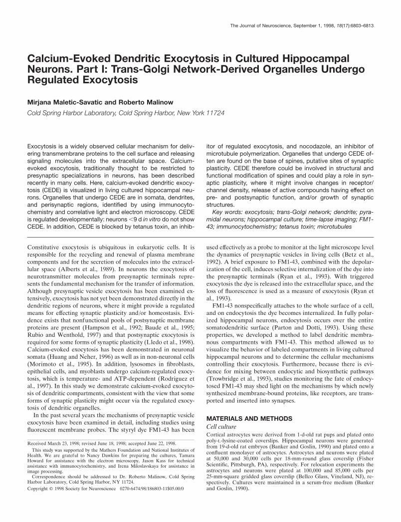
RESULTSFM1-43 labels dendritic compartmentsBrief exposure of cultured neurons to FM1-43, combined withdepolarizing stimuli, can produce a selective loading of presyn-aptic vesicles (Fig. 1A,C) (Ryan et al., 1993). In contrast, we foundthat cultured hippocampal neurons incubated with FM1-43 for16–36 hr in the absence of stimulation loaded intracellular com-partments in all processes and the soma (Figs. 1D, 2, 4). Wefocused our analysis on the characterization of FM1-43-labeledcompartments in processes, anticipating that these may be morelikely to be associated with synaptic plasticity.
To determine the distribution of FM1-43 fluorescence in pre-and postsynaptic compartments after the two labeling protocols,we compared its labeling pattern with that of a presynapticmarker protein, synapsin I (Fig. 1B,E; n 5 3). After standardacute exposure and depolarizing stimuli, FM1-43 and synapsin Ishowed significant colocalization, confirming the presynaptic lo-calization of FM1-43 with this loading protocol (Fig. 1A,B).However, with long exposure and no stimulation, FM1-43 andsynapsin I showed little colocalization (Fig. 1D,E). This indicatesthat the longer exposure selectively labels intracellular compart-ments that are not presynaptic terminals. Quantitated immuno-cytochemical analysis (see Materials and Methods) confirmedthat, with overnight exposure to the dye, FM1-43 fluorescence insynapsin I-labeled compartments accounts for only 10% of thedye (Fig. 3A; R 5 0.13 6 0.06; n 5 3).
Dendritic compartments labeled with FM1-43 often were seenat the base of small filopodial structures, proposed to be synapticspine precursors (Fig. 1F) (Ziv and Smith, 1996). Time-lapseimaging over 1 hr revealed that FM1-43-loaded compartmentsmaintained this association although the filopodial structuresmoved over time (;2 mm/min; Fig. 1F). In contrast, presynapticvesicles labeled with acute loading of FM1-43 did not move within
Figure 1. FM1-43 fluorescent probe labels presynaptic or dendritic compartments in cultured hippocampal neurons, depending on the loading protocol.A, Location of FM1-43 dye after exposing 14 DIV neurons to FM1-43, combined with 90 mM KCl, for 1 min. Scale bar, 10 mm. B, Expression of synapsinI in characteristic punctate labeling pattern indicates presynaptic axon terminals that are closely apposed to the neuronal soma and major dendrite. Themajority of FM1-43-labeled sites in A corresponds to synapsin I immunoreactivity (arrow). C, Time-lapse images of a region after acute FM1-43 loadingof 20 DIV cultured neuron. Little movement of spots is apparent. Spontaneous destaining (;20%) over a 1 hr observation period was typical (displayscale maximum was changed from 110 to 95 arbitrary units to facilitate the comparison of spot locations). The time is indicated in minutes. Scale bar,2 mm. D, Overnight exposure of 14 DIV neurons to FM1-43 extensively labels compartments in soma and dendrites. Scale bar, 10 mm. E, Expressionof synapsin I, again labeling presynaptic sites closely apposed to the major dendrite. FM1-43-labeled compartments and synapsin I do not overlap in themajority of sites, indicating dendritic (arrowheads) and presynaptic (arrows) localization of labeled sites, respectively. F, Time-lapse images of a regionafter overnight FM1-43 loading of 20 DIV cultured neuron. Note the location of FM1-43 spots at the base of small filopodial structures and coordinatemovement of the filopodia and spots. The time is indicated in minutes. Scale bar, 2 mm.
Maletic-Savatic and Malinow • CEDE in Hippocampal Neurons: TGN-Derived Organelles J. Neurosci., September 1, 1998, 18(17):6803–6813 6805

1 hr (n 5 3; Fig. 1C), again supporting the conclusion thatovernight FM1-43 exposure loads compartments that are notpresynaptic terminals.
The dendritic localization of FM1-43-labeled compartmentswas confirmed further by correlative electron and fluorescentmicroscopy (Fig. 2). Neurons loaded with FM1-43 overnight wereimaged under high magnification (1003 lens; Fig. 2-1,6) and thenprocessed for electron microscopy (EM). The same regions wereidentified on the EM preparations (Fig. 2-3,8) and aligned withthe light microscope images by using fiduciary points (Fig. 2-4,9).Under EM, FM1-43-fluorescent sites were dendritic and oftenclose to synapses. The sites included different organelles, such ascoated pits, endosomes, multivesicular bodies, endoplasmic retic-ulum, Golgi apparatus, and mitochondria (Fig. 2-5,10). No densecore granules were observed.
To identify which dendritic organelles are labeled by overnightexposure to FM1-43, we performed high-resolution immunocyto-chemical studies on FM1-43-loaded neurons (Fig. 3A). AfterFM1-43 imaging, the cultures were fixed and labeled with differ-ent organelle-specific markers: transferrin receptor (Moos, 1996),cathepsin D (Nakanishi et al., 1994), and mannose-6-phosphatereceptor (Couce et al., 1992), specific for endosome/lysosomeorganelles; IP3 receptor (Moschella et al., 1995) and BiP (Huovilaet al., 1992), specific for endoplasmic reticulum; and NBD C6-ceramide (Pagano et al., 1989), a trans-Golgi fluorescent marker.Quantitative immunocytochemical analysis (see Materials andMethods) indicated little colocalization between FM1-43-labeledcompartments and transferrin receptor (R 5 0.2 6 0.14; n 5 3),cathepsin D (R 5 0.4 6 0.16; n 5 3), mannose-6-phosphatereceptor (R 5 0.21 6 0.1; n 5 3), IP3 receptor (R 5 0.14 6 0.07;n 5 3), and BiP (R 5 0.3 6 0.09; n 5 3). However, we observeda consistently high correlation of FM1-43-labeled dendritic sitesonly with NBD C6-ceramide (R 5 0.77 6 0.11; n 5 5; Fig. 3). Thisindicates that, after overnight exposure to FM1-43, dendriticorganelles loaded with the FM1-43 are primarily dendritic Golgi-like or Golgi-derived structures.
Dendritic organelles undergo regulated exocytosisTo determine whether FM1-43-labeled compartments exhibitcalcium-evoked exocytosis, we challenged neurons loaded withFM1-43 for 16–36 hr with the calcium ionophore A23187 (calci-mycin) for 1 min. This produced a marked loss of FM1-43fluorescence (Fig. 4A) that was attributable to the exocytosis ofinternalized dye (see below). We measured this CEDE as thepercentage of loss of background-subtracted FM1-43 fluores-cence as compared with pre-ionophore background-subtractedfluorescence (Fig. 4B). Exposure to the carrier solution alone(0.1% DMSO) produced no loss of fluorescence (Fig. 5; n 5 12).Similarly, calcimycin did not have any effect when it was appliedin the presence of 5 mM EGTA (Fig. 5; n 5 6). A23187 inducedCEDE only in the presence of 2.5 mM calcium. It could berecorded within 5 min after ionophore application and persistedfor 15 min.
Ionophore-induced exocytosis from neuronal cultures loadedovernight with FM1-43 was confirmed by recovering dye in theextracellular medium (Henkel et al., 1996). Extracellular mediumaliquots from 10 pooled cultures were obtained before (Apre) and15 min after (Apost ) A23187 application. Dye from pooled sam-ples was extracted with butanol and dissolved in 2% CHAPS.FM1-43 fluorescence intensity was measured in microcapillaries.The relative FM1-43 fluorescence intensity of solution afterA23187 treatment was 3.32 6 0.7 (n 5 7; p , 0.05). This shows
directly that a significant amount of FM1-43 was released bycalcium ionophore treatment and could be recovered in the ex-tracellular medium. Calibration curves generated from 1.5 nM to1.5 mM FM1-43 indicate that the observed exocytosis correspondsto ;35 nM FM1-43. This is ;100 times more than the estimatedFM1-43 release measured after similar experiments conducted onpresynaptic exocytosis (0.3 nM; Henkel et al., 1996).
If the organelles undergoing CEDE are derived from the trans-Golgi network (TGN), then one may expect that endocytosedFM1-43 may require several hours before reaching such compart-ments. To examine this possibility, we measured the amount ofCEDE as a function of the FM1-43 loading time (Fig. 6A).Mature cultures were exposed to FM1-43 for different periods oftime (1–16 hr) and challenged with A23187. Only when neuronswere exposed to FM1-43 for .8 hr was CEDE observed (Fig.6A). Notably, FM1-43 showed little colocalization with NBDC6-ceramide fluorescence after 4 hr of loading (n 5 2) andsignificant colocalization after 16 hr of FM1-43 loading (Fig. 6B).These results indicate that the passive loading of FM1-43 requires8 hr before the dye reaches CEDE-competent organelles, at whichtime the dye colocalizes with NBD C6-ceramide. This resultsupports the view that organelles undergoing CEDE are TGN-like or TGN-derived structures.
We tested if CEDE uses exocytotic machinery with tetanustoxin, a specific inhibitor of exocytosis (Montecucco and Schiavo,1994). CEDE was blocked completely by pretreatment with 10 nM
tetanus toxin (see Fig. 5; n 5 7), indicating regulated exocytosisrather than some nonspecific loss of the dye from intracellularcompartments.
We found that CEDE is regulated developmentally. Althoughneurons of all ages that were examined (5–21 DIV) showedcomparable loading after overnight exposure to FM1-43, onlyneurons that were 9 DIV or older were capable of producingCEDE (see Fig. 5; n 5 11; p , 0.05). At this stage the culturedneurons have already established synaptic contacts and are fullydifferentiated. At earlier ages A23187 occasionally induced re-grouping of labeled sites or intensified movement of fluorescentlylabeled organelles but caused no detectable CEDE.
Dendritic exocytosis of FM1-43 also could be elicited withrepetitive field electrical stimulation (Fig. 7). We tested differentnumbers and frequencies of stimuli. Low-frequency stimuli (1Hz) did not produce any detectable loss of fluorescence (n 5 3).Medium-frequency stimuli (10 Hz) could produce loss of fluores-cence, but only if sufficient stimuli were delivered. High-frequency stimuli (50 Hz) generally produced exocytosis (n 510). Figure 7 shows a pattern of CEDE observed in a neuronstimulated with different parameters. This pattern generally wasobserved in the tested neurons. In addition, the application of 1mM glutamate for 1 min could evoke the exocytosis of somedendritic structures (n 5 2; data not shown). We characterizedCEDE by using calcium ionophore as the stimulating agent,because it gave the most robust effect.
DISCUSSIONFM1-43 labels dendritic compartmentsIn this study we report on a novel biological process of regulateddendritic exocytosis. Cultured hippocampal neurons were ex-posed to FM1-43 for .16 hr with no stimulation. Because FM1-43immerses into the outer leaflet of the plasma membrane, it isinternalized inside the cells whenever there is endocytosis. Onwashout of the uninternalized dye from the outer surface, theremaining FM1-43 fluorescence marks the inner membrane of
6806 J. Neurosci., September 1, 1998, 18(17):6803–6813 Maletic-Savatic and Malinow • CEDE in Hippocampal Neurons: TGN-Derived Organelles

Figure 2. Identification of FM1-43-labeled compartments by electron microscopy (EM). Shown are two example sets (1-5, 6–10) of correlativefluorescent and electron microscopy of mature neurons exposed overnight to FM1-43. Fluorescent micrographs (1, 6 ) show FM1-43-labeled compart-ments. Scale bars, 10 mm. Outlined regions are enlarged (2, 7 ) and are represented on the electron micrographs (3, 8). Scale bars: 1 mm in 3; 1.8 mm in8. Asterisks indicate fiduciary points used to superimpose fluorescent and electron micrographs. FM1-43-labeled compartments in dendrites (whitearrowheads), opposite the presynaptic sites (arrows), and in areas containing SER/TGN organelles (black arrowheads) are evident on the superimposedpseudocolored fluorescent micrographs (4, 9). Note the lack of FM1-43 in regions corresponding to presynaptic terminals. EM sections at other levelsin the same region also failed to show presynaptic terminals in fluorescent regions. Outlined regions on the electron micrographs are enlarged in 5 and10 for better identification of postsynaptic SER/TGN-like structures. Scale bars: 0.3 mm in 5; 0.5 mm in 10. Figure continues.
Maletic-Savatic and Malinow • CEDE in Hippocampal Neurons: TGN-Derived Organelles J. Neurosci., September 1, 1998, 18(17):6803–6813 6807

Figure 2 continued.
6808 J. Neurosci., September 1, 1998, 18(17):6803–6813 Maletic-Savatic and Malinow • CEDE in Hippocampal Neurons: TGN-Derived Organelles

intracellular membranous compartments. Via the fusion of endo-somes and endosome-derived compartments, over time, the dyehas access to numerous intracellular compartments. Because theneurons are exposed continuously to FM1-43 for .16 hr, someFM1-43 can be found in intracellular compartments that arederived from endocytic organelles—like early endosomes (ex-pressing transferrin receptor), late endosomes and lysosomes(expressing cathepsin D and mannose-6-phosphate receptor), andendoplasmic reticulum in soma (expressing IP3 receptor andBiP). However, quantitative analysis of the colocalization be-tween FM1-43-labeled structures and intracellular markers ofvarious organelles shows the greatest degree of colocalizationwith NBD C6-ceramide, indicating the labeling of TGN-like orTGN-derived structures (see Fig. 3).
The immunocytochemical analysis of labeled compartments issupported by correlative electron and light microscopy. Electronmicroscopy of FM1-43-labeled regions indicates that the majorityof labeled sites clearly is in dendrites and not presynaptic termi-nals, has no dense core vesicles, has smooth endoplasmic reticu-lum (SER)/TGN organelles, and often is close to synapses. SER/TGN-like structures have been identified in spine apparatus andparent dendrites (Spacek and Harris, 1997). In addition, numer-ous organelles potentially involved in endocytosis and exocytosishave been visualized in spines under electron microscopic anal-ysis (Harris and Kater, 1994; Spacek and Harris, 1997), althoughtheir function has only been hypothesized. Smooth vesicles fus-ing with the plasma membrane were seen in some spines, andexocytosis was suggested on the basis of these observations(Spacek and Harris, 1997).
In contrast to acute loading protocols, overnight exposure ofneurons to FM1-43 appears to load little dye in presynaptic sites.This is indicated by several observations. First, organelles labeledwith the overnight exposure to FM1-43 do not colocalize withsynapsin I, a marker for presynaptic vesicles (see Fig. 1D,E). Inaddition, in associated electron micrographs the fluorescent re-gions are in dendrites and only occasionally in presynaptic termi-nals (see Fig. 2). Organelles loaded with overnight exposure toFM1-43 show considerable movement (;2 mm/min) when ob-served during the 1 hr period, whereas organelles loaded withbrief exposure to high potassium (which loads presynaptic termi-nals) do not move (see Fig. 1F). After the neurons are exposed toFM1-43 in the absence of depolarization, 8 hr are required beforethe dye reaches exocytosis-competent organelles (see Fig. 6A).Presynaptic vesicles require only seconds after endocytosis tobecome exocytosis-competent (Ryan, 1996). Finally, compart-ments labeled with overnight exposure to FM1-43 colocalize withNBD C6-ceramide, which is found only in soma and dendritesand not in presynaptic terminals (see Figs. 3, 6B). The signifi-cantly lower labeling of presynaptic sites during prolonged dye
4
to eight regions per neuron. Significant correlation was observed only witha trans-Golgi network marker NBD C6-ceramide. B, Colocalization ofFM1-43-labeled compartments and trans-Golgi network, identified by thefluorescent marker NBD C6-ceramide. Most FM1-43 label colocalizeswith the trans-Golgi marker. Quantitative analysis of the correlationbetween FM1-43-labeled compartments and NBD C6-ceramide was per-formed along the indicated lines. Scale bar, 10 mm. C, Quantitativeanalysis of the correlation between FM1-43-labeled compartments andNBD C6-ceramide for the neuron in B. For each pixel on the indicatedlines, intensity in the channel measuring FM1-43 (ordinate) was plottedagainst intensity in the channel measuring NBD C6-ceramide (abscissa).The correlation coefficient, R, was calculated.
Figure 3. Organelles labeled by overnight exposure to FM1-43 colocalizewith NBD C6-ceramide in dendrites, a label of SER/TGN-like structures.A, Quantitative analysis of the correlation between FM1-43-labeled com-partments and specific organelles identified by immunocytochemistry.Bars show mean 6 SEM of the correlation coefficient analyzed in two
Maletic-Savatic and Malinow • CEDE in Hippocampal Neurons: TGN-Derived Organelles J. Neurosci., September 1, 1998, 18(17):6803–6813 6809

exposure may be attributable to their lower volume, mechanicalperturbation (moving the coverslip to the recording chambercould induce presynaptic release), or extensive washing (gener-ally .1 hr may preferentially induce presynaptic loss).
Calcium-evoked dendritic exocytosisWe find that a brief challenge of hippocampal neurons withcalcium ionophore A23187 produces a robust dendritic exocyto-sis. CEDE of FM1-43 was demonstrated directly by measuringdye fluorescence in the extracellular medium. Calcium is re-quired for CEDE, because ionophore did not produce any effectin the presence of the calcium chelator EGTA (see Fig. 5). CEDEwas blocked by tetanus toxin, an agent known to block exocytosis(Montecucco and Schiavo, 1994). This indicates that the target oftetanus toxin, synaptobrevin, cellubrevin, or a homolog (Ya-masaki et al., 1994; Chilcote et al., 1995), is necessary for CEDE.
CEDE can be detected within 5 min after ionophore applica-tion and reaches a plateau ;15 min after stimulation. Individualsites do not disappear suddenly but destain continuously over thistime. This is comparable to what has been described with pre-synaptic destaining, in which a single spot observed by lightmicroscopy destains slowly rather than abruptly (Betz et al.,
Figure 5. CEDE is age-dependent and blocked by tetanus toxin (TT )and nocodazole (noc). CEDE was analyzed in developing cultured neu-rons (5–21 DIV; EGTA, TT, and noc were tested at .9 DIV). Bar graphshows mean 6 SEM of CEDE 15 min after A23187 application. Vehicle,0.1% DMSO; *p , 0.05; **p , 0.01. Sample size in parentheses refers tothe number of cultures examined.
Figure 4. Calcium-evoked dendritic exocytosis (CEDE). A, Fluorescent images of a representative culture showing CEDE in response to ionophoreapplication. Cultured hippocampal neurons (12 DIV) loaded overnight with 1.5 mM FM1-43 were challenged with 1 mM calcium ionophore A23187(arrow) for 1 min after a prechallenge wash of 1 hr. The three images shown were taken at the times indicated, after the removal of FM1-43. Scale bar,10 mm. B, Plot of the mean 6 SEM of CEDE analyzed in two regions of interest outlined in A, with respect to fluorescence observed immediately beforeA23187 application.
6810 J. Neurosci., September 1, 1998, 18(17):6803–6813 Maletic-Savatic and Malinow • CEDE in Hippocampal Neurons: TGN-Derived Organelles

1992). The slow time course is explained by the presence of manysmall (50 nm) structures (labeled presynaptic vesicles) that cannotbe viewed individually by light microscopy (Betz et al., 1992). Theslow time course of CEDE may owe to a similar underlyingprocess. For instance, CEDE destaining is consistent with thebudding of small vesicles from dendritic SER/TGN-like struc-tures and their fusion with the local dendritic plasma membrane.Such a process could occur over a time frame of minutes andwould cause a relative decrease in the fluorescence of the SER/TGN-like compartments, rather than their complete destaining.Although small vesicles may be formed, they appear not to betransported for long distances along the dendrite, because allregions along a dendrite lose fluorescence with a similar timecourse. If vesicles were to move along a dendrite, then one wouldexpect bright regions to lose fluorescence and neighboring darkregions (in the dendrite) to gain fluorescence. However, we seehomogeneous loss of FM1-43 fluorescence over time, which indi-cates a delivery to the immediate surface. Therefore, organellesappear to be released locally.
Intact microtubules are required for CEDE, because nocoda-zole, an agent producing microtubule depolymerization (Goslinet al., 1989), prevents CEDE (see Fig. 5). An association between
CEDE-competent compartments and microtubules is supportedby the observation that these compartments undergo rapid move-ments that appear synchronized to local filopodial movements(see Fig. 1F). Thus, microtubules may provide a delivery systemfor TGN-like organelles to reach appropriate sites as well as adocking station for additional components that may be necessaryto execute the numerous processes required for exocytosis (bud-ding, docking, fusion, etc.).
Exocytosis of FM1-43-labeled sites also was observed in thesoma, which is consistent with the recent publication demonstrat-ing calcium-dependent exocytosis of compounds from the cellbody of dorsal root ganglion cells (Huang and Neher, 1996).
Our experiments indicate that passive loading of FM1-43 re-quires 8 hr before the dye reaches structures that are competentto undergo CEDE. Studies of intracellular trafficking indicatethat 8 hr is sufficient for endocytosed material to reach biosyn-thetic pathways like SER/TGN in cultured cells (Green andKelly, 1992). Thus, these experiments further support the viewthat the organelles undergoing CEDE are TGN-derived struc-tures and part of the biosynthetic pathway.
Under experimental conditions described in this study, inwhich calcium is raised everywhere in the cell, CEDE is wide-spread. However, the homogeneous loss of the dye in a nonho-mogeneously labeled structure indicates that exocytosis is local.These observations suggest the existence of a general mechanismthat is present throughout the cell. It is well known that physio-logical stimuli can produce a localized rise in postsynaptic Ca 21
concentration. Under such conditions one may expect localizedCEDE, thereby ensuring spatial specificity to this process.
Possible Roles of CEDECEDE may underlie several previously described phenomena.Some of them include the release of dopamine from dendrites insubstantia nigra neurons (Heeringa and Abercrombie, 1995) andsomatic release of substance P observed after strong stimulation(Huang and Neher, 1996). Furthermore, there is evidence sup-porting a role for activity or calcium in the release of growthfactors and b-amyloid (Querfurth and Selkoe, 1994; Blochl andThoenen, 1995). It will be interesting to determine whetherCEDE releases such compounds.
In addition, CEDE may be involved in the activity-dependentexpression of transmembrane proteins. There are several reportson the activity-dependent expression of membrane-bound pro-teins, like neural cell adhesion molecules (NCAM) (Kiss et al.,1994). CEDE also might be involved in the redistribution ofsubstance P receptors in spinal neurons after somatosensorystimulation (Mantyh et al., 1995) or the homeostatic control oftransmembrane glucose transporter proteins (Cushman andWardzala, 1980; Suzuki and Kono, 1980). Similarly, CEDE mightplay a role in the distribution of ion channels at the surfacemembrane, observed under different stimulus conditions (Harriset al., 1991; Turrigiano et al., 1994).
Could CEDE be involved in long-term potentiation (LTP)?Indeed, a recent report indicates that postsynaptic exocytosis isrequired to generate LTP (Lledo et al., 1998). Localized calciumentry during LTP induction might trigger the insertion of gluta-mate receptors (stored in intracellular compartments or perisyn-aptic regions) (Hampson et al., 1992; Baude et al., 1995) intosynapses by a CEDE-like process. This would produce largerresponses to a synaptically released transmitter. A similar processtriggered by insulin appears to deliver GABA receptors to thecell surface (Wan et al., 1997). In addition to delivering receptors
Figure 6. FM1-43 reaches CEDE-competent compartments only after 8hr of loading. A, Plot of mean 6 SEM of CEDE observed 15 min aftervehicle (0.1% DMSO; dotted line) or A23187 (solid line) application inmature neurons (.9 DIV) loaded for 1–16 hr with FM1-43 (FM1-43 at 1hr, n 5 13; all other time points, n 5 5). B, Quantitative analysis of thecorrelation between FM1-43-labeled compartments and NBD C6-ceramide in neurons loaded with the FM1-43 for 4 or 16 hr. Highcorrelation is observed only in neurons loaded for 16 hr with FM1-43 (n 55), but not in neurons loaded for 4 hr (n 5 2).
Maletic-Savatic and Malinow • CEDE in Hippocampal Neurons: TGN-Derived Organelles J. Neurosci., September 1, 1998, 18(17):6803–6813 6811

to synapses, substances concentrated inside the postsynapticmembranous structures could be released into the synaptic cleft,potentially affecting presynaptic function.
Finally, CEDE could provide new membrane at postsynapticsites, allowing for the growth and formation of new synapses(Lisman and Harris, 1993). Indeed, SER and smooth vesicleshave been localized at the base and inside dendritic spines(Spacek and Harris, 1997), but their function has not yet beenclarified.
ConclusionIn summary, in this manuscript we have described a novel processof regulated exocytosis in cultured hippocampal neurons—CEDE. We have shown that CEDE has a number of important,physiologically relevant, properties: it is calcium-evoked and itrequires mature neurons, exocytotic machinery, and intact micro-tubules. Compartments that undergo CEDE derive from SER/TGN-like organelles in dendrites and postsynaptic sites. Thisphenomenon thus could play a role in synaptic plasticity, where itmight involve changes in glutamate sensitivity, release of activecompounds having effect on pre- and postsynaptic function,and/or growth of the plasma membrane.
REFERENCESAlberts B, Bray D, Lewis J, Raff M, Roberts K, Watson JD (1989)
Molecular biology of the cell. Hamden, CT: Garland.Banker GA, Goslin K (1990) Culturing nerve cells. Cambridge, MA:
MIT.Baude A, Nusser Z, Molnar E, McIlhinney RA, Somogyi P (1995)
High-resolution immunogold localization of AMPA-type glutamate
receptor subunits at synaptic and nonsynaptic sites in rat hippocampus.Neuroscience 69:1031–1055.
Betz WJ, Mao F, Bewick GS (1992) Activity-dependent fluorescentstaining and destaining of living vertebrate motor nerve terminals.J Neurosci 12:363–375.
Blochl A, Thoenen H (1995) Characterization of nerve growth factor(NGF) release from hippocampal neurons: evidence for a constitutiveand an unconventional sodium-dependent regulated pathway. EurJ Neurosci 7:1220–1228.
Chilcote TJ, Galli T, Mundigl O, Edelmann L, McPherson PS, Takei K,De Camilli P (1995) Cellubrevin and synaptobrevins: similar subcel-lular localization and biochemical properties in PC12 cells. J Cell Biol129:219–231.
Couce ME, Weatherington AJ, McGinty JF (1992) Expression ofinsulin-like growth factor-II (IGF-II) and IGF-II/mannose-6-phosphatereceptor in the rat hippocampus: an in situ hybridization and immuno-cytochemical study. Endocrinology 131:1636–1642.
Cushman SW, Wardzala LJ (1980) Potential mechanism of insulin ac-tion on glucose transport in the isolated rat adipose cell. Apparenttranslocation of intracellular transport systems to the plasma mem-brane. J Biol Chem 255:4758–4762.
Goslin K, Birgbauer E, Banker G, Solomon F (1989) The role of cy-toskeleton in organizing growth cones: a microfilament-associatedgrowth cone component depends upon microtubules for its localization.J Cell Biol 109:1621–1631.
Green SA, Kelly RB (1992) Low-density lipoprotein receptor andcation-independent mannose 6-phosphate receptor are transportedfrom the cell surface to the Golgi apparatus at equal rates in PC12 cells.J Cell Biol 117:47–55.
Hampson DR, Huang XP, Oberdorfer MD, Goh JW, Auyeung A,Wenthold RJ (1992) Localization of AMPA receptors in the hip-pocampus and cerebellum of the rat using an anti-receptor monoclonalantibody. Neuroscience 50:11–22.
Harris Jr HW, Strange K, Zeidel ML (1991) Current understanding of
Figure 7. Field electrical stimulation elicits dendritic exo-cytosis. A, Fluorescent images of a neuron loaded overnightwith FM1-43 before (lef t) and after (right) field electricalstimulation (50 Hz, 500 stimuli). Note the marked loss offluorescence indicating dendritic and somatic exocytosis. B,Plot of dendritic exocytosis in the neuron ( A) as a functionof stimulation frequency. Note the requirement for higherfrequencies and repeated stimuli, reminiscent of the re-quirements for LTP.
6812 J. Neurosci., September 1, 1998, 18(17):6803–6813 Maletic-Savatic and Malinow • CEDE in Hippocampal Neurons: TGN-Derived Organelles

the cellular biology and molecular structure of the antidiuretichormone-stimulated water transport pathway. J Clin Invest 88:1–8.
Harris KM, Kater SB (1994) Dendritic spines: cellular specializationsimparting both stability and flexibility to synaptic function. Annu RevNeurosci 17:341–371.
Heeringa MJ, Abercrombie ED (1995) Biochemistry of somatodendriticdopamine release in substantia nigra: an in vivo comparison withstriatal dopamine release. J Neurochem 65:192–200.
Henkel W, Lubke J, Betz WJ (1996) FM1-43 dye ultrastructural local-ization in and release from frog motor nerve terminals. Proc Natl AcadSci USA 93:1918–1923.
Huang L-YM, Neher E (1996) Ca 21-dependent exocytosis in the so-mata of dorsal root ganglion neurons. Neuron 17:135–145.
Huovila A-P, Eder AM, Fuller SD (1992) Hepatitis B surface antigenassembles in a post-ER, pre-Golgi compartment. J Cell Biol118:1305–1320.
Kiss JZ, Wang C, Olive S, Rougon G, Lang J, Baetens D, Harry D,Pralong WF (1994) Activity-dependent mobilization of the adhesionmolecule polysialic NCAM to the cell surface of neurons and endocrinecells. EMBO J 13:5284–5292.
Lisman JE, Harris KM (1993) Quantal analysis and synaptic anatomy—integrating two views of hippocampal plasticity. Trends Neurosci16:141–147.
Lledo PM, Zhang X, Sudhof TC, Malenka RC, Nicoll RA (1998)Postsynaptic membrane fusion and long-term potentiation. Science279:399–403.
Mantyh PW, DeMaster E, Malhotra A, Ghilardi JR, Rogers SD, MantyhCR, Liu H, Basbaum AI, Vigna SR, Maggio JE (1995) Receptorendocytosis and dendrite reshaping in spinal neurons after somatosen-sory stimulation. Science 268:1629–1632.
Montecucco C, Schiavo G (1994) Mechanism of action of tetanus andbotulinum neurotoxins. Mol Microbiol 13:1–8.
Moos T (1996) Immunohistochemical localization of intraneuronaltransferrin receptor immunoreactivity in the adult mouse central ner-vous system. J Comp Neurol 375:675–692.
Morimoto T, Popov S, Buckley KM, Poo MM (1995) Calcium-dependent transmitter secretion from fibroblasts: modulation by synap-totagmin I. Neuron 15:689–696.
Moschella MC, Watras J, Jayaraman T, Marks AR (1995) Inositol 1,4,5-trisphosphate receptor in skeletal muscle: differential expression inmyofibres. J Muscle Res Cell Motil 16:390–400.
Nakanishi H, Tominaga K, Amano T, Hirotsu I, Inoue T, Yamamoto K
(1994) Age-related changes in activities and localizations of cathepsinsD, E, B, and L in the rat brain tissues. Exp Neurol 126:119–128.
Pagano E, Sepanski MA, Martin OC (1989) Molecular trapping of afluorescent ceramide analogue at the Golgi apparatus of fixed cells:interaction with endogenous lipids provides a trans-Golgi marker forboth light and electron microscopy. J Cell Biol 109:2067–2079.
Parton RG, Dotti CG (1993) Cell biology of neuronal endocytosis.J Neurosci Res 36:1–9.
Querfurth HW, Selkoe DJ (1994) Calcium ionophore increases amyloidbeta peptide production by cultured cells. Biochemistry 33:4550–4561.
Rodriguez A, Webster P, Ortego J, Andrews NW (1997) Lysosomesbehave as Ca 21-regulated exocytic vesicles in fibroblasts and epithelialcells. J Cell Biol 137:93–104.
Rubio ME, Wenthold RJ (1997) Glutamate receptors are selectivelytargeted to postsynaptic sites in neurons. Neuron 18:939–950.
Ryan TA (1996) Endocytosis at nerve terminals: timing is everything.Neuron 17:1035–1037.
Ryan TA, Reuter H, Wendland B, Schweizer FE, Tsien RW, Smith SJ(1993) The kinetics of synaptic vesicle recycling measured at singlepresynaptic boutons. Neuron 11:713–724.
Spacek J, Harris KM (1997) Three-dimensional organization of smoothendoplasmic reticulum in hippocampal CA1 dendrites and dendriticspines of the immature and mature rat. J Neurosci 17:190–203.
Suzuki K, Kono T (1980) Evidence that insulin causes translocation ofglucose transport activity to the plasma membrane from an intracellularstorage site. Proc Natl Acad Sci USA 77:2542–2545.
Trowbridge IS, Collawn JF, Hopkins CR (1993) Signal-dependent mem-brane protein trafficking in the endocytic pathway. Annu Rev Cell Biol9:129–161.
Turrigiano G, Abbott LF, Marder E (1994) Activity-dependent changesin the intrinsic properties of cultured neurons. Science 264:974–977.
Wan Q, Xiong ZG, Man HY, Ackerley CA, Braunton J, Lu WY, BeckerLE, MacDonald JF, Wang YT (1997) Recruitment of functionalGABAA receptors to postsynaptic domains by insulin. Nature388:686–690.
Yamasaki S, Baumeister A, Binz T, Blasi J, Link E, Cornille F, Roques B,Fykse EM, Sudhof TC, Jahn R, Niemann H (1994) Cleavage of mem-bers of the synaptobrevin/VAMP family by types D and F botulinalneurotoxins and tetanus toxin. J Biol Chem 269:12764–12772.
Ziv NE, Smith SJ (1996) Evidence for a role of dendritic filopodia insynaptogenesis and spine formation. Neuron 17:91–102.
Maletic-Savatic and Malinow • CEDE in Hippocampal Neurons: TGN-Derived Organelles J. Neurosci., September 1, 1998, 18(17):6803–6813 6813