calcium: regulation of cell death: the calcium–apoptosis link
TRANSCRIPT

552 | JULY 2003 | VOLUME 4 www.nature.com/reviews/molcellbio
R E V I E W S
A finely tuned regulation of intercellular and intracellu-lar signalling is fundamental for both survival anddeath in biological organisms. Resembling an inte-grated system of traffic lights that ease the rush-hourflow of cars on busy roads, the systems that control ionmovements across cell membranes are essential for cellsurvival. A failure in the red-light switch at a crossroadsmight result in a fatal vehicle pile-up. Similarly, intracel-lular ion overload resulting from the dysregulation ofchannels or pumps can cause a series of catastrophicevents that lead to cell death. Cell demise, however, doesnot always result from a collapse of homeostatic controlsystems. PROGRAMMED CELL DEATH, which is required forembryonic development, and physiological cell death,which is required to control tissue turnover in adultorganisms, are active and highly regulated processes. Inboth of these cases, the maintenance of adequate sig-nalling is required for the effective execution of the celldeath programme. APOPTOSIS, the best known form ofactive cell demise, can be brought about by a loss ofCa2+ homeostatic control, but can also be finely tuned,positively or negatively, by more subtle changes in Ca2+
distribution within intracellular compartments. In itsdual role, Ca2+ can protect cells or cause their demise.
The role of Ca2+ as a death trigger was first suggestedby A. Fleckenstein et al., who proposed that the entry ofexcess Ca2+ into myocytes might be the mechanismunderlying the cardiac pathology that occurs afterISCHAEMIA1. Subsequent studies emphasized the generalimportance of this observation, as both receptor over-stimulation2 and cytotoxic agents3,4 were found to cause
a lethal influx of Ca2+ into cells. As we have learnedmore about intracellular Ca2+ distribution and fluxes(FIG.1), it has become clear that even non-disruptivechanges in Ca2+ signalling could have adverse effects,including alterations in cell proliferation and differenti-ation, as well as the modulation of apoptosis. It is nowknown that Ca2+-dependent processes are interwovenwith the mainstream apoptosis executioners — theCASPASES — and recent findings indicate that interferingwith the sequestration of Ca2+ into intracellular pools,that is, the endoplasmic reticulum (ER), can be suffi-cient to trigger apoptosis as part of a stress response. Inaddition, Ca2+-dependent processes can be recruited toallow the final elimination of dead or dying cells by pro-moting either their phagocytosis or their lysis. Finally,Ca2+ overload and Ca2+-dependent processes haverecently been shown to activate and modulate the exe-cution of a non-apoptotic death programme inCaenorhabditis elegans5.
In this review, we discuss present views on the role ofCa2+ as a trigger and modulator of the apoptotic deathprocess, as well as some of the most recent findings on itsfunction as a central cell death regulator. We summarizeevidence for the importance of Ca2+-dependent mecha-nisms in promoting mitochondria-mediated caspaseactivation, and discuss recent findings that indicate thatthere is a role for the BCL-2 FAMILY of proteins in the regula-tion of intracellular Ca2+compartmentalization andsignalling. Finally, we discuss how caspases might con-tribute to cellular Ca2+ overload and promote NECROSIS bycleaving and inhibiting Ca2+ translocases.
REGULATION OF CELL DEATH:THE CALCIUM–APOPTOSIS LINKSten Orrenius*, Boris Zhivotovsky* and Pierluigi Nicotera‡
To live or to die? This crucial question eloquently reflects the dual role of Ca2+ in living organisms— survival factor or ruthless killer. It has long been known that Ca2+ signals govern a host of vitalcell functions and so are necessary for cell survival. However, more recently it has become clearthat cellular Ca2+ overload, or perturbation of intracellular Ca2+ compartmentalization, can causecytotoxicity and trigger either apoptotic or necrotic cell death.
PROGRAMMED CELL DEATH
A genetically controlled form ofcell death that is important fornormal development.
APOPTOSIS
A morphologically definedmode of cell death that ischaracterized by cell shrinkage,plasma-membrane blebbingand nuclear condensation andfragmentation.
ISCHAEMIA
A condition in which oxygendeprivation of the tissue isaccompanied by inadequateremoval of metabolites becauseof reduced blood flow orperfusion.
*Institute of EnvironmentalMedicine, Division ofToxicology, KarolinskaInstitutet, Box 210,SE-171 77 Stockholm,Sweden.‡MRC Toxicology Unit,Hodgkin Building,University of Leicester,Lancaster Road, LeicesterLEI 9HN, UK.Correspondence to S.O.e-mail:[email protected]:10.1038/nrm1150
C A LC I U M

NATURE REVIEWS | MOLECULAR CELL BIOLOGY VOLUME 4 | JULY 2003 | 553
R E V I E W S
In turn, this leads to exudative inflammation in thesurrounding tissue. The ‘decision’ of the cell to die byeither necrosis or apoptosis is thought to dependlargely on the nature and severity of the insult. Theintracellular ATP concentration and the status of mito-chondrial function are crucial factors that influence themode of death7,8.
Our understanding of the regulation of apoptosis isbased on studies of programmed cell death in C. elegans,as elucidated by H. R. Horvitz and co-workers9.Programmed cell death involves a cell-death protease(CED-3), which is associated with both activating(CED-4) and inhibitory (CED-9) proteins.Homologous proteins have been found in organismsthroughout the animal kingdom, including insects,amphibians and humans. The homologues of CED-3are known as the caspases, and overexpression ofthese proteins results in cell death with an apoptoticmorphology10.
The proteolytic activity of the caspases provides abiochemical basis for the apoptotic phenotype.Structural components, such as nuclear lamins andcytoskeletal proteins, are cleaved by caspases, and thisprecedes nuclear condensation and membrane bleb-bing. Exposure of phosphatidylserine on the cell surface,which is a signal for the phagocytosis of apoptotic cells,is blocked by caspase inhibitors. Furthermore, caspasescleave negative regulators of apoptosis and either inacti-vate them or produce fragments that promote celldeath. However, it should be kept in mind that not allforms of apoptotic cell death involve caspases (BOX 1).
Among the proteins that regulate cell death is theBcl-2 family. At present, more than 30 proteins of thisfamily have been identified, and on the basis of theirrole in apoptosis they can be divided into 2 groups:anti- and pro-apoptotic members11. Importantly, manyof these proteins function at the level of the mitochon-dria, and translocation of pro-apoptotic members fromthe cytosol to mitochondria is a key initiating event inapoptosis.
In addition to necrosis and apoptosis, several otherforms of cell death are known. One of these is AUTOPHAGIC
CELL DEATH, which is thought to have evolved beforeapoptosis. Plants, slime moulds and fungi showautophagy-related processing of dying cells. Althoughthe precise mechanism of autophagy is unknown, it hasrecently been found that a family of cysteine proteases,which are known as autophagins, are involved in theinduction, regulation and execution of autophagy indifferent organisms12. In addition, it was shown thatdeath-associated protein (DAP) kinase and DAPkinase-related protein (DRP)-1, which belong to thefamily of Ca2+/CALMODULIN-regulated serine/threoninedeath kinases, mediate the formation of autophagic vesi-cles as well as membrane blebbing during cell death13.
The endoplasmic reticulum, Ca2+ and apoptosisAn early link that was made between Ca2+ and apoptosiswas the finding that Ca2+ induced a typical apoptotic,ladder-like DNA fragmentation pattern in isolated thy-mocyte nuclei through the activation of a Ca2+- and
Major forms of cell deathApoptosis is a genetically controlled and evolutionar-ily conserved form of cell death that is of importancefor normal embryonic development and for themaintenance of tissue homeostasis in the adultorganism. Apoptotic cell death is triggered by extrinsic,receptor-mediated, or intrinsic, mitochondria-mediated,signalling pathways that induce death-associatedproteolytic and/or nucleolytic activities (BOX 1).Apoptosis occurs in a well-organized sequence ofmorphological events6. The cell first undergoesnuclear and cytoplasmic condensation with BLEBBING
of the plasma membrane. It then breaks up intomembrane-enclosed fragments, known as apoptoticbodies, which are recognized and engulfed rapidly byneighbouring cells or macrophages.
Necrosis, on the other hand, is characterized by theirreversible swelling of the cytoplasm and itsorganelles. Loss of membrane integrity results in celllysis and the release of noxious cellular constituents.
Figure 1 | The regulation of intracellular Ca2+ compartmentalization. Cellular Ca2+ importthrough the plasma membrane occurs largely by receptor-operated (for example, glutamatereceptors), voltage-sensitive and store-operated channels. Once inside the cell, Ca2+ can eitherinteract with Ca2+-binding proteins or become sequestered into the endoplasmic reticulum (ER) ormitochondria. The largest Ca2+ store in cells is found in the ER or sarcoplasmic reticulum, with localCa2+ concentrations reaching millimolar levels. Ca2+ levels in the ER are affected by the relativedistribution of sarco(endo)plasmic reticulum Ca2+-ATPase (SERCA) pumps and of inositol-1,4,5-trisphosphate (Ins(1,4,5)P3) receptors (Ins(1,4,5)P3Rs) and ryanodine receptors (RYRs), as well as bythe relative abundance of Ca2+-binding proteins (calreticulin, calsequestrin) in the ER or sarcoplasmicreticulum136. The cytosolic Ca2+ concentration in unstimulated cells is kept at ~100 nM by bothuptake into the ER and Ca2+ extrusion into the extracellular space by the plasma-membrane Ca2+-ATPase (PMCA). ER Ca2+ release is triggered by agonist stimulation through the generation ofIns(1,4,5)P3 through hydrolysis of phosphatidylinositol-4,5-bisphosphate (PtdIns(4,5)P2) operated by aphospholipase C (PLCγ)137. The mitochondria take up Ca2+ electrophoretically through a uniporttransporter and can release it again through three different pathways: reversal of the uniporter,Na+/H+-dependent Ca2+ exchange, or as a consequence of permeability transition pore (PTP)opening. The PTP can also flicker to release small amounts of Ca2+. Ca2+ efflux from cells is regulatedprimarily by the PMCA, which binds calmodulin and has a high affinity for Ca2+. Ca2+ efflux might alsobe mediated by the Na+/Ca2+-exchanger (NCX). [Ca2+], calcium concentration; DAG, diacylglyceride.
Ca2+[Ca2+]
Ca2+Ca2+
Ca2+
Ca2+
Ca2+
[Ca2+] (1.3 mM)
Ca2+ Ca2+ Ca2+
Ca2+
Ca2+
Na+/H+
Na+
Voltage- sensitivechannels
Store-operatedchannel
Receptor-operatedchannels
PMCA
DAG
SERCApump
RYRCalreticulin/calsequestrin
Ins(1,4,5)P3R
PtdIns(4,5)P2
Ins(1,4,5)P3
(100 nM)
PLCγ
Uniporter
PTP
ER
Mitochondria
Agonist-stimulatedreceptorNCX
Extracellular space
CASPASE
A cysteine-dependent aspartate-specific protease that is activatedduring apoptotic cell death orinflammation.
BCL-2 FAMILY
A family of proteins that eitherpromote or inhibit cell death.
NECROSIS
A mode of cell death that ischaracterized by cell swelling,rupture and inflammation of thesurrounding tissue.
BLEBBING
The formation of blebs, orprotrusions, of the plasmamembrane.
554 | JULY 2003 | VOLUME 4 www.nature.com/reviews/molcellbio
R E V I E W S
AUTOPHAGIC CELL DEATH
A degradative pathway thatterminates in the lysosomalcompartment after theformation of a cytoplasmicvacuole that engulfsmacromolecules and organelles.
CALMODULIN
A calcium-binding protein thathas a role in Ca2+ signalling.
DEATH-INDUCIBLE SIGNALLING
COMPLEX
A protein complex formed at theplasma membrane after death-receptor ligation, which isinvolved in apoptosis signalling.
APOPTOSOME
The complex that is formed byApaf-1, cytochrome c and pro-caspase-9, in the presence ofdATP, which results in caspaseactivation and apoptosis.
GRANZYME A
A serine protease that mediatescaspase-independent cell death.
NM23-H1
A nucleoside diphosphate kinasethat is implicated in thesuppression of tumourmetastasis.
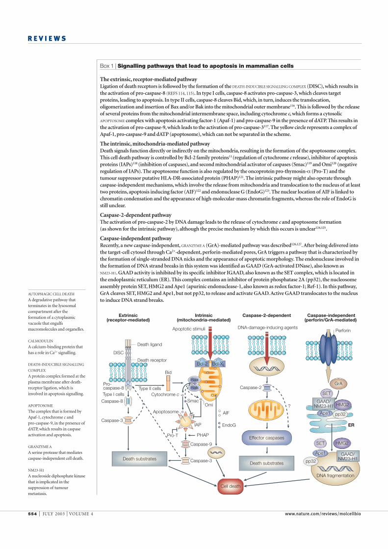
Box 1 | Signalling pathways that lead to apoptosis in mammalian cells
The extrinsic, receptor-mediated pathway Ligation of death receptors is followed by the formation of the DEATH-INDUCIBLE SIGNALLING COMPLEX (DISC), which results inthe activation of pro-caspase-8 (REFS 114, 115). In type I cells, caspase-8 activates pro-caspase-3, which cleaves targetproteins, leading to apoptosis. In type II cells, caspase-8 cleaves Bid, which, in turn, induces the translocation,oligomerization and insertion of Bax and/or Bak into the mitochondrial outer membrane116. This is followed by the releaseof several proteins from the mitochondrial intermembrane space, including cytochrome c, which forms a cytosolicAPOPTOSOME complex with apoptosis activating factor-1 (Apaf-1) and pro-caspase-9 in the presence of dATP. This results inthe activation of pro-caspase-9, which leads to the activation of pro-caspase-3117. The yellow circle represents a complex ofApaf-1, pro-caspase-9 and dATP (apoptosome), which can not be separated in the scheme.
The intrinsic, mitochondria-mediated pathway Death signals function directly or indirectly on the mitochondria, resulting in the formation of the apoptosome complex.This cell death pathway is controlled by Bcl-2 family proteins11 (regulation of cytochrome c release), inhibitor of apoptosisproteins (IAPs)118 (inhibition of caspases), and second mitochondrial activator of caspases (Smac)119 and Omi120 (negativeregulation of IAPs). The apoptosome function is also regulated by the oncoprotein pro-thymosin-α (Pro-T) and thetumour suppressor putative HLA-DR-associated protein (PHAP)121. The intrinsic pathway might also operate throughcaspase-independent mechanisms, which involve the release from mitochondria and translocation to the nucleus of at leasttwo proteins, apoptosis inducing factor (AIF)122 and endonuclease G (EndoG)123. The nuclear location of AIF is linked tochromatin condensation and the appearance of high-molecular-mass chromatin fragments, whereas the role of EndoG isstill unclear.
Caspase-2-dependent pathway The activation of pro-caspase-2 by DNA damage leads to the release of cytochrome c and apoptosome formation (as shown for the intrinsic pathway), although the precise mechanism by which this occurs is unclear124,125.
Caspase-independent pathway Recently, a new caspase-independent, GRANZYME A (GrA)-mediated pathway was described126,127. After being delivered intothe target-cell cytosol through Ca2+-dependent, perforin-mediated pores, GrA triggers a pathway that is characterized bythe formation of single-stranded DNA nicks and the appearance of apoptotic morphology. The endonuclease involved inthe formation of DNA strand breaks in this system was identified as GAAD (GrA-activated DNase), also known as NM23-H1. GAAD activity is inhibited by its specific inhibitor IGAAD, also known as the SET complex, which is located inthe endoplasmic reticulum (ER). This complex contains an inhibitor of protein phosphatase 2A (pp32), the nucleosomeassembly protein SET, HMG2 and Ape1 (apurinic endonuclease-1, also known as redox factor-1; Ref-1). In this pathway,GrA cleaves SET, HMG2 and Ape1, but not pp32, to release and activate GAAD. Active GAAD translocates to the nucleusto induce DNA strand breaks.
Bid
Pro-T
SmacCytochrome c
Apoptotic stimuli
Omi
IAP
PHAP
Apoptosome AIF
EndoG
Type II cellsType I cells
Caspase-8
Pro-caspase-8
Caspase-3
Caspase-3
DISC
Extrinsic(receptor-mediated)
Intrinsic(mitochondria-mediated)
Caspase-2-dependent Caspase-independent(perforin/GrA-mediated)
Death ligand
Death receptor
Caspase-9
Bax
Bak
Bcl-2 Bcl-XL
Death substrates
DNA-damage-inducing agents
?
Caspase-2
Death substrates
Effector caspases
ER
DNA fragmentation
GrA
SET
SET
Ape1
Ape1
GAAD/NM23-H1
GAAD/NM23-H1
pp32
pp32
HMG2
HMG2
Cell death
Perforin

NATURE REVIEWS | MOLECULAR CELL BIOLOGY VOLUME 4 | JULY 2003 | 555
R E V I E W S
plasma membrane17. The expression of Ins(1,4,5)P3
receptor type 3 antisense constructs in the mouse S49T–lymphoma cell line blocked dexamethasone-induced apoptosis. This finding provided an explana-tion for the need for extracellular Ca2+ in lymphocyteapoptosis. Moreover, another study has shown thatT lymphocytes that are deficient in type 1 Ins(1,4,5)P
3
receptors are resistant to apoptosis induced by severaltriggers, and that this resistance could be reversed by
Mg2+-dependent endonuclease14,15. Subsequent dissec-tion of the mechanism of glucocorticoid-inducedapoptosis in thymocytes showed that extracellular Ca2+
was necessary for cell death to occur16. More recentwork has shown that the type 3 INOSITOL-1,4,5-
TRISPHOSPHATE (INS(1,4,5)P3) RECEPTOR was upregulated in
lymphocytes undergoing Immunoglobulin-M-, ordexamethasone-induced apoptosis, and that the aug-mented receptor population was localized to the
INOSITOL-1,4,5-TRISPHOSPHATE
RECEPTOR
A receptor that is localizedmainly in the endoplasmicreticulum. It mediates therelease of Ca2+ into the cytosol.
Figure 2 | ER stress: causes and consequences. The accumulation of unfolded or misfolded proteins stimulates theactivation of several endoplasmic reticulum (ER) proteins, which results in either inhibition of translation or induction of geneexpression. In response to ER stress, unfolded proteins bind to the ER chaperone immunoglobulin heavy-chain binding protein(BiP/Grp78), thereby disrupting its interaction with a transmembrane serine/threonine (Ser/Thr) kinase, Ire1α, which affects generegulation. Ire1α can also be separated from the membrane through limited proteolysis by presenilin 1 (PS1). A homologousprotein, Ire1β, cleaves 28S ribosomal RNA, thereby inhibiting translation. Activating transcription factor-6 (ATF6) and RNA-activated protein kinase (PERK) are released from the ER in response to ER stress and are involved in the inhibition of geneactivation and translation, respectively. Among the affected genes are those that increase protein folding in the ER lumen, suchas the genes encoding BiP/Grp78, calreticulin and the transcription factor CHOP/GADD153. Upregulation of CHOP/GADD153also induces a decrease in Bcl-2 and Bcl-XL expression. Activation of pro-caspase-12 occurs either by the formation of acomplex with Ire1-α and tumour necrosis factor (TNF)-receptor-associated factor-2 (TRAF-2), or through cleavage by m-calpain21,22, which is released from the complex with its inhibitor, calpastatin (FIG. 4). The Ire1α–TRAF-2 complex can activatethe apoptotic signalling kinase 1 (ASK-1), which leads to activation of the Jun N-terminal kinase (JNK) pathway138. This might inturn result in phosphorylation of Bcl-XL and Bcl-2. Bcl-2-associated protein-31 (BAP31) interacts with pro-caspase-8, Bcl-XL andBcl-2 (REF. 24). This interaction is inhibited by Spike, a BH-3-only protein25. Active caspase-8 can cleave BAP31, and the amino-terminal fragment remains in the ER and promotes apoptosis. The putative ion-channel protein A4 was also identified as abinding partner of BAP31139. Changes in the ER Ca2+ concentration (four arrows in the middle of the figure) result in the activationof Ca2+-dependent enzymes (calpains) and reactive oxygen species (ROS) production. The luminal Ca2+ concentration iscontrolled by several other Ca2+ pumps and channels — sarco(endo)plasmic reticulum Ca2+-ATPase (SERCA), inositol-1,4,5-trisphosphate receptor (not shown) and ryanodine receptor (not shown) — as well as by Ca2+-sequestering andBcl-2 proteins (the latter can negatively regulate steady-state Ca2+ levels).
Caspase-3
ATF6
ASK-1
Ire1α
Ire1α
Caspase-12
Activation of JNKpathway
Pro-caspase-12Caspase-7
Caspase-9 (?)
Bcl-2
Bcl-2Bcl-2Bcl-XLBcl-XL
PERKCalpain
Calpastatin
ATF6
BiP/Grp78
PS1
PS1
Cleavage
Cleavage andphosphorylation
Oligomerization andphosphorylation
Regulation ofCa2+ spikes
SERCA
Regulation of ROS
Regulation ofsteady-state Ca2+
TRAF-2Ire1α
TRAF-2Ire1α
ER
Ire1β
Ire1β
Caspase-8
SpikePro-caspase-8
CleavageBAP31
Ca2+
Clustering
Gene activation
PERK
Inhibition oftranslation
Misfolded proteins
BAP31
A4

556 | JULY 2003 | VOLUME 4 www.nature.com/reviews/molcellbio
R E V I E W S
uptake of Ca2+ into mitochondria and mitochondrialrecruitment of a dynamin-related protein that mediatesrupture of the outer mitochondrial membrane(OMM), which has previously been shown to result inmitochondrial division27. Moreover, this fragmentstrongly sensitized mitochondria to caspase-8-inducedCYTOCHROME C release.
Mitochondria, Ca2+ and apoptosisFor almost half a century, mitochondria have beenknown to participate actively in intracellular Ca2+ com-partmentalization28. They take up Ca2+ electrophoreti-cally from the cytosol through a uniport transporterand can release it again through several different routes(FIG. 1). The energy-dependent Ca2+ uptake, coupled tothe release mediated by the exchange systems, consti-tutes an energy-dissipating mitochondrial Ca2+ cycle.
The affinity for Ca2+ of the uniporter is low, and thesize of the mitochondrial Ca2+ pool is small under phys-iological conditions. However, much larger amounts ofCa2+ can accumulate in mitochondria under pathologi-cal conditions, when intracellular Ca2+ concentrationsrise29. In fact, when phosphate is also taken up, Ca2+ isprecipitated in the mitochondrial matrix as insolublehydroxyapatite.
So, for many years mitochondrial Ca2+ uptake wasregarded mainly as a safety device in situations of tem-porary intracellular Ca2+ overload. However, morerecently this view has changed thanks to the develop-ment of new indicators, which can sense changes inCa2+ concentration in specific intracellular compart-ments30. As a result of this technology (see also the arti-cle by Tullio Pozzan and colleagues in this issue), it hasbecome apparent that mitochondrial Ca2+ fluxes areintegrated parts of cellular Ca2+ signalling31. The lowaffinity of the mitochondrial Ca2+ import system isovercome by the proximity of the mitochondria to theER and by the creation of Ca2+ ‘hotspots’ at the mouthof release channels32 (the local Ca2+ concentration atthese sites can reach very high levels). The resultingrapid uptake of Ca2+ by the mitochondria stimulatesthe Ca2+-sensitive matrix dehydrogenases, which arekey sites of NADH production for the respiratory chainand thereby for stimulation of mitochondrial energymetabolism.
Mechanisms of mitochondrial permeabilization.Recruitment of the mitochondrial pathway of apop-tosis signalling results in the permeabilization of theOMM and the release of pro-apoptotic mitochondr-ial proteins, of which cytochrome c has attracted themost attention, owing to its role in the activation ofthe caspase cascade (FIG. 3). Permeabilization of theOMM can be achieved by several different mecha-nisms, including pore formation by pro-apoptoticBcl-2 family proteins and OMM rupture as a result ofmitochondrial swelling. Evidence is accumulatingthat pro-apoptotic Bcl-2 family proteins, Bax andBak, as well as t-Bid and other BH3-only proteins,translocate to the mitochondria in the presence ofapoptotic stimuli. Bax and Bak can integrate into the
pharmacologically raising cytoplasmic Ca2+ levels18.Taken together, these observations indicate that Ca2+ isan important trigger of apoptosis in lymphocytes.
ER stress. Ca2+ storage and signalling,as well as the folding,modifying and sorting of newly synthesized proteins,are among the main functions of the ER in mammaliancells. Disturbances in any of these functions can lead toso-called ER stress (FIG. 2). Both Ca2+ overload anddepletion of the ER Ca2+ pool can result in changes inprotein folding and in ER stress. An enhanced protein-folding capacity of the ER might occur as a result ofthe activation of at least two signalling pathways19.First, the unfolding protein response (UPR) pathwayleads to the induction of ER chaperones, such asimmunoglobulin heavy-chain binding protein, BiP,which is also known as glucose-regulated protein-78(Grp78),Grp94,calreticulin,protein disulphide isomerase(PDI) and the transcription factor CHOP/GADD153.Second, the ER overload response (EOR) pathway leads tothe production of cytokines and interferons through acti-vation of nuclear factor (NF)-kB.Both pathways help cellsto remove the incorrectly folded and/or accumulated pro-teins in the ER.However, in response to severe stress, thesepathways can also contribute to the elimination of cellsthrough apoptosis20.
Prolonged ER stress stimulates the activation ofpro-caspase-12 (REF. 21). This enzyme is localized in theER membrane and is cleaved and activated by m-CALPAIN
during ER stress, or in response to the mobilization ofintracellular Ca2+ stores. Pro-caspase-12 might also beactivated by its recruitment into a complex with theendoribonuclease Ire1α and adaptor protein TRAF-2(tumour necrosis factor (TNF)-receptor-associatedfactor 2), which is formed as part of the UPR pathway22.Once activated, caspase-12 acts on effector caspases toinduce apoptosis. This link with the calpains is of particu-lar interest, because of the coordinated activity betweenthe two families of proteases. So, these findings indicatethat ER stress, as caused by Ca2+ depletion or alterations inthe Ca2+ transport systems (SARCO(ENDO)PLASMIC RETICULUM
CA2+-ATPASE (SERCA) and Ins(1,4,5)P3
receptors), can belinked directly to caspase activation.
As mentioned above, Bcl-2 proteins are key regula-tors of apoptosis. Several ER membrane proteins havebeen reported to interact with Bcl-2 family membersand influence the apoptotic process. Bax inhibitor 1protein, a mammalian apoptosis suppressor, which isalso present in plants and yeast, localizes specifically tothe ER membrane23. Furthermore, BAP31 (Bcl-2-asso-ciated protein-31), a 28-kDa integral membrane proteinof the ER, contains a cytosolic domain, which interactspreferentially with pro-caspase-8, Bcl-X
Land Bcl-2
(REF. 24; FIG. 2). Recently, a new member of the BH3-ONLY
FAMILY, Spike, was isolated and found exclusively in theER, where it inhibits the formation of a complexbetween BAP31 and Bcl-X
L25. Active caspase-8 can
cleave BAP31, with the amino-terminal fragmentremaining integrated in the ER and involved in induc-tion of apoptosis26.Overexpression of this fragment causesan early release of Ca2+ from the ER, with an associated
CALPAIN
A family of Ca2+-activatedcysteine proteases that have arole in cell death.
SARCO(ENDO)PLASMIC
RETICULUM CA2+-ATPASE
(SERCA). A pump located in thesarcoplasmic or endoplasmicreticulum membranes thatcouples ATP hydrolysis to thetransport of Ca2+ from cytosolicto lumenal spaces.
BH3-ONLY FAMILY
Pro-apoptotic Bcl-2 proteinsthat contain the BH3 domain,but lack the BH1, BH2 and BH4domains.
CYTOCHROME C
A haem-containing protein thatfunctions as an electron carrierin the mitochondrial respiratorychain and as a cofactor forcaspase activation duringapoptosis.

NATURE REVIEWS | MOLECULAR CELL BIOLOGY VOLUME 4 | JULY 2003 | 557
R E V I E W S
permeability transition. This involves the opening ofa permeability transition pore (PTP), which consistsof a large proteinaceous complex that comprises theVOLTAGE-DEPENDENT ANION CHANNEL (VDAC), the ADENINE
NUCLEOTIDE TRANSLOCATOR (ANT), mitochondrialcyclophilin D and several other proteins36,37 (FIG. 3).The pore complex has been localized to the contactsites between the inner mitochondrial membrane(IMM) and the OMM. The pore behaves as a voltage-operated channel that becomes activated by a highconcentration of Ca2+ in the mitochondrial matrix,oxidative stress, pyridine nucleotide oxidation, thioloxidation, alkalinization or low transmembranepotential. Initially, rapid and stochastic opening andclosing of the pore is observed. This, however, devel-ops rapidly into persistent pore opening, allowing notonly Ca2+ but also low-molecular-mass matrix com-ponents (M
r<1500) to easily leave the mitochondria.
At this stage, the opening of the pore can still bereversed by agents such as cyclosporin A38.
Apoptosis by mitochondrial permeabilization. Severaltreatments have been found to trigger apoptosisthrough Ca2+-mediated mitochondrial permeabilitytransition in various cell types. Such agents includeCa2+ ionophores and thapsigargin39, neurotoxins40,chemotherapeutics41 and pro-oxidants (for example,arachidonic acid42 and peroxynitrite43). Cell death canoften be prevented by inhibitors of mitochondrial Ca2+
uptake or PTP formation, such as ruthenium red andcyclosporin A. PTP formation results in the releasefrom mitochondria of cytochrome c and other pro-apoptotic proteins and might be associated withosmotic swelling of the mitochondria, owing to highmatrix protein concentration, and OMM rupture.However, this is not always the case, and it has beenspeculated that only a fraction of the mitochondrialpopulation might undergo permeability transition andrelease cytochrome c. Alternatively, resealing of the PTPmight occur, which allows the mitochondria to recoverin spite of the partial loss of cytochrome c and otherproteins from the intermembrane space42. Of particularinterest is the observation that apoptotic stimuli, notablyceramide, can induce a switch in mitochondrial Ca2+ sig-nalling at the beginning of the apoptotic process byfacilitating Ca2+-induced opening of the PTP44. This is inagreement with the recent observation that resistanceof leukaemic cells to 2-chlorodeoxyadenosine (CDA) isassociated with an increased ability of their mitochon-dria to sequester Ca2+ without concomitant PTPinduction. The CDA-resistant cells were selectivelycross-resistant to thapsigargin-induced apoptosis, butnot to staurosporine- or Fas-induced apoptosis45.
So, it seems that at least two distinct mechanismsmight be responsible for the permeabilization of the mito-chondria during the early phase of apoptosis. However,several observations indicate that these two mechanismsmight, in fact, be closely interrelated. For example, Bcl-2overexpression prevents mitochondrial permeability tran-sition and cytochrome c release46, and increases the overallmitochondrial Ca2+-uptake capacity47,48. Bax and Bak can
OMM as oligomers, and this process is stimulated byt-Bid and other BH3-only proteins33. However,sequestration of the pro-apoptotic Bcl-2 proteins bythe anti-apoptotic family members, Bcl-2 and Bcl-X
L,
prevents the oligomerization and insertion of Baxand Bak into the OMM and therefore pore forma-tion34. The finding that mice deficient in both Baxand Bak are resistant to most apoptotic stimuli pro-vides strong support for the importance of thismechanism in the activation of the mitochondrialpathway of apoptosis signalling35.
With the exception of the activation of the Bcl-2protein Bad by CALCINEURIN-catalysed dephosphoryla-tion, there is no known role for Ca2+ in the regulationof the translocation or oligomerization of the Bcl-2family proteins during apoptosis. Instead, Ca2+ seemsto regulate a second mechanism that is responsiblefor OMM permeabilization and the release of pro-apoptotic mitochondrial proteins, which is coupledto a stress response known as the inner-membrane
Figure 3 | Mechanisms of release of intermembrane-space proteins from mitochondria.At present, there are two recognized mechanisms for outer mitochondrial membrane (OMM)permeabilization. The first involves the opening of the permeability transition pore (PTP), whichconsists of voltage-dependent anion channel (VDAC), adenine nucleotide translocator (ANT) andcyclophilin D (CyD), as well as several other proteins37. Pore opening is followed by swelling of themitochondrial matrix, rupture of the OMM and the release of cytochrome c (Cyt c) and otherproteins from the intermembrane space. However, transient pore opening might also occurwhereby a small fraction of mitochondria undergo permeability transition at a given time. In thiscase, mitochondrial protein release occurs without marked large-amplitude swelling or loss ofmembrane potential (∆Ψ) in the entire population (not shown). In both cases, the process can beblocked by cyclosporin A, which binds to CyD and prevents pore opening. The secondmechanism of OMM permeabilization involves members of the Bcl-2 family of proteins, notablyBax and Bak, and t-Bid. Cytochrome c is normally bound to the inner mitochondrial membrane(IMM) by association with the anionic phospholipid cardiolipin. Cardiolipin is unique tomitochondria and is present predominantly, if not exclusively, in the IMM. Dissociation ofcytochrome c from cardiolipin is an important first step in cytochrome c release into the cytosoland is stimulated by reactive oxygen species (ROS) production as well as Ca2+ binding tocardiolipin (not shown)55-57.
Cyt c
Cyt c
Cyt c
Cyt c
Cyt c Cyt c
Cyt c
Cyt c
Cyt cCyt c
Cyt c
Cyt c Cyt c
Bcl-XL,Bcl-2
Cyt c
t-Bid
BaxBak
∆Ψ
∆Ψ
∆Ψ
ROS
Cardiolipin
Membranerupture
VDAC
Solutes,water
Swelling
CyDANT
Cyclosporin A
Bax, Bak
OMM
IMM
Ca2+
Mitochondria
PTP
CALCINEURIN
A Ca2+/calmodulin-activatedprotein phosphatase that isinvolved in Ca2+ signalling.
VOLTAGE-DEPENDENT ANION
CHANNEL
A pore in the outer membraneof the mitochondria thatmediates transport of anionsand certain other smallmolecules in and out of themitochondria.
ADENINE NUCLEOTIDE
TRANSLOCATOR
A carrier that is located in theinner mitochondrial membraneand that transports ADP into,and ATP out of, themitochondrial matrix.

558 | JULY 2003 | VOLUME 4 www.nature.com/reviews/molcellbio
R E V I E W S
active calcineurin A in Ins(1,4,5)P3-receptor-deficient
T cells restored the dephosphorylation of Bad and ren-dered the cells sensitive to apoptosis60.
Nitric oxide synthase. The role of the NITRIC OXIDE SYNTHASE
(NOS) enzymes in apoptosis signalling is complex,since NO• can exert both pro-apoptotic and anti-apoptotic effects. Ca2+-stimulated isozymes are consti-tutively expressed in endothelial cells (eNOS) andneurons (nNOS), and excessive production of NO•following glutamate-stimulated Ca2+entry throughNMDA (N-methyl-D-aspartate)-type glutamatereceptors has been implicated in excitotoxicity in cor-tical neuronal cultures61 and in brain ischaemia62. Theterminal mediator of cytotoxicity might in fact beperoxynitrite (ONOO−), which is readily formed byNO• and O
2−.
Endonucleases. Studies of endonucleases were an earlyapproach to the biochemistry of apoptosis, and severalCa2+-dependent and Ca2+-independent enzymes havesince been reported to be involved in apoptotic DNAdegradation63. The discovery of the caspase-3-activatedendonuclease and its inhibitor DNA fragmentationfactor DFF40/DFF45 (REF. 64; also known as caspase-activated DNase, CAD, and its inhibitor ICAD65), and itsinvolvement in nuclear apoptosis, led to a decrease inthe search for new nucleases, as it seemed that the iden-tity of the apoptotic endonuclease activity had beenresolved. However, the fact remains that the incubationof isolated cell nuclei with Ca2+ and ATP (in the absenceof caspase-mediated DNA cleavage activity) results in achromatin fragmentation pattern that is indistinguish-able from that seen in apoptotic cells66. So, the detailedcontribution of various nucleases to overall DNA degra-dation during apoptosis remains to be explained. Ofconsiderable interest is the recent finding that DNase IIhas a vital role in the phagocytic degradation ofengulfed apoptotic nuclear material67,68.
Phospholipases. The activation of PHOSPHOLIPASE A2
(PLA2) by Ca2+ results in the release of arachidonic acid
and related polyunsaturated fatty acids, which are fur-ther metabolized by lipoxygenases or cyclooxygenaseswith an associated generation of ROS. In addition, PLA
2
activation generates lysophosphatides that alter mem-brane structures. The importance of PLA
2in apoptosis
signalling is unclear, but recent work has established alink between intracellular arachidonic acid accumula-tion, resulting from the inhibition of its furthermetabolism by cyclooxygenases and lipoxygenases, andmitochondrial permeability transition, cytochrome crelease and caspase activation69.
Transglutaminases. TISSUE TRANSGLUTAMINASE has beenimplicated in several physiological processes, includingcrosslinking of integral plasma-membrane proteinswith the cytoskeleton. Expression of transglutaminasemessenger RNA and transglutaminase protein concen-trations both show a marked increase in dying cells,and apoptotic bodies have been found to be resistant
bind to VDAC and stimulate its activity, whereas Bcl-2and Bcl-X
Linhibit this binding49. Bax has been reported
to form a hybrid channel with VDAC that mediatescytochrome c release50. Hexokinase II interferes with theability of Bax to bind to mitochondria and inducecytochrome c release, presumably by competing with Baxfor binding to VDAC51. Furthermore, both Bax and Bcl-2can interact physically with the ANT and modulate itsactivity52. Finally, pretreatment of mitochondria witholigomeric, recombinant Bax has been found to sensi-tize them to Ca2+-induced permeability transition53,and t-Bid has been reported to stimulate mitochondrialCa2+ uptake, possibly by facilitating the passage of Ca2+
through the OMM54.
Ca2+–cardiolipin interaction and cytochrome c release.Cytochrome c is bound to the IMM by its associationwith the anionic phospholipid CARDIOLIPIN. Cardiolipin isunique to mitochondria and is present predominantly,if not exclusively, in the IMM. Evidence indicates thatthe dissociation of cytochrome c from cardiolipin is acrucial first step for cytochrome c release into the cytosoland for the induction of apoptosis55 (FIG. 3). The dissoci-ation of cytochrome c is facilitated by the peroxidationof cardiolipin, which results in a decreased bindingaffinity for the haemoprotein55,56. In addition, Ca2+ canbind to cardiolipin in the IMM, leading to decreasedlipid mobility, formation of cardiolipin-enricheddomains and protein aggregation57. In turn, thisrearrangement leads to an increased production ofreactive oxygen species (ROS) by the respiratory chain,which promotes the oxidation of membrane phospho-lipids and proteins and, as a result, an increase inmembrane permeability. In fact, the Ca2+–cardiolipininteraction might be an early and crucial step in thesequence of events by which Ca2+ triggers mitochondrialmembrane permeabilization.
Ca2+-activated effector mechanismsThere are many potential targets for Ca2+ signalling inapoptosis, some of which are discussed below.
Calcineurin. The activation of Ca2+-dependent proteinkinases and/or phosphatases that leads to alterations ingene transcription represents one possible mechanismby which Ca2+ might regulate apoptosis. The findingthat the immunosuppressant cyclosporin A could blockCa2+-dependent apoptosis in lymphoid cells providedsome of the first evidence for the involvement of cal-cineurin, a Ca2+/calmodulin-dependent protein phos-phatase in this process58. The orphan steroid receptorNur77 and the CD95 ligand represent two moleculartargets of calcineurin in mature T lymphocytes, andtheir induction might mediate the pro-apoptotic effect.Another mechanism for the pro-apoptotic effect of cal-cineurin is by means of the dephosphorylation of theBcl-2 family member, Bad, which results in its transloca-tion from the soluble cytoplasm to the mitochondriaand the stimulation of cytochrome c release and caspaseactivation59. Further support for this mechanism comesfrom the observation that expression of constitutively
CARDIOLIPIN
An anionic phospholipid that isuniquely localized in the innermitochondrial membrane andthat is of vital importance for thefunction of the respiratory chainand the adenine nucleotidetranslocator.
NITRIC OXIDE SYNTHASE
(NOS). A Ca2+-activated enzymein endothelial cells (eNOS) orneurons (nNOS) that formsnitric oxide from the aminoacid, L-arginine. Anotherisoform, inducible (i)NOS, canbe expressed in various cell typesbut is independent of calcium.
PHOSPHOLIPASE A2
A Ca2+-dependent enzyme thatreleases arachidonic acid fromphospholipids in cellularmembranes.
TISSUE TRANSGLUTAMINASE
A Ca2+-dependent enzyme thatis involved in the crosslinking ofproteins in apoptotic cells.

NATURE REVIEWS | MOLECULAR CELL BIOLOGY VOLUME 4 | JULY 2003 | 559
R E V I E W S
proteins (Bid, Bcl-XL
and Bcl-2), structural proteins(actin, α-fodrin, cytokeratin and focal adhesion kinase),neural cell-adhesion molecules (NCAMs), signal-trans-duction proteins (Ca2+/calmodulin-dependent proteinkinase, PKC-α and PKC-δ), DNA-repair and cell-cycleregulatory proteins (poly(ADP)ribose polymerase(PARP), DNA polymerase-ε, cyclin D and p53),glutamatereceptors (α-amino-3-hydroxy-5-methyl-4-isoxazolepropionic acid (AMPA) and NMDA) and several unclas-sified proteins (Ins(1,4,5)P
3receptor, amyloid precursor
protein (APP) and calcineurin).Although both calpains and caspases are cysteine
proteases, their functions and cleavage specificity are dif-ferent. All caspases contain a conserved QACXG pen-tapeptide, where C is the active-site cysteine, and theyhave a unique, strong preference for the cleavage of pep-tide bonds that are carboxy-terminal of aspartateresidues. This cleavage specificity is rare among proteasefamilies, and only the serine protease granzyme B has asimilar cleavage specificity73. Calpains are Ca2+-activatedproteases that include several tissue-specific isoforms(n-calpains) and two ubiquitous isozymes (µ-calpainand m-calpain). For activation, µ- and m-calpainsrequire micromolar and millimolar Ca2+ concentra-tions, respectively74. Compared with caspases, calpainsdo not have a strict cleavage specificity. Calpain activityis regulated by the endogenous inhibitor, calpastatin.Interestingly, in several experimental models of apoptosisit has been shown that calpastatin is cleaved by caspase-3,and that this cleavage is essential for the regulation ofcalpain activity during cell death75, 76.
Calpain can also cleave caspases, and, most often, thiscleavage inactivates caspase function. For example, cal-pain can cleave caspase-7 at sites that are distinct fromthose cleaved by the upstream caspases, generating prote-olytically inactive fragments77. Pro-caspase-8 and pro-caspase-9 can also be cleaved by calpains, and truncatedcaspase-9 is unable to activate pro-caspase-3 (REF. 78).Finally, it has been reported that pro-caspase-3 cleavageby calpain results in the generation of a 29-kDa frag-ment, although it is unclear whether such cleavage causesthe activation or inactivation of caspase-3 function77,78.Arecent study79 reports that, in the early execution phase,calpain downregulates caspase-3 activity and slows pro-gression of the apoptotic nuclear morphology, whereassubsequent calpain activity, facilitated by caspase-medi-ated degradation of calpastatin, contributes to plasma-membrane disruption and secondary necrosis. The bestknown example of caspase activation by calpain is thecleavage of pro-caspase-12 (discussed above), althoughthe precise mechanism of this activation is unclear80.
Several cell survival proteins, including Bcl-2 andBcl-X
L, are cleaved by both caspases and calpain. In both
cases, this cleavage results in fragments that promote celldeath rather than survival. By contrast, a 18-kDa prod-uct of Bax cleavage by calpain or caspases is a morepotent inducer of apoptosis than uncleaved Bax81.Similarly, the pro-apoptotic protein Bid can be cleavedby calpain as well as by caspase-8. Calpain cleaves Bidbetween Gly70 and Arg71, whereas caspase-8 cleavesbetween Asp59 and Gly60 as well as between Asp75 and
to dissolution by detergents and chaotropic agentsas a result of Ca2+-dependent transglutaminase activa-tion70. The resistance of these bodies to proteolysismight allow them to accumulate, and they can bedetected in blood plasma after the induction of apop-tosis in various organs.
Proteases. Although many recent studies have focusedon caspases, there is considerable evidence that otherproteases, including serine proteases and members ofthe calpain family of Ca2+-activated cysteine proteases,participate in apoptosis. Results of inhibitor studiesseem to indicate that the contribution of calpains toapoptosis is more prominent in certain cell types (forexample, thymocytes, monocytes, cardiomyocytes andneuronal cells)71. An increasing number of cellular pro-teins have now been found to be cleaved by calpainsduring apoptosis. In addition, calpain has recently beenreported to cleave the endogenous calcineurin inhibitorcain (also known as cabin 1), which results in the activa-tion of calcineurin and the promotion of Ca2+-triggeredcell death72.
Caspase and calpain crosstalk Both calpains and caspases are cysteine proteases that areimportant in cell death, and accumulated data indicatethat there is crosstalk between calpains and caspases inthe regulation of this process (FIG. 4). Proteins that can becleaved by both calpains and caspases during apoptosisinclude pro-caspase-3 and pro-caspase-7, Bcl-2 family
Figure 4 | Crosstalk between calpains and caspases during apoptosis. a | Cleavage of theendogenous calpain inhibitor calpastatin by caspase-3 (or by calpain itself) is essential foractivation of the Ca2+-dependent protease calpain75,76. This can lead to cleavage of several pro-caspases, which can either activate or inactivate their function77–80. b | Both calpain andcaspases can cleave the same proteins and either activate or inactivate their function. Althoughthe cleavage sites in target proteins are different, the consequences of cleavage induced by thetwo protease families are usually similar. There is accumulating evidence that these proteasesmight in fact work in conjunction to accelerate the cell death process. AMPA, α-amino 3-hydroxy-5-methyl-4-isoxazole proprionic acid; APP, amyloid precursor protein; FAK, focal adhesion kinase;Ins(1,4,5)P3R, inositol-1,4,5-trisphosphate receptor; NCAM, neural cell-adhesion molecule; NMDA,N-methyl-D-aspartate; PARP, poly(ADP)ribose polymerase; PKC, protein kinase C.
Caspase-3 Calpastatin
Pro-caspase-12
Pro-caspase-3
Pro-caspase-7
Pro-caspase-8
Pro-caspase-9
Calpain
Ca2+
Calpain
Caspases
and
Activation
Inactivation
Activation
Inactivation
BaxBid
ActinAPPCalcineurinCyclin DFAKIns(1,4,5)P3RNMDA receptorPKC α and δ
AMPA receptorBcl-XLCalpastatinDNA polymerase εα-FodrinNCAMPARP
a
b

560 | JULY 2003 | VOLUME 4 www.nature.com/reviews/molcellbio
R E V I E W S
finding that it can function as an ion channel whenincorporated into lipid bilayers95 provided a plausiblemechanism for its reduction of the ER Ca2+ pool. Thishypothesis is supported by the finding that Bcl-2increases the permeability of the ER membrane to Ca2+,without any compensatory effects on its Ca2+ uptakecapacity. However, it seems that this mechanism mightnot apply to all cell types, as overexpression of Bcl-2 inbreast epithelial cells has been reported to modulate theER Ca2+ pool by upregulating SERCA expression with-out affecting Ca2+ release from the ER96. This led toaccelerated Ca2+ uptake and enhanced Ca2+ loading bythe ER, which, however, did not seem to be associatedwith an increased risk for mitochondrial permeabilitytransition and apoptosis, as released ER Ca2+ is quicklyretrieved owing to the enhanced activity of the SERCApump.
In addition, pro-apoptotic Bcl-2 family proteinscan affect Ca2+ handling by the ER. The enforcedexpression of either Bax or Bak has been found toresult in the accumulation of these proteins in the ERand mitochondria, and to promote Ca2+ translocationfrom the ER to the mitochondria and cytochrome crelease97. Neither mobilization of ER Ca2+, nor mito-chondrial Ca2+ uptake, occurred in Bax−/− cells.However, in a recent study, double knock out (DKO)cells deficient for both Bax and Bak had a reduced ERCa2+ pool and, secondarily, showed a decreaseduptake of Ca2+ by mitochondria98. The decrease in theER Ca2+ concentration was presumably due toenhanced Ca2+ leakage from the ER and was correlatedwith a marked increase in phosphorylation of theIns(1,4,5)P
3 receptor, which is known to stabilize its
open conformation and therefore enhance Ca2+ leak-age. Transfection of the SERCA pump into DKO cellscorrected the ER Ca2+ level and mitochondrial Ca2+
uptake and restored sensitivity to apoptosis induced byoxidative stress. So, the effects on the ER Ca2+ pool size,mitochondrial Ca2+ uptake and sensitivity to Ca2+-mediated apoptosis that are seen in the DKO cells weresimilar to the findings with Bcl-2-overexpressingcells93,94. However, unlike the latter cells, the DKO cellsdid not show decreased capacitative Ca2+ entry orreduction of calreticulin and SERCA levels. Together,the results of these studies indicate that the Bcl-2 familyproteins are important in the modulation of the ERCa2+ pool, and point to the importance of this pool forthe generation of Ca2+-mediated apoptotic signals.
Ca2+ and the phagocytosis of apoptotic cellsApoptotic cell death is terminated by phagocytosis ofthe dying cell by phagocytes or neighbouring cells. Infact, the efficiency with which apoptotic cells undergophagocytosis compared with necrotic cells has longbeen regarded as a hallmark of apoptosis, althoughrecent studies in C. elegans indicate that a common setof genes function to eliminate cell corpses, irrespectiveof the mode of cell death99.
Exposure of the aminophospholipid phosphatidylser-ine on the outer cell surface is the most important ‘eat me’signal used by apoptotic cells100 (FIG. 5). Phospholipids are
Ser76 (REF. 82). Importantly, in both situations the BH3domain of Bid remains intact, and the pro-apoptoticactivity is maintained. So, it is clear that calpains andcaspases interact, and that cleavage of the same proteinsby two, or more, proteases allows either protease alone, orboth in combination, to activate the apoptotic process.
Caspases and intracellular ion homeostasisAs discussed above, various apoptotic stimuli mightaffect the intracellular Ca2+ concentration,[Ca2+]
i, and,
in turn, Ca2+ signals can trigger apoptosis and lead to theactivation of caspases. Once activated, the caspasesmight act on key components of the signalling systemand perturb Ca2+ homeostasis. For example, it has beenshown that caspase-3 can cleave the type 1 Ins(1,4,5)P
3
receptor in apoptotic Jurkat cells, and that such cleavageresulted in a decrease in channel activity as the recep-tor was degraded83. In addition, recent reports indicatethat caspase-3 can also cleave the plasma-membraneCa2+-ATPase84,85. This enzyme is crucial for the long-term control of cellular Ca2+ homeostasis, and cleavage-associated loss of function during apoptosis results inCa2+ overload and secondary cell lysis or necrosis.Moreover, preliminary observations (P.N., unpublishedobservations) indicate that the Na+/Ca2+-exchanger isalso cleaved by caspase-3 during apoptosis, which fur-ther decreases the ability of the apoptotic cell to main-tain Ca2+ homeostasis.
The Na+/K+-ATPase is another ion pump in theplasma membrane that can function as a caspase sub-strate during apoptosis. Previous studies have shownthat volume shrinkage in apoptotic cells is accompa-nied by a net efflux of ions owing to the inactivation ofthe Na+/K+-ATPase86, and that apoptosis is acceleratedby a Bcl-2-sensitive activation of outward K+
currents87. The β-subunit of the Na+/K+-ATPase, whichregulates the K+ affinity of the enzyme at the extracel-lular site, seems to be the caspase target88. Apart fromits role in cell shrinkage during apoptosis, inactivationof the Na+/K+-ATPase might promote the apoptoticprocess by lowering the intracellular K+ concentrationto levels that are compatible with optimal caspaseactivity89.
Effects of Bcl-2 proteins on ER Ca2+ storageThere is now convincing evidence that the release ofCa2+ from the ER store, followed by its translocationinto the mitochondria, is an important signal for theactivation of the apoptotic process. Early studies indi-cated that depletion of the ER Ca2+ pool was bettercorrelated to the apoptotic outcome of a particulartreatment than the resultant capacitative influx of extra-cellular Ca2+ or the overall increase in the cytosolic Ca2+
concentration90,91. Support for this assumption alsocomes from the observation that Bcl-2 overexpressioncould protect cells from thapsigargin-inducedapoptosis92, and that Bcl-2 reduced the filling of theER Ca2+ store and diminished the extent of capacita-tive Ca2+ entry following its depletion93,94. Bcl-2 hasbeen found to localize to the ER and the nuclear mem-brane, in addition to the mitochondria. The subsequent

NATURE REVIEWS | MOLECULAR CELL BIOLOGY VOLUME 4 | JULY 2003 | 561
R E V I E W S
regulation of phospholipid asymmetry in the plasmamembrane, have not been characterized in detail,although it seems that the aminophospholipid translo-case is crucially dependent on the intracellular ATPconcentration for activity and is highly sensitive toinhibition by SH-reagents. A comparison of phos-phatidylserine exposure in several different cell lines thatundergo Fas-mediated apoptosis has shown that it wasbetter correlated to differences in translocase than inscramblase activity, and that the upregulation of scram-blase activity by either gene transfection or enzymeinduction had little effect on the extent of phos-phatidylserine exposure104.
So, what is the role of Ca2+ in the regulation of phos-pholipid asymmetry in the plasma membrane? As men-tioned above, scramblase activity is dependent on Ca2+,whereas aminophospholipid translocase activity is inhib-ited by Ca2+.Furthermore, in platelets,which also have anapoptosis-like death programme105,106, elevation of thecytosolic Ca2+ concentration with thapsigargin can triggerlarge amounts of phosphatidylserine exposure in min-utes107. In thapsigargin-treated Jurkat cells or U937promonocytes, however, far fewer cells exposed phos-phatidylserine, and it took up to six hours for this to
distributed asymmetrically across the plasma mem-brane, with the bulk of phosphatidylcholine and sphin-gomyelin localized to the outer leaflet, whereas most ofthe phosphatidylethanolamine and all of the phos-phatidylserine are localized to the inner leaflet of theplasma membrane in healthy cells. In most, but not all,apoptotic cells this situation is changed, such that part ofthe phosphatidylserine pool is translocated to the cellsurface the distribution of the other major phospho-lipids is not affected. Phosphatidylserine exposure stimu-lates phagocytic activity through several receptors,including the newly discovered phosphatidylserinereceptor101 and scavenger receptors102.
In the plasma membrane, the distribution of phos-phatidylserine between the two leaflets is regulated bythree enzymes: a Ca2+-dependent scramblase, whichcatalyses the nonspecific randomization of phospho-lipids across the bilayer; an ATP-dependent aminophos-pholipid translocase, which mediates the movement ofaminophospholipids from the outer to the innerleaflet; and a third enzyme, which mediates the energy-dependent transfer of phospholipids from the inner tothe outer leaflet103. With the exception of the scramblase,these enzymes and their relative contribution to the
Figure 5 | Positive and negative recognition signals for phagocytosis of apoptotic cells. a | Phosphatidylserine (PS)accumulates in the outer leaflet of the plasma membrane of apoptotic cells. b | Apoptotic neurons with condensed nuclei (stainedblue with Hoechst 33342) and phosphatidylserine exposed on the cell surface, as visualized by staining with FITC (fluoresceinisothiocyanate)–annexin V (green)140. c | Regulation of phosphatidylserine distribution between the two leaflets of the plasmamembrane. Phospholipid scramblase (PLS) activity is Ca2+ dependent, whereas aminophospholipid translocase (APT) is inhibited byCa2+ and SH-reagents (N-ethylmaleimide, NEM) and is vitally dependent on the intracellular ATP concentration. d | Normal viablecells are not engulfed by macrophages. Active repulsion of viable cells can be achieved by cell-surface molecules, which transmitdetachment signals. Platelet–endothelial cell adhesion molecule-1 (PECAM-1, also known as CD31) normally prevents phagocyterecognition of viable cells. In apoptosis, the function of CD31 changes from repulsive (orange) to adhesive (green) and promotesphagocytosis110. In some cells, CD31 engagement activates a cation-nonspecific channel111, which mediates Ca2+ influx.Changesin the properties of CD31 in apoptotic cells might modulate Ca2+ signals and the subsequent exposure of phosphatidylserine and/orannexin I.
PSPSPS
PSPSPSPSPS
–
PLS
APT
PSPS
PS ATP ADP
NEMCa2+ Ca2+ Annexin I
+ –
Healthy cells Apoptotic cells
Outer leaflet
Inner leaflet
CD31
Phagocyte
?
a b
c
d
562 | JULY 2003 | VOLUME 4 www.nature.com/reviews/molcellbio
R E V I E W S
ConclusionsRecent developments have emphasized the central roleof the calcium ion in the regulation of cell death. Similarto the conductor of an orchestra, it can activate dis-tinct parts of the cell death programme, which canthen function alone or in conjunction with other sub-programmes to kill the cell. Thanks to the developmentof new and highly sensitive methodologies, we can nowappreciate the importance of ER–mitochondrial Ca2+
fluxes in apoptosis signalling and the central function ofmitochondria in the Ca2+-regulated cell death pro-gramme. Recent findings have provided ample evidencefor crosstalk between calpains, caspases and other pro-tease families, and have indicated new pathways that leaddirectly from a Ca2+ signal to caspase activation andapoptosis. The discovery that intracellular Ca2+ compart-mentalization is modulated by the Bcl-2 family proteins,and that caspase cleavage of Ca2+ transporters mightcause perturbation of intracellular Ca2+ homeostasis andtherefore both aggravate the damage and affect the modeof cell death, provides further support for the impor-tance of the Ca2+–apoptosis link. Future research willhave to delineate these pathways in further detail andexplore the function of Ca2+ in other, less well-defineddeath programmes, such as autophagy and granzyme A-mediated cell killing (BOX 1). The potential importance ofthe Ca2+–apoptosis link in disease pathogenesis must befurther substantiated (BOX 2). Furthermore, although wehave already entered the era of developing new pharma-ceuticals that target the Ca2+–apoptosis link, both thebeneficial and the potential adverse effects of such thera-pies must be critically evaluated.
So, is Ca2+ involved in all forms of apoptosis? It couldbe too early to answer this question. Many stimuliinduce apoptosis in the absence of any detectablechanges in Ca2+ fluxes, and therefore no direct role forCa2+ signalling is apparent. On the other hand, researchover the past several years has identified components of
occur108. In addition, in TNF- or Fas-treated Jurkat cells,chelation of intracellular Ca2+ had no effect on phos-phatidylserine exposure,whereas removal of extracellularCa2+ caused about 50% inhibition. So, at present it can beconcluded that phosphatidylserine exposure in apoptosis isCa2+ dependent,but not Ca2+ regulated,whereas the sameprocess in platelets is Ca2+ regulated. However, the exactmechanisms by which Ca2+ regulates phosphatidylserineexposure in platelets remain to be elucidated.
Phagocytosis of apoptotic cells is promoted by sev-eral endogenous protein ligands. A recent study109 hasidentified the Ca2+-dependent, phosphatidylserine-binding protein annexin I as a new engulfment ligand. Itis rapidly upregulated during Fas-mediated apoptosisand recruited to the phosphatidylserine-rich domainson the cell surface, where it mediates the tethering andinternalization of apoptotic cells. The recruitment ofannexin I is both Ca2+ and caspase dependent, and itseems that exposed annexin I stimulates phagocytosisby promoting clustering of the phosphatidylserinereceptor on engulfing cells.
CD31. A further mechanism that might promotephagocytosis of apoptotic cells is the disappearance of‘repulsion’ signals from the cell surface. The surfacemolecule CD31, also known as platelet–endothelial celladhesion molecule-1 (PECAM-1), can change its func-tion in apoptotic cells110. CD31 normally transmits‘detachment’ signals to scavenger cells. Instead, in apop-totic cells, CD31 allows tethering of dying cells tophagocytes (FIG.5d). Notably, CD31 engagement modu-lates a cation channel that is permeable to Ca2+, the acti-vation of which leads to an increased [Ca2+]
i(REF. 111).
This could constitute an interesting signalling loopbetween surface adhesion molecules that are involved inthe recognition of apoptotic cells and Ca2+ signals thatwould promote the associated appearance of phos-phatidylserine and/or annexin I on the cell surface.
Box 2 | Calcium and apoptosis in disease: possible therapeutic approaches
The dysregulation of apoptosis has been implicated in the pathogenesis of several human diseases, including brainischaemia, autoimmune disease and cancer128, and a link between Ca2+ overload and disease development has long beenknown129. Brain ischaemia is a pathological condition in which both Ca2+ overload and the caspase proteases arepotential therapeutic targets. In focal brain ischaemia, the initial insult, the subsequent reperfusion injury and theimmune response all contribute to the final degree of damage. Ion-channel blockers, particularly NMDA (N-methyl-D-aspartate) receptor antagonists, can attenuate the initial neuronal Ca2+ overload and the subsequentneuronal damage130.
Downstream of the NMDA receptor, caspases execute apoptosis, but also promote cytokine release and pro-inflammatory reactions131. Caspase inhibition is effective in reducing excitotoxic and ischaemic injury in isolatedneurons or in animal models of ischaemia132,133, and co-administration of NMDA channel blockers and caspase inhibitorshas a synergistic effect in brain hypoxia134.A possible explanation for this synergy has recently been suggested85.
There are, however, concerns about the use of caspase inhibitors for therapeutic purposes. One such concern is therisk of proliferation of unwanted cells that would normally be removed by caspase-mediated apoptosis. The other is thequestion of non-apoptotic functions of caspases. In stroke therapy, systemic effects of caspase inhibition might beavoided by selectively targeting caspase inhibitors to the central nervous system. However, other possible effects ofcaspase activation remain to be investigated. For example, recent experiments135 indicate a potential role of limitedcaspase-3 activation for preconditioning that is required to achieve neuroprotection. Preconditioning refers tosituations when exposure to a limited insult, such as transient ischaemia, makes neurons less sensitive to subsequentchallenges. If confirmed, these findings show that the limited activation of caspases might indeed have protectivefunctions.

NATURE REVIEWS | MOLECULAR CELL BIOLOGY VOLUME 4 | JULY 2003 | 563
R E V I E W S
observed in several cell types, perhaps most strikingly indeveloping neurons that undergo apoptosis on with-drawal of nerve growth factor. Depolarization of thecells using K+ to open voltage-sensitive Ca2+ channelsprovides the neurons with a survival signal112. Theseobservations led to the formulation of the Ca2+ set pointhypothesis, in which a minimal Ca2+ level is required tomaintain neuron viability113. When the [Ca2+]
imoves
below or above this set point, apoptosis is rapidlyinduced. The mechanisms responsible for the anti-apoptotic effect of Ca2+ have not been conclusively iden-tified but they might involve transcriptional activationand synthesis of anti-apoptotic signals or, simply, itcould be that Ca2+ influx keeps the ER Ca2+ pool filledand prevents its depletion by the apoptotic stimulus orER stress response.
the Ca2+-signalling machinery as important regulatorsof apoptosis, even when there was no prior evidence forCa2+ involvement. These observations indicate that thereis a more general participation of Ca2+ signalling inapoptosis than was previously thought. An example ofthis is the recent finding that, once it has been releasedfrom the mitochondria, cytochrome c binds not only toApaf-1 but also to the Ins(1,4,5)P
3receptor and blocks
Ca2+-mediated inhibition of its function (S. H. Snyder,personal communication). This results in sustained,oscillatory Ca2+ increases, which lead to augmentedcytochrome c release and amplification of the apoptoticsignal.
Finally, in spite of the accumulating evidence of theCa2+–apoptosis link, we should not forget that Ca2+ canalso function as an anti-apoptotic signal. This has been
1. Fleckenstein, A., Janke J., Doring, H. J. & Leder, O.Myocardial fiber necrosis due to intracellular Ca overload a new principle in cardiac pathophysiology.Recent Adv. Stud. Cardiac Struct. Metab. 4, 563–580(1974).
2. Leonard, J. P., & Salpeter, M. M. Agonist-induced myopathyat the neuromuscular junction is mediated by calcium. J. Cell Biol. 82, 811–819 (1979).
3. Schanne, F. A. X., Kane, A. B., Young, E. E. & Farber, J. L.Calcium dependence of toxic cell death. Science 206,700–702 (1979).
4. Trump, B. F. & Berezesky, I. K. Calcium-mediated cell injuryand cell death. FASEB J. 9, 219–228 (1995).
5. Xu, K., Tavernarakis, N. & Driscoll, M. Necrotic cell death inC. elegans requires the function of calreticulin and regulatorsof Ca2+ release from the endoplasmic reticulum. Neuron 31,957–971 (2001).Evidence of Ca2+-mediated necrotic cell death in thesame nematode species that has been the modelorganism for the elucidation of the genetic regulationof programmed cell death.
6. Kerr, J. F. R., Wyllie, A. H. & Currie, A. R. Apoptosis: a basicbiological phenomenon with wide-ranging implications intissue kinetics. Br. J. Cancer 26, 239–257 (1972).This famous report introduced the term apoptosis anddescribed the morphological characteristics andbiological significance of this mode of cell death.
7. Eguchi, Y., Shimizu, S. & Tsujimoto, Y. Intracellular ATP levelsdetermine cell death fate by apoptosis or necrosis. CancerRes. 57, 1835–1840 (1997).
8. Leist, M., Single, B., Castoldi, A. F., Kuhnle, S. & Nicotera, P.Intracellular adenosine triphosphate (ATP) concentration: aswitch in the decision between apoptosis and necrosis. J. Exp. Med. 185, 1481–1486 (1997).
9. Horvitz, H. R. Genetic control of programmed cell death inthe nematode Caenorhabditis elegans. Cancer Res. 59,1701s–1706s (1999).This review summarizes the pioneering studies on thegenetic regulation of programmed cell death in C. elegans.
10. Alnermi, E. S. et al. Human ICE/CED-3 proteasenomenclature. Cell 87, 171 (1996).
11. Cory, S. & Adams, J. M. The Bcl-2 family: regulators of thecellular life-or-death switch. Nature Rev. Cancer 2, 647–656(2002).
12. Bursch, W. The autophagosomal–lysosomal compartmentin programmed cell death. Cell Death Differ. 8, 569–581(2001).
13. Inbal, B., Bialik, S., Sabanay, I., Shani, G. & Kimchi, A. DAP kinase and DRP-1 mediate membrane blebbing and the formation of autophagic vesicles during programmed cell death. J. Cell Biol. 157, 455–468(2002).
14. Wyllie, A. H. Glucocorticoid-induced thymocyte apoptosis isassociated with endogenous endonuclease activation.Nature 284, 555–556 (1980).This report shows that the ‘DNA ladder’ is a hallmarkof apoptosis and can be formed as a result of Ca2+-mediated endonuclease activity.
15. Cohen, J. J. & Duke, R. C. Glucocorticoid activation of acalcium-dependent endonuclease in thymocyte nuclei leadsto cell death. J. Immunol. 132, 38–42 (1984).
16. McConkey, D. J. et al. Glucocorticoids activate a suicideprocess in thymocytes through an elevation of cytosolicCa2+ concentration. Arch. Biochem. Biophys. 269, 365–370(1989).
17. Khan, A. A. et al. Lymphocyte apoptosis: mediation byincreased type 3 inositol 1,4,5-triphosphate receptor.Science 273, 503–507 (1996).This report links apoptosis to upregulation of type 3Ins(1,4,5)P3 receptors in the lymphocyte plasmamembrane.
18. Jayaraman, T. & Marks, A. R. T cells deficient in inositol1,4,5-triphophate receptor are resistant to apoptosis. Mol. Cell. Biol. 17, 3005–3012 (1997).
19. Kaufman, R. J. Orchestrating the unfolded protein responsein health and disease. J. Clin. Invest. 110, 1389–1398(2002).
20. Ferri, K. F. & Kroemer. G. Organelle-specific initiation of celldeath pathways. Nature Cell Biol. 3, E255–E263 (2001).
21. Nakagawa T., et al. Caspase-12 mediates endoplasmic-reticulum-specific apoptosis and cytotoxicity by amyloid-β.Nature 403, 98–103 (2000).The first evidence that caspase-12 is localized to theER and is activated by ER stress.
22. Yoneda, T., et al. Activation of caspase-12, an endoplasmicreticulum (ER) resident caspase, through tumor necrosisfactor receptor-associated factor 2-dependent mechanismin response to the ER stress. J. Biol. Chem. 276,13935–13940 (2001).
23. Xu, Q. & Reed, J. C. Bax inhibitor-1, a mammalian apoptosissuppressor identified by functional screening in yeast. Mol. Cell 1, 337–346 (1998).
24. Ng, F. W., et al. p28 Bap31, a Bcl-2/Bcl-XL- andprocaspase-8-associated protein in the endoplasmicreticulum. J. Cell. Biol. 139, 327–338 (1997).
25. Mund, T. Gewies, A., Schoenfield, N., Bauer, M. K. A. &Grimm, S. Spike, a novel BH3-only protein, regulatesapoptosis at the endoplasmic reticulum. FASEB J.2003 Feb 19 (DOI: 10.1096/2 fj.02–0657fje).
26. Breckenridge, D. G., Stojanovic, M., Marcellus, R. C. &Shore, G. C. Caspase cleavage product of BAP31 inducesmitochondrial fission through endoplasmic reticulumcalcium signals, enhancing cytochrome c release to thecytosol. J. Cell Biol. 160, 1115–1127 (2003).
27. Frank, S. et al. The role of dynamin-related protein 1, a mediator of mitochondrial fission, in apopotosis. Dev. Cell 1, 515–525 (2001).
28. Carafoli, E. Calcium signaling: a tale for all seasons. Proc. Natl Acad. Sci. USA 99, 1115–1122 (2002).
29. Thor, H., Hartzell, P. & Orrenius, S. Potentiation of oxidativecell injury in hepatocytes which have accumulated Ca2+. J. Biol. Chem. 259, 6612–6615 (1984).
30. Rizzuto, R., Simpson, A. W., Brini, M. & Pozzan, T. Rapidchanges of mitochondrial Ca2+ revealed by specificallytargeted recombinant aequorin. Nature 358, 325–327(1992).
31. Rizzuto, R., Brini, M., Murgia, M. & Pozzan, T. Microdomainswith high Ca2+ close to IP3-sensitive channels that aresensed by neighboring mitochondria. Science 262,744–747 (1993).Early report of ER–mitochondrial Ca2+ fluxesoriginating from microdomains with elevated Ca2+ inthe cytoplasm.
32. Hajnoczky, G., Robb-Gaspers, L. D., Seitz, M. B. & Thomas, A. P. Decoding of cytosolic calcium oscillations inthe mitochondria. Cell 82, 415–424 (1995).
33. Eskes, R., Desagher, S., Antonsson, B. & Martinou, J. C.Bid induces the oligomerization and insertion of Bax into theouter mitochondrial membrane. Mol. Cell. Biol. 20, 929–935(2000).
34. Antonsson, B. et al. Inhibition of Bax channel-formingactivity by Bcl-2. Science 277, 370–372 (1997).
35. Lindsten, T. et al. The combined functions of proapoptoticBcl-2 family members Bak and Bax are essential for normaldevelopment of multiple tissues. Mol. Cell 6, 1389–1399(2000).
36. Halestrap, A. P., Kerr, P. M., Javadov, S. & Woodfield, K. Y.Elucidating the molecular mechanism of the permeabilitytransition pore and its role in reperfusion injury of the heart.Biochim. Biophys. Acta 1366, 79–94 (1998).
37. Crompton, M. The mitochondrial permeability transition poreand its role in cell death. Biochem. J. 341, 233–249 (1999).
38. Weis, M., Kass, G. E. & Orrenius, S. Further characterizationof the events involved in mitochondrial Ca2+ release andpore formation by prooxidants. Biochem. Pharmacol. 47,2147–2156 (1994).
39. Korge, P. & Weiss, J. N. Thapsigargin directly induces themitochondrial permeability transition. Eur. J. Biochem. 265,273–280 (1999).
40. Akao, Y. et al. Mitochondrial permeability transition mediatesapoptosis induced by N-methyl(R)salsolinol, an endogenousneurotoxin, and is inhibited by Bcl-2 and rasagiline, N-propargyl-1(R)-aminoindan. J. Neurochem. 82, 913–923(2002).
41. Kidd, J. F. et al. Paclitaxel affects cytosolic calcium signalsby opening the mitochondrial permeability transition pore. J. Biol. Chem. 277, 6504–6510 (2002).
42. Petronili, V., Penzo, D., Scorrano, L., Bernardi, P. & Di Lisa, F.The mitochondrial permeability transition, release ofcytochrome c and cell death. J. Biol. Chem. 276,12030–12034 (2001).
43. Schweizer, M. & Richter, C. Peroxynitrite stimulates thepyridine nucleotide-linked Ca2+ release from intact rat livermitochondria. Biochemistry 35, 4524–4528 (1996).
44. Szalai, G., Krishnamurthy, R. & Hajnoczky, G. Apoptosisdriven by IP3-linked mitochondrial calcium signals. EMBO J.18, 6349–6361 (1999).This paper presents evidence that apoptotic stimulifacilitate Ca2+-induced mitochondrial permeabilitytransition, and that resealing of pores can occur afterdecay of Ca2+ spikes.
45. Chandra, J. et al. Resistance of leukemic cells to 2-chlorodeoxyadenosine is due to a lack of calcium-dependent cytochrome c release. Blood 99, 655–663 (2002).
46. Kowaltowski, A. J., Vercesi, A. E. & Fiskum, G. Bcl-2prevents mitochondrial permeability transition andcytochrome c release via maintenance of reduced pyridinenucleotides. Cell Death Differ. 7, 903–910 (2000).
47. Murphy, A. N., Bredesen, D. E., Cortopassi, G., Wang, E. &Fiskum, G. Bcl-2 potentiates the maximal calcium uptakecapacity of neural cell mitochondria. Proc. Natl Acad. Sci.USA 93, 9893–9898 (1996).
48. Zhu, L. et al. Modulation of mitochondrial Ca2+ homeostasisby Bcl-2. J. Biol. Chem. 247, 33267–33273 (1999).
564 | JULY 2003 | VOLUME 4 www.nature.com/reviews/molcellbio
R E V I E W S
49. Shimizu, S., Narita, M. & Tsujimoto, Y. Bcl-2 family proteinsregulate the release of apoptogenic cytochrome c by themitochondrial channel VDAC. Nature 399, 483–487 (1999).
50. Shimizu, S., Ide, T., Yanagida, T. & Tsujimoto, Y.Electrophysiological study of a novel large pore formed byBax and the voltage-dependent anion channel that ispermeable to cytochrome c. J. Biol. Chem. 275,12321–12325 (2000).
51. Pastorino, J. G., Shulga, N. & Hoek, J. B. Mitochondrialbinding of hexokinase II inhibits Bax-induced cytochrome crelease and apoptosis. J. Biol. Chem. 277, 7610–7618(2002).
52. Marzo, I. et al. Bax and adenine nucleotide translocatorcooperate in the mitochondrial control of apoptosis. Science 281, 2027–2031 (1998).
53. Gogvadze, V., Robertson, J. D., Zhivotovsky, B. & Orrenius, S.Cytochrome c release occurs via Ca2+-dependent and Ca2+-independent mechanisms that are regulated by Bax. J. Biol. Chem. 276, 19066–19071 (2001).
54. Csordas, G., Madesh, M., Antonsson, B. & Hajnoczky, G.tcBid promotes Ca2+ signal propagation to themitochondria: control of Ca2+ permeation through the outermitochondrial membrane. EMBO J. 21, 2198–2206 (2002).
55. Ott, M., Robertson, J. D., Gogvadze, V., Zhivotovsky, B. &Orrenius, S. Cytochrome c release from mitochondriaproceeds via a two-step process. Proc. Natl Acad. Sci. USA99, 1259–1263 (2002).
56. Petrosillo, G., Ruggiero, F. M., Pistolese, M. & Paradies, G.Reactive oxygen species generated from the mitochondrialelectron transport chain induce cytochrome c dissociationfrom beef-heart submitochondrial particles via cardiolipinperoxidation. Possible role in the apoptosis. FEBS Lett. 509,435–438 (2001).
57. Grijalba, M. T., Vercesi, A. E. & Schreier, S. Ca2+-inducedincreased lipid packing and domain formation insubmitochondrial particles. A possible early step in themechanism of Ca2+-stimulated generation of reactiveoxygen species by the respiratory chain. Biochemistry 38,13279–13287 (1999).
58. Shi, Y., Sahai, B. M. & Green, D. R. Cyclosporin A inhibitsactivation-induced cell death in T-cell hybridomas andthymocytes. Nature 339, 625–626 (1989).
59. Wang, H. G. et al. Ca2+-induced apoptosis throughcalcineurin dephosphorylation of BAD. Science 284,339–343 (1999).This report links apoptosis to calcineurin-mediateddephosphorylation and activation of the pro-apoptoticBcl-2 family protein Bad.
60. Jayaraman, T. & Marks, A. R. Calcineurin is downstream ofthe inositol 1,4,5-triphosphate receptor in the apoptotic andcell growth pathways. J. Biol. Chem. 275, 6417–6420(2000).
61. Dawson, V. L., Dawson, T. M., London, E. D., Bredt, D. S. &Snyder, S. H. Nitric oxide mediates glutamate neurotoxicityin primary cortical cultures. Proc. Natl Acad. Sci. USA 88,6368–6371 (1991).
62. Huang, Z. et al. Effects of cerebral ischaemia in micedeficient in neuronal nitric oxide synthase. Science 265,1883–1885 (1994).
63. Robertson, J. D., Orrenius, S. & Zhivotovsky, B. Nuclearevents in apoptosis. J. Struct. Biol. 129, 346–358 (2000).
64. Liu, X. et al. The 40-kDa subunit of DNA fragmentationfactor induces DNA fragmentation and chromatincondensation during apoptosis. Proc. Natl Acad. Sci. USA95, 8461–8466 (1998).
65. Enari, M. et al. A caspase-activated DNase that degradesDNA during apoptosis, and its inhibitor ICAD. Nature 391,43–50 (1998).
66. Jones, D. P., McConkey, D. J., Nicotera, P. & Orrenius, S.Calcium-activated DNA fragmentation in rat liver nuclei. J. Biol. Chem. 264, 6398–6403 (1989).
67. Wu, Y. C., Stanfield, G. M. & Horvitz, H. R. NUC-1, aCaenorhabditis elegans DNase II homolog, functions in anintermediate step of DNA degradation during apoptosis.Genes Dev. 14, 536–548 (2000).
68. Kawane, K. et al. Requirement of DNase II for definitiveerythropoiesis in the mouse fetal liver. Science 292,1546–1549 (2001).
69. Gugliucci, A. et al. Mitochondria are direct targets of the lipoxygenase inhibitor MK886. A strategy for cell killing by combined treatment with MK886 andcyclooxygenase inhibitors. J. Biol. Chem. 277,31789–31795 (2002).
70. Fesus, L. & Piacentini, M. Transglutaminase 2: an enigmaticenzyme with diverse functions. Trends Biochem. Sci. 27,534–539 (2002).
71. Vanags, D. M., Pörn-Ares, M. A., Coppola, S., Burgess, D. H. & Orrenius, S. Protease involvement in fodrincleavage and phosphatidylserine exposure in apoptosis. J. Biol. Chem. 271, 31075–31085 (1996).
72. Kim, M. J. et al. Calpain-dependent cleavage of cain/cabin1activates calcineurin to mediate calcium-triggered cell death.Proc. Natl Acad. Sci. USA 99, 9870–9875 (2002).
73. Thornberry, N. A. et al. A combinatorial approach definesspecificities of members of the caspase family andgranzyme B. Functional relationships established for keymediators of apoptosis. J. Biol. Chem. 272, 17907–17911(1997).
74. Croal, D. E. & DeMartino, G. N. Calcium-activated neutralprotease (calpain) system: structure, function and regulation.Physiol. Rev. 71, 813–847 (1991).The first extensive review of the Ca2+-dependentcalpain proteases.
75. Porn-Ares, M. I., Samali, A. & Orrenius, S. Cleavage of thecalpain inhibitor, calpastatin, during apoptosis. Cell DeathDiffer. 5, 1028–1033 (1998).
76. Wang, K. K. et al. Caspase-mediated fragmentation ofcalpain inhibitor protein calpastatin during apoptosis. Arch. Biochem. Biophys. 356, 187–196 (1998).
77. Chua, B. T., Guo, K. & Li, P. Direct cleavage by the calcium-activated protease calpain can lead to inactivationof caspases. J. Biol. Chem. 275, 5131–5135 (2000).
78. Waterhouse, N. J. et al. Calpain activation is upstream ofcaspases in radiation-induced apoptosis. Cell Death Differ.5, 1051–1061 (1998).
79. Neumar, R. W., Xu, Y. A., Gada, H., Guttmann, R. P. &Siman, R. Cross-talk between calapain and caspaseproteolytic systems during neuronal apoptosis. J. Biol.Chem. 278, 14162–14167 (2003).
80. Nakagawa, T. & Yuan, J. Cross-talk between two cysteineprotease families. Activation of caspase-12 by calpain inapoptosis. J. Cell Biol. 150, 887–894 (2000).
81. Choi, W. S. et al. Cleavage of Bax is mediated by caspase-dependent or independent calpain activation indopaminergic neuronal cells: protective role of Bcl-2. J. Neurochem. 77, 1531–1541 (2001).
82. Mandic, A. et al. Calpain-mediated Bid cleavage andcalpain-independent Bak modulation: two separatepathways in cisplatin-induced apoptosis. Mol. Cell. Biol. 22,3003–3013 (2002).
83. Hirota, J., Furuichi, T. & Mikoshiba, K. Inositol 1,4,5-trisphosphate receptor type 1 is a substrate for caspase-3and is cleaved during apoptosis in a caspase-3-dependentmanner. J. Biol. Chem. 274, 34433–34437 (1999).
84. Paszty, K. et al. Plasma membrane Ca2+-ATPase isoform 4bis cleaved and activated by caspase-3 during the earlyphase of apoptosis. J. Biol. Chem. 277, 6822–6829 (2002).
85. Schwab, B. L. et al. Cleavage of plasma membrane calciumpumps by caspases: a link between apoptosis andnecrosis. Cell Death Differ. 9, 818–831 (2002).
86. Mann, C. L., Bortner, C. D., Jewell, C. D. & Cidlowski, J. A.Glucocorticoid-induced plasma membrane depolarizationduring thymocyte apoptosis: association with cell shrinkageand degradation of the Na+/K+-adenosine triphosphatase.Endocrinology 142, 5095–5068 (2001).
87. Ekhteral, D. et al. Bcl-2 decreases voltage-gated K+ channelactivity and enhances survival in vascular smooth musclecells. Am. J. Cell Physiol. 281, C157–C165 (2001).
88. Dussmann, H., Rehm, M., Kogel, D. & Prehn, J. H. Outermitochondrial membrane permeabilization during apoptosistriggers caspase-independent mitochondrial and caspase-dependent plasma membrane potential depolarization: a single-cell analysis. J. Cell Sci. 116, 525–536 (2003).
89. Vu, C. C., Bortner, C. D. & Cidlowski, J. A. Differentialinvolvement of initiator caspases in apoptotic volumedecrease and potassium efflux during Fas- and UV-inducedcell death. J. Biol. Chem. 276, 37602–37611 (2001).
90. Lam, M., Dubyak, G. & Distelhorst, C. W. Effect ofglucocorticosteroid treatment on intracellular calciumhomeostasis in mouse lymphoma cells. Mol. Endocrinol. 7,686–693 (1993).Early work that showed that depletion of the ER Ca2+
pool might be crucial for Ca2+-mediated apoptosis.91. Wertz, I. E. & Dixit, V. M. Characterization of calcium
release-activated apoptosis of LNCaP prostate cancer cells.J. Biol. Chem. 275, 11470–11477 (2000).
92. Lam, M. et al. Evidence that Bcl-2 represses apoptosis byregulating endoplasmic reticulum-associated Ca2+ fluxes.Proc. Natl Acad. Sci. USA 91, 6569–6573 (1994).
93. Pinton, P. et al. Reduced loading of intracellular Ca2+ storesand downregulation of capacitative Ca2+ influx in Bcl-2-overexpressing cells. J. Cell Biol. 148, 857–862 (2000).
94. Foyouzi-Youssefi, R. et al. Bcl-2 decreases the free Ca2+
concentration within the endoplasmic reticulum. Proc. NatlAcad. Sci USA 97, 5723–5728 (2000).Early evidence that Bcl-2 decreases the ER Ca2+ poolby increasing the permeability of the ER membrane.
95. Schendel, S. L. et al. Channel formation by anti-apoptoticprotein Bcl-2. Proc. Natl Acad. Sci. USA 94, 5113–5118(1997).
96. Kuo, T. H. et al. Modulation of endoplasmic reticulumcalcium pump by Bcl-2. Oncogene 17, 1903–1910 (1998).
97. Nutt, L. K. et al. Bax and Bak promote apoptosis bymodulating endoplasmic reticular and mitochondrial Ca2+
stores. J. Biol. Chem. 277, 9219–9225 (2002).98. Scorrano, L. et al. BAX and BAK regulation of endoplasmic
reticulum Ca2+: a control point for apoptosis. Science 300,135–139 (2003).Evidence that Bax and Bak are required for themaintenance of ER Ca2+ homeostasis, and that certainstimuli (for example, oxidative stress) induceapoptosis by promotion of ER–mitochondrial Ca2+
translocation.99. Chung, S., Gumienny, T. L., Hengartner, M. O. & Driscoll M.
A common set of engulfment genes mediates removal ofboth apoptotic and necrotic cell corpses in C. elegans.Nature Cell Biol. 2, 931–937 (2000).
100. Savill, J. & Fadok, V. Corpse clearance defines the meaningof cell death. Nature 407, 784–788 (2000).
101. Fadok, V. A. et al. A receptor for phosphatidylserine-specificclearance of apoptotic cells. Nature 405, 85–90 (2000).
102. Savill, J., Hogg, N., Ren, Y. & Haslett, C. Thrombospondincooperates with CD36 and the vitronectin receptor inmacrophage recognition of neutrophils undergoingapoptosis. J. Clin. Invest. 90, 1513–1522 (1992).
103. Balasubramanian, K. & Schroit, A. J. Aminophospholipidasymmetry: a matter of life and death. Annu. Rev. Physiol.65, 701–734 (2003).
104. Fadeel, B. et al. Phosphatidylserine exposure duringapoptosis is a cell-type-specific event and does notcorrelate with plasma membrane phospholipid scramblaseexpression. Biochem. Biophys. Res. Commun. 266,504–511 (1999).
105. Vanags, D. M., Orrenius, S. & Aguilar-Santelises, M.Alterations in Bcl-2/Bax protein levels in platelets form partof an ionomycin-induced process that resembles apoptosis.Br. J. Haematol. 99, 824–831 (1997).
106. Brown, S. B., Clarke, M. C. H., Magowan, L., Sanderson, H.& Savill, J. Constitutive death of platelets leading toscavenger receptor-mediated phagocytosis. A caspase-independent cell clearance program. J. Biol. Chem. 275,5987–5996 (2000).
107. Dachary-Prigent, J., Pasquet, J. M., Freyssinet, J. M. &Nurden, A. T. Calcium involvement in aminophospholipidexposure and microparticle formation during plateletactivation: a study using Ca2+-ATPase inhibitors.Biochemistry 34, 11625–11634 (1995).
108. Hampton, M. B., Vanags, D. M., Porn-Ares, M. I. & Orrenius, S.Involvement of extracellular calcium in phosphatidylserineexposure during apoptosis. FEBS Lett. 399, 277–282(1996).
109. Arur, S. et al. Annexin I is an endogenous ligand thatmediates apoptotic cell engulfment. Dev. Cell 4, 587–598(2003).
110. Brown, S. et al. Apoptosis disables CD31-mediated celldetachment from phagocytes promoting binding andengulfment. Nature 418, 200–203 (2002).
111. O’Brien, C. D. et al. PECAM-1 (CD31) engagementactivates a phosphoinositide-independent, nonspecificcation channel in endothelial cells. FASEB J. 15, 1257–1260(2001).
112. Kooike, T., Martin, D. P. & Johnson, E. M. Role of Ca2+
channels in the ability of membrane depolarization toprevent neuronal death induced by trophic factor deprivation evidence that levels of internal Ca2+
determine nerve growth factor dependence of sympatheticganglion cells. Proc. Natl Acad. Sci. USA 86, 6421–6425(1989).
113. Franklin, J. L. & Johnson, E. M. Elevated intracellular calciumblocks programmed neuronal death. Ann. NY Acad. Sci.747, 195–204 (1994).
114. Boldin, M. P., Goncharov, T. M., Goltsev, Y. V. & Wallach, D.Involvement of MACH, a novel MORT1/FADD-interactingprotease, in Fas/APO-1- and TNF receptor-induced celldeath. Cell 85, 803–815 (1996).
115. Muzio, M. et al. FLICE, a novel FADD-homologousICE/CED-3-like protease, is recruited to the CD95(Fas/APO-1) death-inducing signaling complex. Cell 85,817–827 (1996).
116. Schmitz, I., Walczak, H., Krammer, P. H. & Peter, M. E.Differences between CD95 type I and II cells detected withthe CD95 ligand. Cell Death Differ. 6, 821–822 (1999).
117. Li, P. et al. Cytochrome c and dATP-dependent formation ofApaf-1/caspase-9 complex initiates an apoptotic proteasecascade. Cell 91, 479–489 (1997).Discovery of the apoptosome mechanism ofmitochondria-mediated caspase activation.
118. Srinivasula, S. M. et al. A conserved XIAP-interaction motif incaspase-9 and Smac/DIABLO regulates caspase activityand apoptosis. Nature 410, 112–116 (2001).

NATURE REVIEWS | MOLECULAR CELL BIOLOGY VOLUME 4 | JULY 2003 | 565
R E V I E W S
119. Du, C., Fang, M., Li, Y., Li, L. & Wang, X. Smac, a mitochondrial protein that promotes cytochrome c-dependent caspase activation by eliminating IAP inhibition.Cell 102, 33–42 (2000).
120. Hegde, R. et al. Identification of Omi/HtrA2 as amitochondrial apoptotic serine protease that disruptsinhibitor of apoptosis protein-caspase interaction. J. Biol. Chem. 277, 432–438 (2002).
121. Jiang, X. et al. Distinctive roles of PHAP proteins andprothymosin-α in a death regulatory pathway. Science 299,223–226 (2003).
122. Susin, S. A. et al. Bcl-2 inhibits the mitochondrial release ofan apoptogenic protease. J. Exp. Med. 184, 1331–1341(1996).
123. Li, L. Y., Luo, X. & Wang, X. Endonuclease G is an apoptoticDNase when released from mitochondria. Nature 412,95–99 (2001).
124. Robertson, J. D., Enoksson, M., Suomela, M., Zhivotovsky, B.& Orrenius, S. Caspase-2 acts upstream of mitochondria topromote cytochrome c release during etoposide-inducedapoptosis. J. Biol. Chem. 277, 29803–29809 (2002).
125. Lassus, P., Opitz-Araya, X. & Lazebnik, Y. Requirement forcaspase-2 in stress-induced apoptosis before mitochondrialpermeabilization. Science 297, 1352–1354 (2002).
126. Beresford, P. J. et al. Granzyme A activates an endoplasmicreticulum-associated caspase-independent nuclease toinduce single-stranded DNA nicks. J. Biol. Chem. 276,43285–43293 (2001).
127. Fan, Z., Beresford, P. J., Oh, D. Y., Zhang, D. & Lieberman, J.Tumor suppressor NM23-H1 is a granzyme A-activatedDNase during CTL-mediated apoptosis, and thenucleosome assembly protein SET is its inhibitor. Cell 112,659–672 (2003).
128. Robertson J. D., Fadeel, B., Zhivotovsky, B. & Orrenius, S.‘Centennial’ Nobel conference on apoptosis and human disease. Cell Death Differ. 9, 468–475 (2002).
129. Fleckenstein, A. & Fleckenstein-Grun, G. Cardiovascularprotection by Ca2+ antagonists. Eur. Heart J. 1, 15–21(1980).
130. Stieg, P. E., Sathi, S., Warach, S., Le, D. A. & Lipton, S. A.Neuroprotection by the NMDA receptor-associated open-channel blocker memantine in a photothromboticmodel of cerebral focal ischemia in neonatal rat. Eur. J. Pharmacol. 375, 115–120 (1999).
131. Allan, S. M. & Rothwell, N. J. Cytokines and acuteneurodegeneration. Nature Rev. Neurosci. 2, 734–744(2001).
132. Hara, H. et al. Inhibition of interleukin 1β converting enzyme family proteases reduces ischemic and excitotoxicneuronal damage. Proc. Natl Acad. Sci. USA 94,2007–2012 (1997).
133. Leist, M. et al. Caspase-mediated apoptosis in neuronalexcitotoxicity triggered by nitric oxide. Mol. Med. 3, 750–764(1997).
134. Schulz, J. B., Weller, M. & Moskowitz, M. A. Caspases astreatment targets in stroke and neurodegenerative diseases.Ann. Neurol. 45, 421–429 (1999).
135. McLaughlin, B. et al. Caspase-3 activation is essential forneuroprotection in preconditioning. Proc. Natl Acad. Sci.USA 100, 715–720 (2003).
136. Berridge, M. J., Lipp, P. & Bootman, M. D. The versatilityand universality of calcium signalling. Nature Rev. Mol. Cell Biol. 1, 11–21 (2000).
137. Berridge, M. J. Inositol trisphosphate and calcium signalling.Nature 361, 315–325 (1993).
138. Urano, F. et al. Coupling of stress in the ER to activation ofJNK protein kinases by transmembrane protein kinase IRE1.Science 287, 664–666 (2000).
139. Wang, B. et al. Uncleaved BAP31 in association with A4protein at the endoplasmic reticulum is an inhibitor of Fas-initiated release of cytochrome c from mitochondria. J. Biol. Chem. 278, 14461–14468 (2003).
140. Koopman, G. et al. Annexin V for flow cytometric detectionof phosphatidylserine expression on B cells undergoingapoptosis. Blood 84, 1415–1420 (1994).
AcknowledgementsWork in the authors’ laboratories was supported by grants from theSwedish and the UK Medical Research Councils, the SwedishCancer Society and the European Commission. The authors thankS. J. Korsmeyer and S. H. Snyder for sharing unpublished data.
Online links
DATABASESThe following terms in this article are linked online to:LocusLink: http://www.ncbi.nlm.nih.gov/LocusLink/Bax | Ins(1,4,5)P3 receptorSwiss-Prot: http://www.expasy.ch/Bad | Bak | BAP31 | Bcl-2 | Bcl-XL | BiP | calmodulin | CD31 | CED-3 |CED-4 | CED-9 | granzyme A | Ire1α | m-calpain | t-Bid | TRAF-2
FURTHER INFORMATIONInstitute of Environmental Medicine:http://www.imm.ki.se/divisions/toxicology.htmlMRC Toxicology Unit: http://www.le.ac.uk/mrctoxAccess to this interactive links box is free online.