canine bacterial pneumonia - helsinki
TRANSCRIPT

Department of Equine and Small Animal Medicine Faculty of Veterinary Medicine
University of Helsinki Finland
CANINE BACTERIAL PNEUMONIA
ROLE OF ACUTEPHASE PROTEINS AND VIRAL CO-INFECTIONS
Sanna J. Viitanen
ACADEMIC DISSERTATION
To be presented, with the permission of the Faculty of Veterinary Medicine of the University of Helsinki, for public examination in the Walter Lecture Room,
EE Building at Viikki Campus, on 13 January 2017, at 12 noon.
Helsinki 2017

2
Director of studies Professor Thomas Spillmann Department of Equine and Small Animal Medicine Faculty of Veterinary Medicine University of Helsinki, Finland
Supervisors Docent Minna Rajamäki Department of Equine and Small Animal Medicine Faculty of Veterinary Medicine University of Helsinki, Finland
Professor Satu Sankari
Department of Equine and Small Animal Medicine Faculty of Veterinary Medicine University of Helsinki, Finland
Reviewers Professor Andreas Moritz
Department of Clinical Pathophysiology and Clinical Pathology Faculty of veterinary Medicine
Justus-Liebig Universität, Giessen, Germany Simon Priestnall, BSc, BVSc, PhD, DipACVP
Department of Pathology and Pathogen Biology The Royal Veterinary College University of London, United Kingdom
Opponent Private Docent Bianka Schulz Faculty of Veterinary Medicine
Ludwig-Maximilian-University, Munich, Germany
ISBN 978-951-51-2467-8 (paperback) ISBN 978-951-51-2468-5 (PDF)
Unigrafia Oy Helsinki 2017

3
ABSTRACT
Bacterial pneumonia (BP) is an acquired inflammation of the lower airways and lung parenchyma secondary to bacterial infection. BP is difficult to induce experimentally in healthy dogs; the pathogenesis is therefore considered complex, involving several underlying mechanisms. BP was first described in dogs decades ago, but it is still one of the most common systemic bacterial infections in dogs, with a significant morbidity and mortality. Several aspects of BP, including the applicability of inflammatory biomarkers in its diagnosis and follow-up as well as the role of respiratory viruses in its clinical picture and development, warrant further studies.
This thesis aimed to describe clinical findings during the disease and recovery periods in dogs with BP and to evaluate the applicability of acute-phase proteins as diagnostic and follow-up markers in BP. The prevalence and role of viral co-infections in dogs with BP were also investigated.
We evaluated the diagnostic applicability of serum C-reactive protein (CRP) and noted that CRP is significantly elevated in BP relative to dogs with other lower respiratory tract diseases, such as chronic bronchitis, bacterial tracheobronchitis, canine idiopathic pulmonary fibrosis, and eosinophilic bronchopneumopathy, as well as cardiogenic pulmonary edema. Our results indicate that serum CRP concentration may be used as an additional biomarker in the diagnosis of canine BP.
Serum CRP, serum amyloid A (SAA), and haptoglobin (Hp) were followed during the disease and recovery periods. The follow-up study showed that serum CRP and SAA reflected well the recovery process and declined rapidly after initiation of successful therapy and could therefore be used as markers of treatment response in dogs with BP.
Currently, markedly longer antibiotic courses are recommended in dogs with BP than in humans with pneumonia. Since serum CRP is a sensitive inflammatory biomarker, it was hypothesized that normalization of serum CRP could be used as an indicator for the cessation of antimicrobial therapy. In our study, we treated a group of dogs according to conventional recommendations. In another group, antimicrobial therapy was ended 5-7 days after CRP normalization. When the normalization of CRP was used to guide antimicrobial therapy, treatment length was significantly reduced without increasing the number of relapses. According to these results, normalization of serum CRP may be applied to guide the length of antimicrobial therapy in dogs with BP.
Respiratory viruses, primarily canine parainfluenza virus, were found frequently in lower respiratory tract samples in dogs with BP. This indicates that viruses may play an important role in the etiology and pathogenesis of BP. Viral co-infections did not affect disease severity or clinical variables.

4
Our findings add new knowledge about the natural course of BP as well as about the possible applications of acute phase protein measurements in the diagnosis and follow-up of BP. The utilization of acute phase protein measurements may allow a more precise diagnosis of BP, enable the early identification of patients with a poor response to treatment, and diminish the use of antimicrobial drugs.

5
ACKNOWLEDGMENTS
The work presented in this thesis was carried out at the Department of Equine and Small Medicine, University of Helsinki, and at the Veterinary Teaching Hospital. Financial support provided by the Finnish Veterinary Foundation, the Finnish Veterinary Research Foundation, and the ANIWEL doctoral school is gratefully acknowledged.
First and foremost, I thank my wonderful supervisor Docent Minna Rajamäki. Minna, you inspired me to start this thesis and supported me tirelessly throughout the project. Your expertise and your friendly and encouraging way of handling all matters, research-related or not, has made this project a pleasant one. Even though preparing three manuscrips was not quite the same as preparing one – as you optimistically suggested to me at the beginning – you really have smoothed my way and made this project feel easy. I am privileged to have had you as my mentor in clinical work and in research world.
A million thanks are also owed to my supervisor Professor Satu Sankari; you provided a new perspective on laboratory methods and interpretation of results, and your always friendly and supportive attitude meant a lot to me. And additionally your contribution to haptoglobin analysis is gratefully acknowledged.
An important cornerstone in this PhD process has been “my boss” Professor Thomas Spillmann. Without your support and flexibility, this project would never have been finished. You have helped me schedule times off from the clinics to work with the thesis and this has facilitated the process tremendously. You have provided our unit with your vast occupational expertise but also created an inspiring and warm research atmosphere, for which I am most grateful.
I have been also privileged to work with a number of brilliant colleagues during this process. Anu Lappalainen, I thank for the enormous work she has done in analyzing my radiographs and CT studies through the years. Anu, I think you mentioned once it has been the most boring task that you have ever encountered; so thank you very much for putting up with it! I am also grateful to Merja Rantala for her invaluable contribution to microbiology analysis and for helping to revise my manuscripts. Michelle Christensen, I thank for assisting in SAA analysis and also for kind and encouraging words on the manuscript.
Without our wonderful laboratory technicians, probably none of my samples would have ended in the right place. I am indebted to Merja Ranta, Lilia Jääskeläinen, and Suvi Virkkala for all the laboratory work, especially the BALF analyses, and for archiving my samples. Research technician Laura Parikka deserves my never-ending gratitude for always being there when

6
needed, whether it concerned contacting owners or assisting in anesthesia procedures or anything and everything else.
I sincerely thank Professor Andreas Moritz and Dr. Simon Priestnall for reviewing the thesis and providing very valuable comments. I am grateful and honored that Docent Bianka Schulz agreed to serve as my opponent.
This project would not have taken place without the vital contribution of all dog owners. Especially the follow-up study required several visits and the commitment shown by the dog owners astounded me.
My colleagues and research buddies at the University; Susanne, Henna, Karo, Liisa, and Marika, your friendship, help, and support throughout the years have been invaluable! In addition, I want to thank the entire Lung Insight team; Lung Insight has been an excellent forum to share research thoughts, ideas, and advice in a friendly and informal way. Thank you Minna for organizing this!
My friends outside the University world, you have had an important role during the PhD process, although you may not realize this. Thank you so much for your friendship, time spent together, and all the wonderful conversations that were completely unconnected to work or research-related matters. You helped me keep my feet on the ground and my dogs well-exercised.
Last, but not least, I thank my family, Jukka, Anni, and Minni, for putting up with my writing and for holding down the fort while I was preoccupied. Your support has meant the world to me, you are all dear!

7
CONTENTS
Abstract....................................................................................................... 3
Acknowledgments ...................................................................................... 5
Contents ...................................................................................................... 7
List of original publications ..................................................................... 12
Abbreviations ........................................................................................... 13
1 Introduction ..................................................................................... 15
2 Review of the literature ................................................................... 17
2.1 Pulmonary defense mechanisms ............................................ 17
2.1.1 Defense mechanisms of the upper airways ......................... 17
2.1.2 Defense mechanisms of the trachea, lower airways, and alveoli .............................................................................................. 17
Mucociliary clearance ............................................................. 17
Cough reflex ............................................................................ 18
Innate immune defenses ........................................................ 18
Lymphatic tissue and immunoglobulins ................................ 19
2.2 Microbiology of the healthy canine lung ................................ 19
2.3 Canine bacterial pneumonia ................................................... 21
2.3.1 Respiratory tract sampling .................................................. 22
2.3.2 Microbiological findings ...................................................... 23
2.3.3 Predisposing factors ............................................................. 26
Aspiration ................................................................................ 26
Ciliary defects .......................................................................... 27
Immune deficit ........................................................................ 27
Other predisposing factors ..................................................... 27
2.3.4 Clinical findings .................................................................. 28

8
Signalment and clinical signs ................................................ 28
Laboratory findings................................................................ 28
Thoracic imaging.................................................................... 28
Respiratory samples ............................................................... 29
Treatment ............................................................................... 29
2.4 Canine infectious respiratory disease .................................... 30
2.4.1 Respiratory viruses in cird ................................................... 31
Canine parainfluenza virus ..................................................... 31
Canine adenovirus ................................................................. 32
Canine herpesvirus ................................................................ 32
Canine respiratory coronavirus ..............................................33
Canine influenzavirus ............................................................ 34
Canine pneumovirus .............................................................. 34
2.4.2 Bacterial pathogens in CIRD ............................................ 35
Bordetella bronchiseptica ...................................................... 35
Mycoplasma spp. ................................................................... 36
Streptococcus equi sp. zooepidemicus ................................... 37
2.4.3 Respiratory viruses predisposing to bacterial infections ... 39
2.4.4 Viral co-infections in dogs with bacterial pneumonia .... 40
2.5 Acute phase response ............................................................. 40
2.5.1 C–reactive protein (CRP) ..................................................... 41
CRP structure and kinetics ..................................................... 41
CRP as an inflammatory biomarker ...................................... 42
C-reactive protein in humans with community-acquired pneumonia ............................................................................. 43
2.5.2 Serum amyloid A (SAA) ...................................................... 44
2.5.3 Haptoglobin (Hp) ................................................................. 45

9
3 Aims of the thesis............................................................................. 47
4 Materials and methods ................................................................... 48
4.1 Study design ........................................................................... 48
4.2 Study population, diagnostic criteria, and clinical examinations ....................................................................................... 48
4.2.1 Dogs with bacterial pneumonia (Studies I, II, III) ............. 48
4.2.2 Dogs with bacterial tracheobronchitis (Studies I, III) ..... 49
4.2.3 Dogs with chronic bronchitis (Study I) ............................... 49
4.2.4 Dogs with eosinophilic bronchopneumopathy (Study I) .50
4.2.5 Dogs with canine idiopathic pulmonary fibrosis (Study I) .50
4.2.6 dogs with cardiogenic pulmonary edema (Study I) .........50
4.2.7 Healthy control dogs (Studies I, II) ..................................... 51
4.2.8 Exclusion criteria (Studies I-III) ...................................... 51
4.3 Ethical approval of study protocols ........................................ 52
4.4 Clinicopathological examinations .......................................... 52
4.4.1 Analysis of blood and fecal samples .................................... 52
4.4.2 Diagnostic imaging ........................................................... 53
4.4.3 Respiratory sampling ....................................................... 53
Bronchoscopy and bronchoalveolar lavage ............................ 53
Transtracheal wash ................................................................. 54
Transthoracic aspirate biopsy and fresh sputum sample ...... 54
4.4.4 Respiratory cytology ......................................................... 54
4.4.5 Bacterial culture ................................................................... 55
4.5 Acute phase protein measurements ....................................... 56
4.5.1 C-reactive protein (Studies I, II, III) ................................... 56
4.5.2 Serum Amyloid A (Study II) ................................................ 56
4.5.3 Haptoglobin measurement (Study II) ................................. 56

10
4.6 Antimicrobial treatment length (Study II) ............................. 57
4.7 PCR analysis of respiratory pathogens (Study III) ................. 57
4.8 Statistical methods .................................................................. 57
5 Results .............................................................................................. 59
5.1 Clinical findings in bacterial pneumonia................................ 59
5.1.1 History and clinical examination ......................................... 59
5.1.2 Hematology, coagulation, and fecal analysis ....................... 59
5.1.3 Arterial blood gas analysis .................................................. 60
5.1.4 Thoracic imaging ................................................................. 60
5.1.5 Bronchoscopy findings ......................................................... 61
5.1.6 Respiratory cytology ........................................................... 62
5.1.7 Microbiological findingS ..................................................... 63
5.1.8 Factors connected to disease severity .................................. 65
5.1.9 Viral co-infections in bacteral pneumonia (Study III) ....... 68
5.1.10 Etiology of bacterial pneumonia ..................................... 68
5.2 Clinical findings in other disease groups ............................... 70
Dogs with bacterial tracheobronchitis (Studies I, III) ........... 72
Dogs with chronic bronchitis (Study I) .................................. 72
Dogs with eosinophilic bronchopneumopathy (Study I) ....... 72
Dogs with canine idiopathic pulmonary fibrosis (Study I) .... 73
Dogs with cardiogenic pulmonary edema (Study I) ............... 73
5.3 Acute phase proteins in bacterial pneumonia ........................ 73
5.3.1 Effect of possible confounding factors in serum C-reactive protein (Study I) .............................................................................. 73
5.3.2 Correlations between acute phase proteins and clinical variables ........................................................................................... 74
5.3.3 Serum C-reactive protein as a diagnostic biomarker (Study I) .............................................................................................. 75

11
5.3.4 Acute phase proteins as prognostic markers (Study II) ...... 76
5.3.5 Acute phase proteins as markers of treatment response (Study II) .......................................................................................... 77
5.3.6 Serum CRP in aiding the estimation of antimicrobial treatment length (Study II) ............................................................. 77
6 Discussion ........................................................................................ 79
6.1 Bacterial pneumonia – new insights into an old disease? ..... 79
6.1.1 Clinicopathological findings ................................................ 79
6.1.2 Radiographic findings .......................................................... 81
6.1.3 Sampling methods and respiratory cytology ...................... 83
6.1.4 Microbiology results ........................................................... 84
6.1.5 Prognosis and disease severity ........................................... 86
6.1.6 Predisposing factors ............................................................ 88
6.2 Acute phase proteins in Bacterial pneumonia ...................... 90
6.2.1 Diagnostic utility of acute phase proteins .......................... 90
6.2.2 Acute phase proteins as follow-up markers ..................... 91
6.2.3 Utility of serum CRP measurement in assessment of treatment length .............................................................................. 92
6.3 Weaknesses of the study ......................................................... 93
6.4 Further research ..................................................................... 94
7 Conclusions ...................................................................................... 96
References ................................................................................................ 97

12
LIST OF ORIGINAL PUBLICATIONS
This thesis is based on the following publications: I Viitanen, S.J., Laurila, H.P., Lilja-Maula, L.I., Melamies, M.A.,
Rantala, M., Rajamaki, M.M., 2014. Serum C-reactive protein as a diagnostic biomarker in dogs with bacterial respiratory diseases. Journal of Veterinary Internal Medicine / American College of Veterinary Internal Medicine 28, 84-91.
II Viitanen S.J., Lappalainen A.K., Christensen M.B., Sankari S.,
Rajamäki M.M. Utility of acute phase proteins in assessment of treatment response in dogs with bacterial pneumonia. Accepted to be published in the Journal of Veterinary Internal Medicine / American College of Veterinary Internal Medicine November 9th 2016.
III Viitanen, S.J., Lappalainen, A., Rajamaki, M.M., 2015. Co-
infections with respiratory viruses in dogs with bacterial pneumonia. Journal of Veterinary Internal Medicine / American College of Veterinary Internal Medicine 29, 544-551.
These publications are referred to in the text by their Roman numerals. The original publications are reprinted with the kind permission of their copyright holders. In addition, some unpublished material is presented.

13
ABBREVIATIONS
A-aO2 Alveolar arterial oxygen gradient AM Alveolar macrophage ANOVA Analysis of variance regression model AP Aspiration pneumonia APP Acute-phase protein APR Acute-phase response aPTT Activated partial thromboplastin time BAL Bronchoalveolar lavage BALF Bronchoalveolar lavage fluid BP Bacterial pneumonia BTB Bacterial tracheobronchitis CAP Community-acquired pneumonia CAV-2 Canine adenovirus type 2 CB Chronic bronchitis CFU Colony-forming unit CHV Canine herpesvirus CIPF Canine idiopathic pulmonary fibrosis CIRD Canine infectious respiratory disease complex CIV Canine influenza virus CnPnV Canine pneumovirus CPE Cardiogenic pulmonary edema CPIV Canine parainfluenza virus CRCoV Canine respiratory corona virus CRP C-reactive protein EBP Eosinophilic bronchopneumopathy ETW Endotracheal wash Hp Haptoglobin HRCT High-resolution computed tomography IgA Immunoglobulin A IL Interleukin IQR Interquartile range lsBP Less severe bacterial pneumonia requiring <2 days
hospitalization MGG May-Grünwald Giemsa msBP More severe bacterial pneumonia requiring >2 days
hospitalization PaCO2 Partial pressure of arterial carbon monoxide PaO2 Partial pressure of arterial oxygen PCD Primary ciliary dyskinesia PCR Polymerase chain reaction PT Prothrombin time

14
SAA Serum amyloid A TTA Transthoracic needle aspiration TTW Transtracheal wash VTHH Veterinary Teaching Hospital of the University of Helsinki

15
1 INTRODUCTION
Bacterial pneumonia (BP) has been recognized already from the early days of documented human history, as the clinicals signs associated with BP were described accurately by Hippocrates ca. 400 years BC (Pappas et al., 2008). Bacteria were first detected in the lungs of a person having died of pneumonia by Edwin Klebs in 1875 (Klebs, 1875), and soon after that, in the 1880s the most common causative organism in humans, Streptococcus pneumoniae, was identified (Friedländer, 1882). Despite rapid progress in human medicine and advanced intensive care facilities, pneumonia remains an important cause of death due to infectious diseases in the Western world (Nair and Niederman, 2011). Pneumonia accounts for a large proportion of human healthcare resources; the global economic cost of pneumonia has been estimated at 17 billion dollars annually (Nair and Niederman, 2011).
In dogs bacterial pneumonia (BP) was also recognized decades ago. In the 1910s and after that, reports were published of dogs serving as experimental models in attempts to shed light on human bacterial diseases (Lamar and Meltzer, 1912; Wollstein and Meltzer, 1912; Leake and Brown, 1922; Harrison and Blalock, 1926; Coggeshall and Robertson, 1935; Loosli, 1942; Dale et al., 1974a). Naturally occurring BP in dogs was later described in connection with contagious respiratory diseases (Armstrong et al., 1972; Rosendal, 1972; Batey and Smits, 1976; Rosendal, 1978), and thereafter, clinical and microbiological findings in have been reported in retrospective studies (Thayer and Robinson, 1984; Jameson et al., 1995; Wingfield, 1997; Radhakrishnan et al., 2007; Epstein et al., 2010; Proulx et al., 2014;).
BP is currently considered one of the most common systemic bacterial infections in dogs, with a significant morbidity and risk of mortality (Ford, 2009). However, several aspects of BP require further studies. Natural course of BP has not been described in longitudinal studies and prognostic factors are largely unknown. The etiology of BP is complex and multifactorial, and the role of preceding or concurrent viral infections in the development of BP in household dogs has not been established. Additionally, therapeutic aspects of BP in dogs are largely based on clinical experience and extrapolations from human medicine.
Acute phase proteins (APPs) are sensitive markers of inflammation, and especially serum C-reactive protein (CRP) is currently an important diagnostic and follow-up marker in humans with community-acquired pneumonia (CAP) (FAU et al., 2007; Lim et al., 2009; Woodhead et al., 2011). CRP has been shown to be elevated in dogs with BP (Yamamoto et al., 1994b; Christensen et al., 2014), but the utility of serum CRP measurement as a diagnostic, prognostic, or follow-up marker in dogs with BP has not been studied.
This thesis aims to further define clinical features during disease and the recovery period in naturally occurring BP in dogs, to evaluate the applicability

16
of APPs as diagnostic, prognostic, and follow-up markers, and to assess the role of viral co-infections.

17
2 REVIEW OF THE LITERATURE
2.1 PULMONARY DEFENSE MECHANISMS
Hundreds of liters of air move in and out of the respiratory tract of an adult middle-sized dog each hour. Inhalation is the most important route for pathogens to invade the respiratory system, followed by hematogenous spread and infection through a penetrating wound in the thoracic wall. A variety of elaborate defense mechanisms have evolved to protect the respiratory system. These defense mechanisms are highly effective in healthy animals, and therefore, primary respiratory tract infections are uncommon in adult dogs (Ford, 2009).
2.1.1 DEFENSE MECHANISMS OF THE UPPER AIRWAYS In order for air to enter the respiratory tract, it must pass through the nasal passages or the mouth. The nasal passages comprise a large surface area in which large particles (>10 μm) can collide with the respiratory epithelium and may be entrapped in the mucous layer and get removed by the sneezing reflex or by ciliary movement (Brady, 2004).
The pharynx is a common anatomic site for both respiratory and gastrointestinal systems and may therefore be considered a weak point in respiratory defense. During normal swallowing the respiratory tract is protected by the upward movement of the soft palate and the passive dorsal folding of the epiglottis (King, 1997). During vomiting and regurgitation the stimulation of the larynx results in a prompt closure of the arytenoid cartilages, preventing aspiration (King, 1997). Additionally, the gag reflex allows the removal of material from the pharynx.
2.1.2 DEFENSE MECHANISMS OF THE TRACHEA, LOWER AIRWAYS, AND ALVEOLI
In the airways, mechanical defense predominates and consists of the mucociliary escalator and the cough reflex. Particles larger than 3 μm tend to collide with bronchial walls and are removed by these mechanisms (Brady, 2004).
Mucociliary clearance
The trachea, bronchi, and bronchioles are lined by ciliated epithelium, where each epithelial cell has numerous tiny cilia oriented in the same direction.

18
Mucus-producing goblet cells are present in smaller numbers between the ciliated epithelial cells (King, 1997). The function of the cilia is to move in a “whip-like” motion, propelling mucus and substances trapped in the mucous layer towards the pharynx, where they will be swallowed (Munkholm and Mortensen, 2014).
The mucous layer surrounding the cilia consists of a watery periciliary layer and a superficial more viscous mucous layer touching only the tips of the cilia. The watery periciliary layer has two functions; due to its low viscosity, it allows the cilia to move and it also prevents the mucous layer from adhering to the epithelium (Munkholm and Mortensen, 2014). In healthy individuals, the mucous layer is composed of 97% water and 3% solids, allowing effortless clearance of respiratory secretions. In addition to mucin, mucus-producing secretory cells produce several antimicrobial and immunomodulatory molecules (Munkholm and Mortensen, 2014).
Cough reflex
Cough is a nonspecific reflex in response to irritation of the trachea or bronchi. The cough reflex assists the mucociliary clearance in the removal of foreign particles and accumulated mucus from the airways.
The reflex starts with the stimulation of cough receptors, which consist of sensory nerves (Rozanski and Rush, 2004). At least three different cough receptors exist; rapidly adapting stretch receptors, which are located in the mucosa of the tracheobronchial tree, and pulmonary and bronchial C-fibers, which are located close to blood vessels (Rozanski and Rush, 2004). Cough receptors may be stimulated by both mechanical and chemical factors. Stretch receptors react to light mechanical stimuli, whereas C-fibers are more sensitive to chemical stimulus (Rozanski and Rush, 2004).
A cough begins with a deep inhalation followed by closure of the glottis and diaphragmatic contraction. Increased intrathoracic pressure is promptly released and the subsequent rapid air flow allows expulsion of large particles or mucus (King, 1997).
Innate immune defenses
When mechanical defenses fail to remove particles or microbes, innate immune defense is the next line of defense in the lungs (Cohn and Reinero, 2007). Innate immune system does not require prior contact with the potential pathogen and comprises chemical defenses, complement and inflammatory cascades, and phagocytic and natural killer cells (Cohn and Reinero, 2007). The respiratory epithelium and submucosal glands produce several antimicrobial chemicals such as defensins, lactoferrin, lysozyme, and cathelicidins (Munkholm and Mortensen, 2014).

19
The major phagocytic cells of innate defense are neutrophils and macrophages, which bind, ingest, and destroy potential pathogens (Cohn and Reinero, 2007). Particles less than 2 μm in diameter, such as bacteria and viruses, may be carried with inhaled air into the alveoli. Alveolar macrophages (AMs) are the most numerous immune cells in the alveoli and are mainly responsible for the innate immune defense. The alveolar epithelium lacks mucociliary properties, and therefore, relies mainly on the AMs to remove particles and micro-organisms (Pilette et al., 2001). AMs have differentiated from hematogenous monocytes and exhibit significant phenotypic and functional specialization (Byrne et al., 2015). The main functions of the AMs include removal of cellular debris and microbes by phagocytosis and secretion of cytokines in the activation of the inflammatory cascade as well as in the recruitment and activation of neutrophils (King, 1997).
Lymphatic tissue and immunoglobulins
The lung is a major lymphatic organ in the body. The main elements of the pulmonary lymphatic system comprise tracheobronchial and hilar lymph nodes as well as the lymphoid tissue loosely arranged as lymphoid nodules at the branching points of the small airways (King, 1997). This bronchus-associated lymphoid tissue (BALT) is responsible for the production of most immunoglobulins in the respiratory secretions (Randall, 2010). The adaptive immune response requires several days to mature, but is highly pathogen-specific and results in the development of immunological memory (Cohn and Reinero, 2007).
Immunoglobulin A (IgA) inhibits the adherence of pathogens to the epithelium by specific IgA antibodies (Pilette et al., 2001). It has also been shown that IgA can neutralize infectious agents and interfere with bacterial plasmids encoding adherence or antibiotic resistance (Pilette et al., 2001). Research has identified receptors for IgA on the surface of blood leukocytes and AMs, indicating mechanisms of interaction between humoral and cellular immunity (Pilette et al., 2001). IgA is most important in the upper airways, whereas immunoglobulin G and immunoglobulin M are of greater importance in the lower airways and pulmonary parenchyma (Cohn and Reinero, 2007). They are both less effective in the exclusion of pathogens than IgA, but are important opsonins and effective when dealing with an established infection (Cohn and Reinero, 2007).
2.2 MICROBIOLOGY OF THE HEALTHY CANINE LUNG
Bacterial flora of the healthy canine lung was first subjected to experimental animal studies in the 1970s and 1980s, and these early studies concluded that

20
the canine tracheobronchial tree is not always sterile (Pecora, 1976; Lindsey and Pierce, 1978; McKiernan et al., 1984).
Pecora (1976) showed that bacteria were detected in healthy dogs using an enrichment culture method in 76% of transtracheal samples, in 34% of lung puncture samples, and in 63% of lung biopsy samples obtained during thoracic surgery. Later studies demonstrated bacteria slightly less often: in 36-47% of tracheal samples and in 37% of lung biopsy samples (Lindsey and Pierce, 1978; McKiernan et al., 1984). Lindsey and Pierce (1978) examined lung biopsies with a quantitative method and demonstrated that the mean bacterial concentration in healthy dogs was 1.2 x 10³ organisms/gram of lung tissue.
Bacterial species identified in healthy canine respiratory tract are shown in Table 1. Bacteria isolated in 78-80% of tracheal samples and in 74% of the lung biopsy samples were identical to the pharyngeal flora of the same animal (Lindsey and Pierce, 1978; McKiernan et al., 1984). This indicates that recurrent aspiration of the oropharyngeal flora could be a likely source of bacteria identified in the airways (McKiernan et al., 1984). This hypothesis is supported by the finding that bacteria were more often encountered in the trachea than in the lower respiratory tract (Lindsey and Pierce, 1978; McKiernan et al., 1984).
Table 1 Prevalence of various bacterial species isolated in tracheal wash and lung biopsy specimens of healthy dogs.
Pecora et al., 1976
42 healthy dogs TTW, TTA, surgical sampling
Enrichment culture method
Lindsey and Pierce 1978
19 healthy dogs Surgical sampling
Quantitative culture method
McKiernan et al. 1984
33 healthy dogs ETW sampling
Enrichment culture method
Staphylococcus spp.
25%
Staphylococcus aureus
Streptococcus spp.
29%
Enterobacter spp. 24% Klebsiella pneumoniae Staphylococcus spp. 16%
B bronchiseptica 15% Enterobacter spp. Pasteurella multocida 11%
Clostridium spp. 7% Acinetobacter spp. Klebsiella spp. 5%
E. coli 5% Moraxella spp. Enterobacter spp. 4%
Klebsiella spp. 5% Corynebacterium sp. 2%
Streptococcus spp. 3% Pseudomonas spp. 2%
Pseudomonas spp. 2% B. bronchiseptica 2%
E. coli 2%
TTW=transtracheal wash, TTA=transthoracic aspirate, ETW= endotracheal wash

21
Since bacterial cultures from the respiratory tract are important means to identify lower respiratory tract infections in canine patients, effort has been made to establish a quantitative cut-off to distinguish normal bacterial colonization of lower airways from actual bacterial infection.
Bronchoalveolar lavage (BAL) is the main method in lung diagnostics, and a quantitative cut-off point for significant bacterial growth in bronchoalveolar lavage fluid (BALF) was assessed in two studies, which came to slightly different conclusions. Peeters et al. (2000) retrospectively examined patient records and compared quantitative bacterial culture findings in dogs with clinical signs typical of a lower respiratory tract infection with findings in dogs with clinical signs suggestive of chronic bronchitis or other non-infectious lung pathology. A cut-off set at 1.7 x 10³ colony-forming units (CFU)/ml identified dogs with bacterial lower respiratory tract infections with a sensitivity of 86% and a specificity of 100% (Peeters et al., 2000).
A later study by Hirt et al. (2010) identified significantly larger quantities of bacteria in healthy laboratory beagles when comparing the BALF obtained through a laryngeal mask with the BALF obtained through unprotected upper airways. The mean bacterial counts identified in this study were between 22.5 x 10³ and 25.6 x 10³ CFU/ml. However, a study describing microbial culture findings in 13 healthy laboratory beagles (Melamies et al., 2011) did not detect bacterial growth (>x103 CFU/ml) in BALF. Cellularity of the BALF samples described by Hirt et al. (2010) were also higher (691.0-734.0 cells/μl) than the reference ranges described in other studies performed in healthy dogs (Rajamaki et al., 2002; Heikkila et al., 2011), suggesting that there could have been differences in the dog populations, sampling sites, or sampling methods.
The site of sampling is likely to affect the quantitative bacterial culture findings in healthy dogs since bacteria are found more often in the trachea than in the lower airways. A quantitative cut-off point for transtracheal wash (TTW) samples in healthy dogs has not been described.
2.3 CANINE BACTERIAL PNEUMONIA
Bacterial pneumonia (BP) is an acquired inflammation of the lower airways and lung parenchyma caused by bacterial infection (Ford, 2009). In humans, community-acquired pneumonia (CAP) is a major cause of death due to infectious disease and has an estimated incidence of 12-18 cases/1000 persons per population per year (Coelho et al., 2007; Lippi et al., 2011). Although similar numbers are not available for canine patients, BP is considered to be one of the most common systemic bacterial infections, with significant morbidity and risk of mortality.
Although BP is a common and well-known disease entity in dogs, a limited number of published reports describe the naturally occurring disease. The first reports describe BP in dogs infected with distemper or CIRD pathogens (Batey and Smits, 1976; Rosendal, 1978). Currently available publications are mainly

22
retrospective case series focusing on clinical characteristics and microbiological findings in BP (Thayer and Robinson, 1984; Jameson et al., 1995; Wingfield, 1997; Radhakrishnan et al., 2007; Epstein et al., 2010; Sumner et al., 2011; Proulx et al., 2014). In addition to reports on naturally occurring BP, some experimental studies using dogs as models in human research aimed at investigating the pathogenesis of BP and possible treatment options are available (Dale et al., 1974a; Dale et al., 1974b; Dale et al., 1976; Katz et al., 1980; Hicks et al., 2012; Cortes-Puch et al., 2014).
The complexity of the etiology and pathogenesis of BP in both humans and dogs is acknowledged, and several aspects of the disease remain obscure. Especially prognostic factors, follow-up characteristics, and therapeutic aspects of BP have not been properly studied in dogs.
2.3.1 RESPIRATORY TRACT SAMPLING Treatment of BP benefits from identifying the causative bacteria and its antimicrobial resistance profile. However, sampling must be weighed against the possible risks related to the procedure.
Bronchoscopy and BAL are generally considered the most effective means of examining the respiratory system and collecting representative samples (Finke, 2013). In patients with BP, bronchoscopy allows identification of the most affected areas and collection of samples in the area where visible secretions are present. However, patients with BP have a varying degree of hypoxemia (Wingfield, 1997) and risks related to the general anesthesia required for the procedure need to be considered. Since the partial pressure of arterial oxygen (PaO2) may decrease transiently even in healthy dogs after BAL, the susceptibility of dogs with BP to marked hypoxemia following the procedure must be taken into account (Finke, 2013).
Transtracheal wash (TTW) is an alternative method to BAL. It is a minimally invasive procedure used to sample the large airways. TTW is performed on conscious animals with local anesthesia and samples from the lower trachea are collected via a catheter passed through the cricothyroid ligament or between two tracheal rings (Creevy, 2009). Small amounts (10% or less) of wash fluid are usually retrieved (Finke, 2013). The main advantage of TTW in dogs with BP is that it can be performed without general anesthesia, and complications during the procedure are rare (Finke, 2013). However, since tracheal samples may not represent well the pathology in lower airways and lung parenchyma, the sensitivity and specificity of TTW samples is considered relatively poor (Moser et al., 1982). TTW sampling is not recommended in cats or small (<10 kg) dogs and may be substituted by endotracheal wash (ETW) sampling (Finke, 2013). However, ETW requires general anesthesia and endotracheal intubation, and therefore, the risks are comparable to bronchoscopy and BAL. Due to its better sensitivity and specificity, BAL is often preferred over ETW.

23
The applicability of transthoracic needle aspiration (TTA) has been evaluated in an experimental model of canine BP. The method has yielded a sensitivity of 90-100% in identification of the causative organism and a relatively low contamination rate (0-12%) (Moser et al., 1982). Unfortunately, TTA was associated with a 20-30% frequency of pneumothorax of varying severity (Moser et al., 1982). Since dogs with BP are already affected with respiratory compromise, additional pneumothorax may markedly exacerbate clinical signs. Transbronchial needle aspiration was not useful in the diagnosis of BP in dogs with experimentally induced pneumonia (Shure et al., 1985).
In humans, most respiratory cultures are performed from fresh sputum samples, mainly due to the invasiveness of other sampling methods. In dogs, the utility of deep oral swabs as a potential sampling method for respiratory cultures has been investigated, but the bacteria isolated in swabs correlated poorly with tracheal wash results (Sumner et al., 2011).
2.3.2 MICROBIOLOGICAL FINDINGS Bacterial pneumonia is most often caused by opportunistic bacteria belonging to the normal oropharyngeal flora. Most commonly isolated bacteria are Gram-negative Escherichia coli and Pasteurella spp., followed by Gram-positive cocci Streptococcus spp. and Staphylococcus spp. (Thayer and Robinson, 1984; Jameson et al., 1995; Angus et al., 1997; Wingfield, 1997). Detailed microbiological findings in the aforementioned studies are presented in Table 2. Primary bacterial pathogens belonging to the canine respiratory disease (CIRD) complex, such as Bordetella bronchiseptica, Mycoplasma spp., and Streptococcus equi sp. zooepidemicus (Str. zooepidemicus), have also been isolated in dogs with BP, especially when dogs were housed in an environment with increased infection pressure (Batey and Smits, 1976; Radhakrishnan et al., 2007; Zeugswetter et al., 2007; Pesavento et al., 2008; Priestnall et al., 2010).
Microbiologal findings in dogs with aspiration pneumonia (AP) were described in a retrospective study by Tart et al. (2010). Of their samples, 77% were positive for bacterial growth and the distribution of bacteria was similar to that reported in dogs with BP without aspiration etiology (Tart et al., 2010).
The prevalence of Mycoplasma spp. varies greatly, most likely due to the lack of specific culture methods for Mycoplasma spp.. Jameson et al. (1995) retrospectively viewed cases of BP where both aerobic and specific culture methods for Mycoplasma spp. were used. Solely Mycoplasma spp. were detected in 7% of dogs and Mycoplasma spp. accompanied by other aerobic bacteria in 63% of dogs (Jameson et al., 1995). Mycoplasma spp. were isolated in 21% of dogs with AP (Tart et al., 2010).
An infection with a single species of bacteria was encountered most often, in 57-74% of dogs (Thayer and Robinson, 1984; Angus et al., 1997). In dogs with AP, more than one species of bacteria was encountered slightly more often, in 45% of samples (Tart et al., 2010). More than one species was also

24
detected more frequently when Mycoplasma spp. were involved in the pathogenesis (Jameson et al., 1995).
Isolated cases of BP caused by atypical bacteria, such as Nocardia asteroids and Mycobacteria spp., have been reported (Turnwald et al., 1988; Lobetti et al., 1993; Gay et al., 2000; Irwin et al., 2000; Leissinger et al., 2015).
A lethal hemorrhagic pneumonia caused by E. coli has been described in four research dogs and one domestic dog in the USA (Handt et al., 2003 ; Breitschwerdt et al., 2005). All dogs presented with a rapidly deteriorating respiratory disease. Histopathological findings comprised intrapulmonary hemorrhage, fibrinopurulent exudate, necrosis of alveolar septae, and hemorrhagic pleural effusion (Handt et al., 2003 ; Breitschwerdt et al., 2005). In all cases, an extraintestinal E. coli strain isolated possessed a virulence factor called cytotoxic necrotizing factor -1 (CNF-1), well characterized in humans with extraintestinal pathogenic E. coli (ExPEC) infections (Handt et al., 2003 ; Breitschwerdt et al., 2005).
Negative respiratory tract culture results occur frequently in humans with CAP: This has been reported in up to 60% of samples (FAU et al., 2007; Lim et al., 2009). Due to the retrospective nature of available canine studies, the prevalence of negative culture results in dogs with BP has not been consistently evaluated. Murphy et al. (1997) reported negative bacterial culture findings in 58% of dogs undergoing pulmonary lobectomy as treatment for pneumonia, while Proulx et al. (2014) reported negative bacterial culture results in 14% of dogs with a clinical diagnosis of BP.

25
Table 2 Microbiological findings in dogs with bacterial pneumonia. Transtracheal wash (TTW), endotracheal wash (ETW), and bronchoalveolar lavage (BAL) were used as sampling methods.
Ta
rt e
t al.
2010
47 d
ogs
TT
W (4
2), E
TW
(4),
BAL
(1)
sam
plin
g
38%
19%
13%
17%
21%
13%
0%
4%
0%
2%
Angu
s et
al.
1997
116
dogs
TTW
sam
plin
g
18%
13%
7%
5%
3%
4%
7%
5%
0%
0%
Win
gfie
ld e
t al.
1997
62 d
ogs
TT
W sa
mpl
ing
29%
24%
15%
21%
27%
6%
0%
2%
4%
2%
Jam
eson
et a
l. 19
95
93
dog
s
TTW
sam
plin
g
19%
30%
15%
8%
70%
6%
5%
0%
17%
0%
Thay
er a
nd
Rob
inso
n 19
84
42
dog
s
TTW
sam
plin
g
12%
5%
19%
14%
10%
5%
24%
2%
0%
Esch
eric
hia
coli
Past
eure
lla s
pp.
Stre
ptoc
occu
s sp
p.
Stap
hylo
cocc
us s
pp.
Myc
opla
sma
spp.
Kle
bsie
lla s
pp.
Bord
etel
la b
ronc
hise
ptic
a
Pseu
dom
onas
spp
.
Mor
axel
la s
pp.
Hae
mop
hilu
s sp
p.

26
2.3.3 PREDISPOSING FACTORS The high prevalence of infections with opportunistic bacteria stresses the importance of predisposing factors in the development of BP. The physiological protective mechanisms in the lungs are relatively effective, and the development of BP usually requires debilitation of these pulmonary defense mechanisms. Several predisposing factor to the development of BP have been identified in dogs.
Aspiration
The predisposing factor most often reported is aspiration. The injury begins initially as an aseptic inflammation in the airways and pulmonary parenchyma (aspiration pneumonitis), where the severity of injury is highly dependent on the nature of the aspirated fluid (Schulze and Rahilly, 2012). The initial caustic tissue damage triggers the release of inflammatory cytokines and results in the necrosis of alveolar cells, bronchoconstriction, infiltration of neutrophils into the alveoli, increased mucus production, and increased vascular permeability (Schulze and Rahilly, 2012). These changes impair the pulmonary defense mechanisms and predispose to secondary infection via bacterial colonization.
Conditions reported to predispose to AP include laryngeal dysfunction (MacPhail and Monnet, 2001; Mercurio, 2011; Bahr et al., 2014), esophageal motility problems (Dewey et al., 1997; McBrearty et al., 2011), recent anesthesia (Ovbey et al., 2014), and neurological disease (Fransson et al., 2001; Java et al., 2009). Two retrospective studies have examined the prevalence of different predisposing factors in dogs with AP; esophageal dysfunction (in 17-40% of dogs) and vomiting (18-39%) were reported most commonly in both studies, followed by neurologic disease (11-27%), postanesthetic AP (14-16%), and laryngeal disease (10-18%) (Kogan et al., 2008b; Tart et al., 2010).
The prognosis for dogs treated for AP at university hospitals is relatively good; 77-82% of dogs are reported to survive to discharge with an average hospitalization of 3-5 days (Kogan et al., 2008b; Tart et al., 2010). The underlying disease process predisposing to aspiration did not affect survival to hospital discharge (Kogan et al., 2008b). Tart et al. (2010) concluded that, similar to humans, dogs with AP involving more than one lung lobe have a higher mortality rate. However, Kogan et al. (2008b) was unable to establish a connection between the extent of alveolar density in thoracic radiographs and prognosis. A statistical association with a specific medication or supportive treatment and survival has not been noted (Tart et al., 2010).
An overall incidence of 0.5% has been reported for AP in a specialist animal hospital in Sydney, Australia (Greenwell and Brain, 2014). Greenwell et al. (2014) also suspected a possible breed predisposition in the Irish wolfhound based on the high prevalence of presumptive AP in this breed. However, due

27
to the retrospective nature of the study, aspiration etiology could not be fully confirmed in most cases.
Ciliary defects Mucociliary clearance is a critically important feature in pulmonary defense, facilitating the removal of secretions and foreign material from the airways. Repeated BP due to primary ciliary dyskinesia (PCD) is identified in dogs (Dhein et al., 1990; Watson et al., 1999, Merveille et al., 2014).
Since PCD in an uncommon disease, existing research data comprises mainly isolated case reports and case series describing clinical findings. In a recent genetic study by Merveille et al. (2014), the prevalence of bronchopneumonia in Old English Sheepdogs with primary ciliary dyskinesia was 100%.
Immune deficit
Bacterial pneumonia is commonly encountered in dogs with congenital or acquired immune deficit (Blum et al., 1985; Breitschwerdt et al., 1987; Trowald-Wigh et al., 2000). In addition to the commonly encountered bacteria in BP, dogs with dysfunction of the immune system may present with atypical pathogens of low pathogenicity such as Pneumocystis caninii (Lobetti, 2000; Kanemoto et al., 2015).
Other predisposing factors Chronic structural changes in the airways that debilitate function of the mucociliary escalator, such as chronic bronchitis and bronchiectasis, can predispose to BP. Secondary bacterial infections are frequently encountered in dogs with bronchiectasis (Johnson et al., 2016).
Rare congenital defects, such as bronchoesophageal and tracheoesophageal fistulas, enable the passage of fluid and ingested food material to the lungs and are associated with secondary BP (Della Ripa et al., 2010; Kaminen et al., 2014). Tracheobronchial foreign bodies may also cause secondary BP (Caywood et al., 1985; Cerquetella et al., 2013).
Viral infections are also regarded as predisposing factors for secondary BP in dogs (Johnsson, 2010). However, most published reports describe viral infections and BP in dogs living under increased infection pressure (e.g. living in kennels or rescue shelters), and currently there is very little information on the role of viral infections leading to BP in household dogs.

28
2.3.4 CLINICAL FINDINGS
Signalment and clinical signs
Bacterial pneumonia can affect dogs of all sizes and ages, but it has been reported that young medium-sized and large breed dogs are more often affected (Thayer and Robinson, 1984). Thayer and Robinson (1984) reported a male predisposition in dogs with BP, however, later studies did not find differences in gender distribution (Jameson et al., 1995; Wingfield, 1997).
Most common signs of BP are cough (57%), depression (50%), and anorexia (36%). Dyspnea was observed in 33% of dogs with BP (Thayer and Robinson, 1984). Although BP is caused by a bacterial infection, fever was encountered in less than half of the dogs (Thayer and Robinson, 1984; Kogan et al., 2008a). Increased respiratory rate as well as increased breathing sounds or crackles on auscultation were encountered in most dogs (Thayer and Robinson, 1984). Hemoptysis has been reported to occur most commonly in dogs with BP (Bailiff and Norris, 2002). Severe BP may develop into acute respiratory distress syndrome and respiratory failure (Parent et al., 1996).
Laboratory findings Since BP is most often a rapidly developing severe bacterial infection, the extravasation of neutrophils into lung parenchyma may affect markedly the findings in peripheric blood leukogram. The most common changes in leukogram were left shift (in 39-69% of dogs), leukocytosis (66%), and neutrophilia (52-69%) (Thayer and Robinson, 1984; Jameson et al., 1995). Since changes are not consistently present in dogs with BP, a normal hemogram cannot be applied to rule out BP.
The accumulation of secretions and inflammatory cells in the alveoli leads to ventilation-perfusion mismatch and shunting, resulting in hypoxemia of varying severity (Wingfield, 1997). Wingfield (1997) reported a significantly lower PaO2 (mean 61.4 ± SD 12.2 mmHg) and an elevated alveolar arterial oxygen gradient (A-aO2) (mean 26.8 ± SD 16.3) in dogs with BP compared with healthy dogs. Hypoxemia of a similar degree was reported in dogs with AP (Kogan et al., 2008a). Elevated serum C-reactive protein (CRP) has been reported in dogs with BP (Yamamoto et al., 1994b), and elevated serum amyloid A (SAA) has been described in dogs with AP (Christensen et al., 2014).
Thoracic imaging Thoracic radiographs are considered critical for the diagnosis of BP. An alveolar pattern is the hallmark of BP, but also an interstitial pattern or even normal thoracic radiographs may be encountered early in the disease (Brady,

29
2004; Ford, 2009; Dear, 2014). Detailed radiographical findings have been reported only in dogs with AP (Eom et al., 2006; Kogan et al., 2008a; Tart et al., 2010) and in dogs with BP caused by Mycoplasma spp. (Jameson et al., 1995).
Radiographic findings in AP were reported by Kogan et al. (2008a) in 88 dogs and by Tart et al. (2010) in 115 dogs, comprising an alveolar pattern in 74% and 69% and an interstitial pattern in 26% and 14% of dogs, respectively. Of the dogs, 52% and 39% had one lung lobe affected; the rest of the dogs had several lung lobes affected, the most frequently affected areas being the right middle lung lobe (48% and 70%), right cranial lobe (38% and 39%), left cranial-caudal segment (38% and 40%), and left cranial-cranial segment (31% and 40%) (Kogan et al., 2008a; Tart et al., 2010).
Jameson et al. (1995) found an alveolar pattern in 45% of dogs with BP caused by Mycoplasma spp., and 9% of thoracic radiographs were interpreted as normal.
Respiratory samples
The respiratory cytology in dogs with BP has been described exclusively in TTW samples. Neutrophilic inflammation is most commonly encountered in 66-74% of dogs (Thayer and Robinson, 1984; Jameson et al., 1995). Intracellular bacteria were reported in 48% of dogs (Thayer and Robinson, 1984).
The clinical value of BALF enzymatic markers and biochemical values was investigated by Maden et al. (2001) in 12 dogs with BP. BALF concentrations of lactate dehydrogenase (LDH) and alkaline phosphatase (ALP) were significantly increased in dogs with BP, and the authors suggested these parameters as useful markers of lung inflammation (Maden et al., 2001). Concentrations of BALF calcium and zinc are elevated and that of phosphorus is decreased in dogs with BP compared with healthy controls, but the clinical applications of these findings are unclear (Suzuki et al., 2008).
Treatment
The treatment regimens in dogs with BP have been largely adopted from human medicine, and the regimens have not yet been subjected to any clinical trials.
The mainstay of treatment in BP, as in all systemic bacterial infections, is antimicrobials. Antimicrobial treatment should be initiated without delay and respiratory sampling should be performed, if possible, prior to initiation of therapy. Since culture results are available after 1-2 days, the initial antibiotics need to be chosen empirically. In humans with CAP, administration of

30
antibiotics is recommended within 4 hours of presentation to hospital; elays are known to worsen prognosis (Lim et al., 2009).
Antibiotic susceptibility of bacterial isolates from dogs with respiratory diseases has been evaluated in retrospective studies. Rheinwald et al. (2015) reported a susceptibility of 87% to enrofloxacillin and 59% to amoxicillin-clavulanic acid among Gram-negative bacteria. Of Gram-positive bacteria, 92% were susceptible to amoxicillin-clavulanic acid and 83% to enrofloxacin (Rheinwald et al., 2015). Proulx et al. (2014) found that in 26% of dogs with BP bacteria isolated in tracheal samples were resistant to the empirically selected antimicrobials. In vitro bacterial resistance was more common to those antimicrobials that were recently administered (Proulx et al., 2014). Epstein et al. (2010) studied antibiotic susceptibility in dogs with respiratory infections of varying severity and concluded that patients with severe respiratory failure were more likely to have bacterial isolates resistant to the commonly used antimicrobials.
In addition to antibiotics, supportive therapy generally comprises oxygen supplementation and fluid therapy (Brady, 2004). To aid removal of respiratory secretions, saline nebulization and coupage can be applied (Brady, 2004).
Murphy et al. (1997) evaluated pulmonary lobectomy as a treatment modality in dogs with BP of various etiology. A relatively high perioperative mortality rate was noted (20%), and the resolution rate of pneumonia in survivors was low (54%). Surgical lobectomy was most successful in dogs with foreign body pneumonia, where 75% of dogs had resolution of the disease (Murphy et al., 1997).
Therapy with granulocyte transfusions has been shown to be beneficial in leukopenic dogs with an experimentally induced pneumonia (Dale et al., 1974b; Dale et al., 1976).
The function of the hypothalamic-piltuitary-adrenal axis and possible benefits of corticosteroid medications in severe pneumonia have been evaluated in experimental canine models. Hicks et al. (2012) showed a beneficial effect of stress doses of desoxycorticosterone and dexamethasone in experimental pneumonia and sepsis caused by Staphylococcus aureus. Pretreatment with a high dose of methylprednisolone worsened the prognosis in an experimentally induced canine Pseudomonas spp. pneumonia (Katz et al., 1980). In humans with CAP, adjunctive corticosteroid treatment reduces the length of hospitalization and the number of severe complications (Marti et al., 2015). However, the effect on mortality remains uncertain (Marti et al., 2015).
2.4 CANINE INFECTIOUS RESPIRATORY DISEASE
Canine infectious respiratory disease (CIRD) is a contagious respiratory disease with a multiorganism etiology. CIRD, also called infectious

31
tracheobronchitis or kennel cough, is one of the most common infectious diseases in dogs worldwide. CIRD is highly contagious and is most prevalent in dense dog populations such as in kennels and rehoming centers (Buonavoglia and Martella, 2007). CIRD affects the larynx, trachea, bronchi, and also rarely the nasal mucosa, causing acute respiratory signs, mainly cough, which are usually self-limiting (Buonavoglia and Martella, 2007). CIRD has a multifactorial etiology; several respiratory viruses as well as selected bacterial pathogens have been shown to contribute to the disease complex (Priestnall et al., 2014). Infections with multiple CIRD organisms generally cause a more severe clinical disease than an infection with a single CIRD pathogen (Appel and Percy, 1970; Wagener et al., 1984).
CIRD has attracted considerable interest in recent years, and several novel pathogens have been identified in the 21st century. However, at present most studies addressing CIRD describe outbreaks in dense dog populations. The prevalence, epidemiology, and pathophysiology of CIRD as well as the role of vaccinations in the prevention of CIRD remain largely unknown in household dogs.
2.4.1 RESPIRATORY VIRUSES IN CIRD
Canine parainfluenza virus
Simian virus 5 (SV-5), currently known as canine parainfluenza virus (CPIV), was first described in dogs with contagious respiratory disease already in the 1960s (Binn et al., 1967; Crandell et al., 1968). Thereafter, CPIV has been shown to be a prevalent etiologic agent in CIRD worldwide (Binn et al., 1968; Erles et al., 2004; Mochizuki et al., 2008; Ellis et al., 2011; Schulz et al., 2014). CPIV was detected with a PCR method in 38% of dogs with clinical signs suggestive of CIRD in Germany (Schulz et al., 2014) and in 7% of dogs with CIRD signs in Japan (Mochizuki et al., 2008).
In dogs infected with CPIV, symptoms generally occur 2-8 days post-infection and usually last for less than six days (Ellis and Krakowka, 2012). CPIV is excreted from the respiratory tract of infected animals for 8-10 days (Buonavoglia and Martella, 2007). Already the early epidemiologic studies showed that an infection with CPIV alone resulted in mild to moderate upper respiratory signs, and that signs were more severe when dogs were simultaneously infected with other respiratory viruses such as Bordetella bronchiseptica or Mycoplasma spp. (Appel and Percy, 1970; Binn et al., 1979).
The first CPIV vaccines were introduced in the 1970s, and currently both intranasal and parenteral CPIV vaccines are widely used. The efficacy of both vaccine types has been demonstrated in mainly experimental studies (Ellis and Krakowka, 2012). For reasons not fully understood, despite the widespread use of vaccinations, CPIV remains one of the most frequently encountered

32
viral agents in CIRD (Erles et al., 2004; Mochizuki et al., 2008; Schulz et al., 2014). An outbreak of nosocomial CPIV infection has been described in a veterinary hospital (Weese and Stull, 2013).
Canine adenovirus
Canine adenovirus type 2 (CAV-2) was first isolated in dogs affected by a laryngotracheitis in Canada in 1961 (Ditchfield et al., 1962). The clinical signs comprised a mild increase in body temperature and a dry hacking cough. Conjunctivitis was noted in a minority of dogs (Ditchfield et al., 1962). CAV-2 replicates in mucosal cells in the nasal cavity, pharynx, trachea, and bronchi. The viral replication peak is reached by 3-6 days after infection, and usually CAV-2 cannot be isolated beyond 9 days’ post-infection (Buonavoglia and Martella, 2007). Dogs exposed only to CAV-2 may not show spontaneous respiratory disease, but when additional viral or bacterial pathogens are involved, clinical signs of a respiratory disease are generally observed (Buonavoglia and Martella, 2007). Severe secondary bacterial pneumonia has been reported in conjunction with adenoviral infections (Ditchfield et al., 1962; Damian et al., 2005; Chvala et al., 2007; Almes et al., 2010).
The host range of CAV-2 includes a variety of mammalian species, and wild animals may be a source of infection to dogs (Buonavoglia and Martella, 2007). CAV-2 is a close relative of canine adenovirus type 1 (CAV-1), causing canine contagious hepatitis, with nucleotide identity of approximately 75% (Morrison et al., 1997). Vaccination against CAV-2 provides protection against both CAV-1 and CAV-2, likewise CAV-1 vaccinations protect against both viruses (Buonavoglia and Martella, 2007). Modified live CAV-2 vaccines have proven to be highly effective in reducing the prevalence of CAV-2 in the canine population. Currently, CAV-2 is only rarely found in dogs with CIRD (Erles et al., 2004; Mochizuki et al., 2008; Schulz et al., 2014).
Canine herpesvirus
Canine herpesvirus (CHV) was also detected in dogs with respiratory signs decades ago, but its role in CIRD remains controversial. There is evidence that CHV contributes to CIRD in dogs; CHV has been detected in the respiratory tract of sheltered dogs with respiratory signs (Erles et al., 2004), and seroconversion to CHV has been demonstrated in dogs after introduction to a kennel environment (Erles and Brownlie, 2005). However, since dogs can remain latently infected, CHV detected in kennel dogs may represent reactivation of a latent CHV infection during stress rather than an entirely new infection.
Generally, the pathogenicity of CHV has been considered low in adult dogs. In naturally infected dogs, respiratory signs varying from subclinical infection

33
to mild tracheobronchitis have been described (Karpas et al., 1968). Additionally, early experimental infections in young laboratory dogs resulted in either mild clinical signs of rhinitis and pharyngitis or inapparent infections (Appel et al., 1969). CHV can cause a fatal generalized disease in puppies less than two weeks of age, but infections in older animals appear to be restricted to the respiratory tract (Carmichael et al., 1965; Appel et al., 1969). After both symptomatic and asymptomatic infections, dogs can remain latently infected and excrete CHV periodically over several months or years. Reactivation of the virus can be triggered by stress or immunosuppression (Buonavoglia and Martella, 2007).
Serological surveys indicate that CHV is highly prevalent in the canine population; antibodies against CHV were detected in 88% of dogs in England, in 46% in Belgium, and in 39% in the Netherlands (Reading and Field, 1999; Rijsewijk et al., 1999; Ronsse et al., 2002).
Contrary to the commonly reported low pathogenicity of CHV, sporadic fatal CHV infections in adult dogs were recently described and a strain of high virulence suspected (Gadsden et al., 2012; Kumar et al., 2015). CHV can also cause significant disease in adult dogs with altered immune defense; a nosocomial outbreak of CHV among immunocompromised patients caused severe disease in a veterinary hospital in Japan (Kawakami et al., 2010).
Canine respiratory coronavirus
Canine respiratory coronavirus (CRCoV) was first found in 2003 in a rehoming center in the United Kingdom. The facility suffered from ongoing respiratory disease despite regular vaccinations, and when extensive examinations in order to identify the source of infections were carried out CRCoV was discovered (Erles et al., 2003). Coronaviruses had been described previously in dogs with gastroenteritis, but CRCoV proved to be distinct from canine enteric coronaviruses, showing only 69% nucleotide identity (Erles and Brownlie, 2008).
Dogs with CRCoV infection present generally with mild respiratory disease characterized by dry cough and nasal discharge (Erles et al., 2004; Erles and Brownlie, 2005). Since CRCoV was detected in the 21st century, experimental animal studies describing the pathogenesis have not been reported, and all studies note CRCoV in naturally occurring CIRD. Therefore, it needs to be emphasized that currently available literature describing CRCoV reflects more the pathogenesis of multifactorial CIRD than an isolated CRCoV infection.
The discovery of CRCoV initiated prevalence investigations worldwide. Antibodies against CRCoV were demonstrated in 55% of dogs in North America, in 36% in England, in 23% in Italy, and in 13% in South-Korea (Priestnall et al., 2006; Priestnall et al., 2007; An et al., 2010). The earliest evidence of a CRCoV infection in dogs was found in a Canadian post-mortem sample from 1996 (Ellis et al., 2005).

34
CRCoV appears to be a significant causative agent in CIRD. Schulz et al. (2014) reported CRCoV using a PCR method in 9% of upper respiratory tract samples in dogs with CIRD in Germany. A similar study in Japanese dogs with CIRD detected CRCoV in 2% of dogs (Mochizuki et al., 2008).
At present, vaccines against CRCoV infection are not yet available on the market, but vaccine manufacturing is expected in the future.
Canine influenzavirus
Experimental and natural infections of dogs by human influenza viruses (H3N2) have been demonstrated from the 1970s onwards, but transmission within the canine population was not identified (Kilbourne and Kehoe, 1975; Romvary et al., 1975). Similarly, equine influenza virus (H3N8) has caused respiratory disease in dogs that were in close proximity to horses during an outbreak of equine influenza, but dog-to-dog transmission was not observed (Daly et al., 2008; Kirkland et al., 2010)
In 2004, a newly identified virus was isolated during an outbreak of respiratory disease in racing greyhounds in Florida. This virus proved to be an influenza virus originating from equine lineage (H3N8), and evidence emerged that this canine influenza virus (CIV) was capable of transmission from one dog to another (Crawford et al., 2005). Later studies showed that CIV is widespread among racing greyhounds and also pet dogs in the USA (Crawford et al., 2005; Anderson et al., 2013; Wiley et al., 2013). Retrospectively, evidence of the first CIV infections in greyhounds was detected as early as 1999 (Anderson et al., 2012).
The clinical picture of CIV infection is similar to that of other CIRD pathogens. The onset of clinical signs occurs 2-5 days post-infection and includes lethargy, low-grade fever, and dry cough that may last for several weeks (Dubovi and Njaa, 2008). Dogs of all ages appear susceptible and the morbidity rates described (60-80%) are high relative to other CIRD pathogens (Dubovi and Njaa, 2008). Secondary BP has been commonly described in dogs with CIV infections, and BP contributes often to the severe forms of the disease. Fatal hemorrhagic pneumonia has been described in dogs with simultaneous streptococcal infections (Crawford et al., 2005; Dubovi and Njaa, 2008). A vaccine against CIV has been developed (Larson et al., 2011).
Canine pneumovirus
Canine pneumovirus (CnPnV) is the most recent novel CIRD virus, discovered originally in 2010 in dogs with respiratory disease in two animal shelters in the USA (Renshaw et al., 2010). After the initial discovery, CnPnV has been detected by PCR methods in dogs with generally a mild respiratory disease in several locations in the USA as well as in the United Kingdom and in Italy

35
(Renshaw et al., 2011; Mitchell et al., 2013; Decaro et al., 2014). Seroprevalence of 50% was established in the United Kingdom and Northern Ireland (Mitchell et al., 2013).
CnPnV is a close relative of well-known respiratory pathogens, including the murine pneumovirus as well as the bovine and human respiratory syncytial viruses (Renshaw et al., 2011). Despite the evident prevalence of CnPnV in dogs with respiratory disease, the role as a causative agent in CIRD has not yet been thoroughly evaluated. CnPnV has been shown to replicate and elicit inflammatory pathology in mice (Percopo et al., 2011), but the pathogenesis in dogs remains to be investigated in the future.
2.4.2 BACTERIAL PATHOGENS IN CIRD In addition to viral agents, selected primary bacterial pathogens have been shown to contribute to the etiology of CIRD.
Bordetella bronchiseptica
Bordetella bronchiseptica is a primary respiratory pathogen in dogs that is capable of causing respiratory disease without an initiating viral infection (Bemis et al., 1977). However, B. bronchiseptica is also the most frequently identified bacterial organism in CIRD, and simultaneous infections with B. bronchiseptica and other pathogens such as CPIV, CAV-2, CHV, CRCoV, and Mycoplasma spp. are common (Appel and Percy, 1970; Wagener et al., 1984; Schulz et al., 2014).
B. bronchiseptica has several mechanisms that allow the organism to avoid host defenses and that enhance pathogenicity. Fimbriae and the production of hemagglutinins and adhesins enable the bacteria to adhere to ciliated epithelium, and the production of exotoxins suppresses local immunity and contributes to the loss of ciliary function (Keil and Fenwick, 1998). B. bronchiseptica also has the ability to enter nonphagocytic cells, and this feature offers protection from the host immune system (Keil and Fenwick, 1998).
Productive cough and mucopurulent nasal discharge, appearing 2-10 days post-infection, are hallmarks of B. bronchiseptica infection (Bemis et al., 1977). In experimental infections, clinical signs were self-limiting and lasted from a few days to two weeks (Bemis et al., 1977). However, chronic cough lasting for several months has later been reported in naturally infected dogs (Johnson et al., 2013). Typically, B. bronchiseptica infection did not induce elevations in body temperature or white blood cell counts (Bemis et al., 1977). Despite the short duration of signs in experimental infections, B. bronchiseptica bacteria were isolated in the trachea as long as 6-14 weeks after the infection (Bemis et al., 1977).

36
Since most B. bronchiseptica infections are mild and self-limiting, antimicrobial treatment is only warranted in cases of overt clinical signs or chronic cough lasting for several weeks (Bemis, 1992). Modified live B. bronciseptica vaccines are available for intranasal administration. Intranasal vaccines have been shown to induce significant levels of secretory antibodies as soon as 4 days after administration and to offer a protective effect lasting for at least 12 months (Bey et al., 1981; Lehar et al., 2008).
A severe form of B. bronchiseptica infection leading to bronchopneumonia has been described in puppies aged less than one year (Radhakrishnan et al., 2007). Puppies with BP caused by B. bronchiseptica were significantly younger (median 14 weeks) than puppies with BP caused by opportunistic bacteria and had a significantly longer period of hospitalization (mean 7 days). Altogether 94% survived to discharge, and survival did not differ from BP caused by other pathogens (Radhakrishnan et al., 2007).
Mycoplasma spp.
Mycoplasmas are bacteria that lack a cell wall and are enclosed by a lipid membrane. Mycoplasmas belong to the normal oropharyngeal flora in dogs and may be encountered also in the lower respiratory tract of healthy animals (Randolph et al., 1993).
Whether Mycoplasma spp. can cause respiratory disease in dogs remains elusive. Mycoplasma spp. have been isolated from the lower respiratory tract in dogs with CIRD and BP as well as in dogs with noninfectious respiratory diseases (Armstrong et al., 1972; Randolph et al., 1993; Chalker et al., 2004; Zeugswetter et al., 2007). Several different species have been identified in dogs, and notable variation between individual species is likely (Rosendal, 1978; Chalker et al., 2004). Experimental infection with M. cynos induced an inflammatory response in one-week-old puppies, whereas experimental infections with M. canis, M. gateae, and M. spumans failed to produce clinical respiratory disease (Rosendal, 1978).
M. cynos has also been shown to contribute to spontaneous CIRD. Rycroft et al. (2007) reported that 67% of dogs showed a significant antibody response to M. cynos after introduction to a large rehoming kennel, and 80% of these dogs had simultaneous respiratory disease. Chalker et al. (2004) detected a diverse range of Mycoplasma species in kenneled dogs with and without CIRD, however, only M. cynos was significantly associated with respiratory disease. Infections with M. cynos were more likely in dogs under one year of age (Chalker et al., 2004). The connection between Mycoplasma infection and young age has been shown also in other studies; Randolph et al. (1993) investigated the prevalence of Mycoplasma spp. in dogs with any pulmonary disease and found also that Mycoplasma spp. were more often seen in dogs under one year of age. Additionally, a significant association was noted with simultaneous infections with Mycoplasma spp. and B. bronchiseptica

37
(Randolph et al., 1993). This finding was considered likely to be due to the high prevalence of CIRD in young dogs.
Mycoplasma spp. were first isolated from the lungs of a dog with BP in 1972 (Rosendal, 1972). After this initial discovery, Mycoplasma spp. have been frequently encountered in dogs with BP (Bemis, 1992; Jameson et al., 1995; Wingfield, 1997; Angus et al., 1997; Chvala et al., 2007). The definitive role of Mycoplasma spp. in BP is challenging to determine since generally Mycoplasma spp. are accompanied by other bacteria or viruses (Jameson et al., 1995; Priestnall et al., 2014). Therefore, it is unknown whether Mycoplasma spp. infection contributes to the pathogenesis of BP, or whether the pulmonary pathology in BP only predisposes the dog to Mycoplasma spp. colonization.
In BP, a very limited number of studies have addressed the role of Mycoplasma spp.. Jameson et al. (1995) stated that history, clinical findings, and thoracic radiographs were similar in Mycoplasma-positive and Mycoplasma-negative dogs with BP (Jameson et al., 1995). A minor role of Mycoplasma spp. in the pathogenesis of BP was also suggested by the finding that the resolution of BP was unaffected by failure to treat Mycoplasma-positive dogs with drugs efficient against Mycoplasma spp. (Jameson et al., 1995).
Contrary to the generally benign course of Mycoplasma spp. infections in dogs, Zeugswetter et al. (2007) described an outbreak of contagious M. cynos infection leading to lethal bronchopneumonia in young retriever puppies. M. cynos was cultured solely from lung samples, and viral infections with canine distemper virus, CHV, and CAV-1 as well as CAV-2 were excluded (Zeugswetter et al., 2007). However, other factors besides M. cynos infection may have had an impact on the severe outcome; these puppies had a simultaneous heavy ascarid infection and the role of possible CPIV infection was not addressed.
Streptococcus equi sp. zooepidemicus
Streptococcus equi sp. zooepidemicus (Str. zooepidemicus) has been recognized as a sporadic cause of contagious respiratory disease in dogs already decades ago (Garnett et al., 1982). During the last ten years an increasing number of reports have described outbreaks of an acute severe, often fatal, respiratory disease attributed to Str. zooepidemicus infection (Kim et al., 2007; Byun et al., 2009; Pesavento et al., 2008). Affected dogs showed rapidly progressing respiratory distress, hypovolemia, pyrexia, and hemorrhagic oral or nasal discharges. Dogs often deteriorated rapidly and died within 24-48 hours (Kim et al., 2007). Peracute deaths without any preceding clinical signs have also been reported in kenneled dogs (Garnett et al., 1982). Even early appropriate antimicrobial therapy may not be sufficient to stop the initiation of the inflammatory cascade leading to multiorgan dysfunction.

38
Pulmonary edema and hemorrhage were the main features detected in post-mortem examination. In addition, a marked amount of hemorrhagic pleural effusion was often present (Pesavento et al., 2008; Byun et al., 2009).
Currently, insufficient data exists to determine the factors causing this apparent virulence of Str. zooepidemicus. Certain superantigen-encoding genes, known to aggravate streptococcal disease in humans, have been identified in the Str. zooepidemicus strains isolated from dogs, but the influence in disease severity in dogs is currently unknown (Paillot et al., 2010).
The bacteria is evidently able to spread between dogs, especially in dense populations; dogs in contact with affected dogs or their secretions frequently developed pneumonia or a milder respiratory disease characterized by purulent nasal discharge and tonsillitis (Garnett et al., 1982). The genetic analysis of different Str. zooepidemicus strains has demonstrated that some strains isolated from distinct geographic locations were closely related to each other (Webb et al., 2008). Other canine strains are related to equine strains, indicating that horses may serve as reservoirs of the bacteria and infect dogs in close contact with horses (Priestnall and Erles, 2011).
Outbreaks of Str. zooepidemicus are almost exclusively described in dogs housed in dense populations, and it is likely that stress and elevated infection pressure play a role in the development of the disease. Jaeger et al. (2013) recently described Str. zooepidemicus infection in a pack of athletic sledge dogs, where the stress of endurance racing and a recent CIRD vaccination had likely contributed to the development of the disease. Str. zooepidemicus has only been sporadically detected in household dogs with respiratory disease (Chalker et al., 2003; Gibson and Richardson, 2008).
Simultaneous co-infections with other CIRD pathogens, seen frequently in kenneled populations, are considered to influence the severity of the disease. Viral co-infections have frequently been reported in connection with Str. zooepidemicus infection and are likely to aggravate the disease (Garnett et al., 1982; Yoon et al., 2005; Pesavento et al., 2008). This idea is supported by Larson et al. (2011), who described reduced disease severity in CIV-vaccinated dogs experimentally challenged with CIV and Str. zooepidemicus.
Chalker et al. (2003) investigated the association between CIRD and Str. zooepidemicus in a large re-homing kennel known to suffer from endemic respiratory disease. Str. zooepidemicus was isolated from the lungs of 22% of the dogs, and the detection was associated with increasing severity of respiratory disease. However, a significant proportion of dogs with Str. zooepidemicus presented with only mild to moderate respiratory signs (cough, nasal discharge), and some dogs had no clinical signs (Chalker et al., 2003). This finding demonstrates that dogs with a Str. zooepidemicus infection may develop respiratory disease indistinguishable from that caused by other CIRD pathogens, and factors likely related to infection pressure, bacterial virulence, and host immune defense will account for the development of severe hemorrhagic pneumonia.

39
2.4.3 RESPIRATORY VIRUSES PREDISPOSING TO BACTERIAL INFECTIONS
In humans, the association between respiratory viruses and the development of bacterial pneumonia first received attention in the 1918 influenza pandemic, when 40 to 50 million people died and many of them were lost due to secondary BP (Peltola and McCullers, 2004). Later, epidemiological studies have been able to show a link between outbreaks of respiratory viruses and the increased occurrence of BP; a significant increase in the prevalence of BP has been shown during outbreaks of influenza (Joseph et al., 2013) and Kim et al. (1996) demonstrated a seasonal association between viral respiratory infections (influenza and respiratory syncytial virus) and pneumococcal invasive disease. Additionally, the fact that vaccination against influenza effectively prevented bacterial otitis media in children suggests that viruses play a role in predisposing to secondary bacterial infections (Heikkinen et al., 1991).
Experimental studies have addressed the changes in bacterial colonization on the respiratory epithelium during viral infections. Most studies have measured bacterial adherence subsequent to viral infection in various cultured cell lines. The majority of these studies concluded that prior infection with respiratory virus enhanced bacterial adhesion (Hament et al., 1999). Several mechanisms have been suggested to contribute to increased bacterial adherence during viral infections; physical damage to the respiratory epithelium and loss of cilia may facilitate bacterial adhesion, virus-induced immunosuppression may further enhance bacterial infections, and inflammatory response to viruses may also upregulate expression of molecules that bacteria may utilize as receptors (Peltola and McCullers, 2004).
Co-infections with respiratory viruses are increasingly identified as causative or complicating factors in humans with CAP. Novel molecular methods have allowed sensitive and rapid detection of several viral pathogens, and therefore, in recent years increasing new research information has emerged regarding the etiology of CAP and the role of viruses. Co-infections with respiratory viruses are currently identified in up to 40% of humans with CAP (Camps Serra et al., 2008; Choi et al., 2012; Luyt and Kaiser, 2012; Huijskens et al., 2013). Lower respiratory tract samples are considered ideal when diagnosing viral infections in humans with CAP, and virus-positive PCR in BALF has been shown to be associated with human respiratory symptoms (Garbino et al., 2009; Luyt and Kaiser, 2012).
Huijskens et al. (2014) reported that clinical signs and laboratory findings were not useful in the identification of CAP patients with viral co-infections. Data concerning the effect of viral co-infections in CAP severity are contradictory. Some studies have shown that mixed infections with viruses and bacteria induce a more severe clinical disease, whereas others have been unable to demonstrate significant differences (Angeles Marcos et al., 2006; Jennings et al., 2008; Johnstone et al., 2008; Johansson et al., 2011; Cilloniz et al., 2012).

40
2.4.4 VIRAL CO-INFECTIONS IN DOGS WITH BACTERIAL PNEUMONIA
In household dogs, BP is most often caused by opportunistic bacteria belonging to the normal oral flora (Thayer and Robinson, 1984; Jameson et al., 1995; Wingfield, 1997). The high prevalence of opportunistic infections leads to the question of possible predisposing factors. Viral infections have generally been considered as a possible predisposing cause in the development of BP (Ford, 2009), although few published reports have addressed this subject.
Viral-bacterial co-infections have been almost exclusively described in kenneled dogs infected with primary bacterial pathogens known to contribute to CIRD (B. bronchiseptica, Mycoplasma spp., and Str. zooepidemicus). Isolated reports describe the detection of CIRD viruses in dogs with BP caused by opportunistic bacterial infections (Damian et al., 2005; Almes et al., 2010). However, very little information is available on household dogs with BP; the prevalence of viral co-infections, clinical characteristics, and the effect on disease severity have not yet been reported.
2.5 ACUTE PHASE RESPONSE
The acute phase response (APR) is a complex nonspecific inflammatory response that occurs shortly after any tissue injury. The APR is highly nonspecific and may result from a variety of disease processes inducing infectious, immune-mediated, neoplastic, and traumatic diseases (Eckersall and Bell, 2010). APR is triggered by pro-inflammatory cytokines, mainly interleukin (IL) -1, IL-6, and tumor necrosis factor alpha (TNF- ), which are produced by neutrophils and monocytes in response to bacterial toxins or tissue injury (Petersen et al., 2004). APR is a part of the innate immune system and has critical functions in the early stages of infection.
APR induces changes in blood proteins called acute phase proteins (APPs). Some APPs decrease in concentration (so-called negative APPs such as albumin and transferrin) and others increase in concentration (so-called positive APPs such as C-reactive protein [CRP], serum amyloid A [SAA], haptoglobin [Hp], alpha-1-acid glycoprotein, ceruloplasmin, and fibrinogen). APPs have several roles, including regulation of immune response, initiation and maintenance of inflammation, protection against infection, and repair and recovery of damaged tissue (Ceron et al., 2005).
APPs are mainly produced in the liver. However, there is evidence that APPs may be produced also in other tissues and in white blood cells (Ceron et al., 2005). It has been postulated that this extrahepatic production of APPs at the site of the initial injury may contribute to maintaining homeostasis by reducing tissue damage caused by the inflammation (Fournier et al., 2000).
APR occurs rapidly and may be considered one of the earliest markers of a pathologic process. The response patterns of different APPs vary depending

41
on the particular APP and also among species, and they are sub-grouped according to their kinetic properties. The major positive APPs, such as CRP and SAA in dogs, have low physiological levels, but rise rapidly within hours after inflammatory stimulus and normalize quickly when inflammatory stimulus ceases (Eckersall and Bell, 2010). Due to these properties, the major positive APPs have received the most attention as inflammatory biomarkers. The intermediate positive APPs, such as Hp in dogs, show a more delayed rise and decline during the APR (Ceron et al., 2005).
2.5.1 C–REACTIVE PROTEIN (CRP) C-reactive protein was identified in humans as early as in the 1930s, when it was first discovered in humans with pneumonia caused by Pneumococcus spp. (Tillett and Francis, 1930). Later, numerous applications have evolved using CRP as a diagnostic, prognostic, and follow-up marker in human medicine. Increasing research interest in the 21st century has been directed towards the different clinical applications of serum CRP measurement in dogs (Eckersall and Bell, 2010).
CRP structure and kinetics
Canine CRP resembles the human CRP protein and is a cyclic pentamer with five identical non-covalently bound subunits (Caspi et al., 1984). A difference between human and canine CRP is the glycosylation of two subunits in canine CRP (Caspi et al., 1984), which could explain the difficulties in canine CRP measurement when using human antibodies (Ceron et al., 2005).
CRP concentrations in healthy dogs are generally low and are not affected by age or sex (Yamamoto et al., 1994b; Kuribayashi et al., 2003). However, elevations of CRP during inflammation in adult animals seem to be more pronounced than in very young animals (Hayashi et al., 2001). Serum CRP concentrations in dogs do not appear to follow a circadian rhythm, and significant day-to-day variation has not been noted (Otabe et al., 1998). Physiological elevations in serum CRP have been noted in bitches during pregnancy (Kuribayashi et al., 2003), in dogs after road transportation (Fazio et al., 2015), and after strenous exercise in racing sledge dogs and hunting dogs (Wakshlag et al., 2010; Casella et al., 2013). CRP production is inhibited by obesity and insulin resistance, resulting in lower serum CRP concentrations in markedly obese animals (Veiga et al., 2008). Glucocorticoid treatment does not influence CRP levels in dogs (Martinez-Subiela et al., 2004). Several drugs, e.g. angiotensin converting enzyme inhibitors and anti-inflammatory agents, lower serum CRP concentrations in humans (Prasad, 2006).

42
CRP as an inflammatory biomarker
The elevations in serum CRP were first studied after experimentally induced inflammation in laboratory dogs (Yamashita et al., 1994; Hayashi et al., 2001) and in dogs with surgical trauma (Conner et al., 1988; Yamamoto et al., 1993). According to these studies, after the initiation of the inflammatory stimulus, there is a lag-phase of approximately four hours before increasing CRP concentrations are detected in the circulation (Conner et al., 1988; Higgins et al., 2003). Peak concentrations are reached in approximately 24 hours (Conner et al., 1988; Yamamoto et al., 1993).
Elevated serum CRP concentrations have later been detected in various infectious diseases such as bacterial pneumonia (Yamamoto et al., 1994b), Bordetella bronchiseptica bronchitis (Yamamoto et al., 1994a), monocytic erlichiosis (Mylonakis et al., 2011; Rudoler et al., 2015; Shimada et al., 2002), leishmaniosis (Sasanelli et al., 2007, Martinez-Subiela et al., 2003; Martinez-Subiela et al., 2011), babesiosis (Matijatko et al., 2007; Suzuki et al., 2007), esophageal spirocercosis (Mylonakis et al., 2012), heartworm infection (Mendez et al., 2015), parvoviral enteritis (Kocaturk et al., 2010), and pyometra (Fransson et al., 2004; Dabrowski et al., 2009; Jitpean et al., 2014a). Immune-mediated disease processes are also capable of triggering the APR, and CRP elevations have been reported in dogs with immune-mediated hemolytic anemia (Mitchell et al., 2009), immune-mediated polyarthritis (Kjelgaard-Hansen et al., 2006; Ohno et al., 2006), and steroid-responsive arteritis meningitis (Bathen-Noethen et al., 2008). Mild to moderate elevations in serum CRP have been reported in dogs with congestive heart failure due to myxomatous mitral valve disease (Ljungvall et al., 2010; Cunningham et al., 2012).
The magnitude of CRP elevation in neoplastic diseases reflects the amount of inflammation and tissue necrosis present, and elevated concentrations of APPs have been reported in a variety of malignancies (Tecles et al., 2005; Mischke et al., 2007; Planellas et al., 2009; Chase et al., 2012). In dogs with lymphoma, serum CRP concentrations decrease after initiation of chemotherapy, but this cannot be applied to identify dogs in complete remission or dogs with lymphoma relapses during follow-up (Merlo et al., 2007; Nielsen et al., 2007). A recent study by Alexandrakis et al. (2014) showed that an algorithm using serum CRP and Hp was able to identify dogs with a lymphoma relapse before the appearance of lymphadenopathy and showed prognostic potential during the follow-up period. Chemotherapy itself does not affect serum CRP concentrations (Merlo et al., 2007).
The non-specific nature of the APR hinders the wide use of APPs as diagnostic biomarkers. However, it has been shown that serum CRP is useful in discriminating disease processes such as pyometra from endometrial hyperplasia (Fransson et al., 2004) and steroid-responsive meningitis arteritis from other neurological diseases (Bathen-Noethen et al., 2008) as well as in identifying early postoperative complications (Dabrowski et al., 2009).

43
The prognostic value of initial serum CRP concentrations has been studied in dogs with critical illnesses such as acute abdomen syndrome (Galezowski et al., 2010), systemic inflammatory response syndrome and sepsis (Gebhardt et al., 2009), acute pacreatitis (Mansfield et al., 2008), and pyometra (Jitpean et al., 2014c). These studies all concluded that initial serum CRP concentrations were not connected to disease severity or prognosis. Kocaturk et al. (2010; 2015) reported contradictory findings in puppies with a parvovirus infection, where initial serum CRP was associated with disease severity and prognosis. This could be explained by secondary bacterial infections and endotoxemia serving both as major triggers for APR and as major complicating factors leading to a more severe disease in parvoviral enteritis.
Contrary to the initial CRP concentrations, persistent elevations in serum CRP after 48–72 hours of therapy were linked to poor outcome in dogs with acute abdomen syndrome (Galezowski et al., 2010), acute pancreatitis (Mansfield et al., 2008), and systemic inflammatory response syndrome and sepsis (Gebhardt et al., 2009). In addition, in dogs with leishmaniosis and immune-mediated polyarthritis an elevation of serum CRP during the follow-up period preceded a relapse in the clinical disease (Kjelgaard-Hansen et al., 2006; Sasanelli et al., 2007).
C-reactive protein in humans with community-acquired pneumonia
C-reactive protein has been utilized in humans with CAP as a diagnostic, prognostic, and follow-up marker for decades. In addition to other diagnostic procedures, current human guidelines recommend measuring serum CRP in patients suspected with CAP (Lim et al., 2009; Woodhead et al., 2011). According to human guidelines, CAP is considered very likely when CRP is > 100 mg/l in a patient with compatible clinical presentation and unlikely when CRP is <20 mg/l and symptoms have lasted more than 24 hours (Woodhead et al., 2011). Serum CRP measurement can be applied in humans to distinquish bacterial pneumonia from non-bacterial infections and from exacerbations of asthma and COPD (Flood et al., 2008; Bafadhel et al., 2011).
The utility of serum CRP measurement as a follow-up biomarker has been studied widely in humans with CAP. Consecutive CRP measurements within the first week of CAP were found to be useful in assessing treatment response and in identifying patients with a poor response to therapy (Coelho et al., 2007; Bruns et al., 2008). A failure of serum CRP to decline by day three or four after initiation of therapy was connected to a worse outcome (Coelho et al., 2007; Moreno et al., 2010). The pattern of CRP reflections during the first days of hospitalization is also useful when assessing the treatment response; patients with a persistently high serum CRP and those with a biphasic response characterized by a later rise in serum CRP after an initial decline had a poor prognosis (Moreno et al., 2010; Coelho et al., 2012). Daily CRP measurements may allow the early identification of human patients with an

44
inadequate antimicrobial treatment regimen, infectious complications, and a risk of deterioration (Coelho et al., 2012).
Since serum CRP declines rapidly after initiation of successful therapy, it has been hypothesized that the normalization of CRP could indicate a suitable endpoint for antimicrobial treatment. Ehl et al. (1997) showed that when serum CRP was used to individually guide the duration of antibiotic treatment in neonatal septicemia, treatment length was significantly reduced without increasing relapses. Similar studies using CRP in patients with CAP are lacking. Another novel inflammatory biomarker, procalcitonin, has been revealed to be a useful biomarker in safely reducing the antibiotic treatment length in humans with CAP (Christ-Crain et al., 2006; Schuetz et al., 2009; Schuetz et al., 2010; Long et al., 2011).
2.5.2 SERUM AMYLOID A (SAA) Serum amyloid A (SAA) is a major positive APP in dogs. SAA is mainly produced in the liver, but local production in inflammed tissue has also been demonstrated (Kjelgaard-Hansen et al., 2007). Significantly elevated concentrations of SAA have been noted two hours after an experimentally induced inflammation (Higgins et al., 2003). Similar to CRP, SAA may be elevated after stress or strenous exercise in healthy dogs (Casella et al., 2013; Fazio et al., 2015).
SAA has been studied notably less than CRP in dogs. Similar to CRP, elevated SAA concentrations have been observed in dogs with several infectious diseases such as pyometra (Jitpean et al., 2014b), babesiosis (Matijatko et al., 2007), and monocytic erlichiosis (Mylonakis et al., 2011). However, inconsistent elevations were detected in other infectious diseases such as leishmaniosis (Martinez-Subiela et al., 2003) and esophageal spirocercosis (Mylonakis 2012). Immune-mediated disease may elicit SAA elevations as reported in dogs with SRMA (Lowrie et al., 2009). High interindividual variation in SAA concentrations in dogs with neoplastic diseases has been described (Tecles et al., 2005). SAA was significantly elevated in dogs with lymphoma, but did not predict relapse (Merlo et al., 2008).
Christensen et al. (2014) compared the applicability of CRP and SAA measurement in dogs with systemic inflammation and concluded that both biomarkers were sensitive indicators of inflammation. However, SAA showed a wider range of concentrations and was therefore considered inferior to CRP (Christensen et al., 2014).
Despite extensive research focused on CRP in human medicine, SAA has attracted far less research interest in patients with CAP. The utility of SAA measurement has been evaluated in only a very limited number of studies, and SAA measurement is not part of routine clinical practice thus far. CRP and SAA are well correlated in humans, and the diagnostic value of CRP and SAA did not differ in human patients with systemic bacterial infections (Huttunen et

45
al., 2003; Lannergard et al., 2003). Since SAA elevations are induced also by subtle inflammatory stimulii in humans, SAA is considered a more sensitive marker of minor inflammatory activity typical for viral and non-invasive bacterial infections (Lannergard et al., 2003).
2.5.3 HAPTOGLOBIN (HP) Haptoglobin (Hp) has received the most attention in cattle and sheep as a major positive APP and as a useful indicator of infectious diseases (Murata et al., 2004). In dogs, Hp is a moderate positive APP (Conner et al., 1988).
Canine Hp resembles the human Hp, but dogs have only one subtype of Hp, compared with humans, who have three subtypes (Ceron et al., 2005). Hp binds toxic and pro-inflammatory free hemoglobin in plasma and thereby reduces inflammation (Murata et al., 2004). Hp also has multiple immunomodulatory effects: an inhibitory effect on granulocyte chemotaxis, phagocytosis, and bactericidal activity as well as on mast cell and T-cell proliferation (Murata et al., 2004). The glycosylation pattern of Hp varies in different diseases and further studies are needed to elucidate the full clinical applicability of these changes in differentiating and monitoring different pathological processes (Andersson et al., 1998; Andersson and Sevelius, 2001).
After an experimentally or surgically induced injury in dogs, elevated Hp concentrations were first noted at 24 hours and peak concentrations were detected 3-4 days after onset of inflammation (Dabrowski et al., 2007; Bayramli and Ulutas, 2008).
Increases in Hp concentrations have been noted during gestation in dogs (Vannucchi et al., 2002). Guillermo Couto et al. (2009) reported that serum Hp concentrations in greyhounds were lower than in dogs of other breeds. Stress and exercise induce an elevation in serum Hp similiarly as described for other positive APPs (Casella et al., 2013; Fazio et al., 2015).
Significant increases in serum Hp occur during glucocorticoid medications and hyperadrenocorticism (Martinez-Subiela et al., 2004; Caldin et al., 2009). Studies of the utility of Hp measurements in monitoring treatment response in dogs with hyperadrenocorticism concluded that Hp did not provide additional information over ACTH stimulation test results (McGrotty et al., 2005; Arteaga et al., 2010).
Elevated Hp concentrations have been reported in several infectious diseases in dogs such as monocytic erlichiosis (Mylonakis et al., 2011), pyometra (Dabrowski et al., 2009) esophageal spirocercosis (Mylonakis et al., 2012), babesiosis (Matijatko et al., 2007), heartworm infections (Mendez et al., 2015), leishmaniasis (Martinez-Subiela et al., 2002), and parvoviral enteritis (Kocaturk et al., 2010). Various non-infectious diseases, including nasal diseases (Sheahan et al., 2010), immune-mediated polyarthritis (Eckersall, 1995), and SRMA (Lowrie et al., 2009), have been shown to elicit APR and Hp elevations. Variable Hp elevations have been detected in dogs

46
with neoplastic diseases (Mischke et al., 2007; Planellas et al., 2009; Chase et al., 2012).

47
3 AIMS OF THE THESIS
The objective of this thesis was to study dogs with BP, with special reference to the applicability of APPs as diagnostic and follow-up markers, and to evaluate the role of viral co-infections in these dogs. Detailed aims were as follows:
1. To describe the clinical variables, radiographic findings, and serum APP concentrations at presentation and during natural course of BP in dogs (Studies I and II).
2. To evaluate serum CRP concentrations in dogs with different lung diseases and cardiogenic edema and to further assess whether serum CRP measurement could be used as a diagnostic biomarker in bacterial lung diseases (Study I).
3. To assess the utility of serum CRP measurement in the estimation of
antimicrobial treatment length (Study II).
4. To investigate the occurrence of viral respiratory infections in household dogs diagnosed with BP and to assess the possible demographic or clinical variables associated with viral co-infections as well as to evaluate the impact of viral co-infections on the clinical picture of dogs with BP (Study III).

48
4 MATERIALS AND METHODS
4.1 STUDY DESIGN
Studies I and III were conducted as prospective cross-sectional observational studies. Study II was conducted as a prospective longitudinal observational study.
Sample size was calculated by power analysis (confidence level 95%, power 80%) in Study I. A priori sample size calculations were not performed in Studies II and III.
4.2 STUDY POPULATION, DIAGNOSTIC CRITERIA, AND CLINICAL EXAMINATIONS
4.2.1 DOGS WITH BACTERIAL PNEUMONIA (STUDIES I, II, III) Bacterial pneumonia was diagnosed in the presence of typical acute signs (at least three of the following: fever, lethargy, dyspnea, tachypnea, cough) and findings compatible with BP in thoracic radiographs (Dear, 2014; Ford, 2009). A bacterial origin was proven by cytological confirmation of bacterial infection in respiratory samples (>2 intracellular bacteria/50 oil immersion fields), positive bacterial culture (≥103 CFU/ml in airway samples) (Peeters et al., 2000), or a rapid response to antibiotics and full clinical and radiographic normalization with antibiotic treatment. All dogs were followed until full cure, natural death, or euthanasia. If a dog died or was euthanized during the study period, a post-mortem examination was performed to confirm BP.
Twenty-six privately owned pet dogs diagnosed with BP at the Veterinary Teaching Hospital of the University of Helsinki (VTHH), Finland, were prospectively recruited during 2011-2013. The dogs participated in different studies as follows: Study I, 22 dogs; Study II, 19 dogs; and Study III, 20 dogs.
The dog population comprised 12 females and 14 males (mean age 3.6 years ± STD 3.1, range 0.5-10.8 years), and the following breeds were represented: mixed breed (n=3), Great Dane (n=3), Irish wolfhound (n=2), Labrador retriever (n=2), and German shepherd, Doberman pinscher, Chinese crested dog, Weimaraner, West Highland white terrier, Pharaoh hound, Bullmastiff, Spanish water dog, Borzoi, Schnauzer, Scottish deer hound, Lagotto romagnolo, Wire haired dachshund, Beagle, Dutch shepherd, and Rottweiler (one of each).
Respiratory samples were collected for bacterial culture (26/26) and cytological analysis (24/26). Sampling methods included BAL (13/26), TTW (10/26), TTA (2/26), and a fresh sputum sample (1/26). The diagnostic evaluation included also history and physical examination (26/26),

49
hematology (26/26), serum biochemistry (24/26), thoracic radiographs (26/26), arterial blood gas analysis (25/26), fecal examination (19/26), and blood culture (12/26).
4.2.2 DOGS WITH BACTERIAL TRACHEOBRONCHITIS (STUDIES I, III) The diagnosis of bacterial tracheobronchitis (BTB) was based on clinical signs (acute or chronic cough), findings in thoracic radiographs (absence of alveolar density), and a positive bacterial culture (>103 CFU/ml) in BALF (Study I). The diagnosis of prolonged tracheobronchitis caused by B. bronchiseptica (BBTB) was based on clinical signs (cough lasting for more than 30 days) and a positive bacterial culture of B. bronchiseptica (>103 CFU/ml) in BALF (Study III).
Twenty privately owned pet dogs diagnosed with BTB at the VTHH were recruited. Samples were collected during 2007-2012; 17 dogs were included in Study I and 13 in Study III. Bronchoscopy and BAL were performed on all dogs, and other concurrent lung pathology was excluded. Additional diagnostic examinations included thoracic radiographs (20/20), hematology (17/20), arterial blood gas analysis (17/20), and fecal examination (15/20).
Twelve males and 8 females were included (mean age 1.0 years ± STD 1.6, range 0.3-7.9 years). The dogs represented various breeds: Boxer (n=3), Australian terrier (n=2), Shetland sheepdog (n=2), mixed breed (n=2), and Bull terrier, Miniature bull terrier, English bulldog, Irish wolfhound, Papillon, German shepherd, Labrador retriever, Yorkshire terrier, and Toy poodle (one of each).
4.2.3 DOGS WITH CHRONIC BRONCHITIS (STUDY I) The diagnosis of chronic bronchitis (CB) was based on the presence of chronic cough (>2 months of duration), typical bronchoscopic findings (e.g. increased mucus production, irregular bronchial mucosa, bronchomalacia or bronchiectasia) and the exclusion of other causes capable of causing the signs.
Twenty dogs diagnosed with CB at the VTHH during 2007-2012 were included in the study. In addition to bronchoscopy and BAL (20/20), other clinical examinations comprised hematology (19/20), serum biochemistry (20/20), arterial blood gas analysis (20/20), thoracic radiographs (20/20), thoracic high-resolution computed tomography (HRCT) (5/20), echocardiography (8/20), and fecal analysis (16/20). Dogs with both inflammatory (increased total cell count and predominance of non-degenerate neutrophils in BALF) and non-inflammatory cytologic findings were included in the CB group.
The group comprised 13 males and 7 females, and their mean age was 9.3 years (STD 4.0, range 2.2-14.5 years). The following breeds were represented: Shetland sheepdog (n=4), Bichon frise (n=3), mixed breed (n=3), Smooth fox terrier (n=2) and Labrador retriever, Australian shepherd, Griffon bruxellois, Toy poodle, Tibetian spaniel, and Chinese crested dog (one of each).

50
4.2.4 DOGS WITH EOSINOPHILIC BRONCHOPNEUMOPATHY (STUDY I)
The diagnosis of idiopathic eosinophilic bronchopneumopathy (EBP) was based on respiratory signs and the detection of >20% eosinophil count in BALF in the absence of bacterial growth or intracellular bacteria in BALF. Pulmonary parasites were excluded with fecal analysis in all dogs.
Ten privately owned dogs diagnosed with EBP at the VTHH were recruited during 2006-2013. Additionally, banked frozen (-80°C) serum samples collected from 10 privately owned dogs participating in a previously published study describing EBP (Rajamaki et al., 2002) were included in the group. Bronchoscopy and BAL were performed on all dogs. Clinical examinations included also hematology (20/20), serum biochemistry (20/20), thoracic radiographs (20/20), thoracic HRCT (8/20), echocardiography (6/20), and arterial blood gas analysis (18/20).
The group of dogs comprised 5 males and 15 females, and their mean age was 4.2 years (STD 2.8, range 0.8-8.5 years). The following breeds were represented: mixed breed (n=3), Siberian husky (n=2), Labrador retriever (n=2), Finnish lapphund (n=2), and Australian shepherd, Small münsterländer, Jack Russell terrier, Australian terrier, Bichon frise, Doberman pinscher, German shepherd, Golden retriever, Wire haired dachshund, Lapponian herder, and Basset hound (one of each).
4.2.5 DOGS WITH CANINE IDIOPATHIC PULMONARY FIBROSIS (STUDY I)
The diagnosis of canine idiopathic pulmonary fibrosis (CIPF) was based on typical respiratory signs and clinical findings as well as BALF cytology and thoracic HRCT findings or a post-mortem pathology confirmation. Twelve West Highland White terriers (mean age 12.1 years ± STD 1.8, range 10.2-16.3 years) diagnosed with CIPF at the VTHH were recruited during 2007-2012.
The following diagnostic procedures were performed: BALF cytology and bacterial culture (12/12), thoracic radiographs (12/12), thoracic HRCT (10/12), and post-mortem pathology confirmation (9/12). Additional diagnostics included hematology (12/12), echocardiography (11/12), serum biochemistry (12/12), arterial blood gas analysis (11/12), and fecal analysis (12/12).
4.2.6 DOGS WITH CARDIOGENIC PULMONARY EDEMA (STUDY I) The diagnosis of cardiogenic pulmonary edema (CPE) was made in the presence of typical respiratory signs (cough, tachypnea, or dyspnea), interstitial or alveolar infiltration in thoracic radiographs, and confirmed heart disease by echocardiography.
Fifteen dogs diagnosed with CPE at the VTHH were recruited during 2011-2013. Thoracic radiographs were obtained (15/15) and echocardiography was

51
performed on all dogs by one of the authors (M.M. Rajamäki). Additional diagnostics included serum biochemistry (14/15) and hematology (7/15).
Various breeds were represented as follows: Bichon havanese (n=2), mixed breed (n=2), and Pyrenean shepherd, Irish wolfhound, Kerry blue terrier, Bretone, Cavalier King Charles spaniel, Norfolk terrier, Bichon frise, Papillon, Kleinspitz, Labrador retriever, and Great dane (one of each). The group comprised 8 males and 7 females (mean age 10.7 years ± STD 2.8, range 4.4-14.6 years).
4.2.7 HEALTHY CONTROL DOGS (STUDIES I, II) Altogether 93 privately owned healthy blood donor dogs were recruited as controls for serum APP measurements during 2011-2014. Of the dogs, 72 were included in Study I and 64 in Study II. These healthy dogs did not have any history or clinical signs suggestive of illness and had normal hematology and serum biochemistry.
The group generally comprised young (mean age 3.6 years ± STD 1.3, range 1.6-6.8 years) large breed dogs (mean weight 38.1 kg ± STD 9.7 kg). Forty-seven females and 46 males were included, and the following breeds were represented: German shepherd (n=15), mixed breed (n=13), Labrador retriever (n=13), Hovawart (n=7), Rottweiler (n=4), Rough collie (n=4), Golden retriever (n=4), Great dane (n=3), Doberman pinscher (n=3), Belgian shepherd malinois (n=2), Giant schnauzer (n=2), Gordon setter (n=2), White shepherd (n=2), Bullmastiff (n=2), Leonberger (n=2), Dalmatian (n=2), Rhodesian ridgeback (n=2), and Black Russian terrier, Alaskan malamute, Galgo Español, Curly coated retriever, American akita, Borzoi, Bernese mountain dog, Pyrenese mastiff, Tibetan mastiff, Maremmano-abruzzese, Greyhound, Australian shepherd, and Flat coated retriever (one of each).
4.2.8 EXCLUSION CRITERIA (STUDIES I-III) In Studies I and II, where the APR was assessed, other factors possibly affecting APP concentrations needed to be taken into account. Therefore, dogs with other concurrent infectious, inflammatory, or neoplastic diseases were excluded from these studies. Pregnant or lactating bitches and puppies <3 months of age were also excluded (Kuribayashi et al., 2003). Because corticosteroids affect Hp concentrations (Harvey and West, 1987; Martinez-Subiela et al., 2004), dogs with excess endogenous corticosteroids or corticosteroid medication <4 weeks prior to study inclusion were excluded from the Hp measurements in Study III. Dogs with CB, EBP, CIPF, or CPE as well as healthy controls were excluded if they had received antimicrobial treatment <7 days prior to study inclusion.
In Study III, the prevalence of CIRD pathogens in household dogs was assessed, and therefore all dogs living in or recently (<4 weeks ago) exposed to environments with high infection pressure (e.g. boarding kennels, rescue

52
shelters) were excluded. Since recent vaccinations can cause false-positive PCR results (Ruch-Gallie et al., 2016), dogs that had been vaccinated against CDV, CAV-2, CPIV, or Bordetella bronchiseptica <4 weeks prior to inclusion in the study were excluded from Study III.
4.3 ETHICAL APPROVAL OF STUDY PROTOCOLS
The study protocols were approved by the Ethics Committee for Animal Experimentation at the University of Helsinki, Finland. Owners of the diseased dogs provided a written informed consent for participation.
The use of leftover serum samples for research purposes was approved by the owners of healthy blood donor dogs.
4.4 CLINICOPATHOLOGICAL EXAMINATIONS
In all dogs, history was revised and a thorough general examination was performed with a specific focus on the respiratory tract. All dogs were examined initially at presentation to VTHH and thereafter daily during hospitalization. Additionally, 17 dogs surviving to hospital discharge in Study II were examined on control visits every 7-10 days during antimicrobial treatment and 2 weeks after the antimicrobial treatment had ceased.
4.4.1 ANALYSIS OF BLOOD AND FECAL SAMPLES Blood samples were collected from the cephalic or jugular vein in all dogs and placed in EDTA tubes for hematology analysis and in plain blood collection tubes for serum biochemistry analyses. Samples were analyzed without delay after blood collection. Hematology analysis was performed using an automated hematology analyzer (Advia 2120i, Siemens AG, Munchen, Germany) and leukocyte differential count was calculated manually. Serum biochemistry was analyzed with an automated clinical chemistry analyzer (Konelab 30i, Thermo Scientific, Vantaa, Finland).
Arterial blood samples were collected from the femoral artery or the dorsal metatarsal artery from non-sedated dogs breathing room air. Samples were analyzed immediately after sampling with a blood gas analyzer (ABL800 Flex Analyzer, Radiometer Medical ApS, Brønshøj, Denmark) at 37°C. Partial pressures of arterial oxygen (PaO2) and carbon dioxide (PaCO2) as well as alveolar-arterial oxygen gradient (A-aO2), pH, bicarbonate, and acid-base excess were recorded.
Coagulation parameters of prothrombin time (PT) and activated partial thromboplastin time (aPTT) were evaluated using commercial canine test kits (Thrombotest, Nycomed Pharma, Asker, Norway and DG-APTT Synth Kit, Diagnostic Grifols S.A., Barcelona, Spain).

53
Three fecal samples were collected by the owners and examined for parasites with MgSO4 flotation and Baermann’s sedimentation methods.
4.4.2 DIAGNOSTIC IMAGING Thoracic radiographs (a minimum of a single laterolateral and a dorsoventral or ventrodorsal view) were obtained upon presentation in all dogs with respiratory and cardiac diseases. In 17 dogs with BP participating in Study II, thoracic radiographs were obtained also on control visits. Thoracic radiographs in dogs with BP were assessed blinded from the patient data by one of the authors (A.K. Lappalainen). Thoracic radiographs in dogs with other respiratory and cardiac diseases were assessed by hospital veterinarians from the diagnostic imaging team at VTHH.
High-resolution computed tomography (HRCT) was performed under general anesthesia with a helical dual slice scanner (Somatom Emotion Duo, Siemens AG, Munchen, Germany). Images were viewed by a veterinary radiologist (A.K. Lappalainen).
Echocardiography (Philips iE33, Royal Philips, Amsterdam, Netherlands) was performed by one of the authors (M.M. Rajamäki).
4.4.3 RESPIRATORY SAMPLING
Bronchoscopy and bronchoalveolar lavage
Bronchoscopy was performed with a 4.9 mm flexible endoscope (Olympus GIF-N180, Olympus Medical Systems Europa GmbH, Hamburg, Germany). Dogs were preoxygenated for 5-10 minutes prior to the procedure and they received supplemental oxygen via an intratracheally placed oxygen catheter during the procedure. Individually tailored premedications were used in each dog, including intramuscular or intravenous administration of dexmedetomidine (Dexdomitor, Orion Pharma, Espoo, Finland), butophanol (Butordol, Intervet, Boxmeer, Netherlands), or midazolam (Midazolam Accord, Accord Healthcare Ltd., Middlesex, UK), or a combination of the aforementioned. Anesthesia was induced and maintained with intravenous propofol (PropoVet, Abbot Logistics B.V., Breda, Netherlands) in all dogs.
Trachea and all lung lobes were systematically evaluated and changes, such as tracheal collapse, bronchial mucosal irregularity, bronchomalasia, bronchiectasis, erythema, secretions, or other changes, were documented. Two separate lung lobes were lavaged with sterile warm (37°C) saline solution. A weight-adjusted fluid amount was used in each lung lobe (2 ml/kg divided into two aliquots) in all clinically stable dogs. In dogs with BP, one affected

54
lung lobe was lavaged and an aliquot of 0.5-1 ml/kg was initially used and repeated only if sufficient sample was not retrieved with the first aliquot.
Transtracheal wash
When general anesthesia was considered unsafe due to severe BP, respiratory samples were retrieved using TTW.
The dog was restrained in either standing position or lying in ventral recumbency. If needed, premedication with 0.1-0.2 mg/kg butorphanol (Butordol, Intervet, Boxmeer, Netherlands) was administered intravenously. Additional oxygen was delivered through a nasal oxygen catheter during the procedure. Hair was clipped over the ventral neck and the skin was aseptically prepared using a surgical scrub. 2% lidocain (Lidocain, Orion Oy, Espoo, Finland) was used for local anesthesia approximately 10 minutes prior to the procedure. A 14G introduction catheter (Mila Trans-Tracheal Wash Kit, Mila International Inc., Erlanger, Kentucky, USA) was placed through the cricothyroid ligament into the trachea. The needle was removed and a 16G wash catheter was introduced through the introduction catheter into the distal trachea. A warm 0.9% saline solution was used to flush the trachea in 5-10 ml aliquots until sufficient sample was retrieved. A maximum wash fluid volume of 1 ml/kg was not exceeded.
Transthoracic aspirate biopsy and fresh sputum sample
Transthoracic aspirate biopsy was used as a sampling method for cytology and bacterial culture in one dog too small (5.2 kg) for TTW sampling and unable to tolerate general anesthesia and in one dog with a focal lung lesion.
Dogs were positioned either in ventral or lateral recumbency. Hair was clipped and 95% alcohol was applied on the skin. A suitable lung lesion was identified with ultrasonography and sampling was performed under ultrasonographic guidance using a 23G hypodermic needle.
A fresh sputum sample was aseptically collected for cytology and bacterial culture in one dog with severe hypoxia and a very productive BP.
4.4.4 RESPIRATORY CYTOLOGY BALF and TTW samples were filtered through a single layer of cotton gauze prior to analysis. The total cell count was manually calculated using trypan blue stained cells (Trypan Blue Solution 0.4%, Sigma-Aldrich, St. Louis, Missouri, USA) and a Bürker counting chamber (Brand, Wertheim, Germany). A differential cell count was calculated manually (300 cells) after cytocentrifugation (Cytospin4, Thermo Scientific, Vantaa, Finland) and May-

55
Grünwald Giemsa (MGG) staining. The remaining BALF and TTW samples were frozen (-80°C) and later used for PCR analysis (Study III).
Gram stain was performed on a cytocentrifuged sample and examined for the presence of bacteria. When two or more intracellular bacteria were detected per 50 oil immersion fields, the examination was considered indicative of a bacterial infection (Peeters et al., 2000).
A smear was produced from TTA samples and from the fresh sputum sample and stained with MGG. The cytology was assessed under light microscopy and the main cell types as well as the presence of intracellular bacteria were recorded.
4.4.5 BACTERIAL CULTURE Semi-quantitative aerobic bacterial cultures were performed on all respiratory samples immediately after sampling. In addition to aerobic bacterial cultures, anaerobic, Mycoplasma spp., and enrichment cultures were performed on dogs with BP.
For aerobic bacterial cultures, a volume of 10 μl of the specimen was streaked onto Tryptone Soy Agar with Sheep Blood (Oxoid Limited, Hampshire, UK) and onto Chocolate Agar with Vitox (Oxoid Limited, Hampshire, UK). Blood and chocolate agars were incubated at 35°C in 5% CO2 atmosphere for 7 days and were evaluated daily to detect growth. Anaerobic cultures (Fastidious Anaerobe Agar with Horse Blood, Oxoid Limited, Hampshire, UK) were incubated under anaerobic conditions at 35°C for 7 days and were evaluated every second day. Enrichment tubes (Fastidious Anaerobe broth, Oxoid Limited, Hampshire, UK) were incubated at 35°C for 7 days and inspected daily. If cloudiness was detected, a swab from the enrichment tube was streaked onto the above-mentioned agars and only a qualitative result was given.
Species identification was performed by conventional methods. Antimicrobial susceptibility testing was carried out following standardized methods (disk diffusion or E-test). Bacterial counts were quantified by calculating the colonies on a plate and multiplying the number of colonies by the dilution factor. The limit of detection was 1.0 x 10² CFU/ml. Bacterial growth was considered significant when >103 CFU/ml was detected (Peeters et al., 2000).
Samples for Mycoplasma spp. culture were transferred to transport medium (Copan Universal Transport Medium, UTM-RT System, Copan, Italy) and shipped to a national reference laboratory (Evira, Kuopio, Finland) for specific Mycoplasma spp. culture.
Blood cultures were aseptically collected from the jugular vein into a vacuum culture medium (BactAlert FA and BactAlert SN Culture Mediums, Biomerieux Inc., USA) and incubated in an automated microbial detection system (BactAlert 3D60, Biomerieux Inc., USA).

56
4.5 ACUTE PHASE PROTEIN MEASUREMENTS
4.5.1 C-REACTIVE PROTEIN (STUDIES I, II, III) Serum CRP was analyzed using a magnetic two-sided immunoassay (LifeAssays Canine CRP Point of Care Analyzer, LifeAssays AB, Lund, Sweden) previously validated for use in dogs (Ibraimi et al., 2013).
The assay reagent utilizes conjugated superparamagnetic nanoparticles and silica particles, both of which carry covalently bound polyclonal anti-canine CRP antibodies. The reaction is initiated by introducing the sample into the reagent. Anti-canine CRP microparticles bond with serum CRP antigen and form a sandwich complex. The sandwich complexes sediment to the bottom of the reagent vial, separating bound complexes from unbound microparticles. The subsequent increase in magnetic permeability in the pellet caused by the bound magnetic particles is measured.
Serum samples for CRP measurement were either immediately analyzed or stored frozen at -80°C until analyzed. Results below the detection limit for the assay (<10 mg/L) were set to equal 5 mg/L, and results beyond the upper detection limit for the assay (>210 mg/L) were set to equal 211 mg/L.
A CRP ratio (CRP concentrations at 24 h, 48 h, and 72 h divided by the initial concentration), describing the decline in CRP during early recovery, was calculated after initiation of therapy in dogs with samples available for the time-point in question (Study III).
4.5.2 SERUM AMYLOID A (STUDY II) Serum samples for SAA analysis were frozen after collection, stored at -80°C and shipped in dry ice to an external laboratory (Department of Veterinary and Clinical Sciences, University of Copenhagen, Denmark) for analysis.
SAA was analyzed using an automated (Advia 1800, Siemens AG, Munchen, Germany) latex agglutination turbidimetric immunoassay (SAA-1, Eiken Chemical Company, Tokyo, Japan) previously validated for dogs (Christensen et al., 2012). The assay is based on monoclonal anti-human SAA antibodies.
4.5.3 HAPTOGLOBIN MEASUREMENT (STUDY II) Serum samples for Hp analysis were frozen after collection and stored at -80°C until analysis. Hp was analyzed using a commercial assay kit (Phase Haptoglobin Assay Kit TP-801, Tridelta Development Ltd., County Kildare, Ireland) validated for dogs (Eckersall et al., 1999), and the analysis was run with Konelab Pro Selective Chemistry Analyzer (Thermo Scientific, Vantaa, Finland).

57
The assay applies a colorimetric method in the quantification of Hp. Briefly, the test utilizes the following principle: Free hemoglobin has peroxidase activity, which is inhibited at a low pH. When there is Hp present in the sample, Hp combines with free hemoglobin. At a low pH, the peroxidase activity of bound hemoglobin is preserved and is directly proportional to the amount of Hp present. The assay uses a peroxidase chromogen as a reagent, and the increase in absorbance at 600 nm for the reagent is measured. Hp concentrations in unknown samples are derived by comparison with a standard curve.
Three dogs in Study II had received exogenous corticosteroids prior to inclusion and one dog was diagnosed with Cushing’s disease after the study period. These dogs were excluded from Hp measurements.
4.6 ANTIMICROBIAL TREATMENT LENGTH (STUDY II)
The dogs recruited at the beginning of Study II received antimicrobial treatment following conventional recommendations (Dear, 2014; Ford, 2009). These dogs were treated for 3–6 weeks or 1–2 weeks beyond the resolution of alveolar density on the thoracic radiographs. Antimicrobial treatment of dogs entering the study in 2012 was discontinued 5–7 days after serum CRP returned to normal limits (< 25 mg/L). The CRP-guided approach was applied only when owners agreed to its use.
4.7 PCR ANALYSIS OF RESPIRATORY PATHOGENS (STUDY III)
BALF and TTW samples were shipped to external laboratories for PCR analysis for respiratory pathogens. CPIV, CAV-2, CHV, CIV, canine distemper virus (CDV), and CRCoV as well as Mycoplasma spp. were analyzed by a real-time PCR analysis at a commercial reference laboratory (IDEXX GmbH, Germany). CnPnV was analyzed by RT-PCR at an academic facility (Animal Health Diagnostic Center, College of Veterinary Medicine, Cornell University, Ithaca, USA).
4.8 STATISTICAL METHODS
Statistical analyses were performed using commercial statistical software (PASW Statistics 18, SPSS Inc., USA; SAS System for Windows, version 9.3, USA; R version 3.2.3., The R Foundation for Statistical Computing, Austria). Normality of the data was explored using the Shapiro-Wilk test of normality and Normal Q-Q plots. P-values <0.05 were considered statistically significant.

58
The differences in CRP between disease groups in Study I were evaluated with analysis of variance (ANOVA) models. Together with disease variables, age, gender, and weight were used as fixed effects in the model to control for possible confounding effects. CRP did not follow the normal distribution in several disease groups, and therefore, the analysis was performed for Rank-transformed data. Sidak’s correction was used in pair-wise comparisons to control for the family-wise error rate.
The differences between groups in Studies II and III were evaluated with the independent-samples Student’s t-test (normally distributed variables) and the Mann-Whitney U-test (non-normally distributed variables). Bonferroni correction (Study II) was used in pair-wise comparisons.
Correlations between variables were assessed with Spearman’s correlation coefficients (Studies I, II, and III).
In Study I, the effect of antibiotics use prior to entering the study was evaluated with ANOVA models for dogs with bacterial respiratory disease (BTB and BP). The fitted model included fixed effects for disease and use of antibiotics.
In Study III, the mean curves were calculated with the random effects ANOVA. For CRP and SAA, a logarithmic transformation was computed to normalize the distribution and the fitted values estimated from the models were transformed back to the original scale to represent the geometric mean of the response in time. For Hp and PaO2, the absolute values were used as the response, and the mean curves were computed directly from the fitted values. As explanatory variables, the models included the fixed effects of group, day, and the two-way interaction of group*day and the random effects of dog and dog*day.

59
5 RESULTS
5.1 CLINICAL FINDINGS IN BACTERIAL PNEUMONIA
5.1.1 HISTORY AND CLINICAL EXAMINATION All dogs with BP were presented to the VTHH as acute emergency cases of varying severity. The median length of clinical signs prior to presentation was 1.0 days (IQR 1.0-4.0 days, range 0.5-7.0 days). Of 26 dogs, 4 had a history of BP within the last 12 months.
The owners reported the following complaints upon admission to the VTHH: lethargy (25/26), fever (22/26), respiratory distress (6/26), tachypnea (18/26), and cough (23/26).
Four dogs had received corticosteroids (oral prednisolone 2/4, single oral dose of hydrocortisone 1/4, and inhaled fluticazone 1/4) and 13/26 dogs had received antibiotics prior to presentation.
Clinical examination findings comprised lethargy in 20/26, tachypnea (respiratory rate >30/min) in 22/26, expiratory dyspnea in 6/26, cough in 19/26 (spontaneous 15/26 and after tracheal provocation 4/26), abnormal lung auscultation findings in 19/26, fever (>39.3°C) in 14/26, bilateral nasal discharge in 4/26, decreased capillary refill time (≤ 1 second) in 3/26, and joint pain in 2/26 dogs.
5.1.2 HEMATOLOGY, COAGULATION, AND FECAL ANALYSIS Blood samples for hematology analysis were obtained in all dogs with BP,
and the results were the following: leukocytosis (>17.4 x10⁹ /l in 6/26 dogs, leukopenia (<5.4 x10⁹ /L) in 3/26, neutrophilia (>13.8 x10⁹/l) in 7/26, left shift (band neutrophils >0.1 x10⁹/l) in 17/26, monocytosis (>1.1 x10⁹/l) in 10/26, lymphopenia (<1.0 x10⁹/l) in 13/26, and eosinopenia (<0.1 x10⁹/l) in 12/26 dogs. Detailed hematology results are presented in Table 6.
PT and aPTT were measured in 8/26 dogs on 15 different occasions either at presentation or during hospitalization. PT was normal in all measurements. Mildly prolonged aPTT was noted in 3/8 dogs.
Fecal analysis was available for 19 dogs, and all samples were negative for lung and gastrointestinal parasites.

60
5.1.3 ARTERIAL BLOOD GAS ANALYSIS Arterial blood samples were obtained in 25/26 dogs with BP at presentation. Results are shown in Table 6. Arterial oxygenation was followed during the disease and recovery period in 19 dogs with BP (Study II), and the results are displayed in Figure 1.
A B
Figure 1 Partial pressure of arterial oxygen (PaO2) in dogs with bacterial pneumonia (BP) (n=19) during the disease and follow-up period. Each line shows results of a single dog. A shows dogs with a less severe BP requiring < 2 days of hospitalization and B dogs with a more severe BP requiring >2 days of hospitalization.
5.1.4 THORACIC IMAGING Radiographic findings in dogs with BP (n=26) at presentation to the VTHH are shown in Table 3.
The resolution of radiographic changes was followed during the recovery period in 17 dogs participating in Study II. The alveolar pattern found in 13/17 dogs was resolved in a median of 10 days (IQR 8–15 days), and 9/13 dogs had cleared alveolar infiltrates by day 10.
At a follow-up visit 2 weeks after discontinuation of antibiotics, 15/17 dogs still presented a mild to moderate bronchial, bronchointerstitial, or interstitial pattern.

61
Table 3 Thoracic radiographic findings in dogs with bacterial pneumonia (n=26).
Main radiographic pattern Number of dogs Location
Alveolar, single lobe 6/26 Right cranial lobe 1/6
Right middle lobe 1/6
Right caudal lobe 1/6
Left cranial cranial lobe 1/6
Left caudal cranial lobe1/6
Alveolar, multiple lobes 15/26 Right cranial lobe 10/12
Right middle lobe 9/12
Right caudal lobe 1/12
Left cranial cranial lobe 9/12
Left caudal cranial lobe 7/12
Left caudal lobe 2/12
Accessory lobe 1/12
Patchy alveolar 3/12
Bronchointerstitial
5/26
5.1.5 BRONCHOSCOPY FINDINGS Bronchoscopy and BAL were performed on 13 dogs with BP. This group of dogs was generally less severely affected with BP. Anesthetic complications did not occur during the procedure and none of the dogs deteriorated after BAL.
Changes in the trachea were noted in 5/13 dogs; these included purulent secretions 3/5, grade I tracheal collapse in 1/5, and an orifice in the tracheal bifurcation in 1/5 (this dog was later diagnosed with a tracheoesophageal fistula). Abundant purulent secretions were found in the bronchi of 11/13 dogs. Secretions were detected in the same lung lobes as the alveolar radiographic pattern, but also in other lung lobes with a bronchointestitial or interstitial radiographic pattern in 5/13 dogs. Other bronchoscopy findings included mild to moderate bronchial mucosal hyperemia in 7/13, mild bronchial mucosal irregularity in 2/13, bronchiectasis in 2/13, and mild bronchomalacia in 1/13 dogs.

62
5.1.6 RESPIRATORY CYTOLOGY BALF and TTW cytology findings in BP and a comparison between the two sampling methods are presented in Table 4. Cytology analysis was available in 13/13 BALF samples and in 8/10 TTW samples.
Cytology in TTA (n=2) and fresh sputum samples (n=1) consisted mostly of numerous neutrophils, and intracellular bacteria were detected in all cases.
Table 4 Comparison between bronchoalveolar lavage fluid (BALF) and transtracheal wash (TTW) cytology results in dogs with bacterial pneumonia.
BALF
n = 13
TTW
n = 8
p-value
Median (IQR) Median (IQR)
Total cell count (109/l) 1.4 (0.1–10.6) 4.7 (0.2–31.7) 0.536
Neutrophils (%) 66.1 (7.7–95.9) 65.2 (12.0–90.2) 0.185
Eosinophils (%) 0.4 (0.0–9.0) 1.0 (0.1–3.5) 0.860
Mast cells (%) 0.0 (0.0–1.8) 0.0 (0.0–0.0) 0.697
Lymphocytes (%) 5.0 (0.7–16.6) 4.7 (0.4–20.4) 0.238
Macrophages (%) 23.7 (3.7–57.9) 22.1 (8.2–57.6) 0.089
Epithelial cells (%) 0.0 (0.0–0.0) 0.0 (0.0–0.7) 0.016
Basophils (%) 0.0 (0.0–0.0) 0.0 (0.0–0.0)
Plasma cells (%) 0.0 (0.0–0.0) 0.0 (0.0–0.0)

63
5.1.7 MICROBIOLOGICAL FINDINGS Aerobic bacterial culture was performed on all dogs with BP. Additionally, anaerobic bacterial culture and culture with an enrichment broth were performed on 19/26 dogs.
A single species of bacteria was detected in 18/22 dogs with bacterial growth, and two species were detected in four dogs. In three dogs, simultaneous growth of Pasteurella spp. and Mycoplasma spp. was detected, and one dog had simultaneous E. coli and Arcanobacter spp. growth. Four dogs did not have bacterial growth in respiratory samples. Anaerobic bacterial growth was not detected in any of the dogs. A summary of the culture results is provided in Table 5.
Mycoplasma cultures were sent to an external laboratory for 15/26 dogs and were positive for Mycoplasma spp. in 4/15 dogs. As mentioned above, three of these dogs had a concomitant growth of Pasteurella spp.. For one dog, only Mycoplasma spp. were detected. The presence of Mycoplasma spp. was additionally examined using a PCR method in 20 dogs with BP (Study III). Mycoplasma spp. were detected with PCR in 8/20 dogs: Four of these dogs had positive Mycoplasma culture results, two had negative culture results, and two did not have Mycoplasma culture performed. When dogs with positive Mycoplasma PCR or culture results (n=8) were compared with dogs with negative results (n=12), the length of hospitalization, age or weight of the dogs, hematology and arterial blood gas analysis results, and the differential cell count in respiratory samples did not differ significantly among groups. The total cell count in respiratory samples (BALF n=13, TTW n=7) was significantly higher (median 13.9, IQR 3.5-34.0 cells x109/l) in dogs with Mycoplasma spp. detected than in Mycoplasma-negative dogs (median 0.3, IQR 0.1-1.9 cells x109/l, p=0.025). In Mycoplasma-positive dogs, 2/8 samples were collected using TTW and in Mycoplasma-negative dogs 5/12 samples.
Blood cultures were performed on 12/26 dogs and were positive for 4/12 dogs. In two dogs with BP caused by E. coli, blood culture was consistent with respiratory culture findings. In two dogs, Staphylococcus spp. were detected in blood culture.

64
Table 5 Aerobic, anaerobic and Mycoplasma culture results in dogs with bacterial pneumonia (n=26).
Dogs with significant
bacterial growth in
primary culture (n=15)
Dogs with bacterial
growth after
enrichment (n=7)
Dogs with no
bacterial growth
(n=4)
BALF (n=13) 8/13 4/13 1/13
TTW (n=10) 4/10 3/10 2/10
Antimicrobial therapy
prior to sampling
(n=13)
7/15 4/7 2/4
Culture results Pasteurella sp. 4/15
Escherichia. coli 3/15
E. coli, Arcanobacter sp.
1/15
Streptococcus sp. 3/15
Mycoplasma sp. 3/15
Hemophilus sp. 1/15
Actinomyces sp. 1/15
Nocardiopsis sp. 1/15
Actinomyces sp. 2/7
Streptococcus sp. 2/7
Pasteurella sp. 2/7
Haemophilus sp. 1/7
Mycoplasma sp. 1/7
Intracellular bacteria 9/12 1/5 0/2
BALF=Bronchoalveolar lavage fluid, TTW=Transtracheal wash

65
5.1.8 FACTORS CONNECTED TO DISEASE SEVERITY Disease severity varied in dogs with BP, from dogs managed solely with peroral antibiotics at home to dogs dying of severe pneumonia despite hospitalization and intensive care. Of 26 dogs, 20 required hospitalization. All hospitalized dogs received supplemental oxygen using either a nasal oxygen catheter or an oxygen collar, additionally severely affected small and medium-sized dogs were placed in an oxygen chamber. None of the dogs were managed with mechanical ventilation. The median length of hospitalization was 1.8 days (IQR 0.8-4.1, range 0-14 days).
The correlation between demographic and clinical variables at presentation and length of hospitalization was assessed, and the results are shown in Table 6. Additionally, the number of affected lung lobes (lung lobes with an alveolar pattern) was significantly correlated with the length of hospitalization (r=0.683, p<0.001).
Of the 26 dogs, 24 survived to discharge. One dog died due to severe BP and one was euthanized due to refractory BP caused by multiresistant E. coli. Financial factors did not influence treatment decisions in this study; none of the owners refused hospitalization or selected euthanasia for financial reasons.
Dogs with BP caused by E. coli (n=4) were hospitalized significantly (p=0.026) longer (median 4.5 days, range 4.0-5.5 days) than dogs with BP caused by other bacteria (median 1.5 days, range 0-14 days). Three dogs with BP caused by E. coli showed clinical findings suggestive of septic shock (decreased capillary refill time ≤ 1 second, fever ≥ 40°C) at presentation to the VTHH. Mildly prolonged aPTT was noted in 3/4 dogs with E. coli as a causative agent. Of four dogs with BP caused by E. coli, one died and another was euthanized during hospitalization.

66
Table 6 Demographic data, clinical findings, and hematology, arterial blood gas analysis, and respiratory cytology results in dogs with bacterial pneumonia (n=26) and the correlation of these variables with the length of hospitalization.
Dogs with bacterial
pneumonia
Correlation with length of
hospitalization
Mean ± SD or median (IQR) p-value R
Weight, kg 32.0 ± 20.4 0.752 0.065
Body temperature, °C 39.4 ± 0.9 0.529 0.129
Respiratory rate, /min 55.5 ± 20.8 0.068 0.379
Heart rate, /min 103 ± 20 0.847 0.041
Arterial paO2, mmHg 75.3 ± 11.3 0.003 -0.562
Arterial paCO2, mmHg 30.1 ± 3.4 0.850 -0.041
Alveolar-arterial O2 gradient 39.3 ± 12.3 0.013 0.491
Age, years 2.6 (0.8-5.8) 0.467 0.149
Duration of clinical signs, days 1.0 (1.0-4.0) 0.426 -0.163
Serum urea, mmol/l 4.1 ± 1.2 0.310 -0.227
Serum glucose, mmol/l 5.8 ± 0.9 0.620 0.102
Blood leukocyte count, 109/l 12.6 (8.5-18.2) 0.018 -0.461
Segmented neutrophils 9.8 (5.8-14.4) 0.015 -0.472
Band neutrophils 0.5 (0.04-1.4) 0.091 0.339
Lymphocytes 1.0 (0.4-1.7) 0.061 -0.372
Eosinophils 0.1 (0.0-0.5) 0.201 -0.259
Monocytes 0.7 (0.3-1.5) 0.159 -0.285
Basophils 0.0 (0.0-0.0) 0.512 -0.135
BALF total cell count, 109/l 1.4, 0.2-15.2 (0.1-45.2) 0.241 0.275
BALF Neutrophils, % 66.1, 12.2-95.4 (4.7-99.9) 0.014 0.517
BALF Eosinophils, % 0.7, 0.0-4.0 (0.0-24.7) 0.014 -0.526
BALF Mast cells, % 0.0, 0.0-0.0 (0.0-4.4) 0.004 -0.607
BALF Lymphocytes, % 4.7, 0.4-8.9 (0.0-29.4) 0.011 -0.543
BALF Macrophages, % 22.1, 3.9-53.7 (1.1-66.7) 0.006 -0.577
BALF Epithelial cells, % 0.0, 0.0-0.4 (0.0-7.2) 0.976 -0.007
BALF=Bronchoalveolar lavage fluid

67
Table 7 Comparison of demographic data, clinical findings, and hematology, arterial blood gas analysis, and respiratory cytology results between dogs with a less severe bacterial pneumonia requiring < 2 days of hospitalization (lsBP, n=10) and a more severe bacterial pneumonia requiring > 2 days of hospitalization (msBP, n=9).
Dogs with lsBP
Mean ± SD or
Median (IQR)
Dogs with msBP
Mean ± SD or
Median (IQR)
p–value
Duration of symptoms, days 3.0 (1.0–7.0) 1.5 (1.0–3.5) 0.315
Body weight, kg 36.5 ± 17.5 33.6 ± 24.6 0.764
Body temperature, °C 39.3± 0.9 39.6 ± 0.9 0.419
Respiratory rate, / min 43 ± 22 61 ± 17 0.064
Leukocyte count, 109/l 20.2 ± 16.3 12.1 ± 6.5 0.183
Segmented neutrophil count, 109/l 15.9 ± 14.2 9.3 ± 5.5 0.234
Band neutrophil count, 109/l 0.1 (0.0–0.7) 1.4 (0.3–2.8) 0.035
Lymphocyte count, 109/l 2.2 ± 1.6 0.5 ± 0.3 0.009
Eosinophil count, 109/l 0.4 ± 0.4 0.1 ± 0.2 0.078
Monocyte count, 109/l 0.7 (0.4–2.3) 0.4 (0.2–1.4) 0.278
Basophil count, 109/l 0.0 (0.0–0.0) 0.0 (0.0–0.0) 0.720
Arterial PaO2, mmHg 84.6 ± 6.8 69.0 ± 9.8 0.001
Arterial PaCO2, mmHg 30.8 (27.0–31.1) 28.8 (28.4–32.2) 0.842
Alveolar-arterial O2 gradient 30.8 ± 10.8 46.5 ± 9.2 0.003
pH 7.41 ± 0.02 7.41 ± 0.6 0.893
HCO3, mmol/L 18.2 ±1.7 18.7 ± 3.4 0.669
Base excess, mEq/L -4.6 ± 1.3 -4.3 ± 3.9 0.833
Respiratory sample cytology (n=15)
Neutrophils, % 14.7 (5.3–76.2) 96.4 (46.8–97.8) 0.019
Eosinophils, % 3.7 (0.6–13.2) 0.0 (0.0–0.4) 0.008
Mast cells, % 0.0 (0.0–2.7) 0.0 (0.0–0.0) 0.254
Lymphocytes, % 14.6 ±11.2 2.3 ±3.1 0.008
Macrophages, % 42.9 ± 22.9 17.2 ± 27.6 0.077
Epithelial cells, % 0.0 (0.0–0.2) 0.0 (0.0–8.5) 1.000

68
5.1.9 VIRAL CO-INFECTIONS IN BACTERAL PNEUMONIA (STUDY III) The presence of respiratory viruses in BALF or TTW samples was assessed using PCR in 20/26 dogs with BP (Study III). CPIV was detected in 7/20 (35%, 95% confidence interval 14–56%) and CRCoV in 1/20 (5%, 95% confidence interval 0–15%) dogs. CAV-2, CHV, CIV, CDV, or CnPnV was not detected in any of the samples.
Detailed demographic data, clinical findings, and hematology, arterial blood gas analysis, and respiratory cytology results as well as a comparison between dogs with positive and negative PCR results are presented in Table 8. Briefly, dogs with viral co-infections were significantly heavier (p=0.037) than dogs with negative PCR results. Dogs in the PCR-positive group were also younger than dogs with negative PCR results, although this finding did not reach statistical significance (p=0.057). Duration of clinical signs or hospitalization, body temperature, respiratory rate, serum CRP, hematology, arterial blood gas analysis, and BALF and TTW cytology results did not differ significantly between dogs with and without a viral co-infection.
5.1.10 ETIOLOGY OF BACTERIAL PNEUMONIA Other concomitant respiratory diseases or infections possibly predisposing to BP were identified in 14/26 dogs (CIRD viruses 8/26, chronic bronchitis 2/26, aspiration 1/26, oral abscess 1/26, and tracheoesophageal fistula 1/26). A summary of the dogs with possible predisposing factors is presented in Table 9. In addition, a possible breed-related predisposition to BP was suspected in one Irish wolfhound and in one Scottish deerhound with a history of previous BP (Clercx et al., 2003). In these dogs, BP reccurred 3-6 months after the study period.
Laryngeal examination was not consistently performed on dogs with BP, and therefore, aspiration etiology could not be fully excluded. Except for the one dog with a gastric foreign body and vomiting, none of the dogs with BP had a history of vomiting, regurgitation, recent anesthesia, or signs compatible with laryngeal paralysis.

69
Table 8 Demographic data, clinical findings, and hematology, arterial blood gas analysis, and respiratory cytology results in dogs with bacterial pneumonia, comparing dogs with positive (n=8) and negative (n=12) respiratory virus PCR results.
Negative virus PCR Positive virus PCR p-value
Mean ± SD or median (IQR) Mean ± SD or median (IQR)
Weight, kg 25.0 ± 16.4 41.7 ± 15.7 0.037
Body temperature, ºC 39.6 ± 0.9 39.2 ± 0.8 0.31
Respiratory rate, /min 59 ± 21 45 ± 20 0.15
Arterial PaO2, mmHg 74.9 ± 11.1 79.1 ± 14.1 0.49
Arterial PaCO2, mmHg 29.3 ± 3.9 30.9 ± 1.1 0.19
Alveolar-arterial O2 gradient 41.3 ± 11.8 33.5 ± 14.4 0.23
Age, years 5.8 (1.3-8.4) 0.9 (0.7-1.4) 0.057
Duration of clinical signs, days 1.0 (1-3.5) 3.0 (1.0-5.3) 0.48
Duration of hospitalization, days 1.8 (0.0-4.8) 1.0 (0.3-1.5) 0.43
Serum C-reactive protein, mg/L 140 (84-192) 63 (52-178) 0.98
Blood leukocyte count, 109/l 14.6 (12.5-22.0) 10.6 (7.4-22.9) 0.34
Segmented neutrophils 11.7 (9.1-15.6) 7.8 (5.2-17.5) 0.27
Band neutrophils 0.5 (0.08-2.0) 0.3 (0.0-0.9) 0.48
Lymphocytes 0.7 (0.4-1.7) 1.5 (0.4-3.6) 0.52
Eosinophils 0.3 (0.0-0.6) 0.2 (0.0-0.6) 0.91
Monocytes 1.0 (0.3-1.7) 0.7 (0.5-1.5) 0.97
Basophils 0.0 (0.0-0.0) 0.0 (0.0-0.0) 0.68
Respiratory cytology
(BALF n=13, TTW n=7)
Total cell count, 109/l 1.4 (0.1-10.6) 4.7 (0.2-31.7) 0.57
Neutrophils, % 66.1 (7.7-95.9) 65.2 (12.0-90.2) 0.91
Eosinophils, % 0.4 (0.0-9.0) 1.0 (0.1-3.5) 0.62
Mast cells, % 0.0 (0.0-1.8) 0.0 (0.0-0.0) 0.57
Lymphocytes, % 5.0 (0.7-16.6) 4.7 (0.4-20.4) 1.00
Macrophages, % 23.7 (3.7-57.9) 22.1 (8.2—57.6) 1.00
Epithelial cells, % 0.0 (0.0-0.0) 0.0 (0.0-0.7) 0.57
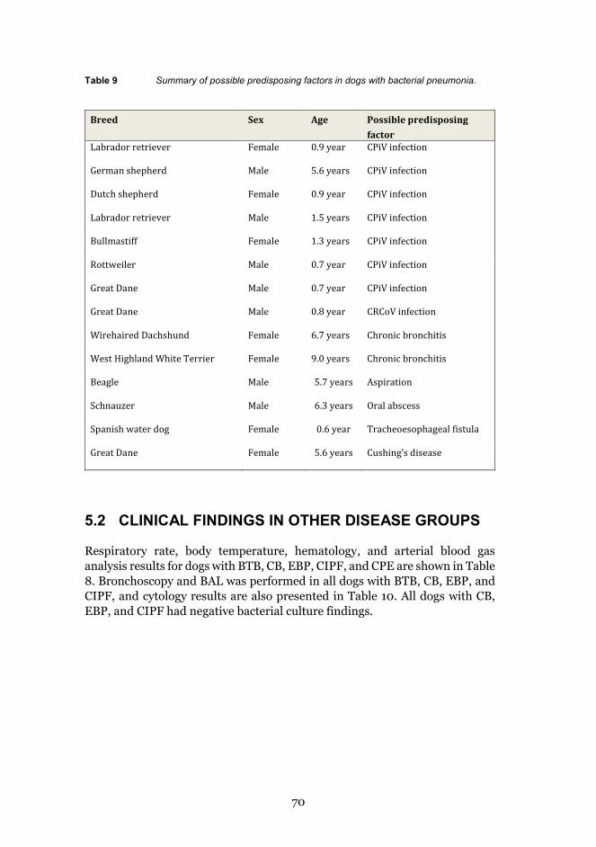
70
Table 9 Summary of possible predisposing factors in dogs with bacterial pneumonia.
Breed
Sex Age Possible predisposing factor
Labrador retriever Female 0.9 year CPiV infection
German shepherd Male 5.6 years CPiV infection
Dutch shepherd Female 0.9 year CPiV infection
Labrador retriever Male 1.5 years CPiV infection
Bullmastiff Female 1.3 years CPiV infection
Rottweiler Male 0.7 year CPiV infection
Great Dane Male 0.7 year CPiV infection
Great Dane Male 0.8 year CRCoV infection
Wirehaired Dachshund Female 6.7 years Chronic bronchitis
West Highland White Terrier Female 9.0 years Chronic bronchitis
Beagle Male 5.7 years Aspiration
Schnauzer Male 6.3 years Oral abscess
Spanish water dog Female 0.6 year Tracheoesophageal fistula
Great Dane Female 5.6 years Cushing’s disease
5.2 CLINICAL FINDINGS IN OTHER DISEASE GROUPS
Respiratory rate, body temperature, hematology, and arterial blood gas analysis results for dogs with BTB, CB, EBP, CIPF, and CPE are shown in Table 8. Bronchoscopy and BAL was performed in all dogs with BTB, CB, EBP, and CIPF, and cytology results are also presented in Table 10. All dogs with CB, EBP, and CIPF had negative bacterial culture findings.

71
Table 10 Clinical findings, hematology, arterial blood gas analysis, and respiratory cytology results in dogs with bacterial tracheobronchitis (BTB), chronic bronchitis (CB), eosinophilic bronchopneumopathy (EBP), canine idiopathic pulmonary fibrosis (CIPF), and cardiogenic pulmonary edema (CPE).
BTB
n= 20
CB
n= 20
EBP
n=20
CIPF
n=12
CPE
n=15
Mean ± SD or Median (IQR)
Body temperature, ºC 38.9 ± 0.5 38.7 ± 0.7 38.7 ± 0.5 38.5 ± 0.4 38.2 ± 0.6
Respiratory rate, /min 39.9 ± 12.6 45.1 ± 23.7 24.8 ± 6.9 57.6 ± 35.1 61.,0 ± 22.9
Arterial paO2, mmHg 88.0 ± 12.3 81.3 ± 12.8 91.7 ± 11.1 63.0 ± 8.7
Arterial paCO2, mmHg 35.3 ± 6.4 30.1 ± 4.7 31.8 ± 3.8 30.5 ± 4.1
Alveolar-arterial O2
gradient
24.8 ± 9.8 34.9 ± 12.8 26.8 ± 8.1 51.3 ± 10.2
Blood leukocyte count,
109/l
13.3 (10.8-17.4) 9.0 (7.1-10.4) 10.0 (8.9-12.4) 9.7 (7.4-11.9) 10.1 (6.0-12.3)
Segmented neutrophils 8.1 (6.7-12.2) 6.4 (4.9-9.7) 6.3 (5.4-7.9) 7.5 (5.2-9.4) 4.0 (3.8-7.5)
Band neutrophils 0 (0-0.1) 0 (0-0) 0 (0-0) 0 (0-0) 0 (0-0)
Lymphocytes 2.8 (2.3-4.2) 1.6 (1.2-3.3) 1.9 (1.3-2.5) 1.4 (1.1-1.5) 1.5 (1.4-1.7)
Eosinophils 0.4 (0.2-0.7) 0.3 (0.2-0.5) 1.2 (0.8-2.1) 0.3(0.1-0.6) 0.35 (0.2-0.7)
Monocytes 1.0 (0.6-1.2) 0.7 (0.4-1.1) 0.5 (0.4-0.7) 0.5 (0.3-1.0) 0.5 (0.2-1.0)
Basophils 0 (0-0.03) 0.02 (0-0.05) 0.15 (0-0.3) 0 (0-0.01) 0.01 (0-0.01)
BALF total cell count,
109/l
0.6 (0.2-1.2) 0.23 (0.1-0.4) 0.99 (0.4-1.7) 1.09 (0.72-1.59)
BALF neutrophils, % 11.6 (3.9-46.2) 5.7 (2.7-14.4) 5.2 (2.4-10.1) 6.5 (4.2-13.0)
BALF eosinophils, % 2.2 (0.4-3.9) 1.7(0.4-6.0) 50.7 (41.2-65.5) 0.2 (0-0.6)
BALF mast cells, % 0.0 (0-0.4) 1.4 (0.7-3.0) 0.9 (0-1.9) 0.7 (0.2-1.0)
BALF lymphocytes, % 11.9 (5.9-22.1) 12.0 (6.4-17.4) 7.9 (3.7-14.2) 6.0 (4.4-6.7)
BALF macrophages, % 59.7 (40.2-68.7) 75.0 (54.0-85.0) 29.7 (16.2-37.2) 85,1 (77.6-89.4)
BALF epithelial cells, % 0 (0-2.9) 0 (0-0.4) 0.4 (0-1.2) 0 (0-0)
BALF basophils, % 0 (0-0-) 0 (0-0) 0 (0-0) 0 (0-0)
BALF plasma cells, % 0 (0-0) 0 (0-0) 0 (0-0) 0 (0-0)

72
Dogs with bacterial tracheobronchitis (Studies I, III)
All dogs with BTB were clinically stable and were presented to the VTHH for subacute or chronic cough. The median duration of clinical signs was 62 days (IQR 30-120 days, range 7-360 days). Of the 20 dogs, seven had received peroral antibiotics <7 days prior to study inclusion. Radiographic findings included bronchial or bronchointestial pattern in 17/20 dogs. Thoracic radiographs were interpreted as normal in 3/20 dogs. Alveolar pattern was not detected in any of the dogs. Fecal samples were available for 15/20 dogs and were negative for lung worms in all dogs. One dog had intestinal parasites (Coccidia spp.).
Significant bacterial growth in aerobic cultures (>103 CFU/ml) was detected in all dogs. Microbiological findings comprised B. bronchiseptica in 15/20, Pasteurella spp. in 2/20, E. coli in 1/20, Haemophilus spp. in 1/20, and Klebsiella pneumoniae and Streptococcus canis in 1/20 dogs.
Respiratory viruses (CPiV, CAV-2, CHV, CRCoV, CIV, CDV, and CnPnV) were not detected with PCR methods in dogs (n=13) with BBTB (Study III).
The presence of Mycoplasma spp. infection was evaluated with a culture method in 4/13 dogs and with a PCR method in 13/13 dogs with BBTB participating in Study III. Culture was positive in 2/4 dogs and PCR was positive in 4/13 dogs. PCR was positive in the two dogs with positive culture findings. PCR was also positive in one dog with negative culture findings and in another dog lacking a Mycoplasma culture.
Dogs with chronic bronchitis (Study I)
All 20 dogs with CB were clinically stable and had chronic cough as their main sign. The median duration of clinical signs was 6 months (IQR 4-12 months, range 2-36 months). Four dogs were treated with glucocorticoids at the time of presentation to the VTHH. Changes in thoracic radiographs were noted in 18/20 dogs, and the most commonly reported abnormality was moderate to severe bronchial or bronchointerstitial pattern. In all dogs, visible changes were evident in bronchoscopy and included bronchial wall irregularity (17/20), bronchomalasia (18/20), increased amount of mucus (5/20), and bronchiectasia (7/20). None of the dogs had significant bacterial growth or eosinophilia in BALF. Fecal analysis was available for 16/20 dogs and was negative in all cases.
Dogs with eosinophilic bronchopneumopathy (Study I) All dogs with EBP suffered from chronic cough. In addition, exercise intolerance of varying degree was commonly noted. The median duration of clinical signs was 5 months (IQR 2.6-6.8 months, range 1.5-24 months). The

73
radiographic changes most commonly detected were moderate to severe bronchial or bronchointerstitial pattern. Solely interstitial pattern was detected in 2/20 dogs, and alveolar pattern was detected in 2/20 dogs. Thoracic radiographs were interpreted as normal in two dogs. BALF eosinophilia (>20%) was present in all dogs, and fecal analysis was negative for lung worms in all dogs. In two dogs, intestinal parasites were detected (Ancylostoma caninum in one dog and Coccidia spp. in the other).
Dogs with canine idiopathic pulmonary fibrosis (Study I)
The dogs with CIPF presented most commonly with either cough, exercise intolerance, or panting, or a combination of these. Detailed clinical, radiographic, and HRCT findings are previously reported in a study describing CIPF in West Highland white terriers (Heikkilä et al., 2011). Fecal analysis was negative in all dogs.
Dogs with cardiogenic pulmonary edema (Study I)
Dogs with cardiogenic pulmonary edema were presented to the VTHH with acute or subacute onset of cough, tachypnea, or respiratory distress of varying severity. The median duration of clinical signs was 5 days (IQR 2-14 days, range 1-30 days), and the clinical signs reported on admission to the VTHH comprised cough (12/15), tachypnea (11/15), respiratory distress (3/15), and collapse (1/15). In thoracic radiographs, an alveolar pattern was detected in 11/15 dogs and a severe intersititial or bronchointerstitial pattern in 4/15 dogs. Echocardiography was performed on all dogs to confirm the underlying cardiac disease. Of the 15 dogs, 11 were diagnosed with myxomatous mitral valve disease and 4 with dilated cardiomyopathy.
5.3 ACUTE PHASE PROTEINS IN BACTERIAL PNEUMONIA
5.3.1 EFFECT OF POSSIBLE CONFOUNDING FACTORS IN SERUM C-REACTIVE PROTEIN (STUDY I)
Age (p=0.15), sex (p=0.37), or body weight (p=0.15) did not affect serum CRP concentrations in the entire material (dogs with BP, BTB, CB, EBP, CIPF, or CPE and healthy dogs). Prior antibiotic treatment did not affect serum CRP in dogs with bacterial diseases (BTB and BP; p=0.58). The duration of clinical signs did not affect CRP concentration in dogs with BP (p=0.17), BTB (p=0.61), or CPE (p=0.15).

74
5.3.2 CORRELATIONS BETWEEN ACUTE PHASE PROTEINS AND CLINICAL VARIABLES
Significant correlations between clinical variables and serum CRP are presented in Table 11.
Table 11 Significant correlations between serum CRP and clinical findings, hematology, arterial blood gas analysis, and respiratory cytology results in the entire patient group (all dogs), dogs with bacterial tracheobronchitis (BTB), dogs with chronic bronchitis (CB), dogs with bacterial pneumonia (BP), and dogs with canine idiopathic pulmonary fibrosis (CIPF) in Study I.
Patient group Correlation to serum CRP
p-value R
Body temperature, ºC All dogs 0.020 0.25
Respiratory rate (/min) All dogs 0.003 0.33
Arterial paCO2 mmHg Dogs with BP 0.022 -0.50
Blood leukocyte count 109/l All dogs 0.018 0.23
Blood segmented neutrophils 109/l All dogs 0.0007 0.34
Band band neutrophils 109/l All dogs <0.0001 0.64
Dogs with BTB 0.012 0.59
Blood lymphocytes 109/l Dogs with CB 0.033 -0.49
Blood eosinophils 109/l All dogs <0.0001 -0.48
Dogs with CB 0.0091 -0.58
Blood basophils 109/l All dogs 0.0002 -0.36
BALF total cell count (109/l)
BALF Neutrophils (%) Dogs with BTB 0.015 0.58
BALF Eosinophils (%) All dogs <0.0001 -0.48
Dogs with CB 0.0091 -0.58
Dogs with CIPF 0.0058 -0.74
BALF Mast cells (%) All dogs 0.012 -0.27
BALF Macrophages (%) Dogs with CB 0.0016 0.67

75
The serum concentrations of positive APPs at presentation to the VTHH were significantly correlated with each other in Study III: CRP and SAA (r=0.60, p=0.015), CRP and Hp (r=0.61, p=0.010), and SAA and Hp (r=0.85, P<0.001). Significant correlations between positive APPs and serum albumin (a negative APP) did not arise in all comparisons: SAA was significantly negatively correlated with albumin (r=-0.53, P=0.035), but CRP (r=-0.22, p=0.40) and Hp (r=-0.15, p=0.58) were not.
5.3.3 SERUM C-REACTIVE PROTEIN AS A DIAGNOSTIC BIOMARKER (STUDY I)
Serum C-reactive protein concentrations in dogs with various respiratory diseases (BP, BTB, CB, EBP, CIPF), in dogs with CPE, and in healthy dogs are presented in Figure 2.
Figure 2 Serum C-reactive protein (CRP) concentrations in dogs with bacterial tracheobronchitis (BTB, n=17), chronic bronchitis (CB, n=20), eosinophilic bronchopneumopathy (EBP, n=20), idiopathic pulmonary fibrosis (CIPF, n=12), cardiogenic pulmonary edema (CPE, n=15), bacterial pneumonia (BP, n=22), and healthy controls (n=72) in Study I.
*) Dogs with BP had significantly higher CRP concentrations (median 121 mg/l, interquartile range 68-178 mg/l) than dogs with BTB (23, 15-38, p=0.0003), CB (13, 8-14 , p<0.0001), EBP (5, 5-15, p<0.0001), CIPF (17, 10-20, p<0.0001) , or CPE (19, 13-32, p <0.0001), and healthy controls (14, 8-20, p<0.0001).
**) Dogs with BTB had significantly higher CRP concentrations than dogs with CB (p=0.0010) or EBP (p<0.0001) or healthy controls (p=0.029).

76
5.3.4 ACUTE PHASE PROTEINS AS PROGNOSTIC MARKERS (STUDY II)
Serum APP concentrations in dogs with BP at presentation to the VTHH and the comparison between dogs with a less severe BP (lsBP) requiring <2 days of hospitalization and dogs with a more severe BP (msBP) requiring >2 days of hospitalization are shown in Figure 3.
A B
C D
Figure 3 Serum acute phase protein concentrations at presentation in healthy dogs (n=64), dogs with a less severe bacterial pneumonia (lsBP) requiring < 2 days of hospitalization (n=10) and in dogs with a more severe bacterial pneumonia (msBP) requiring >2 days of hospitalization (n=9) in Study II. A) C-reactive protein (CRP): Healthy dogs, median 11.0 mg/l, interquartile range 5.0–16.0, n=47; lsBP, 101.5, 62.3–182.5, n=10; msBP, 142.0, 98.0–211.0, n=9. B) Serum amyloid A (SAA): Healthy dogs, median 3.9 mg/l, interquartile range 1.7–9.1, n=64; lsBP, 1048.2, 236.3–1522.2, n=9; msBP, 1336.9, 778.3–3650.0, n=7. C) Haptoglobin (Hp): Healthy dogs, median 0.5 mg/ml, interquartile range 0.2–1.7, n=64, in lsBP, 3.7, 2.5–7.3, n=8; msBP, 6.9, 2.7–13.3, n=6. D) Albumin: Healthy dogs, median 31.4 g/l, interquartile range 29.4–33.0, n=64; lsBP, 30.6, 27.3–32.2, n=9; msBP, 27.3, 24.2–31.9, n=8.

77
Serum APPs at presentation were not significantly correlated with variables reflecting disease severity such as duration of hospitalization (CRP r=0.36, p=0.24; SAA r=0.462, p=0.072; Hp r=0.305, p=0.235), arterial PaO2 (CRP r=0.353, p=0.138; SAA r=-0.450, p=0.080; Hp r=-0.48, p=0.052), or A-aO2 (CRP r=0.35, p=0.14; SAA r=0.42, p=0.11; Hp r=0.42, p=0.090).
CRP ratio (CRP concentrations at 24 hours, 48 hours, and 72 hours divided by the initial concentration) was calculated for hospitalized patients at 24 hours (median 1.0, interquartile range [IQR] 0.6-2.2, n=11), 48 hours (median 0.95, IQR 0.8-1.1, n=8), and 72 hours (median 1.0, IQR 0.8-1.2, n=5). CRP continued to rise in the first 24 hours in 5/11 dogs. CRP ratios after 24 hours (r=0.35, p=0.30), 48 hours (r=0.46, p=0.25), and 72 hours (r=-0.15, p=0.81) were not correlated with duration of hospitalization.
5.3.5 ACUTE PHASE PROTEINS AS MARKERS OF TREATMENT RESPONSE (STUDY II)
Serum CRP, SAA, Hp, and albumin concentrations during hospitalization and the recovery period are shown in Figure 4.
5.3.6 SERUM CRP IN AIDING THE ESTIMATION OF ANTIMICROBIAL TREATMENT LENGTH (STUDY II)
Seventeen of 19 dogs survived to hospital discharge in Study II. In nine of these dogs, CRP was not used for determining the length of antimicrobial treatment, and dogs received antibiotics according to conventional recommendations (median length of treatment 35 days, IQR 29–48 days, range 26-88 days). Of these nine dogs, six had lsBP and three had msBP. Bacteria susceptible to the initially chosen antimicrobials was detected in 15/17 dogs. In two dogs, bacterial growth was not detected and therefore antimicrobial susceptibility could not be addressed. Of these two dogs, one was treated according to conventional recommendations and one was treated CRP-guided.
Antimicrobial treatment was discontinued 5–7 days after CRP normalization in 8/17 dogs (median length of treatment 21 days, IQR 19–29 days, range 18-65 days). lsBP was observed in 4/8 and msBP in 4/8 dogs. CRP-guided treatment was significantly shorter (p=0.015) than conventional treatment.
One dog treated conventionally had a relapse of BP after antibiotic discontinuation, and Cushing’s disease was later found as a predisposing factor. The other 16 dogs did not show clinical signs or findings suggestive of BP relapse at follow-up visits occurring 2 weeks after antibiotic discontinuation or during follow-up phone calls at 4–8 weeks after the follow-up visit.

78
A B
A B
A B
Figure 4 Serum C-reactive protein (CRP), serum amyloid A (SAA), and haptoglobin (Hp) concentrations in dogs with bacterial pneumonia (BP) (n=19) during the disease and follow-up period. Each line shows results of a single dog. A presents dogs with a less severe BP requiring < 2 days of hospitalization and B dogs with a more severe BP requiring >2 days of hospitalization.
A B

79
6 DISCUSSION
Although BP is a common and well-known disease entity in small animal medicine, it has attracted limited research interest, especially during the last decades. Consequently, the clinical and diagnostic findings in BP have been reported almost solely in retrospective case series and follow-up studies have not been published. This thesis summary reports clinical and diagnostic findings as well as serum acute-phase protein concentrations in 26 prospectively recruited dogs diagnosed with BP and follow-up findings during the disease and recovery periods in 19 of these dogs.
6.1 BACTERIAL PNEUMONIA – NEW INSIGHTS INTO AN OLD DISEASE?
6.1.1 CLINICOPATHOLOGICAL FINDINGS Our findings confirmed the previous conclusion (Thayer and Robinson, 1984) that dogs with BP are mainly large breed dogs; in our study, the mean weight was 32 kg and 65% of dogs weighed >20 kg. We found a slight male predominance (54% male, 46% female), as also reported previously in BP (Thayer and Robinson, 1984) and in AP (Kogan et al., 2008b; Tart et al., 2010). However, the male predominance found in our study and in the previous studies is relatively small, and therefore, the clinical relevance is questionable.
Clinical signs and physical examination findings in our study were in line with those reported elsewhere (Radhakrishnan et al., 2007; Thayer and Robinson, 1984). An acute onset of lethargy, cough, tachypnea, and respiratory distress were the most common complaints on admission. In addition to these, abnormal lung auscultation findings were commonly noted in physical examination. Fever was found in 73% of dogs, which is more often than in earlier studies, which reported fever in less than half of dogs (Radhakrishnan et al., 2007; Thayer and Robinson, 1984). We also noted clinical findings suggestive of polyarthritis in a small group of dogs with BP. This had not been reported before, but is not unexpected in dogs with BP; secondary immune-mediated polyarthritis may be triggered by a bacterial respiratory infection (Johnson and Mackin, 2012). According to both our study and previous research (Kogan et al., 2008a; Radhakrishnan et al., 2007; Thayer and Robinson, 1984), typical clinical signs and general examination findings (cough, tachypnea, respiratory distress, fever, and abnormal auscultation) are not consistently present in all dogs with BP and the lack of a single finding, e.g. fever or cough, cannot rule out the diagnosis of BP. However, it has been shown in humans that CAP can be dependably ruled out when all vital signs are normal (Lim et al., 2009). While this has not been

80
addressed in dogs, it is likely that similarly normal vital signs could rule out BP in dogs as well.
The numbers of blood leukocytes are largely affected by extravasation into pulmonary parenchyma during acute bacterial infection, and therefore, hematology appears to be rather insensitive as a diagnostic test in dogs with BP. Leukocytosis and neutrophilia were detected in a minority of dogs (23% and 27% of dogs, respectively) also in our study. Concordant with previous studies, left shift was the most commonly encountered hematological abnormality, occurring in 65% of our dogs (Thayer and Robinson, 1984).
Hypoxia of varying severity was commonly detected in dogs with BP. PaO2 results in our study (mean 75 mmHg) are slightly higher than previously reported (mean 61 mmHg) by Wingfield et al. (1997). Evidently, the most important feature that affects the PaO2 results is the patient group chosen. In our study, also less severely affected dogs, treated with peroral antibiotics without hospitalization, were included. In addition to the patient characteristics in these two studies, the results may be affected by altitude: Wingfield (1997) reported findings in Fort Collins, Colorado, at an altitude of 1500 meters above sea level, whereas our dogs lived in Southern Finland at an altitude of 50 m or less above sea level.
As expected, initial arterial PaO2 reflected disease severity and was significantly correlated with length of hospitalization. Retention of PaCO2 was not encountered, and hypocapnea, most likely due to increased respiratory rate, was commonly detected. This is in accordance with Wingfield (1997), who reported lower PaCO2 in dogs with pneumonia than in healthy dogs.
We reported serial arterial blood gas measurements during disease and the recovery period in 19 dogs with BP (Figure 1). PaO2 returned to normal in less severely affected dogs relatively rapidly, and an increase in PaO2 was noted often already during the first two days. In more severely affected dogs, PaO2
often declined during the first days of hospitalization, and normalization of arterial PaO2 required a markedly longer time. A consistent decline in arterial PaO2 during hospitalization was found in both dogs that did not survive. Considering these findings, a decline in PaO2 during the first days of BP could be indicative of a more severe course of the disease.
Arterial PaO2 had returned to normal reference levels in most dogs by the last control visit 2 weeks after the antimicrobial treatment had ceased. In two dogs with a severe BP requiring extended hospitalization, PaO2 rose slowly during the follow-up period, but was slightly below the normal reference range at the last control visit and returned to normal at a later time-point. Based on this, our results suggest that a slow normalization of PaO2 should not raise concerns, assuming other clinical findings and the general condition of the dog are improving.
Lung worms were not detected in any of the dogs with BP in our study. This was expected since BP has not been described as a feature in parasitic lung infections (Sherding, 2004). Additionally the low prevalence of parasitic lung

81
infections in Finland may have affected the results (personal communication, M.M. Rajamäki).
6.1.2 RADIOGRAPHIC FINDINGS Alveolar pattern is the main feature in thoracic radiographs in dogs with BP (Brady, 2004; Dear, 2014; Ford, 2009). However, prior to our study, detailed radiographic findings have been reported only in dogs with AP (Eom et al., 2006; Kogan et al., 2008b; Tart et al., 2010) and in dogs with BP caused by Mycoplasma spp. (Jameson et al., 1995).
As expected, an alveolar pattern was the most common finding also in our study, in 81% of dogs. Similiar to this, Kogan et al. (2008a) reported an alveolar pattern in 74% of dogs with AP. An alveolar pattern was detected less often, only in 44% of dogs with BP caused by Mycoplasma spp. (Jameson et al., 1995). These findings suggest that an alveolar pattern is less often encountered in BP caused by Mycoplasma infections. However, Jameson et al. (1995) did not detect significant differences in the radiographic pattern between Mycoplasma-positive and Mycoplasma-negative dogs, and therefore, the difference is unlikely to be explained by causative organisms. In our study, solely an interstitial or a bronchointerstitial pattern was discovered in a minority of dogs, as described previously (Jameson et al., 1995; Kogan et al., 2008a; Tart et al., 2010). An alveolar pattern was the most common finding in 75% of our Mycoplasma-positive dogs.
Several lung lobes were most often involved in our dogs with BP (in 57% of dogs), and an alveolar pattern in a single lung lobe was encountered less often (in 23% of dogs). This differs from findings described in AP, where the involvement of a single lung region was most commonly described (Kogan et al., 2008a). Similarily as observed in cases of AP (Eom et al., 2006; Kogan et al., 2008b), cranioventral lung lobes were most often affected in our dogs with BP. Aspiration has been described to occur more often to the right lung (Eom et al., 2006; Kogan et al., 2008b). In our study, both right and left lungs were equally involved.
In our patients, aspiration was considered a likely cause of BP in only one dog, in which BP developed after severe vomiting due to a gastric foreign body. In other dogs, aspiration etiology could not be confirmed or excluded based on history, clinical signs, or general examination findings, but was considered unlikely since none of the dogs had diseases predisposing to aspiration, recent vomiting, or anesthesia or clinical signs suggestive of laryngeal paralysis. However, laryngeal examination was performed on only a minority of dogs, and therefore, subclinical laryngeal dysfunction could have predisposed to aspiration in some dogs.
Radiographic findings in our study were very similiar to those described in AP. We found alveolar density primarily in cranial and ventral lung lobes in dogs, where aspiration etiology was considered unlikely, suggesting that alveolar density in dependant lung lobes could be typical for BP regardless of
82
etiology. It is unclear whether this is due to gravity hindering the removal of secretions in these areas or whether unidentified silent aspiration of small amounts of gastric or oral secretions plays an important role in the development of BP.
A patchy alveolar pattern affecting several lung lobes was detected in 12% of dogs with BP in our study. A patchy alveolar pattern may not be connected to aspiration etiology, as it has not been described in dogs with AP (Eom et al., 2006; Kogan et al., 2008b).
Several human studies have addressed the need for thoracic radiographs when diagnosing CAP. Generally, it has been concluded that clinical signs or symptoms are inefficient in discriminating CAP from other lower respiratory diseases and confirmatory radiographic evidence is needed (Lim et al., 2009). Thoracic radiographs are recommended for all hospitalized patients with suspected CAP (Lim et al., 2009). We did not assess the need for thoracic radiographs in diagnosing BP, but since alveolar density typical for BP was discovered in most dogs at presentation and other typical clinical findings were not consistently present in all dogs with BP, thoracic radiographs can be considered important in the diagnosis of BP in dogs.
The resolution of an alveolar lung pattern was followed during hospitalization and the follow-up visits. Thoracic radiographs were not repeated daily, and therefore, an exact time-point for the resolution could not be determined and the first time-point when alveolar density could no longer be detected was used as the time-point for resolution. Naturally, this slightly overestimates the time required. Alveolar infiltrates resolved relatively rapidly (69% of dogs had cleared alveolar infiltrates by day 10) compared with human studies. It has been reported that only 33% of human patients with CAP had clearance of radiographic infiltrates at day 7 and 62% at day 28 (Bruns et al., 2007). In humans, it has also been shown that radiographic normalization lags behind clinical cure assessed by physicians (Bruns et al., 2010). Mild to moderate bronchial, bronchointerstitial, or interstitial pattern was still noted two weeks after the termination of antimicrobial treatment in the majority of our dogs. This finding appears to be of low importance since all of these dogs recovered eventually.
Current human research has not been able to show benefits for routinely controlling thoracic radiographs during or after hopitalization in CAP (Lim et al., 2009). Especially repeating chest radiographs prior to hospital discharge is not recommended if treatment response is otherwise satisfactory (Lim et al., 2009). Controlling chest radiographs after treatment has been recommended if there a non-satisfactory recovery or a concern of underlying lung malignancy (Lim et al., 2009). Our results revealed that additional information gained by thoracic radiographs, especially after the clearance of alveolar infiltrates, was minimal in dogs with otherwise satisfactory clinical recovery. Consequently, it could be suggested that repeating thoracic radiographs during and after treatment in dogs with BP e reserved for cases with poor response to treatment or suspected underlying lung disease.

83
6.1.3 SAMPLING METHODS AND RESPIRATORY CYTOLOGY BAL was a safe sampling method in our dogs with mild to moderate BP. Bronchoscopy allowed the selection of the sampling site and the detection of possible structural changes predisposing to the development of BP, such as bronchiectasia or TEF. The advantage of choosing the sampling site during bronchoscopy resulted also in a better yield in microbiology analysis; in our dogs bacterial culture was more often positive and intracellular bacteria were more commonly seen in BALF samples than in TTW samples. This is concordant with Hawkins et al. (1995), who detected infectious organisms more often in BALF samples than in TTW samples obtained from the same animal.
Cytological findings in both BALF and TTW samples were similar to the previously described TTW findings (Thayer and Robinson, 1984; Wingfield, 1997); neutrophilic inflammation predominated and intracellular bacteria were commonly detected. Significant differences between BALF and TTW differential cell count were detected only in the percentage of epithelial cells. The higher number of epithelial cells in TTW samples is likely due to the sampling site of the distal trachea compared with the small airways and alveoli in BAL.
In our study, TTA was chosen as a sampling method in two dogs due to a focal lung lesion in one dog and small size of the patient precluding TTW sampling in another dog. In both dogs, TTA sampling enabled the diagnosis of BP as neutrophilic inflammation, intracellular bacteria, and significant bacterial growth were detected. However, the risk of pneumothorax in TTA sampling has been well demonstrated (Teske et al., 1991; Wood et al., 1998), and also one our dogs developed a mild pneumothorax after sampling. The risk for the development of pneumothorax may be greater in dogs with BP due to fast and shallow breathing, which can cause excessive movement of the needle while sampling. It has been reported in humans that spontaneous pneumothorax can develop during CAP (Gayatridevi et al., 2015; Sayar et al., 2004). Whether this may also occur in dogs with BP and further predispose to the development of pneumothorax is unknown. The risk for severe complications after TTA is greater in severely ill dogs (Teske et al., 1991), and as such a minor pneumothorax after TTA may exacerbate clinical signs in BP dogs that already have marked hypoxia prior to sampling.
Sputum culture is the one of the most commonly used method in humans for bacterial culture in respiratory samples. The advantage in humans is the non-invasiveness of this sampling method, but results may be compromised due to inability of patients to produce good specimens, contamination with oropharyngeal flora, or prior antimicrobial treatment (Lim et al., 2009). Understandably, sputum samples are not routinely used in animals since volitional sputum production is not feasible. Therefore, the reliability of sputum samples in the assessment of microbial etiology in dogs with BP has not been addressed in studies. In our study, we decided to include one dog in which initial respiratory culture was performed from a fresh sputum sample

84
obtained after a highly productive cough. In this case, numerous neutrophils, intracellular bacteria, and abundant E. coli growth were detected. Later necropsy confirmed the diagnosis of BP and E. coli bacteremia. Sputum sampling is unlikely to become widely used in dogs, but in our case it was beneficial and enabled the isolation of the causative agent.
6.1.4 MICROBIOLOGY RESULTS Pasteurella spp., E. coli, and Streptococcus spp. were the most common bacteria detected in our study and these results are mostly in agreement with previous reports (Epstein et al., 2010; Jameson et al., 1995; Proulx et al., 2014; Thayer and Robinson, 1984; Wingfield, 1997). Slight differences compared with previous reports included lack of BP caused by Staphylococcus spp. or B. bronchiseptica. Both could be explained by the small number of dogs included here. Additionally, the lack of large kennels and rehoming centers in Finland, typical for B. bronchiseptica infections, could have affected the results.
A growth of a single species was detected in 69% of our dogs. Mycoplasma spp. were the most common second bacterial species encountered when two species were detected in the same sample. Previous reports described growth of a single species in 53% (Wingfield, 1997) and 74% (Thayer and Robinson, 1984) of samples without specifying whether particular culture techniques for Mycoplasma spp. were applied. Jameson et al. (1995) reported growth of a single species less often (in 40% of dogs) in a study where specific Mycoplasma culture methods were used. In addition to culture methods, the results may be affected by sampling site; BAL was the main sampling method in our study compared with previous studies using TTW as the sampling method. Additionally, prior antimicrobial treatment was common in our patients and likely affected culture results.
Negative bacterial cultures were encountered in 15% of dogs. This group comprised equal numbers of dogs with and without earlier antimicrobial treatment, and therefore, prior medications do not fully explain the results. In addition to prior antimicrobials, the sampling site of the distal trachea in TTW samples may have been inadequate for a representative sample in some dogs; in our patients undergoing bronchoscopy, purulent discharge was noted in the trachea in only 23% of dogs with BP. Previously, Proulx et al. (2014) reported negative bacterial culture findings in TTW samples in 14% of dogs with a clinical diagnosis of BP. Murphy et al. (1997) found a markedly higher proportion of negative culture findings (58%) in dogs undergoing pulmonary lobectomy for the treatment of pneumonia. This could be explained by patient characteristics; Murphy et al. (1997) included not only dogs with BP, but also dogs with fungal and parasitic lung infections as well as dogs with pulmonary neoplasia. In humans, the phenomen is well described and studies have shown that up to 60% of respiratory cultures are negative in CAP patients. Hence, we decided to include also the dogs with negative respiratory cultures if the dogs

85
fulfilled other diagnostic criteria for BP and recovered fully with solely antimicrobial treatment.
Mycoplasma spp. were cultured in 27% of samples and additionally detected with PCR in 40% of samples. The clinical relevance of PCR-positive samples is difficult to assess since the method allows the detection of very small quantities, and Mycoplasma spp. may be encountered also in the respiratory tract of healthy dogs (Randolph et al., 1993b). We detected Mycoplasma spp. slightly less often than previous studies reporting Mycoplasma spp. in 70% of dogs with BP (Jameson et al., 1995) and in 64% of dogs with septic lung inflammation (Randolph et al., 1993b). The role of Mycoplasma spp. in BP remains unclear. In our study, there were no significant differences in clinical variables in Mycoplasma-positive and -negative dogs, which is in line with previous reports (Jameson et al., 1995). Furthermore, in our study 3/4 dogs with positive Mycoplasma culture received antimicrobials known to be inefficient against Mycoplasma spp. (amoxycillin-clavulanic acid), but recovered uneventfully. A similar finding has been reported by Jameson et al. (1995). In our study, Mycoplasma-positive dogs had significantly higher total cell counts in respiratory samples than Mycoplasma-negative dogs. This has not been reported previously, and the causes as well as the clinical relevance of this finding are unknown.
Anaerobic culture methods were applied in our study, but anaerobic bacteria were not detected in any of the samples. Our findings contradict the report of Angus et al. (1997), who detected anaerobic bacteria in 22% of samples. The reason for this difference can only be speculated; Angus et al. (1997) reported microbiological findings in 116 dogs with lower respiratory tract infections without specifying whether dogs with pyothorax were included. Since the presence of anaerobes is common in pyothorax (Walker et al., 2000), this could be one explanation for the discrepancy. Another possible factor influencing the results could be that our material comprised very few dogs with AP and did not include dogs with pulmonary foreign bodies; these patient groups could have distinct microbial findings.
In addition to aerobic and anaerobic culture methods, an enrichment broth was used. The utility of an enrichment broth is uncertain. Enrichment may enable the detection of small amounts of causative bacteria, especially in cases of prior antimicrobial treatment and this is particularly beneficial because it allows the determination of antimicrobial susceptibility profile. However, since enrichment allows the detection of small amounts of bacteria, insignificant resident bacteria or oral contaminants may also be detected. The distinction between significant and insignificant bacterial growth is difficult since quantity of bacterial growth cannot be estimated after enrichment. A high number of our dogs had received antimicrobial treatment prior to presentation. Additionally, we were aware of a high incidence of negative culture results in humans with CAP, and consequently, we decided to interpret pure growth of a single species after enrichment as the causative agent of BP.

86
In humans, blood cultures are recommended in moderate or severe CAP and a positive blood culture result is considered a highly specific confirmation of microbial etiology (Lim et al., 2009). However, the sensitivity of blood cultures is rather low, at most only 25%, and prior antimicrobial treatment may further diminish the sensitivity (Lim et al., 2009). In canine patients, 19 % of blood cultures were positive in septic patients (Gebhardt et al., 2009). In our study, 34% of blood cultures were positive, but only 17% were consistent with respiratory culture findings. The finding of Staphylococcus spp. in blood cultures of two of our dogs was inconsistent with respiratory culture findings and was considered to originate from skin contamination during sampling. E. coli was commonly detected from blood cultures in our study and in previous reports (Gebhardt et al., 2009). Blood cultures were positive in severely affected dogs; both blood culture-positive dogs with E. coli growth died or were euthanized due to severe BP. However, the utility of blood cultures in dogs with BP is questionable. Due to low sensitivity, negative results must be interpreted cautiously. Positive results are likely to confirm sepsis, but whether blood cultures provide value beyond respiratory cultures in BP is unknown. Blood cultures require special equipment and are rarely available outside university animal hospitals. Furthermore, the size of the patient may limit the possibility of obtaining serial blood cultures, even if pediatric collection vials are used. If blood cultures are performed, it is advisable to obtain samples prior to the initiation of antimicrobial therapy since antibiotics further reduce the sensitivity (Lim et al., 2009).
6.1.5 PROGNOSIS AND DISEASE SEVERITY The majority of our dogs (77%) required hospitalization and additional oxygen support. The number of dogs with moderate and severe BP may be affected by the patient material in VTHH, where a significant proportion of cases are referred for further investigations or hospital care from first opinion practices.
According to our results, the prognosis for survival in dogs with BP appears to be good. The length of hospitalization varied greatly among dogs, but the majority (92%) survived to hospital discharge and recovered. This is in accordance with previous studies reporting 92-96% survival in dogs with BP (Proulx et al., 2014; Radhakrishnan et al., 2007) . Slightly lower survival rates (77%, 82%, and 83%) have been reported in dogs with AP (Jameson et al., 1995; Proulx et al., 2014; Tart et al., 2010).
Dogs with a more severe BP requiring a longer hospitalization period were characterized by more severe hypoxia, a more pronounced left shift in hematology analysis, and a more neutrophilic inflammation in respiratory cytology. These are not unexpected findings; the degree of hypoxia reflects the extent of BP (Lee and Drobatz, 2004) and the presence of band neutrophils in circulation indicates increased consumption as a consequence of a marked inflammatory process in peripheric tissues and a subsequent immature release of neutrophils from the bone marrow (Day, 1999). Similiar findings have been

87
reported in human studies; low PaO2 has been identified as a risk factor for increased mortality in humans with CAP (Basnet et al., 2015; Fine et al., 1993), and the absolute band neutrophil count was connected to outcome in infants with severe bacterial infections (Bonadio et al., 1992). Furthermore, the extent of neutrophilic lung inflammation in BALF has been associated with a more severe CAP and higher mortality in humans (Lee et al., 2015). It has been proposed that in addition to the organism burden severe respiratory failure may be due to the neutrophil-mediated inflammatory process triggering acute respiratory distress syndrome (Lee et al., 2015; Witko-Sarsat et al., 2000).
Dogs with a more widespread alveolar density in thoracic radiographs were more severely affected with BP, as the number of affected lung lobes was strongly and positively correlated with the length of hospitalization in our dogs. This finding is consistent with Tart et al. (2010), who reported a significant relationship between the number of affected lung lobes and decreased survival in dogs with AP.
E. coli as the causative agent was associated with a more severe BP. This is likely due to the endotoxins produced by E. coli affecting several body systems, including hemodynamics, blood clotting, and humoral immunity (Bone, 1993). A similiar finding of increased disease severity has been reported in humans with CAP caused by E. coli (Marrie, 1998). Both mortalities were associated with severe BP caused by E. coli, and the discovery of an extended-spectrum beta-lactamase (ESBL) strain resulted in euthanasia of one of these dogs. The recognition of multidrug-resistant organisms among companion animals has expanded within the last few years, and challenging infections caused by these organisms are increasingly encountered (Walther et al., 2016). Recently dissemination of multidrug-resistant E. coli has been reported to occur in a veterinary hospital (Timofte et al., 2016). ESBL organisms are a current problem also in human medicine, and high prevalence is reported especially in hospital-acquired infections and in ventilator-associated pneumonia (Charles et al., 2013; Behnia et al., 2014; Dabar et al., 2015).
In humans with CAP, various severity scoring systems and predictive models have been developed in an attempt to help clinicians in identifying patients with a poor prognosis at an early stage. The most widely studied predictive model is the Pneumonia Severity Index (PSI). PSI is based on 20 variables used to derive a score that estimates the risk of mortality within 30 days (Fine et al., 1993). Factors increasing the risk of mortality were, for instance, high respiratory and pulse rate, elevated blood urea or glucose, low PaO2, and other concurrent comorbidities (Fine et al., 1993). The complexity of the PSI calculation limits its use in clinical practice, and a simplified scoring system, the CURB65 score, was developed using only patient’s degree of confusion, elevated blood urea concentration, increased respiratory rate, low blood pressure, and age of 65 years or higher as factors increasing risk of mortality (Lim et al., 2009). In our study, factors increasing the risk of mortality could not be statistically addressed due to the small number of dogs included and the low prevalence of mortality in the material. Factors

88
connected to longer hospitalization were assessed in our study, and, except for low PaO2, factors known to increase the risk of mortality in men did not appear to affect the length of hospitalization in dogs with BP. In our dogs, blood glucose or urea concentrations, body temperature, age of the patient, or pulse rate at presentation did not correlate significantly with the length of hospitalization. Moreover, in dogs pulse rate is largely affected by the size of the animal, and therefore, is unlikely to be a useful parameter. Blood pressure measurements were mostly lacking, and the connection between low blood pressure and more severe BP was thus not evaluated. However, it appears that the principle in human severity scoring systems cannot be directly applied to dogs with BP, and larger studies are required in order to generate canine severity scoring systems.
Co-infections with respiratory viruses or Mycoplasma spp. do not appear to result in a more severe BP; the presence of respiratory viruses or Mycoplasma spp. did not affect the clinical findings or the length of hospitalization. The effect of concurrent infections on disease severity has not been previously addressed in dogs with BP, but Jameson et al. (1995) reported similiar resolution rates for Mycoplasma-positive and -negative dogs with BP. The results in humans are contradictory; some studies have shown that humans with mixed infections with viruses and bacteria have a more severe course of CAP, while others have been unable to demonstrate significant differences (Cilloniz et al., 2012; Jennings et al., 2008; Johansson et al., 2011; Johnstone et al., 2008).
6.1.6 PREDISPOSING FACTORS Bacterial pneumonia is generally caused by opportunistic bacteria belonging to the normal oropharyngeal flora, and therefore, it is important to consider possible predisposing factors leading to the development of BP. The etiology in bacterial pneumonia is likely to frequently be multifactorial, and it is often difficult to ascertain the impact of predisposing factors in individual dogs.
Increased age, underlying systemic illness, poor nutrition, and compromised host defenses are often cited as challenging respiratory defenses and predisposing to the development of BP (Brady, 2004). Contrary to this, in our study the majority of the dogs were young or middle-aged (35% aged < 1 year and 62% <5 years) and systemic illness was detected in only one dog. Simultaneous infections with CIRD viruses, mainly CPIV, were the most common concomitant disease process identified. The role of preceding or concurrent infections with CIRD viruses had not been previously evaluated in household dogs with BP, but in humans respiratory viruses had been shown to predispose to secondary bacterial lung infections (Hament et al., 1999; Joseph et al., 2013; Peltola and McCullers, 2004). Our results suggest that viral co-infections can also predispose household dogs to the development of BP. Dogs with BP and concomittant viral infection were generally young large breed dogs. It is not unexpected that young dogs with possibly inadequate acquired
89
immunity against CIRD viruses may frequently contract an infection. The reason why large dogs with BP more often had co-infections with viruses is more equivocal; one hypothesis is that large dogs are more inefficient in clearing respiratory secretions, therefore being more at risk for the development of BP when infected with CIRD viruses. However, our finding of significantly larger dogs with viral co-infections may be also affected by the structure of the virus-negative group; Four virus-negative dogs with another predisposing factor to the development of BP were small and medium-sized.
The prevalence of aspiration etiology in dogs with BP had not been evaluated previously. In our study, aspiration was considered the underlying factor for the development of BP in only one dog (4%). In humans with CAP, also a minority of patients, approximately 10%, are identified as having AP (Lim et al., 2009). AP refers to a situation where the infectious process in the lungs is a consequence to aspiration of oropharyngeal bacteria or gastic contents. However, with the lack of reliable markers for aspiration, the link between aspiration and development of BP is seldom verified and the term AP is commonly used in situations where a patient with risk factors for aspiration presents with BP. In our study, aspiration etiology was considered unlikely in other dogs since none of them had a history of vomiting, regurgitation, recent anesthesia, or signs compatible with laryngeal paralysis. However, aspiration may have played a role in the development of BP in some dogs, but could not be confirmed or denied based on available clinical data. Silent aspiration is a common problem in men and it may predispose to the development of pneumonia (Dang et al., 2015). The prevalence of silent aspiration is currently unknown in dogs, but it potentially may play a role as a predisposing factor in the development of BP also in dogs.
Chronic bronchitis and structural changes in bronchi causing impaired removal of respiratory secretions were considered a predisposing cause for the development of BP in two dogs (8%). Both were older small breed dogs with a history of chronic bronchitis and an acute exacerbation of clinical signs. Bronchiectasis was identified in bronchoscopy in both dogs, along with bronchial wall irregularities and bronchomalasia. Thoracic HRCT was not performed in these dogs, but it would have allowed a more detailed evaluation of the extent of bronchiectasis. Bronchiectasis is known to increase the risk of bacterial infections in both men and dogs, and it could have influenced the development of BP in these dogs (Hill et al., 2011; Johnson et al., 2016).
In addition to the above-mentioned, a congenital tracheoesophageal fistula was identified as a predisposing factor in one dog and Cushing’s disease in another dog with recurrent BP. An underlying disease process was not identified in 38% of dogs in our study. These dogs had BP caused by opportunistic bacteria belonging to the normal oropharyngeal flora, and therefore it is likely that an underlying factor or disease process, albeit unrevealed, has impaired the pulmonary defense mechanisms and allowed the development of BP. Dogs with bronchial foreign bodies were not present in

90
our material, and the absence of grass awns in our geographic location may have affected this.
6.2 ACUTE PHASE PROTEINS IN BACTERIAL PNEUMONIA
6.2.1 DIAGNOSTIC UTILITY OF ACUTE PHASE PROTEINS Acute phase proteins are sensitive, but non-specific inflammatory markers and the non-specific nature of APPs may limit their diagnostic use. However, serum CRP has been found to be very useful in aiding the diagnosis in humans with CAP, and the measurement of CRP is currently recommended by human guidelines (FAU et al., 1231; Lim et al., 2009; Woodhead et al., 2005). Our aim was to assess whether CRP could also be applied as a diagnostic biomarker in dogs with BP.
We showed that dogs with BP had significantly higher serum CRP concentrations at presentation than healthy dogs or dogs with BTB, CB, EBP, CIPF, or CPE. These results suggest that serum CRP at presentation could be applied as an additional diagnostic biomarker in BP. However, a group of dogs with neoplastic lung diseases or pleural space diseases was not included in the study, and therefore, the applicability of serum CRP in the differentiation of these diseases from BP has not been evaluated. As there is a wider range of concentrations and the magnitude of change is more pronounced in SAA than in CRP, SAA might even be a better diagnostic marker than CRP in dogs with BP, as already suggested by a recent study comparing CRP and SAA as diagnostic markers of systemic inflammation (Christensen et al., 2014). Serum Hp and SAA were also both significantly elevated in dogs with BP relative to healthy dogs, but since a comparison between dogs with other respiratory diseases was not performed the diagnostic utility of these biomarkers remains obscure. At the moment, a commercial bed-side test is only available for CRP measurement.
In our material, BP was diagnosed with 100% specificity when CRP was >100 mg/l and ruled out with 100% specificity when CRP was <20 mg/l and signs had lasted for more than 24 hours. Our findings are highly similar to those in humans with CAP, where CAP is considered likely in a patient with compatible clinical signs and serum CRP >100 mg/l and unlikely when CRP is <20 mg/l and clinical signs have lasted for more than 24 hours (Lim et al., 2009). It is important to remember that after the initiation of inflammation serum CRP concentration reaches its maximum within 24 hours. Therefore, low concentrations in acutely presenting patients do not rule out BP. Nevertheless, low serum CRP in patient with clinical signs lasting >24 hours can be rather reliably used to rule out BP.

91
Similiar to BP, CPE can present with an acute onset of cough, tachypnea, or respiratory distress. In cases where thoracic radiographs or echocardiography are not available, serum CRP has the potential to aid diagnosis. Moreover, EBP occasionally presents with respiratory distress, alveolar density in thoracic radiographs, and lowered arterial PaO2. In these cases, we have found serum CRP to be especially useful; since the cytokines released by eosinophils do not generally trigger CRP production, eosinophilic lung inflammation is typically characterized by a low serum CRP contrary to BP.
CRP appears to be elevated also in bacterial respiratory diseases other than BP; in dogs with BTB, CRP was significantly higher than in dogs with other lower airway diseases presenting with cough (CB, EBP) or in healthy controls. However, increases in CRP were mild, and although significantly higher, there was marked overlap between groups, reducing the diagnostic value of CRP measurement in the diagnosis of BTB.
6.2.2 ACUTE PHASE PROTEINS AS FOLLOW-UP MARKERS Serum CRP and SAA have short half-lifes and a decrease in serum concentrations is expected soon after the inflammatory stimulus ceases (Ceron et al., 2005). Due to these properties, the utility of CRP and SAA as markers of treatment response was assessed in our study; CRP and SAA were followed in 19 dogs with BP during hospitalization and the post-hospitalization recovery period.
Serum CRP and SAA reflected well the recovery process and declined rapidly after initiation of therapy. Faster normalization was seen in dogs with less severe disease and a delay in the normalization of especially CRP was typical for dogs with more severe BP. Our findings indicate that CRP and SAA can be used as markers of treatment response in dogs with BP. Hp showed a more gradual rise and decline, and therefore, did not reflect clinical recovery as well as CRP and SAA.
CRP is known to reach peak concentrations approximately 24 hours after onset of inflammatory stimulus, and therefore, it is not meaningful to interpret possible CRP decline before 24 hours after therapy initiation (Gebhardt et al., 2009). This was seen also in our study; CRP continued to rise within the first 24 hours in 5 dogs with an acute presentation. One of these dogs did not show decline in serum CRP 48 hours after treatment initiation and was later euthanized due to refractory BP. All other dogs showed a decline at 48 hours and recovered uneventfully.
A failure to show a decline in serum CRP 48-72 hours after the initiation of therapy has previously been shown to be associated with a poor prognosis in dogs with systemic inflammatory conditions (Galezowski et al., 2010; Gebhardt et al., 2009; Mansfield et al., 2008). Consecutive measurements of CRP have been found to be useful also in humans with CAP, and the magnitude of CRP decline at days three and four has prognostic value (Coelho et al., 2007;

92
Moreno et al., 2010). Unexpectedly, in our study the CRP ratio, describing the magnitude of decline after 48 or 72 hours, did not correlate with the length of hospitalization. This is most likely due to inherent limitations in our study (discussed later in the ”weaknesses of the study” section), making the interpretation of CRP ratio less informative. Further studies are required in order to assess the prognostic capacity of concecutive CRP measurements in dogs with BP.
A connection has also been shown between the pattern of serum CRP and outcome in humans; patients with persistently high CRP (so-called non-response) or a rise of serum CRP after initial decline (so-called biphasic response) during the first days of hospitalization had a poor prognosis (Coelho et al., 2012). In our study, one dog euthanized due to refractory BP showed a biphasic CPR response pattern similar to that described in connection to poor prognosis in humans with CAP (Coelho et al., 2012).
6.2.3 UTILITY OF SERUM CRP MEASUREMENT IN ASSESSMENT OF TREATMENT LENGTH
In BP, the severity of the infection, characteristics of the causative bacteria, and interactions between the pathogen and host immune system vary individually. Therefore, there is a need to individually customize antimicrobial treatment according to disease severity and response to treatment.
Currently, published clinical studies addressing the optimal duration of antimicrobials in dogs with BP are not available. Antimicrobial treatment is recommended for 3–6 weeks, or 1–2 weeks beyond the resolution of radiographic changes (Dear, 2014; Ford, 2009), but current recommendations may overestimate the treatment length needed, especially in uncomplicated cases. Markedly shorter courses are used in humans with CAP; antibiotics are recommended for 5–10 days in cases of mild to moderate CAP (FAU et al., 1231; Lim et al., 2009; Woodhead et al., 2011). In humans, biomarkers, such as procalcitonin and CRP, have proven useful in estimation of sufficient antimicrobial treatment length (Christ-Crain and Opal, 2010; Ehl et al., 1997; Jaswal et al., 2003; Long et al., 2011).
Since it was likely that conventional recommendations in dogs with BP overestimate the treatment length needed, we wanted to assess whether serum CRP could aid in the estimation of suitable treatment length as described in humans (Ehl et al., 1997; Jaswal et al., 2003). However, because we did not have experience with how well CRP reflects the disease and recovery periods in BP, we decided to first monitor CRP, but treat dogs according to conventional recommendations. When clinical experience was gained and serum CRP was found to reflect the recovery process well, it was considered safe to stop administering antibiotics 5–7 days after CRP normalization. We considered ending antimicrobials at the point of CRP normalization, but it was thought to be too incautious, and we decided to extend the length with 5-7 days in order to decrease the risk of adverse effects to the patient. Later, we noticed

93
that CRP normalized rapidly almost exclusively in dogs with a less severe pneumonia, indicating a faster recovery in these dogs. Since decline in serum CRP seemed to differentiate dogs with a less severe disease, ending antimicrobials at the time of CRP normalization could have been sufficient.
CRP was used to guide the duration of antimicrobial treatment in 8/17 dogs, and it resulted in a significantly reduced treatment length compared with the 9 conventionally treated dogs. These results suggest that the normalization of serum CRP could aid in the determination of sufficient antimicrobial treatment length in dogs with BP.
CRP-guided therapy did not result in relapses of BP, and therefore, the approach appears to be safe. However, since the incidence of relapse was low in both groups, it needs to be emphasized that our study did not identify an optimal endpoint for antimicrobial therapy, and it is possible that even shorter courses of treatment would be sufficient in dogs with BP.
6.3 WEAKNESSES OF THE STUDY
We recruited altogether 26 dogs with BP that participated in different studies as follows: Study I 22 dogs, Study II 19 dogs, and Study III 20 dogs. The small number of dogs was the most important limitation in our studies, reducing statistical power. The number of dogs in each group was especially small in Studies II and III, where the dogs were divided into two groups for further comparison.
The interpretation of microbiological results was hindered by the fact that a cut-off value for significant bacterial growth in TTW, TTA, and sputum samples has not been established in dogs. We decided to apply the same cut-off as established for BALF samples (Peeters et al., 2000), but it is possible that significant bacterial growth in TTW, TTA, or sputum samples may differ from that in BALF. However, the diagnosis of bacterial respiratory infection does not rely solely on the amount of bacterial growth, but also on the bacterial species cultured and whether cytology and other clinical findings support the diagnosis. Additionally, the diagnosis of BP was confirmed with post-mortem examination in 2/3 dogs with TTA or sputum cultures.
The comparison between TTW and BALF results is biased by the fact that dogs chosen to undergo TTW sampling were generally more seriously affected with BP than dogs undergoing BAL. This bias was unavoidable since the most suitable sampling method needed to be chosen for individual dogs according to patient characteristics and whether general anesthesia was considered safe. However, notable differences were not detected between TTW and BALF cytology and the better yield in microbiology analysis in BALF samples is likely due to the sampling method rather than patient characteristics.
The results of virus detection in Study III are affected by the patient characteristics and the geographical location. Because our study was performed in Northern Europe in household dogs with a low infection

94
pressure, it needs to be emphasized that the results may not be applicable in diverse situations.
The studies assessing applicability of APPs (Studies I and II) were intentionally designed to exclude dogs with other concurrent diseases capable of increasing APP concentrations. Therefore, the results of these studies cannot be applied to patients with a respiratory disease and other concurrent infectious, inflammatory, or neoplastic diseases. Additionally, groups of dogs with neoplastic lung diseases or dogs with pleural space diseases were not included in the study, and therefore, the applicability of CRP measurement in the diagnosis of these diseases was not assessed.
The interpretation of the decline in CRP during the first days of hospitalization is impaired by several inherent weaknesses in the study design. Only a small number of samples were available for CRP measurement at 72 hours; five dogs with BP were still alive and hospitalized 72 hours after presentation. Another limitation concerns the method used for CRP measurements; the upper detection limit for the assay used was 210 mg/L, and measurements exceeding this were set to equal 211 mg/L. This will underestimate the elevation of serum CRP, as it has been shown that serum CRP can rise markedly above 211 mg/L in dogs with AP (Christensen et al., 2014). These limitations have affected the results, making the interpretation of CRP ratio in our study less informative.
Small sample size and lack of randomization are weaknesses in Study II, which assessed the applicability of serum CRP measurement in the estimation of antimicrobial treatment length. It would have been ideal to randomize the conventional and CRP-guided antimicrobial treatment groups and to stratify the randomization according to disease severity.
6.4 FURTHER RESEARCH
The etiology of BP is complex and multifactorial, and it is likely that we have not yet discovered all factors involved in the development of BP. It would be very interesting to further evaluate the role of silent aspiration in the development of BP as well as the role of subclinical laryngeal paralysis in causing silent aspiration. As microaspiration has been associated with a variety of chronic respiratory diseases in humans (Houghton et al., 2016), the role played by aspiration is intriguing, not only in dogs with BP, but also in dogs with CB and IPF.
The role of bronchiectasis in dogs with BP requires further research. Permanent abnormal dilation of the airways contributes to the loss of normal mucociliary clearance and predisposes to the development of bacterial infections, but bronchiectasis is also a well-established post-infectious consequence of bacterial or viral pneumonia in humans (Hill et al., 2011). In dogs, bronchiectasis is also commonly encountered (Johnson et al., 2016), but the role of bronchiectasis as a cause or consequence of BP is largely unknown.

95
In the field of diagnostic, prognostic, and follow-up biomarkers in dogs with BP, there is still room for further studies. The prognostic value of repeated serum CRP and SAA measurements in dogs with BP remains to be investigated in future studies. Also it is possible that a combination of different APPs and various other clinical parameters would prove to be most useful as a diagnostic or prognostic marker. Furthermore, in addition to APPs, other novel biomarkers as yet uninvestigated in dogs have been shown useful in humans as diagnostic and follow-up markers in CAP. Procalcitonin has been widely studied in humans with CAP (Christ-Crain et al., 2006; Long et al., 2011), but due to lack of reliable canine laboratory assays (Floras et al., 2014) the utility of procalcitonin has not yet been evaluated in dogs. Amino terminal pro-C-type natriuretic peptide (NT-pro-CNP) has also been suggested but not yet evaluated as a potential biomarker in septic respiratory infections (Smith et al., 2015).
Therapy of BP is largely based on clinical experience and extrapolations from human studies, and clinical studies have not yet addressed therapeutic aspects of BP in dogs. Currently, there is clinical data available on the most common pathogens and antimicrobial susceptibility in BP, enabling general recommendations for empirical antimicrobial treatment. Our study addressed the antimicrobial treatment length and may serve as a pilot study in this field, but larger randomized studies are needed to investigate the true applicability of serum APP measurements in guiding the length of antimicrobial treatment.
Critical illness-related corticosteroid insufficiency has been reported to occur in both human and canine patients with severe sepsis (Creedon, 2015; Marik et al., 2008; Martin, 2011). Additionally, it has been shown in humans with CAP that adjunctive corticotherapy in the management of CAP in hospitalized patients reduces the length of hospital stay and the number of severe complications (Marti et al., 2015) The effect of adjunctive corticosteroid treatment on mortality in patients with CAP remains uncertain (Marti et al., 2015). It would be of interest to investigate the incidence of critical illness-related corticosteroid insufficiency in dogs with BP as well the possible therapeutic applications of adjunctive corticosteroid treatment in dogs with severe BP.
A breed-specific rhinitis-bronchopneumonia has been reported in Irish wolfhounds, but the etiology of this disease is not fully known (Clercx et al., 2003). An increased incidence of AP has been also suggested in this breed (Greenwell and Brain, 2014). The background for this apparent breed predisposition warrants clarification.

96
7 CONCLUSIONS
1. Serum CRP, SAA, and Hp concentrations were significantly elevated at presentation in dogs with BP. CRP and SAA declined rapidly after initiation of therapy and reflected well the recovery process, indicating potential use as markers of treatment response in dogs with BP.
2. Serum CRP is significantly increased in dogs with BP relative to healthy
dogs and dogs with BTB, CB, EBP, CIPF, and CPE, and therefore, serum CRP can be used as an additional diagnostic biomarker in BP. Furthermore, increases in serum CRP are not typical in dogs with CB, EBP, or CIPF; if encountered in these patients, a secondary infectious process may be suspected.
3. When normalization of serum CRP was used to guide antimicrobial
treatment length, treatment length was significantly reduced without increasing the number of relapses. According to these results, normalization of serum CRP may be applied to guide the length of antimicrobial therapy in dogs with BP.
4. Respiratory viruses, primarily CPIV, were found frequently in lower
respiratory tract samples of dogs with BP. This indicates that viruses may play an important role in the etiology of BP. Viral co-infections did not affect disease severity or clinical variables in dogs with BP.

97
REFERENCES
Alexandrakis, I., Tuli, R., Ractliffe, S.C., Tappin, S.W., Foale, R.D., Roos, A., Slater, K.J., 2014. Utility of a multiple serum biomarker test to monitor remission status and relapse in dogs with lymphoma undergoing treatment with chemotherapy. Veterinary and Comparative Oncology.
Almes, K.M., Janardhan, K.S., Anderson, J., Hesse, R.A., Patton, K.M., 2010. Fatal canine adenoviral pneumonia in two litters of Bulldogs. Journal of Veterinary Diagnostic Investigation: Official Publication of the American Association of Veterinary Laboratory Diagnosticians, Inc 22, 780-784.
An, D.J., Jeoung, H.Y., Jeong, W., Chae, S., Song, D.S., Oh, J.S., Park, B.K., 2010. A serological survey of canine respiratory coronavirus and canine influenza virus in Korean dogs. The Journal of Veterinary Medical Science / the Japanese Society of Veterinary Science 72, 1217-1219.
Anderson, T.C., Bromfield, C.R., Crawford, P.C., Dodds, W.J., Gibbs, E.P., Hernandez, J.A., 2012. Serological evidence of H3N8 canine influenza-like virus circulation in USA dogs prior to 2004. Veterinary Journal (London, England: 1997) 191, 312-316.
Anderson, T.C., Crawford, P.C., Dubovi, E.J., Gibbs, E.P., Hernandez, J.A., 2013. Prevalence of and exposure factors for seropositivity to H3N8 canine influenza virus in dogs with influenza-like illness in the United States. Journal of the American Veterinary Medical Association 242, 209-216.
Andersson, M., Sevelius, E., 2001. Abnormal microheterogeneity of haptoglobin in serum from dogs with various diseases. The Veterinary Record 148, 14-17.
Andersson, M., Stenstrom, M., Vatne, M., Sevelius, E., Jonsson, L., 1998. Disease-related variations of the glycosylation of haptoglobin in the dog. Journal of Comparative Pathology 119, 227-238.
Angus, J.C., Jang, S.S., Hirsh, D.C., 1997. Microbiological study of transtracheal aspirates from dogs with suspected lower respiratory tract disease: 264 cases (1989-1995). Journal of the American Veterinary Medical Association 210, 55-58.
Appel, M.J., Menegus, M., Parsonson, I.M., Carmichael, L.E., 1969. Pathogenesis of canine herpesvirus in specific-pathogen-free dogs: 5- to 12-week-old pups. American Journal of Veterinary Research 30, 2067-2073.
Appel, M.J., Percy, D.H., 1970. SV-5-like parainfluenza virus in dogs. Journal of the American Veterinary Medical Association 156, 1778-1781.

98
Armstrong, D., Morton, V., Friedman, M.H., Steger, L., Tully, J.G., 1972. Canine pneumonia associated with mycoplasma infection. American Journal of Veterinary Research 33, 1471-1478.
Arteaga, A., Dhand, N.K., McCann, T., Knottenbelt, C.M., Tebb, A.J., Evans, H., Eckersall, P.D., Ramsey, I.K., 2010. Monitoring the response of canine hyperadrenocorticism to trilostane treatment by assessment of acute phase protein concentrations. Journal of Small Animal Practice 51, 204-209.
Bafadhel, M., Clark, T.W., Reid, C., Medina, M.J., Batham, S., Barer, M.R., Nicholson, K.G., Brightling, C.E., 2011. Procalcitonin and C-reactive protein in hospitalized adult patients with community-acquired pneumonia or exacerbation of asthma or COPD. Chest 139, 1410-1418.
Bahr, K.L., Howe, L., Jessen, C., Goodrich, Z., 2014. Outcome of 45 Dogs with Laryngeal Paralysis Treated by Unilateral Arytenoid Lateralization or Bilateral Ventriculocordectomy. Journal of the American Animal Hospital Association.
Bailiff, N.L., Norris, C.R., 2002. Clinical signs, clinicopathological findings, etiology, and outcome associated with hemoptysis in dogs: 36 cases (1990-1999). Journal of the American Animal Hospital Association 38, 125-133.
Basnet, S., Sharma, A., Mathisen, M., Shrestha, P.S., Ghimire, R.K., Shrestha, D.M., Valentiner-Branth, P., Sommerfelt, H., Strand, T.A., 2015. Predictors of duration and treatment failure of severe pneumonia in hospitalized young Nepalese children. PloS One 10, e0122052.
Batey, R.G., Smits, A.F., 1976. The isolation of Bordetella bronchiseptic from an outbreak of canine pneumonia. Australian Veterinary Journal 52, 184-186.
Bathen-Noethen, A., Carlson, R., Menzel, D., Mischke, R., Tipold, A., 2008. Concentrations of acute-phase proteins in dogs with steroid responsive meningitis-arteritis. Journal of Veterinary Internal Medicine / American College of Veterinary Internal Medicine 22, 1149-1156.
Bayramli, G., Ulutas, B., 2008. Acute phase protein response in dogs with experimentally induced gastric mucosal injury. Veterinary Clinical Pathology 37, 312-316.
Behnia, M., Logan, S.C., Fallen, L., Catalano, P., 2014. Nosocomial and ventilator-associated pneumonia in a community hospital intensive care unit: a retrospective review and analysis. BMC Research Notes 7, 232-0500-7-232.
Bemis, D.A., 1992. Bordetella and Mycoplasma respiratory infections in dogs and cats. The Veterinary Clinics of North America.Small Animal Practice 22, 1173-1186.
Bemis, D.A., Greisen, H.A., Appel, M.J., 1977. Pathogenesis of canine bordetellosis. The Journal of Infectious Diseases 135, 753-762.

99
Bey, R.F., Shade, F.J., Goodnow, R.A., Johnson, R.C., 1981. Intranasal vaccination of dogs with liver avirulent Bordetella bronchiseptica: correlation of serum agglutination titer and the formation of secretory IgA with protection against experimentally induced infectious tracheobronchitis. American Journal of Veterinary Research 42, 1130-1132.
Binn, L.N., Alford, J.P., Marchwicki, R.H., Keefe, T.J., Beattie, R.J., Wall, H.G., 1979. Studies of respiratory disease in random-source laboratory dogs: viral infections in unconditioned dogs. Laboratory Animal Science 29, 48-52.
Binn, L.N., Eddy, G.A., Lazar, E.C., Helms, J., Murnane, T., 1967. Viruses recovered from laboratory dogs with respiratory disease. Proceedings of the Society for Experimental Biology and Medicine.Society for Experimental Biology and Medicine (New York, N.Y.) 126, 140-145.
Binn, L.N., Lazar, E.C., Rogul, M., Shepler, V.M., Swango, L.J., Claypoole, T., Hubbard, D.W., Asbill, S.G., Alexander, A.D., 1968. Upper respiratory disease in military dogs: bacterial, mycoplasma, and viral studies. American Journal of Veterinary Research 29, 1809-1815.
Blum, J.R., Cork, L.C., Morris, J.M., Olson, J.L., Winkelstein, J.A., 1985. The clinical manifestations of a genetically determined deficiency of the third component of complement in the dog. Clinical Immunology and Immunopathology 34, 304-315.
Bonadio, W.A., Smith, D., Carmody, J., 1992. Correlating CBC profile and infectious outcome. A study of febrile infants evaluated for sepsis. Clinical Pediatrics 31, 578-582.
Bone, R.C., 1993. Gram-negative sepsis: a dilemma of modern medicine. Clinical Microbiology Reviews 6, 57-68.
Brady, C.A., 2004. Bacterial Pneumonia in Dogs and Cats. In: King, L.G. (Ed.), Textbook of Respiratory Disease in Dogs and Cats. Saunders Elsevier, St. Louis, Missouri, pp. 412--421.
Breitschwerdt, E.B., Brown, T.T., De Buysscher, E.V., Andersen, B.R., Thrall, D.E., Hager, E., Ananaba, G., Degen, M.A., Ward, M.D., 1987. Rhinitis, pneumonia, and defective neutrophil function in the Doberman pinscher. American Journal of Veterinary Research 48, 1054-1062.
Breitschwerdt, E.B., DebRoy, C., Mexas, A.M., Brown, T.T., Remick, A.K., 2005. Isolation of necrotoxigenic Escherichia coli from a dog with hemorrhagic pneumonia. Journal of the American Veterinary Medical Association 226, 2016-9, 2001.
Bruns, A.H., Oosterheert, J.J., El Moussaoui, R., Opmeer, B.C., Hoepelman, A.I., Prins, J.M., 2010. Pneumonia recovery: discrepancies in perspectives of the radiologist, physician and patient. Journal of General Internal Medicine 25, 203-206.

100
Bruns, A.H., Oosterheert, J.J., Hak, E., Hoepelman, A.I., 2008. Usefulness of consecutive C-reactive protein measurements in follow-up of severe community-acquired pneumonia. The European Respiratory Journal 32, 726-732.
Bruns, A.H., Oosterheert, J.J., Prokop, M., Lammers, J.W., Hak, E., Hoepelman, A.I., 2007. Patterns of resolution of chest radiograph abnormalities in adults hospitalized with severe community-acquired pneumonia. Clinical Infectious Diseases: An Official Publication of the Infectious Diseases Society of America 45, 983-991.
Buonavoglia, C., Martella, V., 2007. Canine respiratory viruses. Veterinary Research 38, 355-373.
Byrne, A.J., Mathie, S.A., Gregory, L.G., Lloyd, C.M., 2015. Pulmonary macrophages: key players in the innate defence of the airways. Thorax 70, 1189-1196.
Byun, J.W., Yoon, S.S., Woo, G.H., Jung, B.Y., Joo, Y.S., 2009. An outbreak of fatal hemorrhagic pneumonia caused by Streptococcus equi subsp. zooepidemicus in shelter dogs. Journal of Veterinary Science 10, 269-271.
Caldin, M., Tasca, S., Carli, E., Bianchini, S., Furlanello, T., Martinez-Subiela, S., Ceron, J.J., 2009. Serum acute phase protein concentrations in dogs with hyperadrenocorticism with and without concurrent inflammatory conditions. Veterinary Clinical Pathology / American Society for Veterinary Clinical Pathology 38, 63-68.
Carmichael, L.E., Squire, R.A., Krook, L., 1965. Clinical and pathologic features of a fatal viral disease of newborn pups. American Journal of Veterinary Research 26, 803-814.
Casella, S., Fazio, F., Russo, C., Giudice, E., Piccione, G., 2013. Acute phase proteins response in hunting dogs. Journal of Veterinary Diagnostic Investigation: Official Publication of the American Association of Veterinary Laboratory Diagnosticians, Inc 25, 577-580.
Caspi, D., Baltz, M.L., Snel, F., Gruys, E., Niv, D., Batt, R.M., Munn, E.A., Buttress, N., Pepys, M.B., 1984. Isolation and characterization of C-reactive protein from the dog. Immunology 53, 307-313.
Caywood, D.D., Kramek, B.A., Feeney, D.A., Johnston, G.R., 1985. Hypertrophic osteopathy associated with a bronchial foreign body and lobar pneumonia in a dog. Journal of the American Veterinary Medical Association 186, 698-700.
Ceron, J.J., Eckersall, P.D., Martynez-Subiela, S., 2005. Acute phase proteins in dogs and cats: current knowledge and future perspectives. Veterinary Clinical Pathology / American Society for Veterinary Clinical Pathology 34, 85-99.

101
Cerquetella, M., Laus, F., Paggi, E., Zuccari, T., Spaterna, A., Tesei, B., 2013. Bronchial vegetal foreign bodies in the dog -localization in 47 cases. The Journal of Veterinary Medical Science / the Japanese Society of Veterinary Science 75, 959-962.
Chalker, V.J., Brooks, H.W., Brownlie, J., 2003. The association of Streptococcus equi subsp. zooepidemicus with canine infectious respiratory disease. Veterinary Microbiology 95, 149-156.
Chalker, V.J., Owen, W.M., Paterson, C., Barker, E., Brooks, H., Rycroft, A.N., Brownlie, J., 2004. Mycoplasmas associated with canine infectious respiratory disease. Microbiology (Reading, England) 150, 3491-3497.
Charles, M.P., Easow, J.M., Joseph, N.M., Ravishankar, M., Kumar, S., Sivaraman, U., 2013. Aetiological agents of ventilator-associated pneumonia and its resistance pattern - a threat for treatment. The Australasian Medical Journal 6, 430-434.
Chase, D., McLauchlan, G., Eckersall, P.D., Pratschke, J., Parkin, T., Pratschke, K., 2012. Acute phase protein levels in dogs with mast cell tumours and sarcomas. The Veterinary Record 170, 648.
Christ-Crain, M., Opal, S.M., 2010. Clinical review: the role of biomarkers in the diagnosis and management of community-acquired pneumonia. Critical Care (London, England) 14, 203.
Christ-Crain, M., Stolz, D., Bingisser, R., Muller, C., Miedinger, D., Huber, P.R., Zimmerli, W., Harbarth, S., Tamm, M., Muller, B., 2006. Procalcitonin guidance of antibiotic therapy in community-acquired pneumonia: a randomized trial. American Journal of Respiratory and Critical Care Medicine 174, 84-93.
Christensen, M., Jacobsen, S., Ichiyanagi, T., Kjelgaard-Hansen, M., 2012. Evaluation of an automated assay based on monoclonal anti-human serum amyloid A (SAA) antibodies for measurement of canine, feline, and equine SAA. Veterinary Journal (London, England: 1997) 194, 332-337.
Christensen, M.B., Langhorn, R., Goddard, A., Andreasen, E.B., Moldal, E., Tvarijonaviciute, A., Kirpensteijn, J., Jakobsen, S., Persson, F., Kjelgaard-Hansen, M., 2014. Comparison of serum amyloid A and C-reactive protein as diagnostic markers of systemic inflammation in dogs. The Canadian Veterinary Journal.La Revue Veterinaire Canadienne 55, 161-168.
Chvala, S., Benetka, V., Mostl, K., Zeugswetter, F., Spergser, J., Weissenbock, H., 2007. Simultaneous canine distemper virus, canine adenovirus type 2, and Mycoplasma cynos infection in a dog with pneumonia. Veterinary Pathology 44, 508-512.
Cilloniz, C., Ewig, S., Polverino, E., Marcos, M.A., Prina, E., Sellares, J., Ferrer, M., Ortega, M., Gabarrus, A., Mensa, J., Torres, A., 2012. Community-acquired

102
pneumonia in outpatients: aetiology and outcomes. The European Respiratory Journal 40, 931-938.
Clercx, C., Reichler, I., Peeters, D., McEntee, K., German, A., Dubois, J., Schynts, E., Schaaf-Lafontaine, N., Willems, T., Jorissen, M., Day, M.J., 2003. Rhinitis/Bronchopneumonia syndrome in Irish Wolfhounds. Journal of Veterinary Internal Medicine / American College of Veterinary Internal Medicine 17, 843-849.
Coelho, L., Povoa, P., Almeida, E., Fernandes, A., Mealha, R., Moreira, P., Sabino, H., 2007. Usefulness of C-reactive protein in monitoring the severe community-acquired pneumonia clinical course. Critical Care (London, England) 11, R92.
Coelho, L.M., Salluh, J.I., Soares, M., Bozza, F.A., Verdeal, J.C., Castro-Faria-Neto, H.C., Lapa e Silva, J.R., Bozza, P.T., Povoa, P., 2012. Patterns of C-reactive protein RATIO response in severe community-acquired pneumonia: a cohort study. Critical Care (London, England) 16, R53.
Coggeshall, L.T., Robertson, O.H., 1935. A Study of Repeated Attacks of Experimental Pneumococcus Lobar Pneumonia in Dogs. The Journal of Experimental Medicine 61, 213-234.
Cohn, L.A., Reinero, C.R., 2007. Respiratory defenses in health and disease. The Veterinary Clinics of North America.Small Animal Practice 37, 845-60.
Conner, J.G., Eckersall, P.D., Ferguson, J., Douglas, T.A., 1988. Acute phase response in the dog following surgical trauma. Research in Veterinary Science 45, 107-110.
Cortes-Puch, I., Hicks, C.W., Sun, J., Solomon, S.B., Eichacker, P.Q., Sweeney, D.A., Nieman, L.K., Whitley, E.M., Behrend, E.N., Natanson, C., Danner, R.L., 2014. Hypothalamic-pituitary-adrenal axis in lethal canine Staphylococcus aureus pneumonia. American Journal of Physiology.Endocrinology and Metabolism 307, E994-E1008.
Crandell, R.A., Brumlow, W.B., Davison, V.E., 1968. Isolation of a parainfluenza virus from sentry dogs with upper respiratory disease. American Journal of Veterinary Research 29, 2141-2147.
Crawford, P.C., Dubovi, E.J., Castleman, W.L., Stephenson, I., Gibbs, E.P., Chen, L., Smith, C., Hill, R.C., Ferro, P., Pompey, J., Bright, R.A., Medina, M.J., Johnson, C.M., Olsen, C.W., Cox, N.J., Klimov, A.I., Katz, J.M., Donis, R.O., 2005. Transmission of equine influenza virus to dogs. Science (New York, N.Y.) 310, 482-485.
Creedon, J.M., 2015. Controversies surrounding critical illness-related corticosteroid insufficiency in animals. Journal of Veterinary Emergency and Critical Care (San Antonio, Tex.: 2001) 25, 107-112.

103
Creevy, K.E., 2009. Airway evaluation and flexible endoscopic procedures in dogs and cats: laryngoscopy, transtracheal wash, tracheobronchoscopy, and bronchoalveolar lavage. The Veterinary Clinics of North America.Small Animal Practice 39, 869-880.
Cunningham, S.M., Rush, J.E., Freeman, L.M., 2012. Systemic inflammation and endothelial dysfunction in dogs with congestive heart failure. Journal of Veterinary Internal Medicine / American College of Veterinary Internal Medicine 26, 547-557.
Dabar, G., Harmouche, C., Salameh, P., Jaber, B.L., Jamaleddine, G., Waked, M., Yazbeck, P., 2015. Community- and healthcare-associated infections in critically ill patients: a multicenter cohort study. International Journal of Infectious Diseases: IJID : Official Publication of the International Society for Infectious Diseases 37, 80-85.
Dabrowski, R., Kostro, K., Lisiecka, U., Szczubiał, M., Krakowski, L., 2009. Usefulness of C-reactive protein, serum amyloid A component, and haptoglobin determinations in bitches with pyometra for monitoring early post-ovariohysterectomy complications. Theriogenology 72, 471-476.
Dabrowski, R., Wawron, W., Kostro, K., 2007. Changes in CRP, SAA and haptoglobin produced in response to ovariohysterectomy in healthy bitches and those with pyometra. Theriogenology 67, 321-327.
Dale, D.C., Reynolds, H.Y., Pennington, J.E., Elin, R.J., Herzig, G.P., 1976. Experimental Pseudomonas pneumonia in leukopenic dogs: comparison of therapy with antibiotics and granulocyte transfusions. Blood 47, 869-876.
Dale, D.C., Reynolds, H.Y., Pennington, J.E., Elin, R.J., Pitts, T.W., Graw, R.G., 1974a. Experimental pneumonia due to Pseudomonas in dogs: controlled trial of granulocyte transfusion therapy. The Journal of Infectious Diseases 130 Suppl, S143-4.
Dale, D.C., Reynolds, H.Y., Pennington, J.E., Elin, R.J., Pitts, T.W., Graw, R.G.,Jr, 1974b. Granulocyte transfusion therapy of experimental Pseudomonas pneumonia. The Journal of Clinical Investigation 54, 664-671.
Daly, J.M., Blunden, A.S., Macrae, S., Miller, J., Bowman, S.J., Kolodziejek, J., Nowotny, N., Smith, K.C., 2008. Transmission of equine influenza virus to English foxhounds. Emerging Infectious Diseases 14, 461-464.
Damian, M., Morales, E., Salas, G., Trigo, F.J., 2005. Immunohistochemical detection of antigens of distemper, adenovirus and parainfluenza viruses in domestic dogs with pneumonia. Journal of Comparative Pathology 133, 289-293.
Dang, T.T., Majumdar, S.R., Marrie, T.J., Eurich, D.T., 2015. Recurrent pneumonia: a review with focus on clinical epidemiology and modifiable risk factors in elderly patients. Drugs & Aging 32, 13-19.

104
Day, M.J., 1999. Basic Immunology. In: Day, M.J. (Ed.), Clinical Immunology of the Dog and Cat, Vol. 1. Iowa State University Press, pp. 9--46.
Dear, J.D., 2014. Bacterial pneumonia in dogs and cats. The Veterinary Clinics of North America.Small Animal Practice 44, 143-159.
Decaro, N., Pinto, P., Mari, V., Elia, G., Larocca, V., Camero, M., Terio, V., Losurdo, M., Martella, V., Buonavoglia, C., 2014. Full-genome analysis of a canine pneumovirus causing acute respiratory disease in dogs, Italy. PloS One 9, e85220.
Della Ripa, M.A., Gaschen, F., Gaschen, L., Cho, D.Y., 2010. Canine bronchoesophageal fistulas: case report and literature review. Compendium (Yardley, PA) 32, E1.
Dewey, C.W., Bailey, C.S., Shelton, G.D., Kass, P.H., Cardinet, G.H.,3rd, 1997. Clinical forms of acquired myasthenia gravis in dogs: 25 cases (1988-1995). Journal of Veterinary Internal Medicine / American College of Veterinary Internal Medicine 11, 50-57.
Dhein, C.R., Prieur, D.J., Riggs, M.W., Potter, K.A., Widders, P.R., 1990. Suspected ciliary dysfunction in Chinese Shar Pei pups with pneumonia. American Journal of Veterinary Research 51, 439-446.
Ditchfield, J., Macpherson, L.W., Zbitnew, A., 1962. Association of Canine Adenovirus (Toronto A 26/61) with an Outbreak of Laryngotracheitis ("Kennel Cough"): A Preliminary Report. The Canadian Veterinary Journal.La Revue Veterinaire Canadienne 3, 238-247.
Dubovi, E.J., Njaa, B.L., 2008. Canine influenza. The Veterinary Clinics of North America.Small Animal Practice 38, 827-35, viii.
Eckersall, P., Duthie, S., Safi, S., Moffatt, D., Horadagoda, N., Doyle, S., Parton, R., Bennett, D., Fitzpatrick, J., 1999. An automated biochemical assay for haptoglobin: prevention of interference from albumin. Comparative Haematology International 9, 117-124.
Eckersall, P.D., 1995. Acute phase proteins as markers of inflammatory lesions. Comparative Haematology International 5, 93-97.
Eckersall, P.D., Bell, R., 2010. Acute phase proteins: Biomarkers of infection and inflammation in veterinary medicine. Veterinary Journal (London, England: 1997) 185, 23-27.
Ehl, S., Gering, B., Bartmann, P., Hogel, J., Pohlandt, F., 1997. C-reactive protein is a useful marker for guiding duration of antibiotic therapy in suspected neonatal bacterial infection. Pediatrics 99, 216-221.
Ellis, J., Anseeuw, E., Gow, S., Bryan, H., Salb, A., Goji, N., Rhodes, C., La Coste, S., Smits, J., Kutz, S., 2011. Seroepidemiology of respiratory (group 2) canine

105
coronavirus, canine parainfluenza virus, and Bordetella bronchiseptica infections in urban dogs in a humane shelter and in rural dogs in small communities. The Canadian Veterinary Journal.La Revue Veterinaire Canadienne 52, 861-868.
Ellis, J.A., Krakowka, G.S., 2012. A review of canine parainfluenza virus infection in dogs. Journal of the American Veterinary Medical Association 240, 273-284.
Ellis, J.A., McLean, N., Hupaelo, R., Haines, D.M., 2005. Detection of coronavirus in cases of tracheobronchitis in dogs: a retrospective study from 1971 to 2003. The Canadian Veterinary Journal.La Revue Veterinaire Canadienne 46, 447-448.
Eom, K., Seong, Y., Park, H., Choe, N., Park, J., Jang, K., 2006. Radiographic and computed tomographic evaluation of experimentally induced lung aspiration sites in dogs. Journal of Veterinary Science 7, 397-399.
Epstein, S.E., Mellema, M.S., Hopper, K., 2010. Airway microbial culture and susceptibility patterns in dogs and cats with respiratory disease of varying severity. Journal of Veterinary Emergency and Critical Care (San Antonio, Tex.: 2001) 20, 587-594.
Erles, K., Brownlie, J., 2008. Canine respiratory coronavirus: an emerging pathogen in the canine infectious respiratory disease complex. The Veterinary Clinics of North America.Small Animal Practice 38, 815-25, viii.
Erles, K., Brownlie, J., 2005. Investigation into the causes of canine infectious respiratory disease: antibody responses to canine respiratory coronavirus and canine herpesvirus in two kennelled dog populations. Archives of Virology 150, 1493-1504.
Erles, K., Dubovi, E.J., Brooks, H.W., Brownlie, J., 2004. Longitudinal study of viruses associated with canine infectious respiratory disease. Journal of Clinical Microbiology 42, 4524-4529.
Erles, K., Toomey, C., Brooks, H.W., Brownlie, J., 2003. Detection of a group 2 coronavirus in dogs with canine infectious respiratory disease. Virology 310, 216-223.
FAU, M.L., FAU, W.R., Anzueto A FAU - Bartlett, John,G., FAU, B.J., FAU, C.G., FAU, D.N., FAU, D.S., File TM Jr FAU - Musher, Daniel,M., FAU, M.D., FAU, N.M., Torres A FAU - Whitney, Cynthia,G., Whitney, C.G., 2007. Infectious Diseases Society of America/American Thoracic Society consensus guidelines on the management of community-acquired pneumonia in adults. Clinical Infectious Diseases: An Official Publication of the Infectious Diseases Society of America JID - 9203213 .
Fazio, F., Casella, S., Giannetto, C., Giudice, E., Piccione, G., 2015. Characterization of acute phase proteins and oxidative stress response to road transportation in the dog. Experimental Animals / Japanese Association for Laboratory Animal Science 64, 19-24.

106
Fine, M.J., Singer, D.E., Hanusa, B.H., Lave, J.R., Kapoor, W.N., 1993. Validation of a pneumonia prognostic index using the MedisGroups Comparative Hospital Database. The American Journal of Medicine 94, 153-159.
Finke, M.D., 2013. Transtracheal wash and bronchoalveolar lavage. Topics in Companion Animal Medicine 28, 97-102.
Flood, R.G., Badik, J., Aronoff, S.C., 2008. The utility of serum C-reactive protein in differentiating bacterial from nonbacterial pneumonia in children: a meta-analysis of 1230 children. The Pediatric Infectious Disease Journal 27, 95-99.
Floras, A.N., Holowaychuk, M.K., Hodgins, D.C., Marr, H.S., Birkenheuer, A., Sharif, S., Bersenas, A.M., Bienzle, D., 2014. Investigation of a commercial ELISA for the detection of canine procalcitonin. Journal of Veterinary Internal Medicine / American College of Veterinary Internal Medicine 28, 599-602.
Ford, R.B., 2009. Bacterial Pneumonia. In: Bonagura J.D, T.D.C. (Ed.), Kirk's Current Veterinary Therapy XIV. 2009, St. Louis, Missouri, pp. 658--662.
Fournier, T., Medjoubi-N, N., Porquet, D., 2000. Alpha-1-acid glycoprotein. Biochimica Et Biophysica Acta 1482, 157-171.
Fransson, B.A., Bagley, R.S., Gay, J.M., Silver, G.M., Gokhale, S., Sanders, S., Connors, R.L., Gavin, P.R., 2001. Pneumonia after intracranial surgery in dogs. Veterinary Surgery: VS 30, 432-439.
Fransson, B.A., Karlstam, E., Bergstrom, A., Lagerstedt, A.-., Park, J.S., Evans, M.A., Ragle, C.A., 2004. C-reactive protein in the differentiation of pyometra from cystic endometrial hyperplasia/mucometra in dogs. Journal of the American Animal Hospital Association 40, 391-399.
Friedländer, C., 1882. Über die Schizomyceten bei der acuten fibrösen Pneumonie. Virchows Archiv 87, 319-324.
Gadsden, B.J., Maes, R.K., Wise, A.G., Kiupel, M., Langohr, I.M., 2012. Fatal Canid herpesvirus 1 infection in an adult dog. Journal of Veterinary Diagnostic Investigation: Official Publication of the American Association of Veterinary Laboratory Diagnosticians, Inc 24, 604-607.
Galezowski, A.M., Snead, E.C.R., Kidney, B.A., Jackson, M.L., 2010. C-reactive protein as a prognostic indicator in dogs with acute abdomen syndrome. Journal of Veterinary Diagnostic Investigation 22, 395-401.
Garbino, J., Soccal, P.M., Aubert, J.D., Rochat, T., Meylan, P., Thomas, Y., Tapparel, C., Bridevaux, P.O., Kaiser, L., 2009. Respiratory viruses in bronchoalveolar lavage: a hospital-based cohort study in adults. Thorax 64, 399-404.
Garnett, N.L., Eydelloth, R.S., Swindle, M.M., Vonderfecht, S.L., Strandberg, J.D., Luzarraga, M.B., 1982. Hemorrhagic streptococcal pneumonia in newly procured

107
research dogs. Journal of the American Veterinary Medical Association 181, 1371-1374.
Gay, G., Burbidge, H.M., Bennett, P., Fenwick, S.G., Dupont, C., Murray, A., Alley, M.R., 2000. Pulmonary Mycobacterium bovis infection in a dog. New Zealand Veterinary Journal 48, 78-81.
Gayatridevi, Y., Usharani, N., Premkumar, A., Sambasivarao, G., Kumari, V.S., Joshua, S., 2015. Clinical Profile of Spontaneous Pneumothorax in Adults: A Retrospective Study. The Indian Journal of Chest Diseases & Allied Sciences 57, 219-223.
Gebhardt, C., Hirschberger, J., Rau, S., Arndt, G., Krainer, K., Schweigert, F.J., Brunnberg, L., Kaspers, B., Kohn, B., 2009. Use of C-reactive protein to predict outcome in dogs with systemic inflammatory response syndrome or sepsis: Original Study. Journal of Veterinary Emergency and Critical Care 19, 450-458.
Gibson, D., Richardson, G., 2008. Haemorrhagic streptococcal pneumonia in a dog. The Veterinary Record 162, 423-424.
Greenwell, C.M., Brain, P.H., 2014. Aspiration pneumonia in the Irish wolfhound: a possible breed predisposition. The Journal of Small Animal Practice 55, 515-520.
Guillermo Couto, C., Cerón, J., Parra, M.D., Martínez-Subiela, S., Lazbik, M.C., 2009. Acute phase protein concentrations in retired racing Greyhounds. Veterinary Clinical Pathology 38, 219-223.
Hament, J.M., Kimpen, J.L., Fleer, A., Wolfs, T.F., 1999. Respiratory viral infection predisposing for bacterial disease: a concise review. FEMS Immunology and Medical Microbiology 26, 189-195.
Handt, L.K., Stoffregen, D.A., Prescott, J.S., Pouch, W.J., Ngai, D.T., Anderson, C.A., Gatto, N.T., DebRoy, C., Fairbrother, J.M., Motzel, S.L., Klein, H.J., 2003. Clinical and microbiologic characterization of hemorrhagic pneumonia due to extraintestinal pathogenic Escherichia coli in four young dogs. Comparative Medicine 53, 663-670.
Harrison, T.R., Blalock, A., 1926. Cardiac Output in Pneumonia in the Dog. The Journal of Clinical Investigation 2, 435-455.
Harvey, J.W., West, C.L., 1987. Prednisone-induced increases in serum alpha-2-globulin and haptoglobin concentrations in dogs. Veterinary Pathology 24, 90-92.
Hawkins, E.C., DeNicola, D.B., Plier, M.L., 1995. Cytological analysis of bronchoalveolar lavage fluid in the diagnosis of spontaneous respiratory tract disease in dogs: a retrospective study. Journal of Veterinary Internal Medicine / American College of Veterinary Internal Medicine 9, 386-392.

108
Hayashi, S., Jinbo, T., Iguchi, K., Shimizu, M., Shimada, T., Nomura, M., Ishida, Y., Yamamoto, S., 2001. A comparison of the concentrations of C-reactive protein and alpha1-acid glycoprotein in the serum of young and adult dogs with acute inflammation. Veterinary Research Communications 25, 117-126.
Heikkilä, H.P., Lappalainen, A.K., Day, M.J., Clercx, C., Rajamaki, M.M., 2011. Clinical, bronchoscopic, histopathologic, diagnostic imaging, and arterial oxygenation findings in West Highland White Terriers with idiopathic pulmonary fibrosis. Journal of Veterinary Internal Medicine / American College of Veterinary Internal Medicine 25, 433-439.
Heikkinen, T., Ruuskanen, O., Waris, M., Ziegler, T., Arola, M., Halonen, P., 1991. Influenza vaccination in the prevention of acute otitis media in children. American Journal of Diseases of Children (1960) 145, 445-448.
Hicks, C.W., Sweeney, D.A., Danner, R.L., Eichacker, P.Q., Suffredini, A.F., Feng, J., Sun, J., Moriyama, B., Wesley, R., Behrend, E.N., Solomon, S.B., Natanson, C., 2012. Beneficial effects of stress-dose corticosteroid therapy in canines depend on the severity of staphylococcal pneumonia. Intensive Care Medicine 38, 2063-2071.
Higgins, M.A., Berridge, B.R., Mills, B.J., Schultze, A.E., Gao, H., Searfoss, G.H., Baker, T.K., Ryan, T.P., 2003. Gene expression analysis of the acute phase response using a canine microarray. Toxicological Sciences: An Official Journal of the Society of Toxicology 74, 470-484.
Hill, A.T., Pasteur, M., Cornford, C., Welham, S., Bilton, D., 2011. Primary care summary of the British Thoracic Society Guideline on the management of non-cystic fibrosis bronchiectasis. Primary Care Respiratory Journal: Journal of the General Practice Airways Group 20, 135-140.
Hirt, R.A., Wiederstein, I., Denner, E.B., Mosing, M., de Arespacochaga, A.G., Spergser, J., van den Hoven, R., 2010. Influence of the collection and oxygenation method on quantitative bacterial composition in bronchoalveolar lavage fluid samples from healthy dogs. Veterinary Journal (London, England: 1997) 184, 77-82.
Houghton, L.A., Lee, A.S., Badri, H., DeVault, K.R., Smith, J.A., 2016. Respiratory disease and the oesophagus: reflux, reflexes and microaspiration. Nature Reviews.Gastroenterology & Hepatology.
Huttunen, T., Teppo, A.M., Lupisan, S., Ruutu, P., Nohynek, H., 2003. Correlation between the severity of infectious diseases in children and the ratio of serum amyloid A protein and C-reactive protein. Scandinavian Journal of Infectious Diseases 35, 488-490.
Ibraimi, F., Ekberg, B., Kriz, D., Danielsson, G., Bulow, L., 2013. Preparation of a portable point-of-care in vitro diagnostic system, for quantification of canine C-reactive protein, based on a magnetic two-site immunoassay. Analytical and Bioanalytical Chemistry 405, 6001-6007.

109
Irwin, P.J., Whithear, K., Lavelle, R.B., Parry, B.W., 2000. Acute bronchopneumonia associated with Mycobacterium fortuitum infection in a dog. Australian Veterinary Journal 78, 254-257.
Jaeger, G., Skogmo, H.K., Kolbjornsen, O., Larsen, H.J., Bergsjo, B., Sorum, H., 2013. Haemorrhagic pneumonia in sled dogs caused by Streptococcus equi subsp. zooepidemicus - one fatality and two full recoveries: a case report. Acta Veterinaria Scandinavica 55, 67-0147-55-67.
Jameson, P.H., King, L.A., Lappin, M.R., Jones, R.L., 1995. Comparison of clinical signs, diagnostic findings, organisms isolated, and clinical outcome in dogs with bacterial pneumonia: 93 cases (1986-1991). Journal of the American Veterinary Medical Association 206, 206-209.
Jaswal, R.S., Kaushal, R.K., Goel, A., Pathania, K., 2003. Role of C-reactive protein in deciding duration of antibiotic therapy in neonatal septicemia. Indian Pediatrics 40, 880-883.
Java, M.A., Drobatz, K.J., Gilley, R.S., Long, S.N., Kushner, L.I., King, L.G., 2009. Incidence of and risk factors for postoperative pneumonia in dogs anesthetized for diagnosis or treatment of intervertebral disk disease. Journal of the American Veterinary Medical Association 235, 281-287.
Jennings, L.C., Anderson, T.P., Beynon, K.A., Chua, A., Laing, R.T., Werno, A.M., Young, S.A., Chambers, S.T., Murdoch, D.R., 2008. Incidence and characteristics of viral community-acquired pneumonia in adults. Thorax 63, 42-48.
Jitpean, S., Holst, B.S., Hoglund, O.V., Pettersson, A., Olsson, U., Strage, E., Sodersten, F., Hagman, R., 2014a. Serum insulin-like growth factor-I, iron, C-reactive protein, and serum amyloid A for prediction of outcome in dogs with pyometra. Theriogenology 82, 43-48.
Jitpean, S., Pettersson, A., Hoglund, O.V., Holst, B.S., Olsson, U., Hagman, R., 2014b. Increased concentrations of Serum amyloid A in dogs with sepsis caused by pyometra. BMC Veterinary Research 10, 273-014-0273-9.
Jitpean, S., Strom-Holst, B., Emanuelson, U., Hoglund, O.V., Pettersson, A., Alneryd-Bull, C., Hagman, R., 2014c. Outcome of pyometra in female dogs and predictors of peritonitis and prolonged postoperative hospitalization in surgically treated cases. BMC Veterinary Research 10, 6-6148-10-6.
Johansson, N., Kalin, M., Hedlund, J., 2011. Clinical impact of combined viral and bacterial infection in patients with community-acquired pneumonia. Scandinavian Journal of Infectious Diseases 43, 609-615.
Johnson, K.C., Mackin, A., 2012. Canine immune-mediated polyarthritis: part 1: pathophysiology. Journal of the American Animal Hospital Association 48, 12-17.

110
Johnson, L.R., Johnson, E.G., Vernau, W., Kass, P.H., Byrne, B.A., 2016. Bronchoscopy, Imaging, and Concurrent Diseases in Dogs with Bronchiectasis: (2003-2014). Journal of Veterinary Internal Medicine / American College of Veterinary Internal Medicine 30, 247-254.
Johnson, L.R., Queen, E.V., Vernau, W., Sykes, J.E., Byrne, B.A., 2013. Microbiologic and cytologic assessment of bronchoalveolar lavage fluid from dogs with lower respiratory tract infection: 105 cases (2001-2011). Journal of Veterinary Internal Medicine / American College of Veterinary Internal Medicine 27, 259-267.
Johnson, L.R., 2010. Bacterial Pneumonia. In: Johnson L.R. (Ed.) Clinical Canine and Feline Respiratory Medicine. Blackwell Publishing, Ames, Iowa, USA, pp. 130-135.
Johnstone, J., Majumdar, S.R., Fox, J.D., Marrie, T.J., 2008. Viral infection in adults hospitalized with community-acquired pneumonia: prevalence, pathogens, and presentation. Chest 134, 1141-1148.
Joseph, C., Togawa, Y., Shindo, N., 2013. Bacterial and viral infections associated with influenza. Influenza and Other Respiratory Viruses 7 Suppl 2, 105-113.
Kaminen, P.S., Viitanen, S.J., Lappalainen, A.K., Kipar, A., Rajamaki, M.M., Laitinen-Vapaavuori, O.M., 2014. Management of a congenital tracheoesophageal fistula in a young Spanish water dog. BMC Veterinary Research 10, 16-6148-10-16.
Kanemoto, H., Morikawa, R., Chambers, J.K., Kasahara, K., Hanafusa, Y., Uchida, K., Ohno, K., Nakayama, H., 2015. Common variable immune deficiency in a Pomeranian with Pneumocystis carinii pneumonia. The Journal of Veterinary Medical Science / the Japanese Society of Veterinary Science 77, 715-719.
Karpas, A., Garcia, F.G., Calvo, F., Cross, R.E., 1968. Experimental production of canine tracheobronchitis (kennel cough) with canine herpesvirus isolated from naturally infected dogs. American Journal of Veterinary Research 29, 1251-1257.
Katz, J.D., Barash, P.G., Tilson, M.D., Gersoff, W., Guinazu, A.R., Baue, A.E., 1980. Pretreatment with methylprednisolone shortens survival in a canine model of Pseudomonas pneumonia. The Journal of Surgical Research 28, 260-268.
Kawakami, K., Ogawa, H., Maeda, K., Imai, A., Ohashi, E., Matsunaga, S., Tohya, Y., Ohshima, T., Mochizuki, M., 2010. Nosocomial outbreak of serious canine infectious tracheobronchitis (kennel cough) caused by canine herpesvirus infection. Journal of Clinical Microbiology 48, 1176-1181.
Keil, D.J., Fenwick, B., 1998. Role of Bordetella bronchiseptica in infectious tracheobronchitis in dogs. Journal of the American Veterinary Medical Association 212, 200-207.

111
Kilbourne, E.D., Kehoe, J.M., 1975. Demonstration of antibodies to both hemagglutinin and neuraminidase antigens of H3N2 influenza A virus in domestic dogs. Intervirology 6, 315-318.
Kim, M.K., Jee, H., Shin, S.W., Lee, B.C., Pakhrin, B., Yoo, H.S., Yoon, J.H., Kim, D.Y., 2007. Outbreak and control of haemorrhagic pneumonia due to Streptococcus equi subspecies zooepidemicus in dogs. The Veterinary Record 161, 528-530.
King, L.G., 1997. Normal Responses of the Respiratory Tract. In: Anonymous Bacterial Infections of the Respiratory Tract in Dogs and Cats, Vol. 1. Bayer AG, USA, pp. 1-9.
Kirkland, P.D., Finlaison, D.S., Crispe, E., Hurt, A.C., 2010. Influenza virus transmission from horses to dogs, Australia. Emerging Infectious Diseases 16, 699-702.
Kjelgaard-Hansen, M., Christensen, M.B., Lee, M.H., Jensen, A.L., Jacobsen, S., 2007. Serum amyloid A isoforms in serum and synovial fluid from spontaneously diseased dogs with joint diseases or other conditions. Veterinary Immunology and Immunopathology 117, 296-301.
Kjelgaard-Hansen, M., Jensen, A.L., Houser, G.A., Jessen, L.R., Kristensen, A.T., 2006. Use of serum C-reactive protein as an early marker of inflammatory activity in canine type II immune-mediated polyarthritis: case report. Acta Veterinaria Scandinavica 48, 9.
Klebs, E., 1875. Beiträge zur Kenntniss der pathogenen Schistomyceten. Archiv Für Experimentelle Pathologie Und Pharmakologie 3, 305-324.
Kocaturk, M., Martinez, S., Eralp, O., Tvarijonaviciute, A., Ceron, J., Yilmaz, Z., 2010. Prognostic value of serum acute-phase proteins in dogs with parvoviral enteritis. The Journal of Small Animal Practice 51, 478-483.
Kocaturk, M., Tvarijonaviciute, A., Martinez-Subiela, S., Tecles, F., Eralp, O., Yilmaz, Z., Ceron, J.J., 2015. Inflammatory and oxidative biomarkers of disease severity in dogs with parvoviral enteritis. The Journal of Small Animal Practice 56, 119-124.
Kogan, D.A., Johnson, L.R., Jandrey, K.E., Pollard, R.E., 2008a. Clinical, clinicopathologic, and radiographic findings in dogs with aspiration pneumonia: 88 cases (2004-2006). Journal of the American Veterinary Medical Association 233, 1742-1747.
Kogan, D.A., Johnson, L.R., Sturges, B.K., Jandrey, K.E., Pollard, R.E., 2008b. Etiology and clinical outcome in dogs with aspiration pneumonia: 88 cases (2004-2006). Journal of the American Veterinary Medical Association 233, 1748-1755.
Kumar, S., Driskell, E.A., Cooley, A.J., Jia, K., Blackmon, S., Wan, X.F., Uhl, E.W., Saliki, J.T., Sanchez, S., Krimer, P.M., Hogan, R.J., 2015. Fatal Canid Herpesvirus 1

112
Respiratory Infections in 4 Clinically Healthy Adult Dogs. Veterinary Pathology 52, 681-687.
Kuribayashi, T., Shimada, T., Matsumoto, M., Kawato, K., Honjyo, T., Fukuyama, M., Yamamoto, Y., Yamamoto, S., 2003. Determination of serum C-reactive protein (CRP) in healthy beagle dogs of various ages and pregnant beagle dogs. Experimental Animals 52, 387-390.
Lamar, R.V., Meltzer, S.J., 1912. Experimental Pneumonia by Intrabronchial Insufflation. The Journal of Experimental Medicine 15, 133-148.
Lannergard, A., Larsson, A., Kragsbjerg, P., Friman, G., 2003. Correlations between serum amyloid A protein and C-reactive protein in infectious diseases. Scandinavian Journal of Clinical and Laboratory Investigation 63, 267-272.
Larson, L.J., Henningson, J., Sharp, P., Thiel, B., Deshpande, M.S., Davis, T., Jayappa, H., Wasmoen, T., Lakshmanan, N., Schultz, R.D., 2011. Efficacy of the canine influenza virus H3N8 vaccine to decrease severity of clinical disease after cochallenge with canine influenza virus and Streptococcus equi subsp. zooepidemicus. Clinical and Vaccine Immunology: CVI 18, 559-564.
Leake, C.D., Brown, T.K., 1922. The Loss of Circulating Erythrocytes in Certain Types of Experimental Pneumonia. The Journal of Experimental Medicine 36, 661-666.
Lee, J.A., Drobatz, K.J., 2004. Respiratory Distress and Cyanosis in Dogs. In: King, L.G. (Ed.), Respiratory Disease in Dogs and Cats, Vol. 1. Saunders, St. Louis, Missouri, pp. 1--12.
Lee, J.Y., Park, H.J., Kim, Y.K., Yu, S., Chong, Y.P., Kim, S.H., Sung, H., Lee, S.O., Kim, M.N., Lim, C.M., Kim, Y.S., Koh, Y., Woo, J.H., Choi, S.H., 2015. Cellular profiles of bronchoalveolar lavage fluid and their prognostic significance for non-HIV-infected patients with Pneumocystis jirovecii pneumonia. Journal of Clinical Microbiology 53, 1310-1316.
Lehar, C., Jayappa, H., Erskine, J., Brown, A., Sweeney, D., Wassmoen, T., 2008. Demonstration of 1-year duration of immunity for attenuated Bordetella bronchiseptica vaccines in dogs. Veterinary Therapeutics: Research in Applied Veterinary Medicine 9, 257-262.
Leissinger, M.K., Garber, J.B., Fowlkes, N., Grooters, A.M., Royal, A.B., Gaunt, S.D., 2015. Mycobacterium fortuitum lipoid pneumonia in a dog. Veterinary Pathology 52, 356-359.
Lim, W.S., Baudouin, S.V., George, R.C., Hill, A.T., Jamieson, C., Le Jeune, I., Macfarlane, J.T., Read, R.C., Roberts, H.J., Levy, M.L., Wani, M., Woodhead, M.A., Pneumonia Guidelines Committee of the BTS Standards of Care Committee, 2009. BTS guidelines for the management of community acquired pneumonia in adults: update 2009. Thorax 64 Suppl 3, iii1-55.

113
Lindsey, J.O., Pierce, A.K., 1978. An examination of the microbiologic flora of normal lung of the dog. The American Review of Respiratory Disease 117, 501-505.
Lippi, G., Meschi, T., Cervellin, G., 2011. Inflammatory biomarkers for the diagnosis, monitoring and follow-up of community-acquired pneumonia: clinical evidence and perspectives. European Journal of Internal Medicine 22, 460-465.
Ljungvall, I., Hoglund, K., Tidholm, A., Olsen, L.H., Borgarelli, M., Venge, P., Haggstrom, J., 2010. Cardiac troponin I is associated with severity of myxomatous mitral valve disease, age, and C-reactive protein in dogs. Journal of Veterinary Internal Medicine / American College of Veterinary Internal Medicine 24, 153-159.
Lobetti, R., 2000. Common variable immunodeficiency in miniature dachshunds affected with Pneumonocystis carinii pneumonia. Journal of Veterinary Diagnostic Investigation: Official Publication of the American Association of Veterinary Laboratory Diagnosticians, Inc 12, 39-45.
Lobetti, R.G., Collett, M.G., Leisewitz, A., 1993. Acute fibrinopurulent pneumonia and haemoptysis associated with Nocardia asteroides in three dogs. The Veterinary Record 133, 480.
Long, W., Deng, X., Zhang, Y., Lu, G., Xie, J., Tang, J., 2011. Procalcitonin guidance for reduction of antibiotic use in low-risk outpatients with community-acquired pneumonia. Respirology (Carlton, Vic.) 16, 819-824.
Loosli, C.G., 1942. The Histogenesis of Cells in Experimental Pneumonia in the Dog. The Journal of Experimental Medicine 75, 657-672.
Lowrie, M., Penderis, J., Eckersall, P.D., McLaughlin, M., Mellor, D., Anderson, T.J., 2009. The role of acute phase proteins in diagnosis and management of steroid-responsive meningitis arteritis in dogs. Veterinary Journal 182, 125-130.
Luyt, C.E., Kaiser, L., 2012. Virus detection in patients with severe pneumonia: still more questions than answers? American Journal of Respiratory and Critical Care Medicine 186, 301-302.
MacPhail, C.M., Monnet, E., 2001. Outcome of and postoperative complications in dogs undergoing surgical treatment of laryngeal paralysis: 140 cases (1985-1998). Journal of the American Veterinary Medical Association 218, 1949-1956.
Maden, M., Altunok, V., Birdane, F.M., Aslan, V., Nizamlioglu, M., 2001. Specific enzyme activities in bronchoalveolar lavage fluid as an aid to diagnosis of tracheobronchitis and bronchopneumonia in dogs. Research in Veterinary Science 71, 141-145.
Mansfield, C.S., James, F.E., Robertson, I.D., 2008. Development of a clinical severity index for dogs with acute pancreatitis. Journal of the American Veterinary Medical Association 233, 936-944.

114
Marik, P.E., Pastores, S.M., Annane, D., Meduri, G.U., Sprung, C.L., Arlt, W., Keh, D., Briegel, J., Beishuizen, A., Dimopoulou, I., Tsagarakis, S., Singer, M., Chrousos, G.P., Zaloga, G., Bokhari, F., Vogeser, M., American College of Critical Care Medicine, 2008. Recommendations for the diagnosis and management of corticosteroid insufficiency in critically ill adult patients: consensus statements from an international task force by the American College of Critical Care Medicine. Critical Care Medicine 36, 1937-1949.
Marrie, T.J., 1998. Community-acquired pneumonia due to Escherichia coli. 4, 717-723.
Marti, C., Grosgurin, O., Harbarth, S., Combescure, C., Abbas, M., Rutschmann, O., Perrier, A., Garin, N., 2015. Adjunctive Corticotherapy for Community Acquired Pneumonia: A Systematic Review and Meta-Analysis. PloS One 10, e0144032.
Martin, L.G., 2011. Critical illness-related corticosteroid insufficiency in small animals. The Veterinary Clinics of North America.Small Animal Practice 41, 767-82.
Martinez-Subiela, S., Bernal, L.J., Ceron, J.J., 2003. Serum concentrations of acute-phase proteins in dogs with leishmaniosis during short-term treatment. American Journal of Veterinary Research 64, 1021-1026.
Martinez-Subiela, S., Ginel, P.J., Ceron, J.J., 2004. Effects of different glucocorticoid treatments on serum acute phase proteins in dogs. The Veterinary Record 154, 814-817.
Martinez-Subiela, S., Strauss-Ayali, D., Ceron, J.J., Baneth, G., 2011. Acute phase protein response in experimental canine leishmaniasis. Veterinary Parasitology 180, 197-202.
Martinez-Subiela, S., Tecles, F., Eckersall, P.D., Ceron, J.J., 2002. Serum concentrations of acute phase proteins in dogs with leishmaniasis. The Veterinary Record 150, 241-244.
Matijatko, V., Mrljak, V., Kis, I., Kucer, N., Forsek, J., Zivicnjak, T., Romic, Z., Simec, Z., Ceron, J.J., 2007. Evidence of an acute phase response in dogs naturally infected with Babesia canis. Veterinary Parasitology 144, 242-250.
McBrearty, A.R., Ramsey, I.K., Courcier, E.A., Mellor, D.J., Bell, R., 2011. Clinical factors associated with death before discharge and overall survival time in dogs with generalized megaesophagus. Journal of the American Veterinary Medical Association 238, 1622-1628.
McGrotty, Y.L., Arteaga, A., Knottenbelt, C.M., Ramsey, I.K., Eckersall, P.D., 2005. Haptoglobin concentrations in dogs undergoing trilostane treatment for hyperadrenocorticism. Veterinary Clinical Pathology / American Society for Veterinary Clinical Pathology 34, 255-258.

115
McKiernan, B., Smith, A., Kissil, M., 1984. Bacterial isolates from the lower trachea of clinically healthy dogs. Journal of the American Animal Hospital Association 20, 139-142.
Melamies, M.A., Jarvinen, A.K., Seppala, K.M., Rita, H.J., Rajamaki, M.M., 2011. Comparison of results for weight-adjusted and fixed-amount bronchoalveolar lavage techniques in healthy Beagles. American Journal of Veterinary Research 72, 694-698.
Mendez, J.C., Carreton, E., Martinez-Subiela, S., Tvarijonaviciute, A., Ceron, J.J., Montoya-Alonso, J.A., 2015. Acute phase protein response in heartworm-infected dogs after adulticide treatment. Veterinary Parasitology 209, 197-201.
Mercurio, A., 2011. Complications of upper airway surgery in companion animals. The Veterinary Clinics of North America.Small Animal Practice 41, 969-80, vi-vii.
Merlo, A., Rezende, B.C., Franchini, M.L., Monteiro, P.R., Lucas, S.R., 2008. Serum amyloid A is not a marker for relapse of multicentric lymphoma in dogs. Veterinary Clinical Pathology / American Society for Veterinary Clinical Pathology 37, 79-85.
Merlo, A., Rezende, B.C., Franchini, M.L., Simoes, D.M., Lucas, S.R., 2007. Serum C-reactive protein concentrations in dogs with multicentric lymphoma undergoing chemotherapy. Journal of the American Veterinary Medical Association 230, 522-526.
Merveille, A.C., Battaille, G., Billen, F., Deleuze, S., Fredholm, M., Thomas, A., Clercx, C., Lequarre, A.S., 2014. Clinical findings and prevalence of the mutation associated with primary ciliary dyskinesia in Old English Sheepdogs. Journal of Veterinary Internal Medicine / American College of Veterinary Internal Medicine 28, 771-778.
Mischke, R., Waterston, M., Eckersall, P.D., 2007. Changes in C-reactive protein and haptoglobin in dogs with lymphatic neoplasia. Veterinary Journal 174, 188-192.
Mitchell, J.A., Cardwell, J.M., Renshaw, R.W., Dubovi, E.J., Brownlie, J., 2013. Detection of canine pneumovirus in dogs with canine infectious respiratory disease. Journal of Clinical Microbiology 51, 4112-4119.
Mitchell, K.D., Kruth, S.A., Wood, R.D., Jefferson, B., 2009. Serum acute phase protein concentrations in dogs with autoimmune hemolytic anemia. Journal of Veterinary Internal Medicine 23, 585-591.
Mochizuki, M., Yachi, A., Ohshima, T., Ohuchi, A., Ishida, T., 2008. Etiologic study of upper respiratory infections of household dogs. The Journal of Veterinary Medical Science / the Japanese Society of Veterinary Science 70, 563-569.

116
Moreno, M.S., Nietmann, H., Matias, C.M., Lobo, S.M., 2010. C-reactive protein: a tool in the follow-up of nosocomial pneumonia. The Journal of Infection 61, 205-211.
Morrison, M.D., Onions, D.E., Nicolson, L., 1997. Complete DNA sequence of canine adenovirus type 1. The Journal of General Virology 78 ( Pt 4), 873-878.
Moser, K.M., Maurer, J., Jassy, L., Kremsdorf, R., Konopka, R., Shure, D., Harrell, J.H., 1982. Sensitivity, specificity, and risk of diagnostic procedures in a canine model of Streptococcus pneumoniae pneumonia. The American Review of Respiratory Disease 125, 436-442.
Munkholm, M., Mortensen, J., 2014. Mucociliary clearance: pathophysiological aspects. Clinical Physiology and Functional Imaging 34, 171-177.
Murata, H., Shimada, N., Yoshioka, M., 2004. Current research on acute phase proteins in veterinary diagnosis: an overview. Veterinary Journal (London, England: 1997) 168, 28-40.
Murphy, S.T., Ellison, G.W., McKiernan, B.C., Mathews, K.G., Kubilis, P.S., 1997. Pulmonary lobectomy in the management of pneumonia in dogs: 59 cases (1972-1994). Journal of the American Veterinary Medical Association 210, 235-239.
Mylonakis, M.E., Ceron, J.J., Leontides, L., Rallis, T.S., Koutinas, A.F., 2012. Serum acute phase proteins in dogs with symptomatic esophageal spirocercosis. Veterinary Parasitology 190, 191-195.
Mylonakis, M.E., Ceron, J.J., Leontides, L., Siarkou, V.I., Martinez, S., Tvarijonaviciute, A., Koutinas, A.F., Harrus, S., 2011. Serum acute phase proteins as clinical phase indicators and outcome predictors in naturally occurring canine monocytic ehrlichiosis. Journal of Veterinary Internal Medicine 25, 811-817.
Nair, G.B., Niederman, M.S., 2011. Community-acquired pneumonia: an unfinished battle. Medical Clinics of North America 95, 1143-1161.
Nielsen, L., Toft, N., Eckersall, P.D., Mellor, D.J., Morris, J.S., 2007. Serum C-reactive protein concentration as an indicator of remission status in dogs with multicentric lymphoma. Journal of Veterinary Internal Medicine 21, 1231-1236.
Ohno, K., Yokoyama, Y., Nakashima, K., Setoguchi, A., Fujino, Y., Tsujimoto, H., 2006. C-reactive protein concentration in canine idiopathic polyarthritis. The Journal of Veterinary Medical Science / the Japanese Society of Veterinary Science 68, 1275-1279.
Otabe, K., Sugimoto, T., Jinbo, T., Honda, M., Kitao, S., Hayashi, S., Shimizu, M., Yamamoto, S., 1998. Physiological levels of C-reactive protein in normal canine sera. Veterinary Research Communications 22, 77-85.

117
Ovbey, D.H., Wilson, D.V., Bednarski, R.M., Hauptman, J.G., Stanley, B.J., Radlinsky, M.G., Larenza, M.P., Pypendop, B.H., Rezende, M.L., 2014. Prevalence and risk factors for canine post-anesthetic aspiration pneumonia (1999-2009): a multicenter study. Veterinary Anaesthesia and Analgesia 41, 127-136.
Paillot, R., Darby, A.C., Robinson, C., Wright, N.L., Steward, K.F., Anderson, E., Webb, K., Holden, M.T., Efstratiou, A., Broughton, K., Jolley, K.A., Priestnall, S.L., Marotti Campi, M.C., Hughes, M.A., Radford, A., Erles, K., Waller, A.S., 2010. Identification of three novel superantigen-encoding genes in Streptococcus equi subsp. zooepidemicus, szeF, szeN, and szeP. Infection and Immunity 78, 4817-4827.
Parent, C., King, L.G., Walker, L.M., Van Winkle, T.J., 1996. Clinical and clinicopathologic findings in dogs with acute respiratory distress syndrome: 19 cases (1985-1993). Journal of the American Veterinary Medical Association 208, 1419-1427.
Pecora, D.V., 1976. Bacteriologic cultural examination of the lower respiratory tract of laboratory dogs. American Journal of Veterinary Research 37, 1511-1513.
Peeters, D.E., McKiernan, B.C., Weisiger, R.M., Schaeffer, D.J., Clercx, C., 2000. Quantitative bacterial cultures and cytological examination of bronchoalveolar lavage specimens in dogs. Journal of Veterinary Internal Medicine / American College of Veterinary Internal Medicine 14, 534-541.
Peltola, V.T., McCullers, J.A., 2004. Respiratory viruses predisposing to bacterial infections: role of neuraminidase. The Pediatric Infectious Disease Journal 23, S87-97.
Percopo, C.M., Dubovi, E.J., Renshaw, R.W., Dyer, K.D., Domachowske, J.B., Rosenberg, H.F., 2011. Canine pneumovirus replicates in mouse lung tissue and elicits inflammatory pathology. Virology 416, 26-31.
Pesavento, P.A., Hurley, K.F., Bannasch, M.J., Artiushin, S., Timoney, J.F., 2008. A clonal outbreak of acute fatal hemorrhagic pneumonia in intensively housed (shelter) dogs caused by Streptococcus equi subsp. zooepidemicus. Veterinary Pathology 45, 51-53.
Petersen, H.H., Nielsen, J.P., Heegaard, P.M., 2004. Application of acute phase protein measurements in veterinary clinical chemistry. Veterinary Research 35, 163-187.
Pilette, C., Ouadrhiri, Y., Godding, V., Vaerman, J.P., Sibille, Y., 2001. Lung mucosal immunity: immunoglobulin-A revisited. The European Respiratory Journal 18, 571-588.
Planellas, M., Bassols, A., Siracusa, C., Saco, Y., Giménez, M., Pato, R., Pastor, J., 2009. Evaluation of serum haptoglobin and C-reactive protein in dogs with mammary tumors. Veterinary Clinical Pathology 38, 348-352.

118
Prasad, K., 2006. C-reactive protein (CRP)-lowering agents. Cardiovascular Drug Reviews 24, 33-50.
Priestnall, S., Erles, K., 2011. Streptococcus zooepidemicus: an emerging canine pathogen. Veterinary Journal (London, England: 1997) 188, 142-148.
Priestnall, S.L., Brownlie, J., Dubovi, E.J., Erles, K., 2006. Serological prevalence of canine respiratory coronavirus. Veterinary Microbiology 115, 43-53.
Priestnall, S.L., Erles, K., Brooks, H.W., Cardwell, J.M., Waller, A.S., Paillot, R., Robinson, C., Darby, A.C., Holden, M.T., Schoniger, S., 2010. Characterization of pneumonia due to Streptococcus equi subsp. zooepidemicus in dogs. Clinical and Vaccine Immunology: CVI 17, 1790-1796.
Priestnall, S.L., Mitchell, J.A., Walker, C.A., Erles, K., Brownlie, J., 2014. New and emerging pathogens in canine infectious respiratory disease. Veterinary Pathology 51, 492-504.
Priestnall, S.L., Pratelli, A., Brownlie, J., Erles, K., 2007. Serological prevalence of canine respiratory coronavirus in southern Italy and epidemiological relationship with canine enteric coronavirus. Journal of Veterinary Diagnostic Investigation: Official Publication of the American Association of Veterinary Laboratory Diagnosticians, Inc 19, 176-180.
Proulx, A., Hume, D.Z., Drobatz, K.J., Reineke, E.L., 2014. In vitro bacterial isolate susceptibility to empirically selected antimicrobials in 111 dogs with bacterial pneumonia. Journal of Veterinary Emergency and Critical Care (San Antonio, Tex.: 2001) 24, 194-200.
Radhakrishnan, A., Drobatz, K.J., Culp, W.T., King, L.G., 2007. Community-acquired infectious pneumonia in puppies: 65 cases (1993-2002). Journal of the American Veterinary Medical Association 230, 1493-1497.
Rajamaki, M.M., Jarvinen, A.K., Sorsa, T., Maisi, P., 2002. Clinical findings, bronchoalveolar lavage fluid cytology and matrix metalloproteinase-2 and -9 in canine pulmonary eosinophilia. Veterinary Journal (London, England: 1997) 163, 168-181.
Randall, T.D., 2010. Bronchus-associated lymphoid tissue (BALT) structure and function. Advances in Immunology 107, 187-241.
Randolph, J.F., Moise, N.S., Scarlett, J.M., Shin, S.J., Blue, J.T., Corbett, J.R., 1993. Prevalence of mycoplasmal and ureaplasmal recovery from tracheobronchial lavages and of mycoplasmal recovery from pharyngeal swab specimens in cats with or without pulmonary disease. American Journal of Veterinary Research 54, 897-900.
Reading, M.J., Field, H.J., 1999. Detection of high levels of canine herpes virus-1 neutralising antibody in kennel dogs using a novel serum neutralisation test. Research in Veterinary Science 66, 273-275.

119
Renshaw, R., Laverack, M., Zylich, N., Glaser, A., Dubovi, E., 2011. Genomic analysis of a pneumovirus isolated from dogs with acute respiratory disease. Veterinary Microbiology 150, 88-95.
Renshaw, R.W., Zylich, N.C., Laverack, M.A., Glaser, A.L., Dubovi, E.J., 2010. Pneumovirus in dogs with acute respiratory disease. Emerging Infectious Diseases 16, 993-995.
Rheinwald, M., Hartmann, K., Hahner, M., Wolf, G., Straubinger, R.K., Schulz, B., 2015. Antibiotic susceptibility of bacterial isolates from 502 dogs with respiratory signs. The Veterinary Record 176, 357.
Rijsewijk, F.A., Luiten, E.J., Daus, F.J., van der Heijden, R.W., van Oirschot, J.T., 1999. Prevalence of antibodies against canine herpesvirus 1 in dogs in The Netherlands in 1997-1998. Veterinary Microbiology 65, 1-7.
Romvary, J., Rozsa, J., Farkas, E., 1975. Infection of dogs and cats with the Hong Kong influenza A (H3N2) virus during an epidemic period in Hungary. Acta Veterinaria Academiae Scientiarum Hungaricae 25, 255-259.
Ronsse, V., Verstegen, J., Onclin, K., Guiot, A.L., Aeberle, C., Nauwynck, H.J., Poulet, H., 2002. Seroprevalence of canine herpesvirus-1 in the Belgian dog population in 2000. Reproduction in Domestic Animals = Zuchthygiene 37, 299-304.
Rosendal, S., 1978a. Canine mycoplasmas: pathogenicity of mycoplasmas associated with distemper pneumonia. The Journal of Infectious Diseases 138, 203-210.
Rosendal, S., 1972. Mycoplasmas as a possible cause of enzootic pneumonia in dogs. Acta Veterinaria Scandinavica 13, 137-139.
Rozanski, E.A., Rush, J.E., 2004. Acute and Chronic Cough. In: King, L.G. (Ed.), Respiratory Disease in Dogs and Cats, Vol. 1. Saunders, St. Louis, Missouri, pp. 42--45.
Ruch-Gallie, R., Moroff, S., Lappin, M.R., 2016. Adenovirus 2, Bordetella bronchiseptica, and Parainfluenza Molecular Diagnostic Assay Results in Puppies After vaccination with Modified Live Vaccines. Journal of Veterinary Internal Medicine / American College of Veterinary Internal Medicine 30, 164-166.
Rudoler, N., Harrus, S., Martinez-Subiela, S., Tvarijonaviciute, A., van Straten, M., Ceron, J.J., Baneth, G., 2015. Comparison of the acute phase protein and antioxidant responses in dogs vaccinated against canine monocytic ehrlichiosis and naive-challenged dogs. Parasites & Vectors 8, 175-015-0798-1.
Rycroft, A.N., Tsounakou, E., Chalker, V., 2007. Serological evidence of Mycoplasma cynos infection in canine infectious respiratory disease. Veterinary Microbiology 120, 358-362.

120
Sasanelli, M., Paradies, P., de Caprariis, D., Greco, B., De Palo, P., Palmisano, D., Carelli, G., 2007. Acute-phase proteins in dogs naturally infected with Leishmania infantum during and after long-term therapy with allopurinol. Veterinary Research Communications 31 Suppl 1, 335-338.
Sayar, A., Turna, A., Metin, M., Kucukyagci, N., Solak, O., Gurses, A., 2004. Simultaneous bilateral spontaneous pneumothorax report of 12 cases and review of the literature. Acta Chirurgica Belgica 104, 572-576.
Schuetz, P., Batschwaroff, M., Dusemund, F., Albrich, W., Burgi, U., Maurer, M., Brutsche, M., Huber, A.R., Muller, B., 2010. Effectiveness of a procalcitonin algorithm to guide antibiotic therapy in respiratory tract infections outside of study conditions: a post-study survey. European Journal of Clinical Microbiology & Infectious Diseases: Official Publication of the European Society of Clinical Microbiology 29, 269-277.
Schuetz, P., Christ-Crain, M., Thomann, R., Falconnier, C., Wolbers, M., Widmer, I., Neidert, S., Fricker, T., Blum, C., Schild, U., Regez, K., Schoenenberger, R., Henzen, C., Bregenzer, T., Hoess, C., Krause, M., Bucher, H.C., Zimmerli, W., Mueller, B., ProHOSP Study Group, 2009. Effect of procalcitonin-based guidelines vs standard guidelines on antibiotic use in lower respiratory tract infections: the ProHOSP randomized controlled trial. Jama 302, 1059-1066.
Schulz, B.S., Kurz, S., Weber, K., Balzer, H.J., Hartmann, K., 2014. Detection of respiratory viruses and Bordetella bronchiseptica in dogs with acute respiratory tract infections. Veterinary Journal (London, England: 1997) .
Schulze, H.M., Rahilly, L.J., 2012. Aspiration pneumonia in dogs: pathophysiology, prevention, and diagnosis. Compendium (Yardley, PA) 34, E5.
Sheahan, D., Bell, R., Mellanby, R.J., Gow, A.G., Friend, E., Heller, J., Bence, L.M., Eckersall, P.D., 2010. Acute phase protein concentrations in dogs with nasal disease. The Veterinary Record 167, 895-899.
Sherding, R.G., 2004. Parasites of the Lung. In: King, L.G. (Ed.), Respiratory Disease in Dogs and Cats, Vol. 1. Saunders, St. Louis, Missouri, pp. 548--559.
Shimada, T., Ishida, Y., Shimizu, M., Nomura, M., Kawato, K., Iguchi, K., Jinbo, T., 2002. Monitoring C-reactive protein in beagle dogs experimentally inoculated with Ehrlichia canis. Veterinary Research Communications 26, 171-177.
Shure, D., Moser, K.M., Konopka, R., 1985. Transbronchial needle aspiration in the diagnosis of pneumonia in a canine model. The American Review of Respiratory Disease 131, 290-291.
Smith, K.F., Quinn, R.L., Rahilly, L.J., 2015. Biomarkers for differentiation of causes of respiratory distress in dogs and cats: Part 2--Lower airway, thromboembolic, and inflammatory diseases. Journal of Veterinary Emergency and Critical Care (San Antonio, Tex.: 2001) 25, 330-348.

121
Sumner, C.M., Rozanski, E.A., Sharp, C.R., Shaw, S.P., 2011. The use of deep oral swabs as a surrogate for transoral tracheal wash to obtain bacterial cultures in dogs with pneumonia. Journal of Veterinary Emergency and Critical Care (San Antonio, Tex.: 2001) 21, 515-520.
Suzuki, K., Wakabayashi, H., Takahashi, M., Fukushima, K., Yabuki, A., Endo, Y., 2007. A Possible treatment strategy and clinical factors to estimate the treatment response in Bebesia gibsoni infection. The Journal of Veterinary Medical Science / the Japanese Society of Veterinary Science 69, 563-568.
Suzuki, K., Yamaya, Y., Kanzawa, N., Chiba, M., Sera, K., Asano, R., 2008. Trace and major elements status in bronchoalveolar lavage fluid in dogs with or without bronchopneumonia. Biological Trace Element Research 124, 92-96.
Tart, K.M., Babski, D.M., Lee, J.A., 2010. Potential risks, prognostic indicators, and diagnostic and treatment modalities affecting survival in dogs with presumptive aspiration pneumonia: 125 cases (2005-2008). Journal of Veterinary Emergency and Critical Care (San Antonio, Tex.: 2001) 20, 319-329.
Tecles, F., Spiranelli, E., Bonfanti, U., Ceron, J.J., Paltrinieri, S., 2005. Preliminary studies of serum acute-phase protein concentrations in hematologic and neoplastic diseases of the dog. Journal of Veterinary Internal Medicine / American College of Veterinary Internal Medicine 19, 865-870.
Teske, E., Stokhof, A., Ingh, T.v.d., Wolvekamp, W., Slappendel, R., De Vries, H., 1991. Transthoracic needle aspiration biopsy of the lung in dogs with pulmonic diseases. The Journal of the American Animal Hospital Association (USA).
Thayer, G., Robinson, S., 1984. Bacterial bronchopneumonia in the dog-a review of 42 cases. Journal of the American Animal Hospital Association 20, 731-735.
Tillett, W.S., Francis, T., 1930. Serological Reactions in Pneumonia with a Non-Protein Somatic Fraction of Pneumococcus. The Journal of Experimental Medicine 52, 561-571.
Timofte, D., Maciuca, I.E., Williams, N.J., Wattret, A., Schmidt, V., 2016. Veterinary Hospital Dissemination of CTX-M-15 Extended-Spectrum Beta-Lactamase-Producing Escherichia Coli ST410 in the United Kingdom. Microbial Drug Resistance (Larchmont, N.Y.).
Trowald-Wigh, G., Ekman, S., Hansson, K., Hedhammar, A., Hard af Segerstad, C., 2000. Clinical, radiological and pathological features of 12 Irish setters with canine leucocyte adhesion deficiency. The Journal of Small Animal Practice 41, 211-217.
Turnwald, G.H., Pechman, R.D., Turk, J.R., Hribernik, T.N., Cox, H.U., Waldron, D.R., Roy, A., Burnett, L., Smith, E., 1988. Survival of a dog with pneumonia caused by Mycobacterium fortuitum. Journal of the American Veterinary Medical Association 192, 64-66.

122
Vannucchi, C.I., Mirandola, R.M., Oliveira, C.M., 2002. Acute-phase protein profile during gestation and diestrous: proposal for an early pregnancy test in bitches. Animal Reproduction Science 74, 87-99.
Veiga, A.P., Price, C.A., de Oliveira, S.T., Dos Santos, A.P., Campos, R., Barbosa, P.R., Gonzalez, F.H., 2008. Association of canine obesity with reduced serum levels of C-reactive protein. Journal of Veterinary Diagnostic Investigation: Official Publication of the American Association of Veterinary Laboratory Diagnosticians, Inc 20, 224-228.
Wagener, J.S., Sobonya, R., Minnich, L., Taussig, L.M., 1984. Role of canine parainfluenza virus and Bordetella bronchiseptica in kennel cough. American Journal of Veterinary Research 45, 1862-1866.
Wakshlag, J.J., Stokol, T., Geske, S.M., Greger, C.E., Angle, C.T., Gillette, R.L., 2010. Evaluation of exercise-induced changes in concentrations of C-reactive protein and serum biochemical values in sled dogs completing a long-distance endurance race. American Journal of Veterinary Research 71, 1207-1213.
Walker, A.L., Jang, S.S., Hirsh, D.C., 2000. Bacteria associated with pyothorax of dogs and cats: 98 cases (1989-1998). Journal of the American Veterinary Medical Association 216, 359-363.
Walther, B., Tedin, K., Lubke-Becker, A., 2016. Multidrug-resistant opportunistic pathogens challenging veterinary infection control. Veterinary Microbiology.
Watson, P.J., Herrtage, M.E., Peacock, M.A., Sargan, D.R., 1999. Primary ciliary dyskinesia in Newfoundland dogs. The Veterinary Record 144, 718-725.
Webb, K., Jolley, K.A., Mitchell, Z., Robinson, C., Newton, J.R., Maiden, M.C., Waller, A., 2008. Development of an unambiguous and discriminatory multilocus sequence typing scheme for the Streptococcus zooepidemicus group. Microbiology (Reading, England) 154, 3016-3024.
Weese, J.S., Stull, J., 2013. Respiratory disease outbreak in a veterinary hospital associated with canine parainfluenza virus infection. The Canadian Veterinary Journal.La Revue Veterinaire Canadienne 54, 79-82.
Wiley, C.A., Ottoson, M.C., Garcia, M.M., Wiley, L.E., Otto, C.M., 2013. The seroprevalence of canine influenza virus H3N8 in dogs participating in a flyball tournament in Pennsylvania in 2010: a follow-up study. Journal of Veterinary Internal Medicine / American College of Veterinary Internal Medicine 27, 367-370.
Wingfield, W.E., 1997. Arterial Blood Gases in Dogs with Bacterial Pneumonia. 7, 75-78.
Witko-Sarsat, V., Rieu, P., Descamps-Latscha, B., Lesavre, P., Halbwachs-Mecarelli, L., 2000. Neutrophils: molecules, functions and pathophysiological

123
aspects. Laboratory Investigation; a Journal of Technical Methods and Pathology 80, 617-653.
Wollstein, M., Meltzer, S.J., 1912. Experimental Bronchopneumonia by Intrabronchial Insufflation. The Journal of Experimental Medicine 16, 126-138.
Wood, E.F., O'Brien, R.T., Young, K.M., 1998. Ultrasound-guided fine-needle aspiration of focal parenchymal lesions of the lung in dogs and cats. Journal of Veterinary Internal Medicine / American College of Veterinary Internal Medicine 12, 338-342.
Woodhead, M., Blasi, F., Ewig, S., Garau, J., Huchon, G., Ieven, M., Ortqvist, A., Schaberg, T., Torres, A., van der Heijden, G., Read, R., Verheij, T.J., Joint Taskforce of the European Respiratory Society and European Society for Clinical Microbiology and Infectious Diseases, 2011. Guidelines for the management of adult lower respiratory tract infections--full version. Clinical Microbiology and Infection: The Official Publication of the European Society of Clinical Microbiology and Infectious Diseases 17 Suppl 6, E1-59.
Woodhead, M., Blasi, F., Ewig, S., Huchon, G., Ieven, M., Ortqvist, A., Schaberg, T., Torres, A., van der Heijden, G., Verheij, T.J., European Respiratory Society, European Society of Clinical Microbiology and Infectious Diseases, 2005. Guidelines for the management of adult lower respiratory tract infections. The European Respiratory Journal: Official Journal of the European Society for Clinical Respiratory Physiology 26, 1138-1180.
Yamamoto, S., Shida, T., Honda, M., Ashida, Y., Rikihisa, Y., Odakura, M., Hayashi, S., Nomura, M., Isayama, Y., 1994a. Serum C-reactive protein and immune responses in dogs inoculated with Bordetella bronchiseptica (phase I cells). Veterinary Research Communications 18, 347-357.
Yamamoto, S., Shida, T., Miyaji, S., Santsuka, H., Fujise, H., Mukawa, K., Furukawa, E., Nagae, T., Naiki, M., 1993. Changes in serum C-reactive protein levels in dogs with various disorders and surgical traumas. Veterinary Research Communications 17, 85-93.
Yamamoto, S., Shida, T., Okimura, T., Otabe, K., Honda, M., Ashida, Y., Furukawa, E., Sarikaputi, M., Naiki, M., 1994b. Determination of C-reactive protein in serum and plasma from healthy dogs and dogs with pneumonia by ELISA and slide reversed passive latex agglutination test. The Veterinary Quarterly 16, 74-77.
Yamashita, K., Fujinaga, T., Miyamoto, T., Hagio, M., Izumisawa, Y., Kotani, T., 1994. Canine acute phase response: relationship between serum cytokine activity and acute phase protein in dogs. The Journal of Veterinary Medical Science / the Japanese Society of Veterinary Science 56, 487-492.
Yoon, K.J., Cooper, V.L., Schwartz, K.J., Harmon, K.M., Kim, W.I., Janke, B.H., Strohbehn, J., Butts, D., Troutman, J., 2005. Influenza virus infection in racing greyhounds. Emerging Infectious Diseases 11, 1974-1976.

124
Zeugswetter, F., Weissenbock, H., Shibly, S., Hassan, J., Spergser, J., 2007. Lethal bronchopneumonia caused by Mycoplasma cynos in a litter of golden retriever puppies. The Veterinary Record 161, 626-627.