cardiolipin mediates membrane and channel...
TRANSCRIPT

SC I ENCE ADVANCES | R E S EARCH ART I C L E
B IOCHEM ISTRY
1Department of Molecular and Cell Biology, University of Connecticut, 91 NorthEagleville Road, Storrs, CT 06269–3125, USA. 2Department of Physiological Chem-istry, Biomedical Center Munich, Ludwig-Maximilian University of Munich, 82152Planegg-Martinsried, Germany.*Corresponding author. Email: [email protected]
Malhotra et al., Sci. Adv. 2017;3 : e1700532 1 September 2017
Copyright © 2017
The Authors, some
rights reserved;
exclusive licensee
American Association
for the Advancement
of Science. No claim to
original U.S. Government
Works. Distributed
under a Creative
Commons Attribution
NonCommercial
License 4.0 (CC BY-NC).
Cardiolipin mediates membrane and channelinteractions of the mitochondrial TIM23 protein importcomplex receptor Tim50
Ketan Malhotra,1 Arnab Modak,1 Shivangi Nangia,1 Tyler H. Daman,1 Umut Gunsel,2Victoria L. Robinson,1 Dejana Mokranjac,2 Eric R. May,1 Nathan N. Alder1*
http://advanD
ownloaded from
The phospholipid cardiolipin mediates the functional interactions of proteins that reside within energy-conservingbiological membranes. However, the molecular basis by which this lipid performs this essential cellular role isnot well understood. We address this role of cardiolipin using the multisubunit mitochondrial TIM23 proteintransport complex as a model system. The early stages of protein import by this complex require specific interactionsbetween the polypeptide substrate receptor, Tim50, and the membrane-bound channel-forming subunit, Tim23.Using analyses performed in vivo, in isolated mitochondria, and in reductionist nanoscale model membranesystems, we show that the soluble receptor domain of Tim50 interacts with membranes and with specific siteson the Tim23 channel in a manner that is directly modulated by cardiolipin. To obtain structural insights into thenature of these interactions, we obtained the first small-angle x-ray scattering-based structure of the solubleTim50 receptor in its entirety. Using these structural insights, molecular dynamics simulations combined with a rangeof biophysical measurements confirmed the role of cardiolipin in driving the association of the Tim50 receptorwith lipid bilayers with concomitant structural changes, highlighting the role of key structural elements in mediatingthis interaction. Together, these results show that cardiolipin is required to mediate specific receptor-channelassociations in the TIM23 complex. Our results support a new workingmodel for the dynamic structural changes thatoccur within the complex during transport. More broadly, this work strongly advances our understanding of howcardiolipin mediates interactions among membrane-associated proteins.
ces
on September 17, 2018
.sciencemag.org/
INTRODUCTIONIn addition to generating most of the adenosine 5′-triphosphate (ATP)within eukaryotic cells, mitochondria regulatemany essential processes,including lipid synthesis, calcium homeostasis, and apoptosis. The vastmajority of proteins that reside within mitochondria (approximately1000 in yeast; 1500 in humans) are encoded within the nuclear genome,translated on cytosolic ribosomes, and targeted to the different com-partments of the organelle by mitochondria-resident translocons (1–4).Most of these polypeptides are routed via the TIM23 (translocase of theinnermitochondrialmembrane 23) complex, whichmediates the trans-location of soluble proteins into the matrix and the integration ofhydrophobic proteins into the inner membrane (IM), as well as thebiogenesis of some soluble proteins in the intermembrane space (IMS)and the outer membrane (OM) (5–10). The TIM23 complex is a dy-namic, multisubunit machinery for which 11 subunits have now beenidentified (fig. S1A). The membrane-bound core of the complex in-cludes two essential proteins, which are the focus of the present work:the central subunit Tim23, which forms at least part of the polypeptide-conducting channel (11–14), and Tim50, the central receptor of thecomplex (15–20).
Polypeptide substrates of the TIM23 complex are synthesized withan N-terminal cleavable presequence that consists of an amphipathic ahelix with a positively charged face (21). After binding specific receptorsof the TOM (translocase of the outer mitochondrial membrane)complex, the unfolded preprotein is directed across the TOM complexchannel to the IMS where the presequence then engages the TIM23
machinery. Together, the Tim23 channel and the Tim50 receptorme-diate the early stages of TIM23 complex–mediated protein import.Upon interaction with the Tim23-Tim50 receptor complex, the pre-cursor is transferred to the TIM23 import channel in a manner thatdepends on the membrane potential (Dym) across the IM. The Dym
is sufficient to drive lateral integration of membrane proteins into thebilayer. By comparison, solublematrix-directed preproteins are drivenacross the IM by the adenosine triphosphatase (ATPase) import motor(also termed the presequence translocase–associated motor or PAMcomplex) (fig. S1A).
Current models of the Tim23-Tim50 association indicate that thetwo subunits assemble in a highly dynamicmanner throughmultiplepotential interactive sites. Tim23 has a bipartite domain organiza-tion, consisting of a channel-forming domain at the C terminus withfour predicted transmembrane helices and a region of approximately100 residues in the IMS (Tim23IMS) at the N terminus that is intrin-sically disordered (22, 23). Tim50 consists of a large globular receptordomain in the IMS (Tim50IMS) that is anchored to the IMbya single trans-membrane segment. The crystal structure of the trypsin-resistant coreof the Saccharomyces cerevisiae Tim50 IMS receptor (Tim50CORE)revealed a phosphatase-like fold, with conserved features including ahighly basic protruding b-hairpin proximal to an acidic groove thatmay serve to bind presequences (24, 25). Tim50 of fungal species alsocontains a C-terminal presequence binding domain (PBD) (18–20),although the structure of this region is currently unknown. The Tim23-Tim50 association is mediated, at least in part, by interaction betweenthe heptad leucine repeat region in the Tim23IMS unstructured domain(residues 68 to 85; S. cerevisiae numbering) and the IMS domain ofTim50 (15–17, 23, 26–28). This interaction is critical for TIM23 complexfunction and is likely stabilized as a coiled coil; however, the corre-sponding interactive site on Tim50IMS has yet to be unambiguously
1 of 24
SC I ENCE ADVANCES | R E S EARCH ART I C L E
on Septem
ber 17, 2018http://advances.sciencem
ag.org/D
ownloaded from
identified. It has also been shown that the conserved b-hairpin of theTim50CORE promotes interaction with Tim23 (25). Finally, it hasbeen shown that the IMS domain of Tim50makes contact with the firsttransmembrane helix of Tim23 (26). A graphical summary of theknown Tim23-Tim50 interactive sites is shown in fig. S1B.
Membrane proteins do not operate in isolation; there is anincreasing awareness of the importance of specific lipids in regulatingthe dynamics and associations of proteins in biomembranes (29–32),including the operation of mitochondrial protein import machineries(33). Cardiolipin (CL) is the signature phospholipid of energy-transducingmembranes, which include the mitochondrial IM (34, 35). CL is unusualamong phospholipids in that it contains two phosphate groups and fourhydrocarbon chains. This molecular architecture renders CL capable offorming inverted hexagonal structures in isolationunder certain conditions(for example, low pH and with divalent cations). Hence, CL can havenonbilayer propensity in the context of biomembranes, promoting localregions of high curvature (36). CL is synthesized within the mitochon-drion to yield a nascent form of the lipid with nonuniform acyl chains,followed by a remodeling transacylation process to produce matureCL with highly unsaturated acyl chains whose composition is largelyspecies- or tissue-specific (fig. S1C) (37–42). The importance of thisprocess is underscored by the known diseases that are linked to defectsin CL biogenesis, including diabetes, cardiovascular disease, neuro-degenerative disease, and the multisystem disorder Barth syndrome(43–45). It has long been known that CL mediates protein-proteininteractions. Most notably, it plays a predominant role in mediatingthe association of respiratory complexes of the IM to form supercom-plexes (46–48). This lipid has also been implicated in the functionalinteractions of the TIM23 complex. For example, CL promotes theinteractions of Tim23IMS (49) and the peripheral membrane subunitTim44 (50, 51) with lipid bilayers, and the reconstitution of the motor-free TIM23 complex requires CL for optimal function (52). Many studieshave shown that CL is required for the functional integrity of the TIM23complex; two enzymes now known to be involved in CL biogenesis,namely, Tam41 (53–56) and Ups1 (fig. S1C) (57–59), were originallyidentified on the basis of the effects that their abrogation had on thegeneral assembly status of the TIM23 complex. Moreover, it has beenshown that the lack of CL causes measureable decreases in the proteinimport rates of TIM23 substrates (60).
Therefore, it is accepted that CL is required to maintain the overallfunctional integrity of the TIM23 complex. However, the mechanisticbasis that underpins this critical role for CL is poorly understood. Here,we show that CLmodulates the interaction between the Tim50 receptorand the Tim23 channel in a manner that is functionally relevant forTIM23 complex operation. A confluence of site-specific fluorescence,structural analysis, and molecular dynamics (MD) simulations showsthat the soluble Tim50IMS receptor domain interacts with lipid bilayersin aCL-dependentmanner, providing amechanistic basis for the receptor-channel interaction. Thiswork provides new insights into howa specificlipid mediates functionally critical protein interactions within a multi-component machinery.
RESULTSCL plays a role in stabilizing the Tim23-Tim50 interactionOn the basis of several different studies, Tim23 appears to be the mainsubunit that recruits theTim50 receptor to theTIM23 complex (15–17, 27).Moreover, it has been demonstrated in yeast that the soluble receptordomain of Tim50 alone (Tim50IMS) is sufficient to support the function
Malhotra et al., Sci. Adv. 2017;3 : e1700532 1 September 2017
of the full-length Tim50 receptor (27). Nonetheless, throughout alleukaryotes, Tim50 contains a transmembrane segment, which promptedus to address the potential structural or functional role that anchorage ofthe receptor to the IMmight have.We first compared the interactionof Tim23 to full-length Tim50 and to Tim50IMS using coimmuno-precipitation (Fig. 1A). Mitochondria isolated from a wild-type (WT)strain or from a strain containing Tim50IMS as the only version of thereceptor were subjected to digitonin solubilization and incubationwith affinity-purified antibodies to Tim23 or Tim50 or with preim-mune immunoglobulins bound to protein A–Sepharose (PAS) beads,followed by SDS–polyacrylamide gel electrophoresis (SDS-PAGE) ofbound and unbound fractions and immunoblotting with antibodiesagainst different TIM23 subunits. In WT mitochondria (Fig. 1A, left),aTim23 immunodepleted virtually all Tim23 and Tim17 and immuno-precipitated a significant fraction of Tim50, whereas aTim50 immuno-depleted Tim50 and immunoprecipitated part of the Tim23 and Tim17population, consistent with previous results (27). In samples containingTim50IMS (Fig. 1A, right), we detected the same robust Tim23-Tim17interaction observed with WT; however, aTim23 did not precipitateTim50IMS, nor did aTim50 immunoprecipitate Tim23. Hence, althoughthe IMS regions of Tim23 and Tim50 functionally interact, the lackof transmembrane attachment in Tim50 renders its association withTim23 less stable when evaluated by this method.
To obtain further evidence for the role of Tim50 membrane as-sociation in its interaction with Tim23, we reasoned that a targeteddisruption of the association between the Tim23 and Tim50 IMS do-mains would show a stronger effect in strains containing Tim50IMS
than in strains with full-length Tim50. Specific mutations in the pre-dicted heptad leucine repeat of the Tim23 IMS domain (Y70A,L71A)have been shown to destabilize the interaction between the Tim23 andTim50 IMS domains and to cause a temperature-sensitive growthphenotype in vivo (23). Thus, we generated strains with chromosomaldeletions ofTim23 in aTim50 orTim50IMS background and introducedplasmid-borne copies of WT Tim23 or Tim23 Y70A,L71A (tim23yl)(Fig. 1B). We found that whereas the tim23yl double mutant was viablein theTim50 background, we did not recover any viable cells containingthe tim23yl mutant in the Tim50IMS background, either at 30°C(commonly used for yeast growth) or at 24°C (which should allowthe growth of the temperature-sensitive mutants). As positive controls,we obtained viable cells whenWTTim23was used in rescue experimentswith either Tim50 or Tim50IMS. As negative controls, when empty plas-mids were used, no viable cells were recovered in either of the twobackgrounds, confirming the specificity of the assay. These resultsindicate that the anchorage of the soluble Tim50 receptor domain tothemembrane can, at least partially, compensate for a destabilizationin the interaction between the IMS domains of Tim23 and Tim50.
Given this apparent role of Tim50IMS membrane association, wethen addressed whether the lipid composition of the IM may affectthe interaction between Tim23 and Tim50. Because derangementsin CL biogenesis have been shown to affect TIM23 complex stability(53–59), we asked whether tim23yl shows a genetic interaction withCRD1, the gene encoding CL synthase (fig. S1C). Thus, we analyzedthe growth of yeast strains containing WT Tim23 or tim23yl in abackground of normal CL biosynthesis or with a knockout of theCRD1 gene (Fig. 1C). On fermentable medium [yeast extract, pep-tone, and dextrose (YPD)], in comparison to either of the singlemutants (tim23yl or Dcrd1), the tim23yl/Dcrd1 cells grew slowerat 24° and 30°C and had a marked growth defect at 37°C. Whengrown on a nonfermentable carbon source [yeast extract, peptone,
2 of 24

SC I ENCE ADVANCES | R E S EARCH ART I C L E
on Septem
ber 17, 2018http://advances.sciencem
ag.org/D
ownloaded from
and lactate (YPLac)], tim23yl/Dcrd1 cells showed greatly reducedviability in comparison with either of the single mutants. This strongsynthetic growth defect of tim23yl and Dcrd1 suggests that CL couldplay a role in the Tim23-Tim50 interaction. However, further coim-munoprecipitation tests showed that the presence of CL does notaffect the association of Tim50IMS with the TIM23 complex (fig. S2A).In addition, we did not observe a synthetic growth defect betweenTim50IMS and the Dcrd1 knockout (fig. S2B). This latter result is likelydue, in part, to the ability of phosphatidylglycerol (PG), the biosyntheticprecursor of CL, to compensate for the lack of CL in cell growth underthese conditions. Thus, to more directly test the potential role of CL inthe Tim23-Tim50 interaction, we proceeded using both in organelloand model membrane systems.
CL modulates the interactive sites between Tim23and Tim50To further explore the possible CL dependence of the Tim23-Tim50interaction, we used a strategy based on chemical cross-linking andimmunoprecipitation developed previously to probe subunit inter-actions of Tim23 in activemitochondria (26). By this approach, radio-labeled monocysteine variants of S. cerevisiae Tim23 ([35S]Tim23)were translated in a cell-free system and imported into mitochondria
Malhotra et al., Sci. Adv. 2017;3 : e1700532 1 September 2017
isolated from different yeast strains. It has been amply demonstratedthat, by this approach, in vitro–translated Tim23 can properly inte-grate into the mitochondrial IM and assemble with endogenous TIM23complex subunits (26, 61). Using these samples, chemical cross-linkingwith homobifunctional thiol-reactive reagents was performed to createcross-linked adducts with subunits containing native cysteine sitesproximal to the cysteine site within each Tim23 variant, and immu-noprecipitation was performed to identify the cross-linked partner.It has been shown by this method that the single native cysteine inthe S. cerevisiae Tim50 IMS domain (C268, near the acidic groove andb-hairpin of Tim50CORE) is proximal to both the C-terminal part ofthe Tim23 IMS domain and the first predicted transmembrane seg-ment of Tim23 (fig. S1B) (26). For the present analysis, we drew fromour Tim23 monocysteine variant library those constructs having cy-steines at key sites within the IMS domain and in the first two trans-membrane segments of Tim23 (Fig. 2A).
Because the goal of this work was to evaluate the effect of CL on theTim23-Tim50 interaction, we used mitochondria that were isolatedfrom yeast strains deficient in specific steps of the CL biosynthesisand remodeling pathway (fig. S1C), which have been described andcharacterized elsewhere (62). Mitochondria from yeast lacking CLsynthase (Dcrd1) are devoid of CL and have increased concentration
A
αTim17
αTim23
αTim50
Tota
lP
IαT
im23
αTim
50P
IαT
im23
αTim
50
Sup. Pellets
Tota
lP
IαT
im23
αTim
50P
IαT
im23
αTim
50
Sup. Pellets
WT Tim50IMS
BTim50/Δtim23
Tim50IMS/Δtim23
+tim23yl
+tim23yl
+Tim23
+Tim23
+Emptyplasmid
+Emptyplasmid
30ºC
Tim50/Δtim23
Tim50IMS/Δtim23
+tim23yl
+tim23yl
+Tim23
+Tim23
+Emptyplasmid
+Emptyplasmid
24ºC
CYPD YPLac
24ºC
30ºC
37ºC
Tim23
tim23yl
Tim23/Δcrd1
tim23yl/Δcrd1
Tim23
tim23yl
Tim23/Δcrd1
tim23yl/Δcrd1
Tim23
tim23yl
Tim23/Δcrd1
tim23yl/Δcrd1
Fig. 1. In vivo and in organello analysis of the CL dependence of the Tim23-Tim50 interaction. (A) Coimmunoprecipitation analysis of the Tim23-Tim50 inter-action. Digitonin lysates of mitochondria from yeast strains containing full-length Tim50 (left) or Tim50IMS (right) were immunoprecipitated with affinity-purified anti-bodies to Tim23 (aTim23) or Tim50 (aTim50) or preimmune immunoglobulins (PI). Unbound (Sup.) and bound (Pellets) fractions were analyzed by SDS-PAGE andimmunodecoration with antibodies against Tim50, Tim23, or Tim17, as indicated. Total and Sup. lanes indicate 20% of starting material; all other lanes representundiluted samples. (B) Synthetic lethality of tim23yl and Tim50IMS. Yeast cells containing a chromosomal deletion of TIM23 complemented with a TIM23-containingURA3 plasmid were generated in a Tim50 or Tim50IMS background (delineated by dotted boxes). Cells were transformed with centromeric plasmids carrying eitherTim23 or tim23yl, as indicated, and subjected to URA3 plasmid loss by plating on glucose-containing medium with 5-fluoroorotic acid (5-FOA). Cells were incubated at 30°C(upper plate) or 24°C (lower plate). (C) Genetic interaction of Dcrd1 and tim23yl. Tenfold serial dilutions were prepared from yeast cells containing WT Tim23 or thetim23yl mutant in a background of normal CL synthesis or CL synthase knockouts (Dcrd1). Cells were spotted onto YPD or YPLac medium as shown and grown at theindicated temperatures.
3 of 24

SC I ENCE ADVANCES | R E S EARCH ART I C L E
on Septem
ber 17, 2018http://advances.sciencem
ag.org/D
ownloaded from
A
B
D
30.0
1 2 3 4 5 6 7 98 10 11 12 13 14 15 16
WT Δcrd1 Δtaz1 Δcld1
Total Immunoprecipitation
46.055.469.097.4
Mr(kDa)
G113C F114C S115C A152C G153C I154C A156C
TMS1 TMS2
23 yl 23 yl 23 yl 23 yl
WT Δcrd1 Δtaz1 Δcld123 yl 23 yl 23 yl 23 yl
TMS1
G113F114S115
A152
I154G153
A156
S80
Y70L71
Tim50
Tim23C268
C
E F
Cro
ss-li
nkin
g ef
ficie
ncy
WT
Δcrd1
Δtaz1
Δcld1
WT
Δcrd1
Δtaz1
Δcld1
Tim23 Tim23 yl
Cro
ss-li
nkin
g ef
ficie
ncy
WT
Δcrd1
Δtaz1
Δcld1
WT
Δcrd1
Δtaz1
Δcld1
Tim23 Tim23 yl
TMS1 TMS2
G113
F114
S115
A152
G153
I154
A156
TMS2
1 2 3 4 5 6 7 8
WT Δcrd1 Δtaz1 Δcld123 yl 23 yl 23 yl 23 yl
Fig. 2. In organello cross-linking–detected Tim23-Tim50 interactions. (A) Topology diagram of Tim23 and Tim50 in the mitochondrial IM. Tim23 (yellow) containsfour predicted transmembrane segments (TMS1 to TMS4) and an intrinsically disordered IMS region with a heptad leucine repeat (thickened line). Sites in white depictresidues changed to cysteine for thiol-based cross-linking. Sites in red depict the region of the Y70A,L71A mutations. Tim50 (cyan) contains a single native cysteine site(C268) in the IMS domain. (B) Cross-linking and immunoprecipitation (IP)–detected interaction of Tim50 with Tim23IMS. [35S]Tim23(S80C) (“23”) or [35S]Tim23(S80C) withthe Y70A,L71A mutations (“yl”) was imported into mitochondria isolated from the strains indicated (WT, Dcrd1, Dtaz1, and Dcld1) and subjected to chemical cross-linkingwith BMOE. One subset of samples was retained for analysis of the cross-linking reaction (“Total”), and the other was subjected to immunoprecipitation with antibodies toTim50 to identify Tim23-Tim50 adducts (“Immunoprecipitation”). The band corresponding to non–cross-linked [35S]Tim23(S80C) (or [35S]Tim23(S80C)yl) is indicated by anopen arrowhead on the Total gel; cross-linked adducts between radiolabeled Tim23 constructs and Tim50 [used for quantitation in (C)] are indicated by the closed arrow-heads on the Immunoprecipitation gel. (C) Quantitation of cross-linking efficiency between Tim50 and Tim23IMS. Means represent values from three independentexperiments (normalized relative to [35S]Tim23 in WT mitochondria); error bars represent SDs. Dots indicate significant differences in comparison to results from control([35S]Tim23(S80C) in WT mitochondria) by Student’s t test (•P < 0.05; ••P < 0.01; •••P < 0.0001). (D) Interaction between Tim50 and Tim23 transmembrane segments. Left:Representative cross-linking and IP-detected interaction between Tim50 and the indicated sites on TMS1 and TMS2, showing the higher–molecular weight immunopre-cipitated band. Right: Helical wheel projections of TMS1 and TMS2 indicating sites used for cross-linking. Sites in yellow indicate those with highest cross-linking to Tim50.The red arc on TMS2 indicates the aqueous channel–facing, substrate-interactive face of the helix. (E) IP-detected interaction of Tim50with Tim23 TMS1. [35S]Tim23(F114C)(“23”) or [35S]Tim23(F114C)yl (“yl”) was subjected to chemical cross-linking and IP inmitochondria from the indicated strains exactly as in (B). Cross-linked adducts betweenradiolabeled Tim23 constructs and Tim50 [used for quantitation in (F)] are indicated by the closed arrowheads. (F) Quantitation of cross-linking efficiency between Tim50and Tim23 TMS1. Means represent values from three independent experiments (normalized relative to [35S]Tim23 in WT mitochondria); error bars represent SDs. Dotsindicate significant differences in comparison to results from control ([35S]Tim23(F114C) in WT mitochondria) by Student’s t test (••P < 0.01; •••P < 0.0001).
Malhotra et al., Sci. Adv. 2017;3 : e1700532 1 September 2017 4 of 24

SC I ENCE ADVANCES | R E S EARCH ART I C L E
on Septem
ber 17, 2018http://advances.sciencem
ag.org/D
ownloaded from
of its biosynthetic precursor, PG. Mitochondria from yeast lackingthe CL-specific deacylase (Dcld1) contain unremodeled CL with acylchains that are more saturated and heterogeneous than those of re-modeled CL. In addition, mitochondria from yeast lacking the trans-acylase tafazzin (Dtaz1) contain CL that is at a decreased concentrationand contain unremodeled acyl chain composition, as well as an ac-cumulation of monolysocardiolipin (MLCL). To confirm that cell-freetranslated [35S]Tim23 can be imported into mitochondria from eachof these strains, we performed diagnostic proteolysis of intact mitochon-dria and of mitochondria subject to osmotic rupture of the OM to ex-pose the IMS (fig. S3) (63). [35S]Tim23was importedwith roughly equalefficiency into mitochondria from all four strains, judging by the pro-tease protection of intact mitochondria. When integration efficiencywas evaluated by the ~14-kDa proteolytic fragment following osmoticswelling,mitochondria from the CL biogenesismutants showed slightlydecreased efficiencies to different extents. We note that blocking CLsynthesis has been previously shown to result in measurable decreasesin mitochondrial protein import, for instance, in the decreased importof TIM23 substrates in mitochondria from Dcrd1 yeast (60).
We first evaluated the cross-linking–detected interaction betweenthe Tim50IMS receptor and the Tim23IMS domain in different CL back-grounds using Tim23 constructs with a single cysteine at position 80(Fig. 2B, with cross-linking band intensities quantified in Fig. 2C). Inmitochondria with aWTCL background, [35S]Tim23(S80C) generatedadducts to Tim50, consistent with previous work showing the inter-action between the IMS region of Tim50 and the C-terminal half of theTim23 IMS domain (Fig. 2B, lanes 1 and 9) (15–17, 26). In addition,as shown previously (17, 26), two immunoprecipitated adducts areobserved: a prominent band at ~97 kDa and a weaker band at ~70 kDathat likely represents a proteolysis product. Control samples in whichthe [35S]Tim23(S80C) construct contained the Y70A,L71Amutationsdid not yield cross-linked adducts to Tim50 (Fig. 2B, lanes 2 and 10),confirming that our cross-linking–based assay was a good reporter ofthe native interaction between the IMS domains of these subunits (23).When we performed the same analysis in mitochondria lacking CL(Dcrd1), we found that [35S]Tim23(S80C) cross-linked Tim50 withsignificantly higher efficiency (Fig. 2B, compare lanes 1 and 3 andlanes 9 and 11). In mitochondria synthesizing CL but defective in re-modeling at the transacylase (Dtaz1) or phospholipase (Dcld1) steps, thedegree of cross-linking was still higher (Fig. 2B, compare lane 1 withlanes 5 and 7 and lane 9with lanes 13 and 15). As observedwith theWTlipid complement, in all of the CL knockout backgrounds, negligibleTim50 cross-linking of Tim23 with the yl mutations was observed(Fig. 2B, lanes 4, 6, 8, 12, 14, and 16). These results suggest that whentheTim23-Tim50coiled-coil interaction is intact, defects inCLbiogenesismay actually promote the interaction between the soluble IMS domainof Tim23 and the Tim50 receptor region in the IMS.
We extended our cross-linking–based analysis to test Tim50IMS
interactions with the two N-terminal transmembrane segments ofTim23 (Fig. 2A, TMS1 and TMS2). Notably, the Tim50IMS domainwas previously shown to contact residues within Tim23 TMS1 (26).This suggests that the Tim50 receptor domain might protrude into thelipid bilayer and/or that Tim23 has the conformational freedom tomove this transmembrane segment into the aqueous IMS. Consistentwith these observations, we observed cross-linking between Tim50IMS
and Tim23 TMS1 sites near the center of the predicted transmembranehelix, with the highest efficiency on the helical face in which F114 resides(Fig. 2D). Continuing this analysis forTim23TMS2,whichhas not beenaddressed before, we again observed Tim50IMS site-specific cross-links
Malhotra et al., Sci. Adv. 2017;3 : e1700532 1 September 2017
that are highest on a particular helical face, the side in which A152 andA156 reside (Fig. 2D). These observations are notable for two reasons.First, Tim23 TMS1 and TMS2 have both been shown to be in contactwith the polypeptide substrate in transit across the aqueous channel ofthe TIM23 complex (11). Second, both TMS1 (26) and TMS2 (13) gainexposure to the aqueous IMS in a manner coupled to a reduction inDym across the IM. Together with our cross-linking data, these obser-vations suggest potential mechanisms for the Tim50-Tim23 interaction(see Discussion). For the remainder of this study, we focused on theTim50IMS–Tim23 TMS1 interaction because this has been more exten-sively characterized.
We then extended our analysis to address the CL dependence of theinteraction between Tim50IMS and Tim23 TMS1 (Fig. 2E, with cross-linking band intensities quantified in Fig. 2F). Because Tim50IMS
showed greatest proximity to F114, we used [35S]Tim23(F114C) as atest construct. This analysis yielded two surprising findings. First,whereas theTim23Y70A,L71Amutations (inwhich the IMS coiled-coilinteraction is obstructed) almost completely blocked the cross-linking–detected interaction between Tim23IMS and Tim50IMS (Fig. 2, B andC),these mutations had a much less marked effect on the interaction be-tween Tim50IMS and Tim23 TMS1 in mitochondria with a normal CLcomposition (Fig. 2E, compare lanes 1 and 2). Hence, the interactionbetween Tim50IMS and the Tim23 transmembrane segment appearsto be stabilized, at least in part, by factors other than the IMS coiled-coilinteraction. Second, mitochondria lacking CL (from the Dcrd1 strains)supported much reduced interaction between Tim50IMS and TMS1compared with theWT strain (Fig. 2E, compare lanes 1 and 3 and lanes2 and 4), in stark contrast to the increased Tim50IMS-Tim23IMS inter-action observed in the absence of CL (Fig. 2, B and C). Strains synthe-sizing CL but with defects in remodeling (Dtaz1 and Dcld1) showedvarying degrees of recovery of the Tim50IMS–Tim23 TMS1 interactionto WT levels (Fig. 2E, lanes 5 to 8).
Notably, we have previously shown that the magnitude of theDym affects the cross-linking–detected Tim23-Tim50 interaction.Specifically, a decrease in theDymwas shown to increase the interactionof the IMS regions of Tim23 and Tim50 (26), consistent with theenhanced interactions of the IMS domains with CL biogenesis de-fects (Fig. 2, B andC). Thus, in principle, a lowered Dym accompanyinga block in CL synthesis (64) could, in part, account for our observedcross-linking profiles. However, we also observe a progressive increasein the interaction between the Tim50 IMS domain and Tim23 TMS1when the Dym is reduced by titration of the protonophore CCCP (car-bonyl cyanidem-chlorophenyl hydrazone) (fig. S4), which is in contrastto the respective interaction observed in the presence of CL biogenesisdefects (Fig. 2, E and F). Hence, whereas decreasing Dym causes a gen-eral, site-independent increase in Tim50 interaction with Tim23, thealteration in cross-linking profiles with defective CL biogenesis issite-dependent (increasing the interaction of the Tim50 receptor do-mainwith the Tim23 IMS domain and decreasing its interactionwith theTim23 channel region). These results support the idea that our observedcross-linking patterns result from CL-mediated Tim23-Tim50 interac-tion and do not result from an indirect effect of different Dym amongthe strains used, although it cannot be ruled out that the Dym couldplay a minor role in our observed interactions.
Together, this cross-linking analysis suggests that CL modulates thesite-specific interactions between the Tim23 and Tim50 subunits.Whereas the association between Tim50IMS and Tim23IMS is stabi-lized by the predicted coiled-coil interaction, the association betweenTim50IMS and Tim23 TMS1 is additionally stabilized by the presence
5 of 24

SC I ENCE ADVANCES | R E S EARCH ART I C L E
on Septem
ber 17, 2018http://advances.sciencem
ag.org/D
ownloaded from
of CL. This finding is consistent with a model in which defective CLsynthesis reduces the interaction of Tim50IMS with Tim23 TMS1,thereby concurrently increasing its interaction with Tim23IMS. How-ever, given the complexities of this in organello experimental system(for example, differences in [35S]Tim23 import efficiencies amongstrains and indirect effects of genetic knockouts, including possibleeffects of Dym), we next more directly addressed CL-mediated Tim23-Tim50 interactions using a reductionist approach with model mem-brane systems.
Analysis of Tim23-Tim50 interactions in nanoscalemodel bilayersNanodiscs are discoidal patches of lipid bilayer that are bound byannuli of membrane scaffold protein (MSP) (65–67). As platforms forthe reconstitution ofmembrane proteins in a lamellar bilayer of definedlipid composition, they are excellent experimental systems for probingthe lipid dependence of membrane protein interactions. Thus, to inves-tigate interactions between Tim50IMS and full-length Tim23 in thisreductionist system, we incorporated [35S]Tim23monocysteine con-structs into nanodiscs and performed site-directed cross-linking withpurified soluble Tim50IMS (Fig. 3A). To prepare Tim23-containingnanodiscs, [35S]Tim23 variants were translated in a cell-free systemand added to a reaction containing the MSP1E3D1 scaffold protein(66), mixed micelles of cholate, and the desired phospholipid(s),and disc self-assembly was initiated by the addition of hydrophobicadsorbants. [35S]Tim23-containing nanodiscs ([35S]Tim23-ND) werepurified from the rest of the assembly mixture on a Ni–nitrilotriaceticacid (NTA) matrix that bound the 6×His tag on the MSP1E3D1 (fig.S5A). The fraction of [35S]Tim23 that did not assemble into nanodiscscame out in the flow-through; the fraction of [35S]Tim23 assembledinto nanodiscs coelutedwith theMSP1E3D1 protein (fig. S5A, compareupper and lower panels). To confirm that nonincorporated [35S]Tim23did not nonspecifically bind to theNi-NTAmatrix, we performedmockassembly reactions in the absence of MSP1E3D1 and found that theradiolabeled protein eluted in early fractions (fig. S5B). To addresswhether Tim23 was incorporated into nanodiscs with proper topology,we cotranslationally labeled cysteine positions in the soluble domain(Tim23 T24C) or in TMS1 (Tim23 I111C) with the environment-sensitive fluorescent probe NBD exactly as previously described (11, 13)before incorporation into nanodiscs (yielding NBD-Tim23-ND).Samples with the NBD probe in the transmembrane helix had a higheremission yield and a blue-shifted lmax value compared with samplescontaining the probe in the soluble domain (Fig. 3B). These differencesin fluorescence parameters are nearly identical to a similar comparisonfor NBD-Tim23 integrated into the mitochondrial IM (11, 13),supporting the proper integration of the transmembrane region intonanodiscs. Finally, we conducted several controls to confirm that ournanodisc system could be used to evaluate physiologically relevantcross-linking patterns (Fig. 3C). In the presence of the chemicalcross-linker, [35S]Tim23(T94C)-ND formed cross-links with thesingle cysteine in Tim50IMS (lane 1), revealing the same type ofTim23–Tim50IMS domain interaction observed in organello. How-ever, no such cross-linked adduct was generated in the absence ofTim50IMS (lane 2) or the cross-linker (lane 3) or when the [35S]Tim23construct lacked a Cys site (lane 4) or contained the Y70A,L71A mu-tations (lane 5). On the basis of these controls, we conclude that our[35S]Tim23-NDpreparations containTim23 assembled into nanodiscswith the correct topology, suitable for direct probing of Tim50IMS inter-actions by site-directed cross-linking.
Malhotra et al., Sci. Adv. 2017;3 : e1700532 1 September 2017
Leveraging the ability to reconstitute Tim23 into nanodiscs con-taining or lacking CL, we used this experimental system to directlyanalyze the effect of CL on stabilizing the interaction between Tim50IMS
and nanodisc-bound Tim23, both at the Tim23 IMS domain (Cys80)and at Tim23TMS1 (Cys114).We compared the cross-linking efficiencyof Tim50IMS to [35S]Tim23(F114C)-ND in nanodiscs with and withoutCL (Fig. 3D). We found that the efficiency of reconstitution of Tim23into nanodiscs containing 1′,3′-bis[1,2-dioleoyl-sn-glycero-3-phospho]-sn-glycerol (tetraoleoyl CL, or TOCL) was approximately twice theefficiency of reconstitution into nanodiscs containing 1-palmitoyl-2-oleoyl-sn-glycero-3-phosphocholine (POPC) only (Fig. 3D, openarrowhead), consistent with our previous observations that CL en-hances incorporation of in vitro–translated proteins into model mem-branes (68). However, we also found that the cross-linking–detectedinteraction of Tim50IMS with the Tim23 transmembrane regionwas approximately sixfold higher in the presence of TOCL than in itsabsence (Fig. 3D, closed arrowhead), supporting a CL-enhanced in-teraction between the Tim50 receptor and the Tim23 channel region.To further investigate the site-specific effects of CL on the Tim23-Tim50 interaction, we developed a strategy to factor out any lipid-dependent effects of Tim23 reconstitution into nanodiscs. Namely,[35S]Tim23(S80C) and [35S]Tim23(F114C) constructs with or with-out the Y70A,L71A mutations were reconstituted into nanodiscs withor without TOCL, and the dependence of the Tim50IMS interaction onthe ylmutations was measured (Fig. 3E and quantitation in Fig. 3F). Asexpected, the ylmutations obliterated the cross-linking–detected inter-action between the IMS domains of Tim50 and Tim23 (Fig. 3E, lane 2),and this interaction was not enhanced by the presence of TOCL in thenanodiscs (lane 4). By contrast, whereas the yl mutations strongly re-duced the interaction between Tim50IMS and Tim23 TMS1 in the pres-ence of POPConly (lane 6), this interactionwas significantly enhancedbythe presence of TOCL (lane 8).Hence, in agreementwith our in organellowork, this reductionist system shows that the presence of CL in the bi-layer specifically promotes the interaction between the Tim50 receptordomain and TMS1 of Tim23.
Analysis and modeling of the Tim50IMS solution structureThe S. cerevisiae Tim50IMS receptor (residues 133 to 476) contains ahighly conserved core (Tim50CORE; residues 164 to 361) and aC-terminalPBD (residues 395 to 476) (fig. S6A) (15–20). Several regions withinTim50IMS are predicted to mediate coiled-coil interactions (fig. S6B),and mutation of key sites within heptad leucine repeat regions ofTim50CORE has been shown to inhibit its interaction with Tim23IMS
(28). Advances in our understanding of the structure of S. cerevisiaeTim50 have focused on the highly ordered and protease-resistant“conserved core” (Tim50CC; residues 176 to 361). The crystal structureof this region revealed a structure homologous to C-terminal repeatdomain phosphatases, specifically the Scp1 transcriptional repressor(24, 25, 69). Key features of this structure include a highly exposed basicb-hairpin that is required for its interaction with Tim23 and a proximalpocket with negative charge density that may serve as a binding groovefor positively charged presequences. In addition, nuclear magneticresonance (NMR) analysis of a small segment of the C-terminal PBD(sPBD; residues 400 to 450) confirmed that this site functions to bindpresequences and also binds the Tim50CORE (19). Obtaining structuralinformation of the Tim50IMS receptor domain in its entirety couldprovide further insights into the mechanism by which Tim50IMS mightinteract with CL-containing bilayers. Measurements of the Tim50IMS
secondary structure in solution confirm that it is highly structured
6 of 24

SC I ENCE ADVANCES | R E S EARCH ART I C L E
on Septem
ber 17, 2018http://advances.sciencem
ag.org/D
ownloaded from
FE
D
A
C
B
Wavelength (nm)
Em
issi
on in
tens
ity (R
U) I111C
T24C
Mr(kDa)
97.4
69.0
55.446.0
30.0
[35S]Tim23(T94C)-ND1 2 3 4 5+ + + − −− − − + −− − − − ++ − + + ++ + − + +
[35S]Tim23(ΔCys)-ND[35S]Tim23yl(T94C)-NDTim50IMS
BMOE
1 2
[35S]Tim23(S80C)
23 yl
3 4 5 6 7 823 yl
[35S]Tim23(F114C)
23 yl 23 yl
Total
Immunoprecipitation
PC PC + CL PC + CLPC
1 2
[35S]Tim23(S80C)
23 yl
3 4 5 6 7 823 yl
[35S]Tim23(F114C)
23 yl 23 yl
PC PC + CL PC + CLPC
9.7 6.3 24.0 46.3
12.5 11.5 15.5 67.5
Tim
23 yl c
ross
-link
ing
effic
ienc
y(%
of c
orre
spon
ding
WT)
S80C F114C S80C F114CTotal Immunoprecipitation
PC onlyPC + CL
F114
Tim50IMS
Tim23-ND
C268S80
Y70L71
T1 T2 T3 T4I111
T24
MSP
T94
1 2 3 4− + +
BMOE− Tim50IMS
− + +−
PC onlyPC + CL
Nanodiscreconst.
Rel
ativ
e ef
ficie
ncy
X-link
PC
PC
+ C
L
PC
PC
+ C
L
Fig. 3. Interactions between Tim50IMS and full-length Tim23 in nanodiscs. (A) Diagram of the experimental design. Full-length Tim23 (yellow) was reconstitutedinto nanodiscs (gray disc) containing an experimentally defined lipid composition and bound by MSP (black rings). Sites in white depict residues changed to cysteine for thiol-based cross-linking or fluorescence analysis. Sites in red depict the region of the Y70A,L71Amutation. Purified soluble Tim50IMS (cyan) contains a single cysteine site (C268).(B) Fluorescence analysis of NBD-Tim23-ND. NBD was cotranslationally incorporated into the Tim23 IMS region (Tim23 T24C) or TMS1 (Tim23 I111C) during in vitro translation,assembled into nanodiscs, and purified for spectral analyses. On the basis of emission scans, samples with the NBD probe in TMS1 [NBD-Tim23(I111C)-ND, cyan] had a 3.1-foldhigher fluorescence yield and a 10-nm blue-shifted lmax relative to samples with the probe in the IMS domain [NBD-Tim23(T24C)-ND, red]. RU, relative units. (C) Quality controltests with the nanodisc-based cross-linking system. Purified nanodiscs containing [35S]Tim23(T94C) (lanes 1 to 3), [35S]Tim23DCys (lane 4), or [35S]Tim23yl(T94C) (lane 5) weresubjected to reactions in the presence or absence of a cross-linking agent [bis-maleimidoethane (BMOE)] and Tim50IMS, as indicated, followed by IP with aTim50. The bandcorresponding to the non–cross-linked [35S]Tim23(T94C)-ND is indicated by the open arrowhead, and the cross-linked adduct between [35S]Tim23(T94C)-ND and Tim50IMS isindicated by the closed arrowhead. (D) Relative efficiency of nanodisc incorporation and Tim50IMS-[35S]Tim23-ND cross-linking. [35S]Tim23(F114C) was reconstituted into nano-discs containing only POPC (“PC”) or a binary lipid composition containing 20 mole percent (mol %) TOCL (“PC + CL”), as indicated. Samples were resolved by SDS-PAGE in theabsence or presence of cross-linkingwith Tim50IMS, as shown. The bands corresponding to the non–cross-linked [35S]Tim23(F114C)-ND are indicated by the open arrowhead, andthe cross-linked adducts between [35S]Tim23(F114C)-ND and Tim50IMS are indicated by the closed arrowhead. The panel to the right shows the quantitation (means with SDs) ofrelative incorporation efficiencies of [35S]Tim23 into nanodiscs (“Nanodisc reconst.”) and Tim50IMS cross-linking (“X-link”) for both types of nanodisc lipid compositions (meansof three independent experiments with SDs are shown). Dots indicate significant differences between PC + CL and the cognate and PC-only samples by Student’s t test (••P <0.01; •••P< 0.0001). (E) Cross-linking and IP-detected interactionbetween Tim50IMS and [35S]Tim23-ND. [35S]Tim23(S80C) or [35S]Tim23(F114C) in the absence (“23”) or presence(“yl”) of the Y70A,L71A mutations was reconstituted into nanodiscs containing only POPC (“PC”) or with a binary lipid composition containing 20 mol % TOCL (“PC + CL”), asindicated. Samples from cross-linking reactions with Tim50IMS were resolved directly by SDS-PAGE (Total; top) or subjected to IP with aTim50 and resolved by SDS-PAGE(Immunoprecipitation; bottom). Cross-linked adducts between radiolabeled Tim23 constructs and Tim50 (used for quantitation) are indicated by the closed arrowheads.Numbers above each “yl” lane indicate the percent cross-linking efficiency relative to the corresponding experiment with WT Tim23 (lane to the immediate left), showngraphically in (F). (F) Quantitation of cross-linking efficiency between Tim50IMS and [35S]Tim23-ND. Means represent values from three independent experiments; error barsrepresent SDs. Dots indicate significant differences between PC + CL and the cognate and PC-only samples by Student’s t test (•••P < 0.0001).
Malhotra et al., Sci. Adv. 2017;3 : e1700532 1 September 2017 7 of 24

SC I ENCE ADVANCES | R E S EARCH ART I C L E
Dow
nloaded
(23); however, in comparison with the core domain, Tim50IMS maycontain intrinsically disordered regions (fig. S6C) (70), consistent withits role in mediating protein-protein interactions (71). The predictedconformational flexibility and molecular mass (~41 kDa) of Tim50IMS
would make it recalcitrant to x-ray crystallography or NMR spectros-copy, respectively. We therefore used small-angle x-ray scattering(SAXS) to obtain structural information on the entire Tim50IMS insolution.
A series of SAXS scattering curves was collected for three concentra-tions of purifiedTim50IMS (fig. S6D). Guinier analyses of scattering datashowed linear fits, confirming that the protein was monodisperse at allconcentrations (fig. S6D, inset). On the basis of Guinier plots, thecalculated radii of gyration (Rg) were concentration-independent, withan average value of 32.7 ± 1.0 Å, and the average molecular mass was42.0 ± 1.4 kDa (table S1), which agreed closely with the value calculatedfrom the primary sequence of Tim50IMS (40.3 kDa). The pair distancedistribution function P(r) of Tim50IMS revealed a maximum at ~30 Åwith a tail toward longer distances, suggesting the presence of flexibleregions and/or an elongated structure with a maximum dimensionDmax of ~120 Å (fig. S6E). The envelope of the Tim50IMS structure gen-erated by ab initio reconstructions demonstrated its elongated rodlike
Malhotra et al., Sci. Adv. 2017;3 : e1700532 1 September 2017
shape (Fig. 4A). The normalized spatial discrepancy (NSD) for the 10independent reconstructions fromDAMMIFwas calculated to be 0.658for Tim50IMS (0.5 mg/ml), suggesting high quantitative similarityamong the different bead models.
Next, a model for the full-length Tim50IMS was constructed bycombining the known coordinates of the conserved core region [ProteinData Bank (PDB) 3QLE] with homology-modeled structures of theflanking regions (Fig. 4A). The structures of the N-terminal (residues133 to 175) andC-terminal (residues 362 to 476) regions were predictedby the I-TASSER (Iterative Threading ASSEmbly Refinement) webserver. As described in Materials and Methods, the structure wasenergy-minimized in the CHARMM (Chemistry at HARvard Macro-molecular Mechanics) program (72) using the CHARMM22 force field(73) with CMAP correction (74) to relieve local steric strains. Themini-mized structure was then rigidly docked in the SAXS-derived densitymap using the Situs package (75). The theoretical scattering profilefor the homologymodelwas comparedwith the experimental scatteringvalues of Tim50IMS using the program CRYSOL (76). The modeldisplayed an excellent fit to the experimental scattering data (c2 =1.12) (Fig. 4B). Moreover, the homology model contained a coiled-coilconformation at the C terminus of Tim50IMS, in agreement with the
on Septem
ber 17, 2018http://advances.sciencem
ag.org/from
q (Å−1)
Log[I(q
)]
SAXS dataHomology model
A
Tim50CC
133
164
176
361
400
450
476
Tim50CORE
sPBDC-termN-term
90º 90º
Residuenumber
Tim50IMS
B
Fig. 4. Ab initio SAXS reconstruction of Tim50IMS. (A) Tim50IMS SAXS molecular envelope aligned with the homology model. The ab initio envelope generated byDAMAVER analysis of SAXS data from Tim50IMS (0.5 mg/ml; gray) is shown in comparison with the Tim50IMS homology model (ribbon structure), aligned using SITUS.The domains of the Tim50IMS homology model are color-coded with respect to the linear domain representation shown above. (B) Analysis of the fit between theenvelope and the homology model. The experimental SAXS scattering data for Tim50IMS (0.5 mg/ml; black) and theoretical scattering data for the Tim50IMS homologymodel (red) were fit using the CRYSOL package, yielding a c2 value of 1.12.
8 of 24

SC I ENCE ADVANCES | R E S EARCH ART I C L E
on Septem
ber 17, 2018http://advances.sciencem
ag.org/D
ownloaded from
predicted coiled-coil propensity of that region (fig. S6B), as well as thepreviously reported NMR and circular dichroism spectra of the sPBD,which indicated a domain rich in a-helical content (19).
To further validate our SAXSmodel for Tim50IMS, we extended ourSAXS analysis to include just the Tim50CORE domain (residues 165 to361) in comparison with the known crystal structure of Tim50CC (25).The distance distribution function from Tim50CORE SAXS scatteringdata revealed a parabolic profile (fig. S7A), consistent with the globularstructure of Tim50CC. On the basis of Tim50CORE SAXS data, 10independent reconstructions in DAMMIF were highly consistent (witha lowNSDof 0.468), and the resulting ab initiomodel of the Tim50CORE
molecular envelope confirmed its globular shape, consistent with thecrystal structure (fig. S7B). As confirmation, the theoretical scatteringprofile of Tim50CC showed excellent agreement with the experimentalSAXS scatter of Tim50CORE (fig. S7C).
This SAXS analysis has therefore provided the first low-resolutionstructure of the Tim50IMS receptor in its entirety, which was subse-quently used to inform a Tim50IMS homology model consistent withthe SAXS envelope. With these structural insights, we then sought tounderstand our observed CL dependence of the Tim50-Tim23 interac-tion, which could arise from an effect of CL on Tim23 conformationand/or an effect of CL on Tim50IMS-bilayer interactions.We proceededto directly investigate the latter using both in silico and experimentalapproaches.
MD analysis of the interaction between Tim50IMS andlipid bilayersWe next sought to characterize Tim50IMS-bilayer interactions via MDsimulations using a coarse-grained (CG) model to generate structuralmodels for the orientation of Tim50IMS bound to membranes and therole of CL in mediating these interactions. We initiated our investiga-tion of the membrane binding properties of Tim50IMS by performingspontaneous membrane binding simulations. In these simulations, ourTim50IMS structuralmodel (Fig. 4) was placed in the aqueous phase andallowed to spontaneously adsorb to a bilayer.We initiated the simulationswith five different random orientations of Tim50IMS and performedMD simulations in the presence of a pure POPC bilayer and in thepresence of a TOCL-containing bilayer (POPC/TOCL molar ratio,80:20). When the bilayer contained only POPC lipids, we did not ob-serve a stable association between Tim50IMS and the bilayer over anyof the 4-ms simulations (Fig. 5A andmovie S1). However, whenTOCLwas present in the bilayer, a stable interaction between Tim50IMS andthe bilayer was observed under 3 ms for all five simulations (Fig. 5B andmovie S2). These results suggest a role for CL in mediating the rapidand stable interaction of Tim50IMS with lipid bilayers.
The binding orientation (mode) of Tim50IMS to CL-containing bi-layers differed among the simulations, although one orientationwas ob-served in two of the simulations. The four unique binding orientationsare shown in Fig. 5C and can be described as follows: mode 1 involvedthe binding of the C-terminal, N-terminal, and core regions, but theb-hairpin was not bound; mode 2 involved only the C-terminal region;mode 3 involved the N-terminal region and the core, including theb-hairpin; andmode 4 involved the N-terminal and C-terminal regionswith the core, including the b-hairpin. With these binding modesdefined, we first addressed whether there was an enrichment of TOCLin the vicinity of Tim50IMS. On the basis of the molar concentration ofTOCL in the bilayer and the fact that CL has a two-phosphate head-group, a random distribution of TOCL in the area of Tim50IMS contactwould produce aTOCL/POPCphosphate contact ratio of 0.5.However,
Malhotra et al., Sci. Adv. 2017;3 : e1700532 1 September 2017
whenwe analyzed the ratio of TOCL/POPCheadgroup phosphate con-tacts to Tim50IMS (averaged over the final 800 ns of each simulation),the ratios ranged from 1.6 to 3.8, indicating a significant enrichment ofCL at the region of Tim50IMS binding. A snapshot of mode 4 with thesurrounding POPC and TOCL phosphates is shown in Fig. 5D, whichillustrates the enrichment of TOCL at the bilayer binding interface ofTim50IMS. A detailed view of the b-hairpin in proximity to the inter-facial region for binding mode 4 is shown in Fig. 5E.
The binding strengths of the four unique binding modes were alsoevaluated by performing umbrella sampling unbinding calculations todetermine theDG of binding (Fig. 5F). The bindingmodes involving theb-hairpin (modes 3 and 4) showed significantly stronger binding thanthose not involving the b-hairpin (modes 1 and 2). The rationale for theobservation of multiple binding modes is that the weaker bindingmodes are metastable orientations that are kinetically trapped, andgiven the limited time scales of the simulations, we do not observeunbinding to adopt a lower free energy state (for example, mode 4).Wethen endeavored to identify residues of Tim50IMS that are critical inmediating bilayer interaction using rationalmutation of key sites. Giventhat the b-hairpin is implicated in the strongest bindingmodes, wemu-tated residues W207, H211, W213, and R214 of this structural elementto alanine residues and performed umbrella sampling unbinding ofmode 4, as described above. The introduction of these mutations re-duced the DG of Tim50IMS binding by more than half (Fig. 5F), under-scoring the role of b-hairpin residues in stabilizing the bilayerinteraction. Together, the results of our CG MD analysis indicate thatTim50IMS binds to lipid bilayers in a CL-dependent manner and with abinding mode that is strongly stabilized by the protruding b-hairpin.We next tested these computational predictions using in vitro bindingand mutational studies.
Empirical characterization of the CL dependence of theTim50IMS-bilayer interactionThe results of our in silico analyses prompted us to empirically test theCL dependence of the interaction between the soluble receptor domainof Tim50 and model membrane systems. To this end, we first used anestablished cosedimentation approach (77), which has been used toassay the binding of peripheral membrane proteins to lipid bilayers(Fig. 6). In this technique, soluble proteins are added to oligosaccharide-loaded liposomes of specific lipid composition, followed by an ultra-centrifugation step in which vesicle-bound proteins are separated fromunbound proteins in the supernatant. Cosedimentation analysis ofTim50IMS with sucrose-loaded large unilamellar vesicles (LUVs)composed solely of POPC showed negligible binding with increasingliposome concentration (Fig. 6A, “PC”). However, when LUVs con-tained 20 mol % TOCL, Tim50IMS showed a robust interaction withthe vesicles (Fig. 6A, “PC + CL”), consistent with our MD analyses(Fig. 5). We then performed cosedimentation experiments to testwhether lipids with properties similar to CL might also recruit Ti-m50IMS to the bilayer. To test the general requirement for a negativelycharged headgroup,we repeated the cosedimentation analysiswith LUVscontaining 20mol % 1-palmitoyl-2-oleoyl-sn-glycero-3-phosphoglycerol(POPG), which, like CL, is an anionic phospholipid. The presence ofPOPG enhanced the binding of Tim50IMS to LUVs relative to thosewith POPC only but not to the level observed in the presence of CL(Fig. 6A, “PC + PG”). To test whether the presence of a nonbilayerpreferring lipid might affect Tim50IMS binding, we performed thecosedimentation analysis with LUVs containing 1-palmitoyl-2-oleoyl-sn-glycero-3-phosphoethanolamine (POPE), which, like CL, has the
9 of 24

SC I ENCE ADVANCES | R E S EARCH ART I C L E
on Septem
ber 17, 2018http://advances.sciencem
ag.org/D
ownloaded from
propensity to form hexagonal II (HII) structures in isolation under cer-tain conditions (78). However, even when the POPE concentration wasas high as 50 mol %, we still did not observe an increase in Tim50IMS
binding relative to that in the presence of POPC only (Fig. 6A, “PC +PE”). Together, these results indicate that Tim50IMS binds lipid bilayersin a CL-dependent manner and suggest that the mode of binding de-
Malhotra et al., Sci. Adv. 2017;3 : e1700532 1 September 2017
pends, at least in part, on electrostatic interactions with negativelycharged headgroups.
The membrane affinity of Tim50IMS is reminiscent of anotherTIM23 complex subunit, the peripheral membrane protein Tim44,which also binds bilayers in a CL-dependent manner (50, 51). Usinga Tim44 truncation series, it was shown that deleting a helices A1
A B
Tim
50IM
S-b
ilaye
r dis
tanc
e (n
m)
Tim
50IM
S-b
ilaye
r dis
tanc
e (n
m)
C
Mode 1 Mode 2
Mode 3
Mode 4
D E F
44
Fig. 5. Analysis of Tim50IMS-bilayer interactions from CG MD simulations. (A and B) Time course profiles of the Tim50IMS-bilayer distance. The minimum distancebetween Tim50IMS and the bilayers composed of pure POPC (A) or POPC with 20 mol % TOCL (B) is shown for five MD simulations each. (C) Four unique modes ofTim50IMS binding to TOCL-containing bilayers. N-terminal region, green; Tim50CORE, blue; b-hairpin, purple; C-terminal region, red; lipid headgroups, yellow; lipid hy-drocarbon chains, gray. (D) Lipid headgroup phosphate contacts with Tim50IMS in binding mode 4. Tim50IMS is shown in space-filling representation with proteincoloring the same as that in (C). POPC phosphates, cyan; TOCL phosphates, orange. (E) Tim50IMS b-hairpin–bilayer interactions in bindingmode 4. Detailed image of key b-hairpinside chains shown to stabilize the Tim50IMS-bilayer interaction (W207, H211, W213, and R214) and the site of extrinsic fluorescent probe attachment (S208; see Fig. 7). POPCheadgroup beads, cyan; TOCL headgroup beads, orange; lipid hydrocarbon tails, gray. (F) Free energy binding curves. Umbrella sampling results are shown for the four uniquebinding modes as well as for the four-point mutant of mode 4. The abscissa is a normalized coordinate such that x = 0 when Tim50IMS is bound and x = 1 when Tim50IMS is fullydissociated. This coordinate was used for clarity because the different modes required different absolute displacements from the bilayer to become fully dissociated.
10 of 24

SC I ENCE ADVANCES | R E S EARCH ART I C L E
on Septem
ber 17, 2018http://advances.sciencem
ag.org/D
ownloaded from
and A2 of the C-terminal domain abolished binding to CL-containingbilayers, a result corroborated by MD analysis (50). Informed by ourown MD analyses of Tim50IMS, we sought to empirically analyzeby cosedimentation which regions may be important for bilayer inter-actions (Fig. 6B). Compared with intact Tim50IMS, the core region ofTim50 (Tim50CORE) displayed only about 15% binding efficiency,and this binding was not improved by the presence of the C-terminalregion (Tim50IMSDN). This result is consistent with the role of theN-terminal domain in Tim50IMS-bilayer interactions indicated by MDanalysis (Fig. 5).Unfortunately, wewere unable to test the binding of theTim50 core with only the N-terminal region (Tim50IMSDC) becausethis protein was very unstable, degrading soon after purification. How-ever, we did successfully generate a construct with alanine substitutionsat sites within the b-hairpin that were identified by MD to contributesignificantly to the binding energy (Tim50IMSW207A,H211A,W213A,and R214A), and this mutant showed a significantly reduced cose-dimentation compared with WT Tim50IMS (Fig. 6B). These results
Malhotra et al., Sci. Adv. 2017;3 : e1700532 1 September 2017
not only confirm the importance of the b-hairpin in mediating theTim50IMS-bilayer interaction but also point to an important role of theN-terminal region. In line with ourMD analyses, this empirical analysisindicates that Tim50IMS likely has multiple surfaces that bind to CL-containing bilayers.
Finally, to analyze the importance of Coulombic attraction in thebinding of Tim50IMS to CL-containing bilayers, we then examinedthe effect of ionic strength on the cosedimentation-detected interaction.Taking binding in the absence of added salt as the baseline, we observeda slight increase in Tim50IMS binding at moderate salt concentrations([NaCl] = 50 to 100 mM), followed by a significant decline in bindingefficiency because ionic strength was increased further ([NaCl] up to1 M) (fig. S8). The effect of ionic strength on the binding of basicperipheral membrane proteins to bilayers with acidic lipids has beenwell characterized and likely results from electrostatic screening of theCoulombic interaction (79). Yet, the fact that significant Tim50IMS
binding remained even at the highest salt concentration suggests that
Liposome concentration (mg lipid/ml)
A
Rel
ativ
e Ti
m50
IMS b
indi
ng
PCPC + CL
PC + PEPC + PG
PC
PC + CL
PC + PG
PC + PE
Total 0 0.10
0.25
0.50
0.75
1.00
Liposome concentration(mg lipid/ml)
25
37
50
25
37
50
25
37
50
25
37
50
Mr(kDa)
B
Rel
ativ
e Ti
m50
IMS b
indi
ng
Tim50
IMS
Tim50
IMS
(WHW
R)
Tim50
CORE
Tim50
IMS ΔN
Fig. 6. Cosedimentation analysis of the CL-dependent association of Tim50IMS with lipid bilayers. (A) Liposome cosedimentation analysis of Tim50IMS. Left: SDS-PAGE/SYPRO Orange stain profiles of Tim50IMS (1 mM) cosedimented with sucrose-loaded LUVs of defined lipid composition and increasing lipid concentration (up to1.0mg lipid/ml). Lipid compositions include PC (100mol % POPC), PC + CL (80mol % POPC and 20mol % TOCL), PC + PG (80mol % POPC and 20mol% POPG), and PC + PE(50 mol % POPC and 50 mol % POPE). Bands used for quantitation are shown by the closed arrowheads. Right: Quantitation of relative Tim50IMS liposome cosedimentation foreach lipid composition. Means represent values from three independent experiments (normalized with respect to band intensity in the absence of added liposomes); error barsrepresent SDs. (B) Cosedimentation analysis of Tim50IMS variants. Cosedimentation experiments conducted as above for 1 mM Tim50IMS, Tim50IMS W207A H211AW213A R214A(Tim50IMS WHWR), Tim50CORE (residues 165 to 361), and Tim50IMSDN (residues 165 to 476) in the presence of PC + CL LUVs (final concentration, 0.5 mg lipid/ml). Means arenormalized relative to Tim50IMS binding for three independent experiments; error bars represent SDs.
11 of 24

SC I ENCE ADVANCES | R E S EARCH ART I C L E
on Septem
ber 17, 2018http://advances.sciencem
ag.org/D
ownloaded from
other binding modes (for example, hydrophobic interactions) play arole in Tim50IMS recruitment to CL-containing bilayers. In summary,these results provide empirical evidence that Tim50IMS binds lipidbilayers in a CL-dependent manner; that specific regions, includingthe b-hairpin, contribute significantly to the strength of the interaction;and that the protein-lipid association is mediated, at least in part, byelectrostatic interactions.
CL-mediated structural changes and points of bilayercontact in Tim50IMS
Having demonstrated the CL dependence of the Tim50IMS-bilayer as-sociation using both CG MD and experimental approaches, we nextsought to characterize the nature of this protein-lipid interaction inmore detail. We first performed limited proteolysis assays to ascertainwhether the binding of Tim50IMS toCL-containing bilayersmight resultin altered proteolytic profiles of the protein (Fig. 7A). In the absence oflipid vesicles, Tim50IMS revealed a number of protease-resistant bands(Fig. 7A, lanes 1 to 5), consistent with previous results (23), indicatingthe presence of tightly folded domains. The highest protease concentra-tion yielded a band of approximately 27 kDa (marked by “*”), slightlylarger than the 23-kDa Tim50CORE generated by trypsin digestion (25).The proteolysis profile of Tim50IMS preincubated with POPC vesicles(Fig. 7A, lanes 6 to 10) was identical to that of the protein in the absenceof liposomes. However, proteolysis in the presence of vesicles con-taining 20 mol % TOCL (Fig. 7A, lanes 11 to 15) revealed a markedlydifferent profile, with a predominant protected band (marked by “**”),whichwas larger than the predominant band of Tim50IMS alone or withPOPC vesicles. These data show that upon binding to CL-containingbilayers, Tim50IMS has different solvent accessibility of proteinase Ksites, which may result from a structural change and/or from burialof specific regions of the protein in the membrane.
To further address whether Tim50IMS might undergo conforma-tional changes upon binding to CL-containing bilayers, we performedaccessibility measurements of the single native cysteine at position 268by labeling with the thiol-reactive reagent TMM(PEG)12 (Fig. 7B). Inthe presence of increasing concentrations of liposomes composed ofPOPC only (Fig. 7B, lanes 1 to 5), the accessibility of Cys268 remainedmoderate, with a consistent labeling efficiency of approximately 30%.However, in the presence of increasing concentrations of vesicles with20 mol % TOCL (Fig. 7B, lanes 6 to 10), the degree of labeling increasedsignificantly, suggesting that a structural alteration may have increasedthe aqueous exposure ofCys268 to the labeling reagent. Together, the pro-teolysis and thiol labeling results point to the possibility that Tim50IMS
may undergo a significant conformational change upon binding to CL-containing bilayers.
We next addressed the possibility that a part of Tim50IMS may be-come exposed to a low dielectric environment upon binding to CL-containing bilayers. Given the strong contribution of the protrudingb-hairpin to the bilayer binding energy revealed by our CGMD simula-tions (Fig. 5), we reasoned that this structural element may insert intothe membrane in a CL-dependent manner. We therefore generatedTim50IMSS208C, a constructwith a single cysteine at position 208near thetip of the b-hairpin (Fig. 5E), and labeled this site with NBD to generateNBD-Tim50IMSS208Cas a fluorescent reporter of themicroenvironmentof this region (Fig. 7C). As expected, in the absence of liposomes, thespectrum of the b-hairpin probe revealed a low emission yield and red-shifted lmax (centered at 542 nm), characteristic of NBD exposed to anaqueous environment (Fig. 7C, black trace). The presence of POPC ve-sicles (Fig. 7C, blue trace) caused a 7-nm blue shift in lmax and amodest
Malhotra et al., Sci. Adv. 2017;3 : e1700532 1 September 2017
(twofold) increase in fluorescence yield. By contrast, the presence ofvesicles with 20mol % TOCL (Fig. 7C, red trace) caused an 18-nm blueshift in lmax and a much larger (5.7-fold) increase in the fluorescenceyield. Therefore, these results show that the propensity of the Tim50IMS
b-hairpin to become exposed to amore hydrophobic environment in thepresence of lipid bilayers is strongly potentiated by the presence of CL. Inaddition, we used this assay to address the physicochemical propertiesof CL that might promote Tim50IMS interaction with bilayers. The 2′-deoxycardiolipin (dCL) variant, which lacks the secondary hydroxyl ofthe central glycerol (the only hydrogen bond donor of the CL head-group), consistently promoted the shift of the b-hairpin to a slightlymore nonpolar environment than CL (Fig. 7C, green trace). More sig-nificantly, the monolysocardiolipin (MLCL) variant, which lacks oneacyl chain, promoted this b-hairpin nonpolar exposure to amuch lowerextent than intact TOCL (Fig. 7C, yellow trace).
To evaluate the changes in the exposure of the Tim50IMS b-hairpincaused by these different CL variants with increasing amounts of lipo-somes, we titrated liposomes of different lipid composition in the pres-ence of a constant concentration of NBD-Tim50IMSS208C andquantified the probe environment change as the fractional increase inNBD emission (Fig. 7D). Increasing amounts of liposomes composedsolely of POPC caused a slight increase in fluorescence intensity that didnot saturate up to 1.5 mg lipid/ml. However, vesicles with 20 mol %TOCLor dCL both showed robust increases inNBDemission that beganto saturate at higher lipid concentrations, suggesting the near-completebinding of Tim50IMS to the bilayers. In contrast, vesicles containing20mol%MLCL showed amuchweaker shift to a nonpolar environmentover the same concentration range. Together, these results demonstratethat CL strongly promotes the exposure of the Tim50IMS b-hairpin to anonpolar environment and that the lyso form of CL, which becomesabundant with defects in the tafazzin (62), promotes this interactionmuch more weakly. Finally, to directly evaluate the CL dependence ofthe Tim50IMS b-hairpin exposure to a hydrophobic environment, wemeasured the fractional increase of NBD-Tim50IMSS208C in the pres-ence of liposomes at a single lipid concentrationbutwith increasingmo-lar amounts of TOCL (Fig. 7E). These results reveal a sigmoidal bindingcurve, indicating a threshold CL concentration of about 10 mol %required to promote Tim50IMS interaction, and a cooperative effectof CL on binding.
To determine the binding parameters of the Tim50IMS membraneinteraction, we fit our cosedimentation (Fig. 6A) and fluorescence(Fig. 7, D and E) data to binding isotherms (fig. S9). To specificallyquantify the interaction of Tim50IMS with CL, we analyzed binding ef-ficiency as a function of themolar concentration of CL accessible on theouter leaflet. Binding curves involving increases in the total lipid (lipo-some) concentrationwithout any change in the surface density of CL (fig.S9,A andB) resulted in a hyperbolic saturation curve,whose fit to a Lang-muir isotherm yielded TOCL dissociation constants (Kd) of 10.5 mM(from cosedimentation data; fig. S9A) and 15.1 mM (fluorescence data;fig. S9B). As expected, the affinity for dCL was similar (Kd = 9.7 mM),whereas the affinity for MLCL was significantly diminished (Kd =33.5 mM). Binding curves involving increases in the surface densityof TOCL (fig. S9C) showed a sigmoidal curve whose fit using the Hillequation revealed a Hill coefficient of n≈ 5. This positive cooperativityis consistent with the interaction of Tim50IMS with multiple CL mole-cules (Fig. 5D). This value, which should be considered an apparent co-operativity, represents a minimal stoichiometry that can be subject tononallosteric phenomena (for example, dimensionality effects), as de-scribed in Discussion.
12 of 24

SC I ENCE ADVANCES | R E S EARCH ART I C L E
on Septem
ber 17, 2018http://advances.sciencem
ag.org/D
ownloaded from
Wavelength (nm)
A
Em
issi
on in
tens
ity (R
U)
C
No lipidPCPC + MLCL
PC + CLPC + dCL
D
F/F
0
B
PC PC + CL
[Lipid] (mg/ml) [Lipid] (mg/ml)
% L
abel
ing
% L
abel
ing
E
37.0
25.0
Mr(kDa)
37.0
25.0
37.0
25.0
37.0
25.0
Mr(kDa)
1 2 3 4 5 6 7 98 10 11 12 13 14 15
LUV(PC only)
PK PK PK
LUV(PC + CL)
No LUV
1 2 3 4 5 6 7 8 9 10
***
PC
PC + CL
PC + dCL
PC + MLCLF
/F0
F/F
0F
/F0
[Lipid] (mg/ml)
F/F
0
[CL] (mol %)
[Lipid] (mg/ml)
[Lipid] (mg/ml) [Lipid] (mg/ml)
Fig. 7. Analysis of CL-mediated changes in Tim50IMS proteolysis, labeling, and bilayer exposure. (A) Limited proteolysis of Tim50IMS. Tim50IMS (1 mM) was pre-incubated without lipid vesicles (lanes 1 to 5) or with vesicles composed of POPC only (“PC”; lanes 6 to 10) or POPC with 20 mol % TOCL (“PC + CL”; lanes 11 to 15).Samples were incubated in the absence of protease (lanes 1, 6, and 11) or in the presence of increasing proteinase K (“PK”; 0.67 nM, lanes 2, 7, and 12; 2.0 nM, lanes 3, 8,and 13; 5.0 nM, lanes 4, 9, and 14; 100 nM, lanes 5, 10, and 15) and resolved by SDS-PAGE. Intact Tim50IMS is indicated by the arrowhead, the smallest product detectedwith Tim50IMS alone or with POPC vesicles is indicated by “*,” and the predominant band protected in the presence of POPC + TOCL vesicles is indicated by “**.” (B) Lipid-dependent cysteine accessibility of Tim50IMS. Tim50IMS (1 mM) was incubated in the presence of vesicles composed of POPC only (“PC”) or POPC with 20 mol % TOCL (“PC +CL”) at the indicated lipid concentrations and subjected to labeling with the thiol-reactive reagent TMM(PEG)12. Representative gels (below) show unlabeled (open arrowhead)and labeled (closed arrowhead) Tim50IMS. Quantification of the labeling efficiency (percent labeled relative to total labeled and unlabeled; means of three independentsamples with SDs) is shown for each lipid concentration in plots above. Dots indicate significant differences in labeling compared to results from control (no liposomes)by Student’s t test (•P < 0.05; ••P < 0.01; •••P < 0.0001). (C) Fluorescence-detected interaction between Tim50IMS and lipid bilayers. NBD-Tim50IMS(S208C) (500 nM) was incubatedin the absence of lipid vesicles or in the presence of liposomes (1 mg lipid/ml) with differing lipid composition [POPC only (“PC”) or POPC with 20 mol % TOCL (“PC + CL”), dCL(“PC + dCL”), or MLCL (“PC + MLCL”), as indicated] and samples were subjected to fluorescence emission scans. (D) Tim50IMS-bilayer interactions with different CL variants.NBD-Tim50IMS(S208C) was incubated with varying concentrations of vesicles containing the indicated lipid compositions [described in (C)] and used for spectral analysis. Theextent of interaction is reported as the fractional increase in emission intensity at the lmax (F/F0). (E) Tim50IMS-bilayer interaction with increasing TOCL concentration.NBD-Tim50IMS(S208C) was incubated with liposomes containing up to 40 mol % TOCL, and the fractional increase in emission intensity was measured.
Malhotra et al., Sci. Adv. 2017;3 : e1700532 1 September 2017 13 of 24

SC I ENCE ADVANCES | R E S EARCH ART I C L E
on Septem
berhttp://advances.sciencem
ag.org/D
ownloaded from
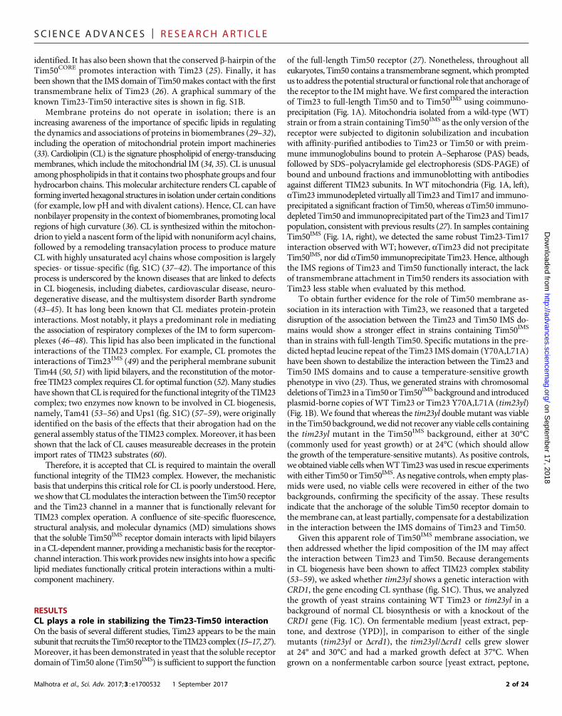
CL affects Tim23-Tim50IMS interactions duringsubstrate transportOur results show that CL promotes the strong interaction of theTim50 receptor with the lipid bilayer, which is critical for its interactionwith the membrane domain of the Tim23 channel. What implicationsdoes this have for protein transport by the TIM23 complex? To addressthis question, we performed our cross-linking–based analysis in thepresence of translocation intermediates. To this end, mitochondriaisolated from WT or Dcrd1 yeast strains were incubated with theTIM23 substrate pSu9-DHFR, a recombinant protein that containsan N-terminal presequence fused to dihydrofolate reductase (DHFR).In the presence of methotrexate, DHFR remains tightly folded andcannot traverse the TOM complex, thereby forming a translocationintermediate engaged with the TIM23 complex (80, 81). Site-directedcross-linking carried out with increasing concentrations of TIM23translocation intermediate allowed us to address how the presence ofCL (WTmitochondria) versus the absence of CL (Dcrd1mitochondria)affected interactions betweenTim50 and different sites of Tim23 duringactive import (Fig. 8).
When we analyzed the interaction between Tim50 and theTim23 IMS region (using the Tim23 S80C variant), we found thatincreasing pSu9-DHFR concentration tended to increase cross-linkingefficiency in both WT mitochondria (Fig. 8A, left) and mitochondrialacking CL (Fig. 8B, left) within each given titration experiment. A gen-eral trend toward enhancing Tim23-Tim50 interaction with increasingsubstrate may be expected, given that translocation promotes activeremodeling of the TIM23 complex (82) and/or leads to increases in theTim50-Tim23 association (18, 20). However, when we analyzed thesubstrate-dependent interaction betweenTim50 and the channel regionof Tim23 (using theTim23 F114C variant), we observed amarked effectof CL. In WT mitochondria, increasing substrate significantly pro-moted the cross-linking–detected interaction between Tim50 and theTim23 channel region (Fig. 8A, right). However, in the absence ofCL, increasing substrate had the opposite effect of further decreasingthe interaction of Tim50 with the Tim23 channel (Fig. 8B, right). Theseresults suggest that the translocation-active form of the TIM23 complexpromotes the interaction of the Tim50 receptor with the Tim23 channelregion in a manner that is facilitated by CL.
17, 2018
DISCUSSIONThe key outcome of this study is the discovery that the soluble recep-tor domain of Tim50 interacts with membranes in a CL-dependentmanner and that this interaction promotes the association betweenthe Tim50 receptor and the Tim23 channel. It has been previouslyshown that Tim50IMS makes several points of contact with Tim23,both (i) at the Tim23 IMS domain, likely via a coiled-coil interaction(15–17, 23, 26–28), and (ii) with Tim23 TMS1 (26). It has also beenshown that CL is important for the functional integrity of the TIM23complex (53–59). Our results here provide a conceptual frameworkto bridge these two phenomena.In the first phase of this work, both biochemical and in vivo geneticevidence indicated that membrane anchorage plays a role in stabilizingthe interaction of Tim50 with other TIM23 complex subunits and thatthe presence of CL is a requirement for this anchorage (Fig. 1). Theseanalyses were predicated on the previous observation that, in yeast, thesoluble Tim50 receptor domain can substitute for full-length Tim50(27). We found that mutations in the Tim23 IMS domain that de-stabilize the Tim50-Tim23 interaction (tim23yl) not only were synthet-
Malhotra et al., Sci. Adv. 2017;3 : e1700532 1 September 2017
ically lethal with a version of Tim50 lacking the transmembrane domain(Tim50IMS) but also had a genetic interactionwith theCL synthase gene(Dcrd). From these results, we concluded that, in addition to theestablished Tim23-Tim50 interaction through their respective IMS do-mains, there are additional stabilizing interactions between Tim50IMS
and a membrane-bound component of the import machinery. Thisconclusion is consistent with a previous cross-linking–based analysis,which revealed an interaction between Tim50IMS and a specific helicalface of Tim23 TMS1 (26). Here, site-directed cross-linking performedin organello (Fig. 2) and in model membranes (Fig. 3) confirmed thatthis interaction occurs in a CL-dependent manner.
The next phase of this work focused on the structural analysis ofTim50IMS. Todate, high-resolution structural informationon S. cerevisiaeTim50 has been limited to the crystal structure of a region of Tim50CORE
(residues 176 to 361) (24, 25). However, the structure of Tim50IMS inits entirety has remained elusive. Understanding the structure of theC-terminal PBD of yeast Tim50 is particularly important because itsdeletion blocks TIM23-mediated protein import, resulting in a lethalphenotype (20). We used SAXS to generate an ab initio envelope ofTim50IMS, which was then used to produce a homology model of theentire receptor domain (Fig. 4). In this model, Tim50IMS forms anelongated structure with Tim50CORE at the center of the N-terminaland C-terminal domains. The C terminus forms a protruding three-helix bundle, consistent with the high a-helical content detected forthe sPBD region (residues 400 to 450) (19). Informed by structuralcharacterization, the current model for substrate recognition by yeastTim50 includes both Tim50CORE and sPBD domains. Specifically,incoming presequences bind to the N-terminal half of sPBD, and then,the affinity of the sPBD for the core region allows the transfer of sub-strate to Tim50CORE, followed by the transfer to the Tim23 channelalong a substrate binding affinity gradient (18–20). The putative acidicbinding pocket at the base of the b-hairpin in Tim50CORE appears to bestructurallymalleable (24), enabling interaction with both presequencesand other sites on Tim50. Consistent with this model of substrate inter-action,NMR-detected sites in sPBD that interactwith presequences andTim50CORE all reside on the second a helix of our homology model,proximal to the core region (closest distances ranging from ca. 9 to17 Å); therefore, a ternary complex of Tim50CORE, sPBD, and the pre-sequence could be formedwith a relativelyminor structural adjustment.
The third phase of the study was aimed at investigating the natureof the CL-dependent membrane interactions of Tim50IMS. MD simula-tions based on our homology model indicated that Tim50IMS interac-tion with lipid bilayers requires CL and that themost stabilizedmode ofinteraction occurs via the face of Tim50IMS that contains the protrudingb-hairpin (Fig. 5). Biochemical and fluorescence-based experimentsthen corroborated the results of the computational simulations, showingthat (i) Tim50IMS binds to model membranes in a CL-dependent man-ner, compared with PG- and PE-containing bilayers; (ii) the b-hairpinbecomes exposed to the nonpolar bilayer core in a CL-dependentmanner,withMLCLsupportingmuch less interaction anddCLsupportingslightly greater interaction; and (iii) the membrane binding surface ofTim50IMS is composed of multiple sites, including the b-hairpin ofTim50CORE and the C-terminal domain (Figs. 6 and 7).
Our analyses of the interaction between Tim50IMS and membranesof many different lipid compositions have provided insights into themolecular basis of this protein-bilayer association. Our results indicatethat like othermembrane-interactive amphitropic proteins, the bindingof Tim50IMS entails both an electrostatic and a hydrophobic component(Fig. 9A) (83). Although yeast Tim50IMS has a negative net charge of
14 of 24
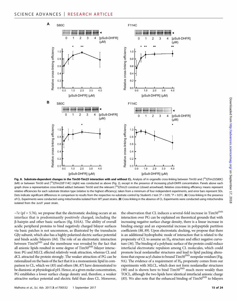
SC I ENCE ADVANCES | R E S EARCH ART I C L E
on Septem
ber 17, 2018http://advances.sciencem
ag.org/D
ownloaded from
−7e (pI = 5.76), we propose that the electrostatic docking occurs at aninterface that is predominantly positively charged, including theb-hairpin and other basic surfaces (fig. S10A). The ability of overallacidic peripheral proteins to bind negatively charged bilayer surfacesvia basic patches is not uncommon, as illustrated by the transducinGbg subunit, which also has a highly polarized electric surface potentialand binds acidic bilayers (84). The role of an electrostatic interactionbetween Tim50IMS and the membrane was revealed by the fact thatall anionic lipids resulted in some degree of Tim50IMS-bilayer interac-tion: PG andMLCL effected relatively weak attraction, whereas CL anddCL attracted the protein strongly. The weaker attraction of PG can berationalized on the basis of the fact that it is amonoanionic lipid in com-parison to CL, which we (85) and others (86, 87) have demonstrated tobe dianionic at physiological pH.Hence, at a givenmolar concentration,PG establishes a lower surface charge density and, therefore, a weakerattractive surface potential and electric field than does CL. Moreover,
Malhotra et al., Sci. Adv. 2017;3 : e1700532 1 September 2017
the observation that CL induces a several-fold increase in Tim50IMS
interaction over PG can be explained on theoretical grounds that withincreasing negative surface charge density, there is a linear increase inbinding energy and an exponential increase in polypeptide partitioncoefficients (88, 89). Upon electrostatic docking, we propose that thereis an additional hydrophobic mode of interaction that is related to thepropensity of CL to assume an HII structure and effect negative curva-ture (36). The binding of a polybasic surface of the protein could reduceinterfacial electrostatic repulsion among CL molecules, which couldpromote local nonlamellar structures and lead to lipid packing altera-tions that expose acyl chains to boundTim50IMS nonpolar residues (Fig.9A). The evidence of a requirement of HII propensity comes from ourexperiments with MLCL, which does not form nonlamellar structures(90) and is shown here to bind Tim50IMS much more weakly thanTOCL, although the two lipids have identical interfacial anionic charge(85). We also note that the enhanced binding of Tim50IMS to bilayers
A
B
Rel
ativ
e cr
oss-
linki
ng e
ffici
ency
[pSu9-DHFR] (μM)
S80C
[pSu9-DHFR] (μM)
[pSu9-DHFR] (μM) [pSu9-DHFR] (μM)
S80C
F114C
F114C
Rel
ativ
e cr
oss-
linki
ng e
ffici
ency
Rel
ativ
e cr
oss-
linki
ng e
ffici
ency
Rel
ativ
e cr
oss-
linki
ng e
ffici
ency
[pSu9-DHFR](μM)
0 1 2 3 4
[pSu9-DHFR](μM)
0 1 2 3 4
[pSu9-DHFR](μM)
0 1 2 3 4
[pSu9-DHFR](μM)
0 1 2 3 4
Fig. 8. Substrate-dependent changes in the Tim50-Tim23 interaction with and without CL. Analysis of in organello cross-linking between Tim50 and [35S]Tim23(S80C)(left) or between Tim50 and [35S]Tim23(F114C) (right) was conducted as above (Fig. 2), except in the presence of increasing pSu9-DHFR concentration. Panels above eachgraph show a representative cross-linked adduct between Tim50 and the relevant [35S]Tim23 construct (closed arrowhead). Relative cross-linking efficiency means representrelative efficiencies for each substrate titration type (relative to the highest efficiency), taken from a minimum of four independent experiments, and error bars represent SDs.Dots indicate significant differences in comparison to results from the respective no-substrate control by Student’s t test (•P < 0.05; ••P < 0.01). (A) Cross-linking in the presenceof CL. Experiments were conducted using mitochondria isolated from WT yeast strains. (B) Cross-linking in the absence of CL. Experiments were conducted using mitochondriaisolated from the Dcrd1 yeast strain.
15 of 24

SC I ENCE ADVANCES | R E S EARCH ART I C L E
on Septem
ber 17, 2018http://advances.sciencem
ag.org/D
ownloaded from
containing dCL may be related to a greater propensity of this lipid tosupport HII structures relative to CL. Finally, the lipid-dependent cross-linking profiles of our in organello experiments are notable (Fig. 2).Whereas the cross-linking–detected interaction between the Tim50 re-
Malhotra et al., Sci. Adv. 2017;3 : e1700532 1 September 2017
ceptor and the Tim23 channel domain wasmore severely altered by theabsence of CL (Dcrd1) than by defects in CL remodeling (Dtaz1 andDcld1) (Fig. 2, E and F), the remodeling mutants resulted in a strongerdeviation from WT for the interaction between Tim50 and the Tim23
SolubleTim50IMS
Electrostaticattraction
A
B
With cardiolipin Without cardiolipin
Substrate Substrate
Membrane-boundTim50IMS Hydrophobic
interaction
Key
Tim50
Tim23
TIM23 substrate
Zwitterionicphospholipid
Cardiolipin
Fig. 9. Working models for Tim50IMS-bilayer and Tim50-Tim23 interactions. (A) Interaction between Tim50IMS and CL-containing bilayers. In solution, Tim50IMS isinitially attracted to the negatively charged bilayer by long-range Coulombic forces. The electric field orients a basic face of the protein, including the b-hairpin, towardthe bilayer. Upon electrostatic docking, hydrophobic interactions between the nonpolar residues at the binding interface and the nonpolar bilayer core stabilize the interaction ina manner stimulated by the HII propensity of CL. Coupled to membrane association are changes in Tim50IMS conformation relative to its water-solvated state. Membrane asso-ciation of Tim50IMSwill be promoted if these Coulombic and hydrophobic interactions are favorable enough to offset the energetic penalty of desolvation. Note that the left paneldisplays the Tim50IMS homology model of this study; subsequent panels show hypothetical outlines of the Tim50IMS structure in cyan, either in the membrane bound state or inthe presence of the TIM23 complex. (B) CL- and substrate-dependent Tim50 dynamics in the context of the TIM23 complex. The presence of CL (left) favors the membrane-associated state of Tim50IMS, which facilitates interaction with the channel domain of Tim23. In the presence of substrate, Tim50IMS retains its interaction with both the Tim23IMS
and Tim23 channel regions. The absenceof CL (right) favors themembrane-dissociated state of Tim50IMS, whichblocks its interactionwith the Tim23 channel region. Thepresenceof substrate further promotes the membrane-dissociated state of Tim50IMS. Note that the stippled region of Tim50 depicts the b-hairpin.
16 of 24

SC I ENCE ADVANCES | R E S EARCH ART I C L E
on Septem
ber 17, 2018http://advances.sciencem
ag.org/D
ownloaded from
IMS domain (Fig. 2, B and C). These disparate responses illustrate thecomplex nature of the relationship between the acyl chaincomposition of CL and protein interactions in the mitochondrialmembrane. For example, the accumulation of unremodeled CL orMLCL resulting from the Dcld1 and Dtaz1 yeast mutants, respectively(62), may produce a stronger effect on the particular Tim50IMS–Tim23TMS1 interaction than does the accumulation of PG resulting from theDcrd1 mutant.
Our results indicate important roles of the conserved b-hairpin andN-terminal region of the Tim50 receptor in promoting membraneassociation. Specifically, b-hairpin structures are known to facilitatebilayer penetration. Highly basic antimicrobial peptides are knownto assume b-hairpin conformations and penetrate bilayers in a mannerdependent on anionic lipids (91), with some also containing conservedTrp residues for cation-p interactions, which promotemembrane inter-action (92), consistent with the conserved sequence of the Tim50IMS
b-hairpin. Our data show that the b-hairpin of Tim50 is exposed to anonpolar milieu upon binding to CL-containing membranes; knowingexactly how deeply it may penetrate the nonpolar core awaits futureexperimentation. Moreover, the C-terminal region of our homologymodel contains multiple segments predicted to be amphipathic, bury-ing hydrophobic faces in the protein interior (fig. S10B). Such a helixbundle fold is a commonmotif for lipid-binding proteins to sequesternonpolar residues while in solution and to undergo dynamic confor-mational changes that expose these residues upon encountering a lipidsurface to promote nonpolar interactions (93). It is possible that theTim50IMS region undergoes a similar structural rearrangement uponmembrane binding that orients these nonpolar faces toward the bi-layer. Our limited proteolysis and site-directed labeling analyses(Fig. 7) strongly suggest that Tim50IMS undergoes a structural changeupon binding to CL-containing bilayers. Exploring which specific re-gions undergo these membrane-coupled dynamics is the subject of anongoing investigation.
Having established the role of CL in promoting the interaction of theTim50 receptor with the bilayer and also with the channel region ofTim23, we propose a working model for CL-dependent receptor-channel dynamics (Fig. 9B). In a translocon not actively transportinga substrate, the presence of CL favors the association of the Tim50receptor domain with the membrane-bound region of Tim23. In theabsence of CL, the association between the Tim50 receptor and theIMS region of Tim23 is favored (Fig. 9B, top). Although CL plays a crit-ical role inmitochondrial energetics (38), theCL-modulated interactionbetween Tim50 and Tim23 does not appear to be directly dependenton the Dym because it is also observed in model membranes lacking atrans-bilayer electric field (Fig. 3). Further, although the CL-dependentmembrane association of Tim50IMS seems to be amajor determinant indirecting its interaction with Tim23, we cannot currently discount thelikely possibility that CL also modulates the conformation of theTim23 channel region (not shown in the model), which may contrib-ute to our observations. On the basis of our in organello experimentswith the TIM23 substrate (Fig. 8), in CL-containing membranes, thepresence of a substrate further stabilizes the interaction between theTim50 receptor and the Tim23 channel region, whereas in membranesdevoid of CL, the presence of a substrate further suppresses this inter-action (Fig. 9B, bottom). One of the key functions of CL is to maintainthe functional associations ofmembrane proteins. Ourmodel suggests amechanism by which CL could promote TIM23-mediated import: bystabilizing the receptor-channel interactions that would otherwise bedestabilized by a substrate in the absence of CL.
Malhotra et al., Sci. Adv. 2017;3 : e1700532 1 September 2017
Some central features of this model warrant further consideration.First, why would a receptor domain with inherent membrane affinityalso be anchored to the bilayer by a transmembrane segment? Onepossibility is that this tethering by a transmembrane element couldmaintain the proximity of the receptor region to the bilayer, effectivelyserving as a mechanism for dimensionality reduction in its diffusionspace to enhance the kinetics of membrane association (94). Anotherpossibility is that the transmembrane segment is required to impartspecificity for assembly with other subunits of the TIM23 complex.Finally, as a structural link between Tim50IMS and the as-yet poorlycharacterized matrix-facing domain of Tim50, the transmembranesegment could transmit signals across the topologically distinct do-mains of this receptor during different stages of the import cycle. Bycomparison, we note that Fis1 (mitochondrial fission 1 protein) ofthe mitochondrial OM is a tail-anchored protein with a soluble cy-tosolic domain that also interacts with bilayers and undergoes confor-mational changes in a lipid-specific manner (95). Second, what couldbe the physiological relevance for the receptor domain to interactwith the membrane in a manner promoted by a specific lipid, in thiscase, CL? One possibility is that spatiotemporal variation of CL concen-tration in the IM could modulate the activity of the receptor. If thedistribution of CL across the IM were homogeneous, then accordingto this model, Tim50IMS would assume a constant membrane-interactivepose regardless of its location. However, given that CL appears to beheterogeneously distributed, for example, more enriched at membranecontact sites (96), these regionsofhigh local [CL]couldpromoteTim50IMS-bilayer association and the resulting conformational alterations atregions near active translocons. By contrast, when the local concentrationof free CL is lower, this could promote a more membrane-dissociatedstate of Tim50IMS, perhaps down-regulating complex activity. We notein this respect that the observed role of Tim50 in regulating the gat-ing of the Tim23 channel (97) or in recruiting Pam17 to transportDy-hypersensitive substrates (98) could also be modulated by CL.
The implications that this model has for mitochondrial proteintransport could come on several fronts that could be verified experi-mentally. First, structural studies investigating CL-dependent confor-mational dynamics will elucidate exactly which regions of the Tim50receptor are membrane-interactive and which domains may revealaltered substrate or subunit binding sites, work that could be carriedout in parallel with atomistic MD simulations. Second, binding studieswill reveal how the affinity of Tim50IMS for substrates may be altered inthe presence of CL-containing bilayers and how CLmay alter the affin-ity of Tim50IMS for subunits other than the Tim23 channel. Finally,comprehensive analysis of substrate import conducted in organello orin reconstituted systems could test how disruptions in CL metabolismcould affect the transmembrane transport versus the lateral integrationfunctions of the TIM23 complex.
MATERIALS AND METHODSYeast strains and growth conditions and isolationof mitochondriaThe yeast strain containing the Tim50IMS truncation mutant was gen-erated by transforming a haploid YPH499-based TIM50 deletion straincontaining aWTcopy of Tim50 on aURAplasmidwith the centromer-ic yeast plasmid pRS315 encodingTIM50IMS and subsequently selectingfor the cells lacking the URA plasmid with 5-FOA, as previously de-scribed (27). To generate yeast strains containing WT Tim23 orTim23 Y70A,L71A in a Tim50 or Tim50IMS background, cells were
17 of 24

SC I ENCE ADVANCES | R E S EARCH ART I C L E
on Septem
ber 17, 2018http://advances.sciencem
ag.org/D
ownloaded from
transformedwith pVT-Uplasmids containingTIM23, and the chromo-somal copy of TIM23 was deleted by homologous recombination, aspreviously described (23). Cells were subsequently transformed withempty pRS314 or pRS314 encoding Tim23 or Tim23 Y70,L71 underthe control of the endogenous promoter and 3′ untranslated regionand selected for loss of URA-containing plasmids with 5-FOA, as de-scribed above. Yeast strains containing genetic knockouts Dcrd1, Dcld1,and Dtaz1 were generated by replacing the open reading frame of thegene using polymerase chain reaction (PCR)–based gene replacement.Yeast cells were grown at 24°C in lactate medium containing 0.1% glu-cose, unless indicated otherwise. For the preparation of activemitochondria, yeast cells were cultivated to mid-log phase, and themitochondria were isolated by lysis of spheroplasts and differential cen-trifugation, as previously described (26).
CoimmunoprecipitationCoimmunoprecipitation experiments using detergent-solubilizedmitochondria were performed as previously described (27). Briefly,mitochondria isolated from yeast strains containing full-length Tim50or Tim50IMSwere incubated at 2mg/ml in solubilization buffer [20mMtris-HCl (pH 8.0), 80 mM KCl, 1 mM phenylmethylsulfonyl fluoride(PMSF), and 1% (w/v) digitonin] at 4°C. Following a clarifying spin,solubilized material was added to protein A–Sepharose beads pre-coupled with antibodies (aTim23 or aTim50) and incubated underagitation for an additional 45min. Following centrifugation, supernatantswere collected, beadswerewashed three times, and bound proteinswereeluted with nonreducing Laemmli buffer for 3 min at 95°C. Both elutedproteins and supernatants were resolved by SDS-PAGE and analyzedby immunoblotting with antibodies against Tim50, Tim23, and Tim17.
Cell-free transcription and protein synthesisThe SP6-based pSP65 plasmid library of Tim23monocysteine variants hasbeen described previously (11, 26). mRNA was transcribed from PCR-generated fragments using 5′ and 3′ oligonucleotides complementary tothe plasmid SP6 promoter and Tim23 sequence downstream of the stopcodon, respectively. Purified PCR products were transcribed in vitro withSP6 polymerase at 37°C for 1.5 hours in reactions containing 100 mMHepes-KOH (pH 7.5), 20 mM MgCl2, 2.5 mM spermidine, 12 mMdithiothreitol (DTT), 4 mM ATP, 4 mM CTP (cytidine 5′-triphosphate),4 mM UTP (uridine 5′-triphosphate), 0.4 mM GTP (guanosine 5′-triphosphate) G(5′)ppp(5′)GRNA cap analog (0.013U/ml; NewEnglandBiolabs), RNasin (0.5 U/ml), and pyrophosphatase (6U/nl); supplemen-ted with 4 mMGTP; and allowed to incubate for an additional 30 min.mRNA was precipitated overnight at −20°C in ethanol and 90 mM so-dium acetate (pH 5.2), washed in 70% (v/v) ethanol, and reconstitutedin TE buffer [10 mM tris-HCl and 1 mM EDTA (pH 7.5)].
In vitro translation reactions were programmed with purified mRNAtranscripts encoding Tim23 variants. Translation reactions {20 mMHepes-KOH (pH 7.5), 100 mM potassium acetate (pH 7.5), 1 mMDTT, 200 mM spermidine, 8 mM S-adenosylmethionine, protease in-hibitors [0.25 mg/ml each of leupeptin, chymostatin, antipain, and pep-tatin A and 0.025% (v/v) aprotinin], RNasin (0.2 U/ml), 1.2 mM ATP,1.2 mM GTP, 64 mM creatine phosphate, creatine phosphokinase(9.6 U/nl), 30 mM amino acids (but lacking methionine for incorpora-tion of [35S]methionine and lacking cysteine for incorporation of NBD-[14C]Cys), and 20% (v/v) wheat germ extract} were incubated at 26°Cfor 5 min before the addition of mRNA and either [35S]methionine(0.25 mCi/ml; for synthesis of [35S]Tim23), 0.6 mM [14C]Cys-tRNACys
(for synthesis of blank samples in fluorescence experiments), or
Malhotra et al., Sci. Adv. 2017;3 : e1700532 1 September 2017
0.6 mM NBD-[14C]Cys-tRNACys (for synthesis of NBD-labeledsamples in fluorescence experiments) and then continued for 40 minat 26°C. The synthesis of the aminoacyl transfer RNA (tRNA) analogNBD-[14C]Cys-tRNACys, designed to recognize single in-frame 5′-UGC-3′ codons of Tim23 monocysteine variants for site-specificNBD probe incorporation, has been previously described (11).
Heterologously expressed proteins: Expression,purification, and labelingFor the expression of soluble domains of Tim50, sequences encodingTim50IMS (residues 133 to 476), Tim50CORE (residues 165 to 361),Tim50IMSDN (residues 165 to 476), and Tim50IMSDC (residues 133to 361) were PCR-amplified using either S. cerevisiae genomic DNAor existing plasmids as a template and subcloned into pET28a vectors,introducing a tobacco etch virus (TEV) protease cleavage site betweenan N-terminal 6×His tag and the coding region. Site-directed mutagen-esis was performed byPCRusing primers designedwithmutations. Theplasmid for the expressionof pSu9-DHFR[thepresequenceof F0-ATPasesubunit 9 fused with DHFR with a 6×His tag in pET30a(+)] has beenpreviously described (26). The plasmid for the expression of membranescaffolding protein (MSP1E3D1 in pET28a vectorwith aTEV-cleavableN-terminal 7×His tag) was obtained from Addgene. All plasmid se-quences were verified by sequencing.
For expression of Tim50 and pSu9-DHFR constructs, competentBL21(DE3) cells (Invitrogen) were transformedwith the relevant plas-mid by heat shock, and cells were grown in 2 liters of YT medium sup-plemented with kanamycin (50 mg/ml) at 37°C to an A600 (absorbanceat 600 nm) of ~0.8. Expression was induced by the addition of 1 mMisopropyl b-D-1-thiogalactopyranoside and incubation at 26°C for2.5 hours. For purification, cells were collected by centrifugation(4000g for 10 min at 4°C); resuspended in resuspension buffer [50 mMmonobasic sodium phosphate (pH 7.5), 300 mM NaCl, and 5 mMimidazole] supplemented with lysozyme (1.25 mg/ml), deoxyribonuclease(0.025 mg/ml), and 0.1 mM PMSF; incubated on ice for 30 min; anddisrupted by probe sonication. After a clarifying spin (30,000g for30 min at 4°C) to remove insoluble material, the soluble fraction wasbatch-incubated with Ni2+-NTA agarose (Qiagen) pre-equilibratedwith resuspension buffer and loaded onto a column, followed by addi-tion of column buffer [50 mMmonobasic sodium phosphate (pH 7.5)and 300 mM NaCl] with step gradients of imidazole (up to 400 mM).Fractions enriched in protein were pooled and dialyzed against dialysisbuffer [20 mM tris-HCl (pH 7.4), 100 mMNaCl, and 0.5 mM EDTA],followed by treatment with His-tagged TEV protease (0.07 U/ml)overnight at 16°C. The cleaved protein was passed over a secondNi2+-NTA column to remove the TEV protease and uncleaved protein.Protein-enriched fractions were then subjected to size exclusion chro-matography on a Superdex 200 10/300 column (Amersham Bio-sciences) equilibrated with protein buffer [10 mM Hepes-KOH (pH7.4), 100 mM NaCl, and 100 mM KCl] and collected at a flow rate of0.5 ml/min. The expression and purification of MSP1E3D1 were simi-lar, except for the fact that cells were incubated with 1% (v/v) TritonX-100 before sonication; column buffers contained 40 mM tris-HCl(pH 8.0) with 1% (v/v) Triton X-100 and 50mMNa-cholate in the col-umn wash steps; and following affinity purification, protein was dia-lyzed directly into MSP buffer [20 mM Tris-HCl (pH 7.5), 100 mMNaCl, and 0.5 mM EDTA], based on described protocols (99, 100),and the 7×His tag was not cleaved to allow for nanodisc purification.
For the site-specific labeling of Tim50IMS with NBD, a 500-ml reac-tion with purified Tim50IMS (S208C) in protein buffer with a 100×molar
18 of 24

SC I ENCE ADVANCES | R E S EARCH ART I C L E
on Septem
ber 17, 2018http://advances.sciencem
ag.org/D
ownloaded from
excess of IANBD (Invitrogen) was incubated for 2 hours at room tem-perature (RT) with continuous rotation, and labeling reaction wasquenched by the addition of 20 mM DTT. Labeled protein wasseparated from free dye by gel filtration with Sephadex G25 resin,and labeling efficiency was quantified as
%Efficiency ¼ ðA280=eTim50IMSÞðA481=eNBDÞ � 100
whereeNBD481 nm ¼23,700M−1 cm−1 andeTim50IMS
280 nm ¼52,830M−1 cm−1. Forall preparations, IANBD labeling efficiency was ~60%.
Assembly of liposomes and nanodiscsSynthetic phospholipids were purchased as chloroform stocks fromAvanti Polar Lipids, including POPC, POPE, POPG, and variants ofCL with 18:1 acyl chains, including TOCL, dCL, and MLCL. All stockswere stored at −20°C until use.
Unilamellar liposomes of specific lipid composition and loaded withsucrose were prepared as previously described (77). Chloroform stocksof lipids blended at the desiredmolar ratios were dried under a nitrogenstream in a Pyrex tube, followed by overnight evaporation in a vacuumdesiccator to remove all traces of organic solvent. Dried lipid films werehydrated in buffer A [10mMNa-Hepes (pH 7.4) and 500mM sucrose]to a concentration of 6 mg lipids/ml, and the resulting multilamellarliposomes were passed 17 times through aMini-Extruder (Avanti) with0.1-mm polycarbonate membranes to produce LUVs. The liposomestock was diluted to 4 ml in buffer B [10 mM Na-Hepes (pH 7.4),100mMNaCl, and 100mMKCl], aliquoted proportionally into four1.5-ml polycarbonate centrifuge tubes, and centrifuged (137,000g for1 hour at 22°C) in a S120-AT2 rotor (Beckman). LUV pellets wereeach reconstituted in 100 ml of buffer B, pooled, and quantified forlipid concentration using the ammonium ferrothiocyanate colorimetricanalysis (101).
To prepare nanodiscs containing [35S]Tim23, chloroform stocksof lipid blends at the desiredmolar ratios were dried under nitrogen andvacuum-desiccated as above. The lipid film was solubilized in MSPbuffer [20mM tris-HCl (pH 7.4), 100mMNaCl, and 0.5mMEDTA]containing 59 mM Na-cholate at a molar lipid concentration thatvaried depending on the lipid blend used. The 2-ml assembly mixturethat included MSP buffer with solubilized lipids, 50 mM purifiedMSP1E3D1 (at a molar ratio of 120:1), [35S]Tim23 at a final concentra-tion of 12.5% (v/v), and protease inhibitor cocktail (Sigma-Aldrich) wasthen prepared. Following a 30-min incubation at RT, prehydrated hy-drophobic adsorbent Bio-Beads SM (Bio-Rad) were added to removedetergent and allow nanodisc assembly to proceed for an additional2hours.Assemblednanodiscswere thenpurified byNi2+-NTAchroma-tography on a column equilibrated with MSP buffer with step gradientsof imidazole (up to 400 mM). The first two elution fraction columnswere combined and dialyzed into ND buffer [20 mM Na-Hepes (pH7.5), 200 mM NaCl, 50 mM KOAc, 5 mM Mg(OAc)2, and 5% (v/v)glycerol] and concentrated in Amicon Ultra centrifugal filter units (3 ml;molecular weight cutoff, 3000).
Chemical cross-linking, thiol modification,and immunoprecipitationCross-linking reactionswith isolatedmitochondria containing [35S]Tim23were conducted exactly as previously described (26). Briefly, in vitro–synthesized [35S]Tim23was imported intomitochondria by performing
Malhotra et al., Sci. Adv. 2017;3 : e1700532 1 September 2017
reactions (26°C for 20 min) in import buffer [20 mM Hepes-KOH(pH 7.5), 250 mM sucrose, 80 mM KCl, 5 mM MgCl2, 2 mM potas-sium phosphate (pH 7.5), fatty acid–free bovine serum albumin (BSA;3 mg/ml), 3 mM NADH, and 1.5 mM ATP] containing isolatedmitochondria (0.25mg/ml) and8%(v/v) translation reaction.Mitochon-dria were isolated by sedimentation (12,500g for 5 min) and re-suspended in cross-linking buffer (CB; import buffer lacking BSA).Where indicated, pSu9-DHFR was added to a final concentration ofup to 4 mM in the presence of methotrexate. Chemical cross-linkingreactions contained mitochondria (final concentration, 0.13 mg/ml)incubated in CB with the homobifunctional thiol-reactive reagentBMOE fromdimethyl sulfoxide (DMSO) stocks to a final concentrationof 50 mM{final [DMSO]= 2.5% (v/v)}. Following incubation at 26°C for20min, the reaction was quenched by the addition of 40mMDTT. Forimmunoprecipitation, mitochondria from cross-linking reactions(25 mg) were sedimented; solubilized in 50 mM tris-HCl (pH 7.5),1mMEDTA, and 1% (v/v) SDS; and then diluted 10-fold in lysis buffer[10 mM tris-HCl (pH 7.5), 15 mM NaCl, 5 mM EDTA, 1 mM PMSF,and 1% (v/v) Triton X-100], followed by a clarifying spin (18,000g for10 min). Supernatants were incubated with PAS beads precoupled toaTim50, rocked for 2 hours at RT, washed three times in 1 ml of lysisbuffer and once in 1 ml of 10 mM tris-HCl (pH 7.5), and eluted with30 ml of 2× SDS sample buffer before SDS-PAGE.
Cross-linking reactions with nanodiscs containing [35S]Tim23([35S]Tim23-ND) were conducted in ND buffer containing 46% (v/v)[35S]Tim23-ND and 50 mM BMOE. Following incubation at 26°C for60 min, the reaction was quenched as described above. For immuno-precipitation, reactions were transferred to a new tube and PAS beadsprecoupled to aTim50were added and incubated at RT for 2 hours. Beadswere then washed three times with 1 ml of radioimmunoprecipitationassay buffer [50 mM tris-HCl (pH 7.4), 150 mM NaCl, 1 mM EDTA,1% (v/v) NP-40, and 0.25 (v/v) Na-deoxycholate] and once in 1 ml of10mM tris-HCl (pH 7.5) and eluted with 30 ml of 2× SDS sample bufferbefore SDS-PAGE.
For site-specific sulfhydryl modification of Tim50IMS, reactions with1 mM Tim50IMS, 5 mM TMM(PEG)12 (Thermo Fisher Scientific), andvariable concentrations of liposomes in CB were incubated at 26°Cfor 5min before quenching with 200mMDTT. Control (prequench)samples were incubated with 200 mM DTT before the addition ofTMM(PEG)12. An equal volume of 2× SDS sample buffer was addedbefore SDS-PAGE.
Cross-linking reactions (“Total” and “Immunoprecipitation”) andthiol modification reactions were resolved on 12% SDS-PAGE gels,which were subsequently fixed, dried, and placed on phosphor imagingK-screens (Kodak). Samples were visualized by radioisotope scan on aPharos FXPlusMolecular Imager (Bio-Rad), andbandswere quantifiedusing Image Lab v3.0 software (Bio-Rad). Cross-linking efficiency wascalculated as the ratio of the intensities of the cross-linked [35S]Tim23 tothe non–cross-linked [35S]Tim23 bands for each experiment. Samplesfrom CL biosynthesis mutants were normalized relative to the bandscorresponding to [35S]Tim23 in WT mitochondria.
Limited proteolysisTim50IMS (1 mM) was incubated in buffer B with or without LUVs(1mg lipid/ml) with the specified lipid composition for 30min at RT ina total volume of 180 ml. Following incubation, 25 ml of the sample wasadded to microfuge tubes containing proteinase K at the indicated finalconcentrations, samples were incubated on ice for 30 min, and pro-teolysis was quenched by the addition of 2.8 mM PMSF. Samples were
19 of 24

SC I ENCE ADVANCES | R E S EARCH ART I C L E
on Septem
ber 17, 2018http://advances.sciencem
ag.org/D
ownloaded from
resolved by SDS-PAGE and visualized by SYPRO Orange staining andscanning on a Pharos FX Plus Molecular Imager.
Liposome cosedimentation analysisReactions (150 ml) with buffer B containing purified Tim50 constructs(final concentration, 1 mM) and varying concentrations of sucrose-loaded LUVs were incubated at RT for 60 min, followed by centrifuga-tion (137,000g at 22°C for 60min). The supernatant was discarded, andpelletswere resuspended in 40 ml of 2× SDS sample buffer. Sampleswereresolved on 12% SDS-PAGE gels, and Tim50 was visualized by stainingwith SYPRO Orange protein stain (Invitrogen). Bands were visualizedon a Pharos FX Plus Molecular Imager (protein stain, SYPRO Orangesetting), and bands were quantified using Image Lab v3.0 software.
Fluorescence spectroscopySteady-state fluorescence measurements were performed with a Fluor-olog 3-22 spectrofluorometer (HORIBA Jobin-Yvon) equipped withphoton-counting electronics, double-grating excitation and emissionmonochromators, and a 450-W xenon lamp. In all cases, fluorescentsamples were measured in parallel with blank samples (lacking dyebut otherwise identical to the fluorescent sample) for signal subtractionto obtain the emission originating from the probe. Emission scans (5-nmbandpass) to measure NBD fluorescence were conducted with lex =470 nm and lem = 490 to 590 nm (2-s integration times with 1-nmsteps). For all sample types, spectra were measured from aminimumof three independently prepared samples.
Spectral measurements of nanodisc samples (recorded in 4-mm×4-mm microcells) included separate readings of NBD-Tim23(T24C)-ND and NBD-Tim23(I111C)-ND (synthesized in the presence ofNBD-[14C]Cys-tRNACys), as well as the corresponding blank samples(synthesized in the presence of [14C]Cys-tRNACys). Measurements ofNBD-Tim50IMS in the presence of liposomes (recorded in 1-cm quartzcuvettes with continuous stirring) contained NBD-Tim50IMS (finalconcentration, 0.5 mM) and the indicated concentration of LUVs ofdefined lipid composition. Fractional changes in maximum fluores-cence intensity (F/F0) for a given concentration of lipid were calculatedby averaging the fluorescence intensity (F) centered about the lmax (530to 535 nm) relative to the intensity in the absence of LUVs (F0). All datapoints were fit using KaleidaGraph (Synergy Software).
Quantitative analysis of bindingOn the basis of Tim50IMS-liposome binding data generated by cose-dimentation analysis (Fig. 6A) and fluorescence-detected binding(Fig. 7, D and E), binding parameters were evaluated using fits toLangmuir binding isothermsorHill expansions thereof. The generalizedequation
½P�B½P�T
¼ ½CLacc�n½Kd�n þ ½CLacc�n
was used to fit hyperbolic binding curves (n = 1) and sigmoidal bindingcurves (n > 1). Here, the variable P represents Tim50IMS, the subscriptB represents bound, and the subscript T represents total; therefore,the ratio [P]B/[P]T is the fraction of Tim50IMS bound. [CLacc] is theTim50IMS-accessible CL concentration, where it is assumed, to a firstapproximation, that all CL of the liposomal outer leaflet is accessible.Kd is the apparent dissociation constant with respect to CL, and nrepresents the Hill coefficient. To analyze cooperative binding, a linear
Malhotra et al., Sci. Adv. 2017;3 : e1700532 1 September 2017
form of this equation was used
logQ
1� Q
� �¼ �n logðKdÞ þ n log½CLacc�
where Q, the fraction of bound Tim50IMS, plotted as a function oflog[CLacc], yields a straight line with a slope equal to the Hill co-efficient. To evaluate binding data specific for the Tim50IMS-CL inter-action, the background (nonspecific) binding to liposomes containingonly POPC was subtracted out from the binding data for liposomeswith CL or CL variants.
SAXS data collection and analysisSAXS scattering data were acquired at the Cornell High Energy Syn-chrotron source G1 station, with the beamline operating at 11.75 keV.Purified protein samples were centrifuged at 14,000 rpm for 10 minbefore data collection and protected from radiation damage by oscil-lating the x-ray beam using a computer-controlled syringe pump.The x-ray beam (wavelength, 1.055 to 1.244 Å) was collimated to250 mm× 500 mm, and images were collected at 4°C with a Dual Pilatus100 K-S detector with a momentum transfer interval of 0.007 Å−1 < q <0.7 Å−1 (where the scattering vector q = 4sinq/l, with q being thescattering angle and l being the wavelength). Sample-to-detector dis-tance was calibrated using silver behenate powder (Gem Dugout).Ten exposures of 2 s each were averaged for both protein-containingsamples and background (buffer B only) samples. Buffer profiles weresubtracted from the averaged protein solution profiles and reducedusing BioXTAS RAW software (102). Guinier analysis was performedusing PRIMUS (103, 104), and the pair-distance distribution functionP(r) and values for I0, Rg, and Dmax were obtained using the programGNOM (105, 106). Rg values calculated from GNOMwere consistentwith those estimated from the Guinier region (table S1). The intensityof a standard (lysozyme, 4.14 mg/ml) at zero scattering angle I0 wasdetermined by Guinier analysis and used to estimate sample molecularweight by comparison (107). The theoretical scattering data from thehomology model and the crystal structure for comparison with theexperimental scattering data were calculated using theCRYSOL program(76). To generate ab initio envelopes, 10 models were constructed inDAMMIF (108), which were aligned and averaged in DAMAVER(109). Final refinement was performed using DAMMIN (110).
Tim50IMS homology model generationAmodel for the full-length Tim50IMS domain was constructed by com-bining the known coordinates of the core region residues (Tim50CC,PDB 3QLE), with homology-modeled structures of the N-terminaland C-terminal regions. Given that the conserved core structure (resi-dues 176 to 361) was solved by x-ray crystallography (25), we chose tomodel the terminal regions independently, rather than generate acomplete homology model for the entire Tim50IMS. The sequence ofthe N-terminal region (residues 133 to 195) and the C-terminal region(residues 343 to 476) was supplied to the I-TASSER web server (111) togeneratemodels of those regions. The terminal regionmodels containedoverlapping regions with the core to facilitate structural alignmentswhen constructing the full-length Tim50IMS model. The top-rankingmodels for both the N terminus and C terminus were built onto thecrystal structure coordinates of Tim50CC. The model building wascarried out in CHARMM (72) by performing a local alignment of res-idues 176 to 180 between the core and the N terminus and then
20 of 24

SC I ENCE ADVANCES | R E S EARCH ART I C L E
on Septem
ber 17, 2018http://advances.sciencem
ag.org/D
ownloaded from
copying in the coordinates of residues 132 to 175. Similarly, the C-terminal coordinates were generated by aligning residues 355 to 361and then copying in the coordinates of residues 362 to 476 into the fullmodel. Hydrogen atoms were constructed with the HBuild command,and steepest descentminimizationwas performed in theGBSW implic-it solventmodel (112). During theminimization, the backbone atoms ofresidues 132 to 360 were harmonically restrained with a force constantof 50 kcal mol−1 Å−2 for 500 steps, followed by 500 steps of unrestrainedminimization.
When this initial model was compared with the Tim50IMS SAXSreconstruction, it was evident that themodel (which predicted a dimericcoiled-coil structure of the C-terminal region) was too elongated,extending outside the envelope. To generate a model consistent withthe experimentally derived SAXS envelope, we replaced the C-terminalregionwith the second highest ranked homologymodel from I-TASSER,which predicted a trimeric coiled-coil structure. The trimeric C-terminalmodel was minimized in the GBSW implicit solvent model while thebackbone atoms of residues 132 to 360 were harmonically restrained.The restraint force constant was set to 200 kcal mol−1 Å−2, and 500steepest descent steps were performed, followed by 500 steps of adoptedbasis Newton-Raphson (ABNR) minimization. The restraint forcewas lowered to 100 kcal mol−1 Å−2, and another 500 steps of ABNRwere performed. This model displayed an angular architecture, whichwas not consistent with the linear SAXS envelope. To improve theagreement between the model and the SAXS envelope, the model wasmanually manipulated with the UCSF Chimera Program (113) to alterthe backbone angles in residues 359 to 360 to generate a more linearconformation. Following the modifications in Chimera, the model wasagain minimized in CHARMM using GBSW solvent while restrainingbackbone atoms in residues 332 to 358 with an initial force constant of200 kcal mol−1 Å−2 for 500 steepest descent steps, followed by 500 ABNRsteps. The restraints were reduced to 100 kcal mol−1 Å−2, and 500 stepsof steepest descent and ABNR were performed. Finally, all restraintswere released, and 500 steps of steepest descent and ABNR minimiza-tion were performed.
CG MD simulationsCGMD simulations were used to characterize Tim50IMS-bilayer inter-actions and to elucidate the influence of CL on Tim50 membranebinding properties. All simulations were performed using the MARTI-NI CGmodel (114) using force field version 2.2P (115, 116) and withthe polarizable water model (117). Bilayers were constructed usingthe insane.py python script (118) to generate a homogeneous POPCbilayer and an 80:20 POPC/CLmolar ratio bilayer. The pure POPC bi-layer contained 722 lipids, whereas the mixed bilayer contained 576POPC and 144 TOCL lipids. The charge distribution and all other para-meters for CL were consistent with the previous study by Dahlberg andMaliniak (119), assigning each phosphate a −0.5 charge to modelproton equilibration between the phosphates. The bilayers weresolvated with polarizable water CG particles, and CG Na+ and Cl− ionswere added to neutralize the systemand to bring the ionic concentrationto 0.1M. The bilayer systems were energy-minimized using the steepest-descents algorithm for 1500 steps. The bilayer systems were thenequilibrated in the NPT ensemble for 1.6 ms.
Tim50IMS was modeled on the basis of the homology model thatwas generated and found to be consistentwith the SAXS envelope. Themartinize.py script was used to generate a CG model of Tim50. Thetertiary structure of the protein wasmaintained through the applicationof an elastic network on top of the force field. In thismethod (ElNeDyn)
Malhotra et al., Sci. Adv. 2017;3 : e1700532 1 September 2017
(120), the distance between pairs of nonbonded backbone beads,separated by less than 0.9 nm, is harmonically restrained with a forceconstant of 500 kJ mol−1 nm−2. However, no elastic restraints wereplaced on the N-terminal region (residues 133 to 195) of Tim50, givenits nature to adopt disordered structures and undergo conformationalchanges.
Protein-bilayer systems were constructed by taking the equilibratedbilayers and placing the protein ~10 nm above the bilayer and solvatingand ionizing to neutralize and bring the ion concentration to 0.1M. Thesizes of the systems were ~115,000 CG particles for the POPC systemsand ~140,000 CG particles for the POPC/CL system. The systems wereenergy-minimized for 1500 steepest descent steps, followed by 800-nsNPT equilibration. We performed five simulations using differentorientations of Tim50IMS for both the POPC andmixed bilayer systems.The same five orientations were used for the POPC and mixed bilayersystems. Production simulations were run for 4 ms.
All simulations were performed using GROMACS v4.6 (121, 122).Electrostatic and Lennard-Jones (LJ) interactions were cut off at 1.2 nm,with electrostatics shifted from 0 to the cutoff, and LJ interactions wereshifted from 0.9 nm to the cutoff. The relative dielectric was set to 2.5, asrecommended when using polarizable water (117). All production runswere simulated in the NPT ensemble using the Berendsen weak cou-pling scheme, with the temperature maintained at 323 K and pressurekept at 1.0 bar with semi-isotropic coupling. The time constants for thepressure and temperature couplings were 3.0 and 1.0 ps, respectively,and the compressibility value was 3 × 10⁻⁵ bar−1. All simulation runswere performed using periodic boundary conditions in the x,y and zdirections. The simulation time step was 0.020 ps. All reported times(with the exception of the time step) were multiplied by the standardMARTINI speed-up factor (4×) to account for the faster dynamics inthe CGmodel. System configurations were visualized, and images weregenerated using VMD software (123).
To compute the potential of mean force (PMF) of Tim50IMS-bilayerbinding, we performed umbrella sampling (124) simulations along thecenter-of-mass (com) separation coordinate between the bilayer andthe protein. From the bound configurations, the protein was pulledoff the bilayer during a steeredMD (SMD) simulation. The SMDpullrate was 0.001 nm/ps, and the force constant was 1000 kJ mol−1 nm−2;intermediateswere collected at 0.1-nm spacing intervals along the z axis,perpendicular to the bilayer plane. Umbrella sampling was performedby simulating each intermediate state for 1.2 ms with a harmonic re-straint on the com separation distance; the restraining potential had aforce constant of 2000 kJ mol−1 nm−2. The last 800 ns of each umbrellasimulation was used to calculate the PMF using the g_wham tool avail-able in GROMACS.
SUPPLEMENTARY MATERIALSSupplementary material for this article is available at http://advances.sciencemag.org/cgi/content/full/3/9/e1700532/DC1fig. S1. The TIM23 complex and CL biogenesis.fig. S2. The effect of CL synthase knockout on Tim50IMS functionality and association with theTIM23 complex.fig. S3. Import and integration of [35S]Tim23 into isolated mitochondria.fig. S4. Response of [35S]Tim23 cross-linking to decreased Dym.fig. S5. Incorporation of [35S]Tim23 into nanodiscs: control tests.fig. S6. Tim50IMS structural predictions and SAXS analyses.fig. S7. SAXS analysis of Tim50CORE.fig. S8. Effect of ionic strength on the Tim50IMS-bilayer interaction.fig. S9. Quantitative analysis of Tim50IMS binding.fig. S10. Structural features of the Tim50IMS homology model.
21 of 24

SC I ENCE ADVANCES | R E S EARCH ART I C L E
table S1. SAXS structural parameters.movie S1. Tim50IMS-bilayer no CL.movie S2. Tim50IMS-bilayer with CL.
on Septem
ber 17, 2018http://advances.sciencem
ag.org/D
ownloaded from
REFERENCES AND NOTES1. A. Chacinska, C. M. Koehler, D. Milenkovic, T. Lithgow, N. Pfanner, Importing
mitochondrial proteins: Machineries and mechanisms. Cell 138, 628–644 (2009).2. W. Neupert, J. M. Herrmann, Translocation of proteins into mitochondria. Annu. Rev.
Biochem. 76, 723–749 (2007).3. L. S. Wenz, Ł. Opaliński, N. Wiedemann, T. Becker, Cooperation of protein machineries in
mitochondrial protein sorting. Biochim. Biophys. Acta 1853, 1119–1129 (2015).4. D. Stojanovski, M. Bohnert, N. Pfanner, M. van der Laan, Mechanisms of protein sorting
in mitochondria. Cold Spring Harb. Perspect. Biol. 4, a011320 (2012).5. M. Marom, A. Azem, D. Mokranjac, Understanding the molecular mechanism of protein
translocation across the mitochondrial inner membrane: Still a long way to go.Biochim. Biophys. Acta 1808, 990–1001 (2011).
6. J. Song, Y. Tamura, T. Yoshihisa, T. Endo, A novel import route for an N-anchormitochondrial outer membrane protein aided by the TIM23 complex. EMBO Rep.15, 670–677 (2014).
7. C. Schulz, A. Schendzielorz, P. Rehling, Unlocking the presequence import pathway.Trends Cell Biol. 25, 265–275 (2015).
8. P. Dolezal, V. Likic, J. Tachezy, T. Lithgow, Evolution of the molecular machines forprotein import into mitochondria. Science 313, 314–318 (2006).
9. T. Endo, K. Yamano, S. Kawano, Structural insight into the mitochondrial protein importsystem. Biochim. Biophys. Acta 1808, 955–970 (2011).
10. L.-S. Wenz, Ł. Opaliński, M.-H. Schuler, L. Ellenrieder, R. Ieva, L. Böttinger, J. Qiu,M. van der Laan, N. Wiedemann, B. Guiard, N. Pfanner, T. Becker, The presequencepathway is involved in protein sorting to the mitochondrial outer membrane.EMBO Rep. 15, 678–685 (2014).
11. N. N. Alder, R. E. Jensen, A. E. Johnson, Fluorescence mapping of mitochondrial TIM23complex reveals a water-facing, substrate-interacting helix surface. Cell 134, 439–450(2008).
12. T. A. Lohret, R. E. Jensen, K. W. Kinnally, Tim23, a protein import component of themitochondrial inner membrane, is required for normal activity of the multipleconductance channel, MCC. J. Cell Biol. 137, 377–386 (1997).
13. K. Malhotra, M. Sathappa, J. S. Landin, A. E. Johnson, N. N. Alder, Structural changes inthe mitochondrial Tim23 channel are coupled to the proton-motive force. Nat. Struct.Mol. Biol. 20, 965–972 (2013).
14. K. N. Truscott, P. Kovermann, A. Geissler, A. Merlin, M. Meijer, A. J. M. Driessen, J. Rassow,N. Pfanner, R. Wagner, A presequence- and voltage-sensitive channel of the mitochondrialpreprotein translocase formed by Tim23. Nat. Struct. Biol. 8, 1074–1082 (2001).
15. A. Geissler, A. Chacinska, K. N. Truscott, N. Wiedemann, K. Brandner, A. Sickmann,H. E. Meyer, C. Meisinger, N. Pfanner, P. Rehling, The mitochondrial presequencetranslocase: An essential role of Tim50 in directing preproteins to the import channel.Cell 111, 507–518 (2002).
16. D. Mokranjac, S. A. Paschen, C. Kozany, H. Prokisch, S. C. Hoppins, F. E. Nargang,W. Neupert, K. Hell, Tim50, a novel component of the TIM23 preprotein translocase ofmitochondria. EMBO J. 22, 816–825 (2003).
17. H. Yamamoto, M. Esaki, T. Kanamori, Y. Tamura, S.-i. Nishikawa, T. Endo, Tim50 is asubunit of the TIM23 complex that links protein translocation across the outer and innermitochondrial membranes. Cell 111, 519–528 (2002).
18. O. Lytovchenko, J. Melin, C. Schulz, M. Kilisch, D. P. Hutu, P. Rehling, Signal recognitioninitiates reorganization of the presequence translocase during protein import.EMBO J. 32, 886–898 (2013).
19. B. Rahman, S. Kawano, K. Yunoki-Esaki, T. Anzai, T. Endo, NMR analyses on theinteractions of the yeast Tim50 C-terminal region with the presequence and Tim50 coredomain. FEBS Lett. 588, 678–684 (2014).
20. C. Schulz, O. Lytovchenko, J. Melin, A. Chacinska, B. Guiard, P. Neumann, R. Ficner,O. Jahn, B. Schmidt, P. Rehling, Tim50’s presequence receptor domain is essential forsignal driven transport across the TIM23 complex. J. Cell Biol. 195, 643–656 (2011).
21. D. Roise, G. Schatz, Mitochondrial presequences. J. Biol. Chem. 263, 4509–4511 (1988).22. L. de la Cruz, R. Bajaj, S. Becker, M. Zweckstetter, The intermembrane space domain
of Tim23 is intrinsically disordered with a distinct binding region for presequences. ProteinSci. 19, 2045–2054 (2010).
23. L. Gevorkyan-Airapetov, K. Zohary, D. Popov-Čeleketić, K. Mapa, K. Hell, W. Neupert,A. Azem, D. Mokranjac, Interaction of Tim23 with Tim50 is essential for protein translocationby the mitochondrial TIM23 complex. J. Biol. Chem. 284, 4865–4872 (2009).
24. J. Li, B. Sha, The structure of Tim50(164–361) suggests the mechanism by which Tim50receives mitochondrial presequences. Acta Crystallogr. F Struct. Biol. Commun. 71 (Pt. 9),1146–1151 (2015).
Malhotra et al., Sci. Adv. 2017;3 : e1700532 1 September 2017
25. X. Qian, M. Gebert, J. Höpker, M. Yan, J. Li, N. Wiedemann, M. van der Laan, N. Pfanner,B. Sha, Structural basis for the function of Tim50 in the mitochondrial presequencetranslocase. J. Mol. Biol. 411, 513–519 (2011).
26. N. N. Alder, J. Sutherland, A. I. Buhring, R. E. Jensen, A. E. Johnson, Quaternary structureof the mitochondrial TIM23 complex reveals dynamic association between Tim23pand other subunits. Mol. Biol. Cell 19, 159–170 (2008).
27. D. Mokranjac, M. Sichting, D. Popov-Čeleketić, K. Mapa, L. Gevorkyan-Airapetov,K. Zohary, K. Hell, A. Azem, W. Neupert, Role of Tim50 in the transfer of precursorproteins from the outer to the inner membrane of mitochondria. Mol. Biol. Cell 20,1400–1407 (2009).
28. Y. Tamura, Y. Harada, T. Shiota, K. Yamano, K. Watanabe, M. Yokota, H. Yamamoto,H. Sesaki, T. Endo, Tim23–Tim50 pair coordinates functions of translocators and motorproteins in mitochondrial protein import. J. Cell Biol. 184, 129–141 (2009).
29. O. S. Andersen, R. E. Koeppe, Bilayer thickness and membrane protein function: Anenergetic perspective. Annu. Rev. Biophys. Biomol. Struct. 36, 107–130 (2007).
30. T. A. Cross, D. T. Murray, A. Watts, Helical membrane protein conformations and theirenvironment. Eur. Biophys. J. 42, 731–755 (2013).
31. A. G. Lee, Lipid–protein interactions in biological membranes: A structural perspective.Biochim. Biophys. Acta 1612, 1–40 (2003).
32. A. G. Lee, How lipids affect the activities of integral membrane proteins. Biochim.Biophys. Acta 1666, 62–87 (2004).
33. L. Böttinger, L. Ellenrieder, T. Becker, How lipids modulate mitochondrial protein import.J. Bioenerg. Biomembr. 48, 125–135 (2016).
34. S. M. Claypool, C. M. Koehler, The complexity of cardiolipin in health and disease. TrendsBiochem. Sci. 37, 32–41 (2012).
35. A. S. Joshi, J. Zhou, V. M. Gohil, S. Chen, M. L. Greenberg, Cellular functions of cardiolipinin yeast. Biochim. Biophys. Acta 1793, 212–218 (2009).
36. R. N. A. H. Lewis, R. N. McElhaney, The physicochemical properties of cardiolipin bilayersand cardiolipin-containing lipid membranes. Biochim. Biophys. Acta 1788, 2069–2079(2009).
37. M. G. Baile, Y.-W. Lu, S. M. Claypool, The topology and regulation of cardiolipinbiosynthesis and remodeling in yeast. Chem. Phys. Lipids 179, 25–31 (2014).
38. M. Ren, C. K. L. Phoon, M. Schlame, Metabolism and function of mitochondrialcardiolipin. Prog. Lipid Res. 55, 1–16 (2014).
39. M. Schlame, Cardiolipin synthesis for the assembly of bacterial and mitochondrialmembranes. J. Lipid Res. 49, 1607–1620 (2008).
40. M. Schlame, Cardiolipin remodeling and the function of tafazzin. Biochim. Biophys. Acta1831, 582–588 (2013).
41. M. Schlame, M. Ren, Y. Xu, M. L. Greenberg, I. Haller, Molecular symmetry inmitochondrial cardiolipins. Chem. Phys. Lipids 138, 38–49 (2005).
42. C. Ye, Z. Shen, M. L. Greenberg, Cardiolipin remodeling: A regulatory hub for modulatingcardiolipin metabolism and function. J. Bioenerg. Biomembr. 48, 113–123 (2016).
43. G. J. Gaspard, C. R. McMaster, Cardiolipin metabolism and its causal role in theetiology of the inherited cardiomyopathy Barth syndrome. Chem. Phys. Lipids 193,1–10 (2015).
44. Y.-W. Lu, S. M. Claypool, Disorders of phospholipid metabolism: An emerging class ofmitochondrial disease due to defects in nuclear genes. Front. Genet. 6, 3 (2015).
45. Z. Shen, C. Ye, K. McCain, M. L. Greenberg, The role of cardiolipin in cardiovascularhealth. Biomed. Res. Int. 2015, 891707 (2015).
46. R. Arias-Cartin, S. Grimaldi, P. Arnoux, B. Guigliarelli, A. Magalon, Cardiolipin binding inbacterial respiratory complexes: Structural and functional implications. Biochim. Biophys.Acta 1817, 1937–1949 (2012).
47. Y. Chaban, E. J. Boekema, N. V. Dudkina, Structures of mitochondrial oxidativephosphorylation supercomplexes and mechanisms for their stabilisation. Biochim.Biophys. Acta 1837, 418–426 (2014).
48. E. Mileykovskaya, W. Dowhan, Cardiolipin-dependent formation of mitochondrialrespiratory supercomplexes. Chem. Phys. Lipids 179, 42–48 (2014).
49. R. Bajaj, F. Munari, S. Becker, M. Zweckstetter, Interaction of the intermembrane spacedomain of Tim23 protein with mitochondrial membranes. J. Biol. Chem. 289,34620–34626 (2014).
50. M. Marom, R. Safonov, S. Amram, Y. Avneon, E. Nachliel, M. Gutman, K. Zohary, A. Azem,Y. Tsfadia, Interaction of the Tim44 C-terminal domain with negatively chargedphospholipids. Biochemistry 48, 11185–11195 (2009).
51. C. Weiss, W. Oppliger, G. Vergères, R. Demel, P. Jenö, M. Horst, B. de Kruijff, G. Schatz,A. Azem, Domain structure and lipid interaction of recombinant yeast Tim44. Proc. Natl.Acad. Sci. U.S.A. 96, 8890–8894 (1999).
52. M. van der Laan, M. Meinecke, J. Dudek, D. P. Hutu, M. Lind, I. Perschil, B. Guiard,R. Wagner, N. Pfanner, P. Rehling, Motor-free mitochondrial presequence translocasedrives membrane integration of preproteins. Nat. Cell Biol. 9, 1152–1159 (2007).
53. M. R. Gallas, M. K. Dienhart, R. A. Stuart, R. M. Long, Characterization of Mmp37p, aSaccharomyces cerevisiae mitochondrial matrix protein with a role in mitochondrialprotein import. Mol. Biol. Cell 17, 4051–4062 (2006).
22 of 24

SC I ENCE ADVANCES | R E S EARCH ART I C L E
on Septem
ber 17, 2018http://advances.sciencem
ag.org/D
ownloaded from
54. S. Kutik, M. Rissler, X. Li Guan, B. Guiard, G. Shui, N. Gebert, P. N. Heacock, P. Rehling,W. Dowhan, M. R. Wenk, N. Pfanner, N. Wiedemann, The translocator maintenanceprotein Tam41 is required for mitochondrial cardiolipin biosynthesis. J. Cell Biol.183, 1213–1221 (2008).
55. Y. Tamura, Y. Harada, S.-i. Nishikawa, K. Yamano, M. Kamiya, T. Shiota, T. Kuroda, O. Kuge,H. Sesaki, K. Imai, K. Tomii, T. Endo, Tam41 is a CDP-diacylglycerol synthase required forcardiolipin biosynthesis in mitochondria. Cell Metab. 17, 709–718 (2013).
56. Y. Tamura, Y. Harada, K. Yamano, K. Watanabe, D. Ishikawa, C. Ohshima, S.-i. Nishikawa,H. Yamamoto, T. Endo, Identification of Tam41 maintaining integrity of the TIM23protein translocator complex in mitochondria. J. Cell Biol. 174, 631–637 (2006).
57. M. Connerth, T. Tatsuta, M. Haag, T. Klecker, B. Westermann, T. Langer,Intramitochondrial transport of phosphatidic acid in yeast by a lipid transfer protein.Science 338, 815–818 (2012).
58. C. Potting, C. Wilmes, T. Engmann, C. Osman, T. Langer, Regulation of mitochondrialphospholipids by Ups1/PRELI-like proteins depends on proteolysis and Mdm35.EMBO J. 29, 2888–2898 (2010).
59. Y. Tamura, T. Endo, M. Iijima, H. Sesaki, Ups1p and Ups2p antagonistically regulatecardiolipin metabolism in mitochondria. J. Cell Biol. 185, 1029–1045 (2009).
60. F. Jiang, M. T. Ryan, M. Schlame, M. Zhao, Z. Gu, M. Klingenberg, N. Pfanner,M. L. Greenberg, Absence of cardiolipin in the crd1 null mutant results in decreasedmitochondrial membrane potential and reduced mitochondrial function. J. Biol.Chem. 275, 22387–22394 (2000).
61. U. Bömer, M. Meijer, A. C. Maarse, A. Hönlinger, P. J. T. Dekker, N. Pfanner, J. Rassow,Multiple interactions of components mediating preprotein translocation across theinner mitochondrial membrane. EMBO J. 16, 2205–2216 (1997).
62. M. G. Baile, M. Sathappa, Y.-W. Lu, E. Pryce, K. Whited, J. M. McCaffery, X. Han, N. N. Alder,S. M. Claypool, Unremodeled and remodeled cardiolipin are functionallyindistinguishable in yeast. J. Biol. Chem. 289, 1768–1778 (2014).
63. A. J. Davis, K. R. Ryan, R. E. Jensen, Tim23p contains separate and distinct signals fortargeting to mitochondria and insertion into the inner membrane. Mol. Biol. Cell 9,2577–2593 (1998).
64. V. Koshkin, M. L. Greenberg, Oxidative phosphorylation in cardiolipin-lacking yeastmitochondria. Biochem. J. 347 (Pt. 3), 687–691 (2000).
65. K. Malhotra, N. N. Alder, Advances in the use of nanoscale bilayers to study membraneprotein structure and function. Biotechnol. Genet. Eng. Rev. 30, 79–93 (2014).
66. M. A. Schuler, I. G. Denisov, S. G. Sligar, Nanodiscs as a new tool to examine lipid–proteininteractions. Methods Mol. Biol. 974, 415–433 (2013).
67. I. G. Denisov, S. G. Sligar, Nanodiscs for structural and functional studies of membraneproteins. Nat. Struct. Mol. Biol. 23, 481–486 (2016).
68. A. R. Long, C. C. O’Brien, N. N. Alder, The cell-free integration of a polytopicmitochondrial membrane protein into liposomes occurs cotranslationally and in alipid-dependent manner. PLOS ONE 7, e46332 (2012).
69. M. Zhang, J. Liu, Y. Kim, J. E. Dixon, S. L. Pfaff, G. N. Gill, J. P. Noel, Y. Zhang, Structural andfunctional analysis of the phosphoryl transfer reaction mediated by the human smallC-terminal domain phosphatase, Scp1. Protein Sci. 19, 974–986 (2010).
70. C. J. Oldfield, Y. Cheng, M. S. Cortese, C. J. Brown, V. N. Uversky, A. K. Dunker, Comparing andcombining predictors of mostly disordered proteins. Biochemistry 44, 1989–2000 (2005).
71. P. Tompa, E. Schad, A. Tantos, L. Kalmar, Intrinsically disordered proteins: Emerginginteraction specialists. Curr. Opin. Struct. Biol. 35, 49–59 (2015).
72. B. R. Brooks, C. L. Brooks III, A. D. Mackerell Jr., L. Nilsson, R. J. Petrella, B. Roux, Y. Won,G. Archontis, C. Bartels, S. Boresch, A. Caflisch, L. Caves, Q. Cui, A. R. Dinner, M. Feig,S. Fischer, J. Gao, M. Hodoscek, W. Im, K. Kuczera, T. Lazaridis, J. Ma, V. Ovchinnikov,E. Paci, R. W. Pastor, C. B. Post, J. Z. Pu, M. Schaefer, B. Tidor, R. M. Venable,H. L. Woodcock, X. Wu, W. Yang, D. M. York, M. Karplus, CHARMM: The biomolecularsimulation program. J. Comput. Chem. 30, 1545–1614 (2009).
73. A. D. MacKerell Jr., D. Bashford, M. Bellott, R. L. Dunbrack Jr., J. D. Evanseck, M. J. Field,S. Fischer, J. Gao, H. Guo, S. Ha, D. Joseph-McCarthy, L. Kuchnir, K. Kuczera, F. T. K. Lau,C. Mattos, S. Michnick, T. Ngo, D. T. Nguyen, B. Prodhom, W. E. Reiher, B. Roux,M. Schlenkrich, J. C. Smith, R. Stote, J. Straub, M. Watanabe, J. Wiórkiewicz-Kuczera,D. Yin, M. Karplus, All-atom empirical potential for molecular modeling and dynamicsstudies of proteins. J. Phys. Chem. B 102, 3586–3616 (1998).
74. A. D. Mackerell Jr., M. Feig, C. L. Brooks III, Extending the treatment of backboneenergetics in protein force fields: Limitations of gas-phase quantum mechanics inreproducing protein conformational distributions in molecular dynamics simulations.J. Comput. Chem. 25, 1400–1415 (2004).
75. W. Wriggers, P. Chacón, Using Situs for the registration of protein structures withlow-resolution beadmodels from X-ray solution scattering. J. Appl. Cryst. 34, 773–776 (2001).
76. D. I. Svergun, M. H. J. Koch, Small-angle scattering studies of biological macromoleculesin solution. Rep. Prog. Phys. 66, 1735–1782 (2003).
77. M. Marom, A. Azem, The use of cardiolipin-containing liposomes as a model system tostudy the interaction between proteins and the inner mitochondrial membrane.Methods Mol. Biol. 1033, 147–155 (2013).
Malhotra et al., Sci. Adv. 2017;3 : e1700532 1 September 2017
78. D. P. Siegel, R. M. Epand, The mechanism of lamellar-to-inverted hexagonal phasetransitions in phosphatidylethanolamine: Implications for membrane fusionmechanisms. Biophys. J. 73, 3089–3111 (1997).
79. T. Heimburg, D. Marsh, Protein surface-distribution and protein-protein interactions inthe binding of peripheral proteins to charged lipid membranes. Biophys. J. 68, 536–546(1995).
80. M. Eilers, G. Schatz, Binding of a specific ligand inhibits import of a purified precursorprotein into mitochondria. Nature 322, 228–232 (1986).
81. D. Rapaport, K.-P. Künkele, M. Dembowski, U. Ahting, F. E. Nargang, W. Neupert, R. Lill,Dynamics of the TOM complex of mitochondria during binding and translocation ofpreproteins. Mol. Cell. Biol. 18, 5256–5262 (1998).
82. D. Popov-Čeleketić, K. Mapa, W. Neupert, D. Mokranjac, Active remodelling of theTIM23 complex during translocation of preproteins into mitochondria. EMBO J. 27,1469–1480 (2008).
83. A. Mulgrew-Nesbitt, K. Diraviyam, J. Wang, S. Singh, P. Murray, Z. Li, L. Rogers,N. Mirkovic, D. Murray, The role of electrostatics in protein–membrane interactions.Biochim. Biophys. Acta 1761, 812–826 (2006).
84. D. Murray, S. McLaughlin, B. Honig, The role of electrostatic interactions in theregulation of the membrane association of G protein bg heterodimers. J. Biol. Chem.276, 45153–45159 (2001).
85. M. Sathappa, N. N. Alder, The ionization properties of cardiolipin and its variants inmodel bilayers. Biochim. Biophys. Acta 1858, 1362–1372 (2016).
86. E. E. Kooijman, L. A. Swim, Z. T. Graber, Y. Y. Tyurina, H. Bayır, V. E. Kagan, Magic anglespinning 31P NMR spectroscopy reveals two essentially identical ionization states forthe cardiolipin phosphates in phospholipid liposomes. Biochim. Biophys. Acta 1859,61–68 (2017).
87. G. Olofsson, E. Sparr, Ionization constants pKa of cardiolipin. PLOS ONE 8, e73040(2013).
88. A. Arbuzova, L. Wang, J. Wang, G. Hangyás-Mihályné, D. Murray, B. Honig, S. McLaughlin,Membrane binding of peptides containing both basic and aromatic residues.Experimental studies with peptides corresponding to the scaffolding region of caveolinand the effector region of MARCKS. Biochemistry 39, 10330–10339 (2000).
89. S. McLaughlin, S. O. Smith, M. J. Hayman, D. Murray, An electrostatic engine model forautoinhibition and activation of the epidermal growth factor receptor (EGFR/ErbB)family. J. Gen. Physiol. 126, 41–53 (2005).
90. G. L. Powell, D. Marsh, Polymorphic phase behavior of cardiolipin derivatives studied byphosphorus-31 NMR and x-ray diffraction. Biochemistry 24, 2902–2908 (1985).
91. M. Tang, M. Hong, Structure and mechanism of b-hairpin antimicrobial peptides in lipidbilayers from solid-state NMR spectroscopy. Mol. Biosyst. 5, 317–322 (2009).
92. D. I. Chan, E. J. Prenner, H. J. Vogel, Tryptophan- and arginine-rich antimicrobialpeptides: Structures and mechanisms of action. Biochim. Biophys. Acta 1758, 1184–1202(2006).
93. V. Narayanaswami, R. S. Kiss, P. M. M. Weers, The helix bundle: A reversible lipid bindingmotif. Comp. Biochem. Physiol. A Mol. Integr. Physiol. 155, 123–133 (2010).
94. A. Nag, A. R. Dinner, Enhancement of diffusion-controlled reaction rates by surface-induced orientational restriction. Biophys. J. 90, 896–902 (2006).
95. R. C. Wells, R. B. Hill, The cytosolic domain of Fis1 binds and reversibly clusters lipidvesicles. PLOS ONE 6, e21384 (2011).
96. D. Ardail, J.-P. Privat, M. Egret-Charlier, C. Levrat, F. Lerme, P. Louisot, Mitochondrialcontact sites. Lipid composition and dynamics. J. Biol. Chem. 265, 18797–18802 (1990).
97. M. Meinecke, R. Wagner, P. Kovermann, B. Guiard, D. U. Mick, D. P. Hutu, W. Voos,K. N. Truscott, A. Chacinska, N. Pfanner, P. Rehling, Tim50 maintains the permeabilitybarrier of the mitochondrial inner membrane. Science 312, 1523–1526 (2006).
98. A. B. Schendzielorz, C. Schulz, O. Lytovchenko, A. Clancy, B. Guiard, R. Ieva,M. van der Laan, P. Rehling, Two distinct membrane potential–dependent steps drivemitochondrial matrix protein translocation. J. Cell Biol. 216, 83–92 (2017).
99. T. K. Ritchie, Y. V. Grinkova, T. H. Bayburt, I. G. Denisov, J. K. Zolnerciks, W. M. Atkins,S. G. Sligar, Reconstitution of membrane proteins in phospholipid bilayer nanodiscs.Methods Enzymol. 464, 211–231 (2009).
100. C. T. Schwall, V. L. Greenwood, N. N. Alder, The stability and activity of respiratoryComplex II is cardiolipin-dependent. Biochim. Biophys. Acta 1817, 1588–1596 (2012).
101. J. C. M. Stewart, Colorimetric determination of phospholipids with ammoniumferrothiocyanate. Anal. Biochem. 104, 10–14 (1980).
102. S. S. Nielsen, K. N. Toft, D. Snakenborg, M. G. Jeppesen, J. K. Jacobsen, B. Vestergaard,J. P. Kutter, L. Arleth, BioXTAS RAW, a software program for high-throughput automatedsmall-angle X-ray scattering data reduction and preliminary analysis. J. Appl. Cryst.42, 959–964 (2009).
103. A. Guinier, G. Fournet, Small-Angle Scattering of X-rays (Structure of Matter Series)(John Wiley & Sons, Inc., 1955).
104. P. V. Konarev, V. V. Volkov, A. V. Sokolova, M. H. J. Koch, D. I. Svergun, PRIMUS: Awindows PC-based system for small-angle scattering data analysis. J. Appl. Cryst. 36,1277–1282 (2003).
23 of 24

SC I ENCE ADVANCES | R E S EARCH ART I C L E
http://advances.scienceD
ownloaded from
105. A. V. Semenyuk, D. I. Svergun, GNOM—A program package for small-angle scatteringdata processing. J. Appl. Cryst. 24, 537–540 (1991).
106. D. I. Svergun, Determination of the regularization parameter in indirect-transformmethods using perceptual criteria. J. Appl. Cryst. 25, 495–503 (1992).
107. E. Mylonas, D. I. Svergun, Accuracy of molecular mass determination of proteins insolution by small-angle X-ray scattering. J. Appl. Cryst. 40, s245–s249 (2007).
108. D. Franke, D. I. Svergun, DAMIFF, a program for rapid ab-initio shape determination insmall-angle scattering. J. Appl. Cryst. 42, 342–346 (2009).
109. V. V. Volkov, D. I. Svergun, Uniqueness of ab initio shape determination in small-anglescattering. J. Appl. Cryst. 36, 860–864 (2003).
110. D. I. Svergun, Restoring low resolution structure of biological macromolecules fromsolution scattering using simulated annealing. Biophys. J. 76, 2879–2886 (1999).
111. Y. Zhang, I-TASSER server for protein 3D structure prediction. BMC Bioinformatics 9, 40 (2008).112. W. Im, M. S. Lee, C. L. Brooks III, Generalized born model with a simple smoothing
function. J. Comput. Chem. 24, 1691–1702 (2003).113. E. F. Pettersen, T. D. Goddard, C. C. Huang, G. S. Couch, D. M. Greenblatt, E. C. Meng,
T. E. Ferrin, UCSF Chimera—A visualization system for exploratory research and analysis.J. Comput. Chem. 25, 1605–1612 (2004).
114. S. J. Marrink, H. J. Risselada, S. Yefimov, D. P. Tieleman, A. H. de Vries, The MARTINI forcefield: Coarse grained model for biomolecular simulations. J. Phys. Chem. B 111,7812–7824 (2007).
115. D. H. de Jong, G. Singh, W. F. D. Bennett, C. Arnarez, T. A. Wassenaar, L. V. Schäfer,X. Periole, D. P. Tieleman, S. J. Marrink, Improved parameters for the Martinicoarse-grained protein force field. J. Chem. Theory Comput. 9, 687–697 (2013).
116. L. Monticelli, S. K. Kandasamy, X. Periole, R. G. Larson, D. P. Tieleman, S.-J. Marrink, TheMARTINI coarse-grained force field: Extension to proteins. J. Chem. Theory Comput.4, 819–834 (2008).
117. S. O. Yesylevskyy, L. V. Schäfer, D. Sengupta, S. J. Marrink, Polarizable water model forthe coarse-grained MARTINI force field. PLOS Comput. Biol. 6, e1000810 (2010).
118. T. A. Wassenaar, H. I. Ingólfsson, R. A. Böckmann, D. P. Tieleman, S. J. Marrink,Computational lipidomics with insane: A versatile tool for generating custom membranesfor molecular simulations. J. Chem. Theory Comput. 11, 2144–2155 (2015).
119. M. Dahlberg, A. Maliniak, Mechanical properties of coarse-grained bilayers formed bycardiolipin and zwitterionic lipids. J. Chem. Theory Comput. 6, 1638–1649 (2010).
120. X. Periole, M. Cavalli, S.-J. Marrink, M. A. Ceruso, Combining an elastic network with acoarse-grained molecular force field: Structure, dynamics, and intermolecularrecognition. J. Chem. Theory Comput. 5, 2531–2543 (2009).
Malhotra et al., Sci. Adv. 2017;3 : e1700532 1 September 2017
121. B. Hess, C. Kutzner, D. van der Spoel, E. Lindahl, GROMACS 4: Algorithms for highlyefficient, load-balanced, and scalable molecular simulation. J. Chem. Theory Comput.4, 435–447 (2008).
122. D. Van Der Spoel, E. Lindahl, B. Hess, G. Groenhof, A. E. Mark, H. J. C. Berendsen,GROMACS: Fast, flexible, and free. J. Comput. Chem. 26, 1701–1718 (2005).
123. W. Humphrey, A. Dalke, K. Schulten, VMD: Visual molecular dynamics. J. Mol. Graph.14, 33–38 (1996).
124. G. M. Torrie, J. P. Valleau, Nonphysical sampling distributions in Monte Carlo free-energyestimation: Umbrella sampling. J. Comp. Phys. 23, 187–199 (1977).
Acknowledgments: We thank S. Claypool for providing CL biosynthesis–defective yeaststrains and J. Landin, P. Robisch, and Z. Stanic for expert technical assistance. Funding: Thiswork was supported by the NIH [grants 1R01GM113092 (to N.N.A.) and K22-A1099163 (toE.R.M.)], the NSF (grant MCB-1330695 to N.N.A.), and the Deutsche Forschungsgemeinschaft(grants MO1944/1-1 and MO1944/1-2 to D.M.). Computational resources were provided on theHPC Hornet cluster located at the University of Connecticut at Storrs. Author contributions:K.M. and N.N.A. conceived the original idea of the project. K.M. and A.M. carried out cross-linking and immunoprecipitation analyses. K.M. carried out membrane interaction analyses.D.M. and U.G. generated yeast strains and carried out coimmunoprecipitation and yeastgrowth experiments. K.M., T.H.D., and V.L.R. carried out SAXS measurements and datainterpretation. E.R.M. and S.N. carried out MD simulations and homology modeling. N.N.A.supervised the research. N.N.A., K.M., D.M., and E.R.M. wrote the manuscript. All authorscontributed to the editing of the manuscript. Competing interests: The authors declare thatthey have no competing interests. Data and materials availability: All data needed toevaluate the conclusions in the paper are present in the paper and/or the SupplementaryMaterials. Additional data related to this paper may be requested from the authors.
Submitted 17 February 2017Accepted 4 August 2017Published 1 September 201710.1126/sciadv.1700532
Citation: K. Malhotra, A. Modak, S. Nangia, T. H. Daman, U. Gunsel, V. L. Robinson,D. Mokranjac, E. R. May, N. N. Alder, Cardiolipin mediates membrane and channelinteractions of the mitochondrial TIM23 protein import complex receptor Tim50. Sci. Adv. 3,e1700532 (2017).
ma
24 of 24
on Septem
ber 17, 2018g.org/

import complex receptor Tim50Cardiolipin mediates membrane and channel interactions of the mitochondrial TIM23 protein
R. May and Nathan N. AlderKetan Malhotra, Arnab Modak, Shivangi Nangia, Tyler H. Daman, Umut Gunsel, Victoria L. Robinson, Dejana Mokranjac, Eric
DOI: 10.1126/sciadv.1700532 (9), e1700532.3Sci Adv
ARTICLE TOOLS http://advances.sciencemag.org/content/3/9/e1700532
MATERIALSSUPPLEMENTARY http://advances.sciencemag.org/content/suppl/2017/08/28/3.9.e1700532.DC1
REFERENCES
http://advances.sciencemag.org/content/3/9/e1700532#BIBLThis article cites 123 articles, 34 of which you can access for free
PERMISSIONS http://www.sciencemag.org/help/reprints-and-permissions
Terms of ServiceUse of this article is subject to the
registered trademark of AAAS.is aScience Advances Association for the Advancement of Science. No claim to original U.S. Government Works. The title
York Avenue NW, Washington, DC 20005. 2017 © The Authors, some rights reserved; exclusive licensee American (ISSN 2375-2548) is published by the American Association for the Advancement of Science, 1200 NewScience Advances
on Septem
ber 17, 2018http://advances.sciencem
ag.org/D
ownloaded from