cardiovascular system - nikolai.lazarov.pronikolai.lazarov.pro/files/angiology_eng/heart.pdf ·...
TRANSCRIPT
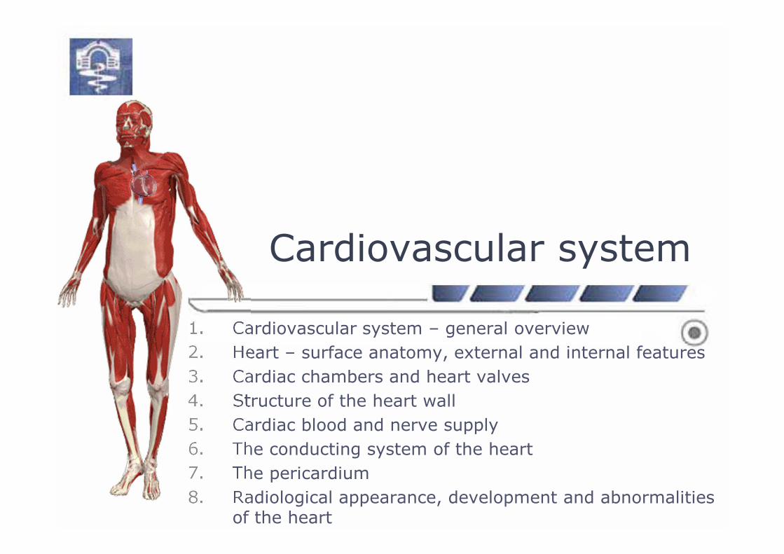
Cardiovascular system
1. Cardiovascular system – general overview
2. Heart – surface anatomy, external and internal features
3. Cardiac chambers and heart valves
4. Structure of the heart wall
5. Cardiac blood and nerve supply
6. The conducting system of the heart
7. The pericardium
8. Radiological appearance, development and abnormalities of the heart

Cardiovascular system
2
� Angiology (Gr. ἀγγεῖον, angeīon, „vessel"; и -λογία, -logia)= vascular medicine
� Cardiovascular system:� central organ
– heart
� blood vessels� arteries� capillaries� veins

Circulatory system� Two parts (systems):
� blood-vascular system
� arterial system
� venous system
� lymphatic system
3

Central organ
4
� Heart – cor, (Gr. cardia):
� ‘left (arterial)’ heart
� ‘right (venous)’ heart
� Functional compartments:
� atria – left and right
� chambers – left and right
� Blood circulation:
� systemic circuit
� portal circulation
� pulmonary circuit

5
� Blood flow (circulation)
� Blood vessels, vasa sanguinea:
� arteries, arteriae
� arterial system
� capillaries
�microcirculatory compartment
� veins, venae
� venous system
� Collateral circulation
Blood-vascular system

6
Arterial system� Arterial tree, Gr. ἀρτηρία (artēria); αήρ, air (Galen)
� principle of divergence
� “arterial tree”
� arterial branches – monotomous
and dichotomous branching
o collateral arteries, vas collaterale
o terminal branches vs. end-arteries
� anastomoses, vas anastomoticum
� intrasystemic and intersystemic
� arteriovenous anastomoses (shunts)
o anastomosis arteriovenosa simplex
o anastomosis arteriovenosa glomeruloformis
o obturator arteries

7
Microcirculatory compartment
� Microcirculation:
� material exchange
� anastomoses� end (terminal) arteries
� Microcirculatory bed:
� precapillary (terminal)
arterioles – 30-50 µm � metarterioles
� capillaries
� venules:
� postcapillary – 8-30 µm
� collecting
� Capillaries:
� anastomoses – capillary networks
� transcapillary vs.
� juxtacapillary route(arteriole-venule shunt)

8
Venous system� Venous bed:
� principle of convergence � tributaries(excl. portal venous system)
� have thinner wall and valves
� low blood pressure and flow� “store” blood – 3 l. blood available
� larger number and more anastomoses� venous networks and plexuses
� superficial and deep veins

Heart – topography
9
� Location – asymmetric position
� in middle mediastinum
� upon centrum tendineum
� Somatotopy:
� lateral – pericardium and
mediastinal pleura
� at the front – sternum and
IV-VIth rib cartilage
� at the back – esophagus and thoracic aorta

Cardiac surface anatomy
10

Heart – external surface
� External morphology:
� shape – an irregular cone
� weight – ~300 g (♂); 220 g (♀)
� size:
� longitudinal – 10-12 cm
� transverse – 9-10.5 cm
� anterior-posterior – 6-7 cm
� Cavities – four chambers:
� two atria – left and right
� two chambers – left and right
11

Cardiac chambers� The right atrium, atrium dextrum:
� auricula dextra with mm. pectinati
� v. cava superior et v. cava inferior
� tuberculum intervenosum
� valvula v. cavae inferioris
� ostium et valvula sinus coronarii cordis
� foramina venarum minimarum (Thebesii)
� septum interatriale with fossa ovalis
12

Cardiac chambers
13
� The right ventricle, ventriculus dexter:
� the outflow tract – conus arteriosus
� ostium trunci pulmonalis;
valva trunci pulmonalis
� inflow tract – body, corpus �
ostium atrioventriculare dextrum;
valva antrioventricularis dextra
� trabeculae carneae
� trabecula septomarginalis (da Vinci)
– right A-V bundle (of His)
� crista supraventricularis
� septum interventriculare

Cardiac chambers
� The left atrium, atrium sinistrum:
� auricula sinistra with mm. pectinati
� vv. pulmonales
� septum interatriale with fossa ovalis
14

Cardiac chambers
� The left ventricle, ventriculus sinister:
� an outflow orifice and tract �
ostium aortae with valva aortae
� an inflow orifice and tract �
ostium atrioventriculare sinistrum;
valva antrioventricularis sinistra
15
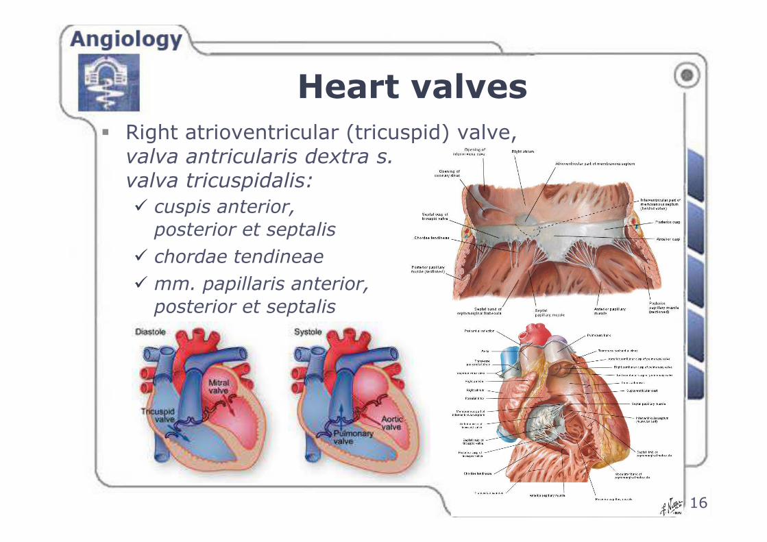
Heart valves
� Right atrioventricular (tricuspid) valve, valva antricularis dextra s. valva tricuspidalis:
� cuspis anterior,
posterior et septalis
� chordae tendineae
� mm. papillaris anterior,
posterior et septalis
16

Heart valves
� Pulmonary valve, valva trunci pulmonalis:
� valvula semilunaris anterior,
dextra et sinistra
� lunula et nodulus
valvulae semilunaris
17

Heart valves
18
� Left atrioventricular (bicuspid, mitral) valve, valva antricularis sinsitra s. valva bicuspidalis (mitralis):
� cuspis anterior et posterior
� chordae tendineae
� mm. papillaris anterior et posterior
� trabeculae carneae

Heart valves
� Aortic valve, valva aortae:
� valvula semilunaris
posterior, dextra et sinistra
� lunula et nodulus
valvulae semilunaris
� sinus aortae (Valsalva)
19

Heart valves
20
� Function and biomechanics:
� diastole
� systole

Structure of the heart wall� internal layer, endocardium:
� stratum endotheliale
� stratum subendotheliale
� stratum myoelasticum
� atrioventricular valves
� tela subendocardialis
� cells of Purkinje
� middle layer, myocardium
� external layer, epicardium
� pericardium
21

Microscopic structure ofatrioventricular valve
22
� AV valve leaflet:� endocardium
�elastic fibers�smooth muscle cells
� fibrous skeleton –dense connective tissue �chordae tendineae
� endocardium

The cardiac muscle, myocardium
23
� Cardiac muscle tissue, myofibrae cardiacae
� labor cardiac musculature
� impulse-conducting system
� cardiac myoendocrine cells
(atrial cardiomyocytes)
� atrial natriuretic peptide (cardiodilatin)

Labor cardiac musculature� Structural features:
� defines the heart thickness
� separate for the atria and ventricles
� atria – two layers
(superficial and deep layers)
� ventricles – three layers
� substantially thicker in the ventricles
� attached to the fibrous skeleton
24

Fibrous skeleton of the heart
25
� The fibrous skeleton:
� two fibrous rings:
� right AV ring, anulus fibrosus dexter
� left AV ring, anulus fibrosus sinister
� two fibrous trigones:
� right trigone, trigonum fibrosum dexter
� left trigone, trigonum fibrosum sinister

Cardiac blood supply
26
� Arteries of the heart, aa. cordis:
� left coronary artery
� right coronary artery
� beginning of aortic sinuses
� Varieties in arterial blood supply:

Blood supply of the heart
� The right coronary artery, a. coronaria dextra:� ramus coni arteriosus
� ramus nodi sinuatrialis
� rami atriales
� ramus marginalis dexter
� ramus atrialis intermedius
� ramus interventricularis posterior
� rami interventriculares septales
� ramus nodi atrioventricularis
� Areas of blood supply:� the right ventricle
� the right atrium
� posterior part of the left ventricle
� posterior papillary muscle
� posterior ⅓ of the interventricular septum
27

Blood supply of the heart� The left coronary artery,
a. coronaria sinistra:� ramus interventricularis anterior
� ramus coni arteriosus
� ramus lateralis (a. diagonalis)
� rami interventriculares septales
� ramus circumflexus
� ramus atrialis anastomoticus (артерия на Kugel)
� rami atriales
� rami atrioventriculares
� ramus marginalis sinister
� variable branches
� Areas of blood supply:� anterior wall of the right ventricle� left atrium� most of the left ventricle� anterior ⅔ of the interventricular septum
28

Blood supply of the heart
29
� Normal type:
� in 75% of human population
� Left coronary artery
dominance:
� in 11% of cases
� Right coronary artery
dominance:
� in 14% of cases
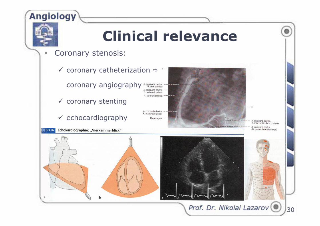
Clinical relevance� Coronary stenosis:
� coronary catheterization �
coronary angiography
� coronary stenting
� echocardiography
30

Venous drainage of the heart
31
� The cardiac veins, venae cordis:
� coronary sinus, sinus coronarius
� v. cardiaca magna et media
� vv. ventriculi dextri anteriores
� vv. cardiacae minimae (Thebesius)
� vv. cardiacae parvae:
� vv. atriales
� vv. ventriculares
� vv. atrioventriculares

The coronary sinus
32
� The coronary sinus,
sinus coronarius:
� a remnant of the left duct of Cuvier
� length 2-3 cm
� opens into the right atrium
� tributaries:
� v. cardiaca magna
� vv. cardiacae anteriores
� v. obliqua atrii sinistri
� v. posterior ventriculi sinistri
� v. cardiaca media
� v. cardiaca parva

Lymphatic drainage of the heart
33
� Cardiac lymphatic networks:
� subendocardial
� myocardial
� subepicardial networks
� Main lymphatic vessels:
� the left marginal vein
� the right marginal vein
� Regional lymph nodes:
� mediastinal nodes
� left tracheal nodes
� tracheobronchial lymph nodes
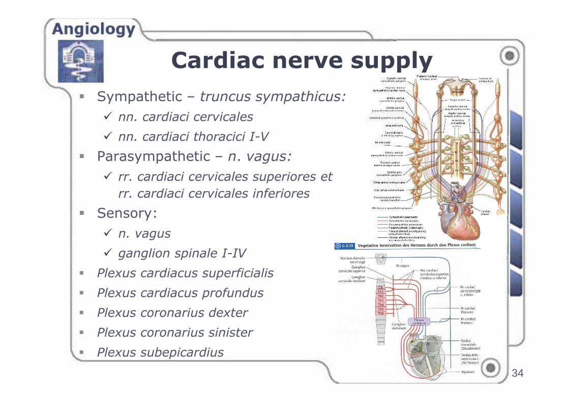
Cardiac nerve supply
34
� Sympathetic – truncus sympathicus:
� nn. cardiaci cervicales
� nn. cardiaci thoracici I-V
� Parasympathetic – n. vagus:
� rr. cardiaci cervicales superiores et
rr. cardiaci cervicales inferiores
� Sensory:
� n. vagus
� ganglion spinale I-IV
� Plexus cardiacus superficialis
� Plexus cardiacus profundus
� Plexus coronarius dexter
� Plexus coronarius sinister
� Plexus subepicardius
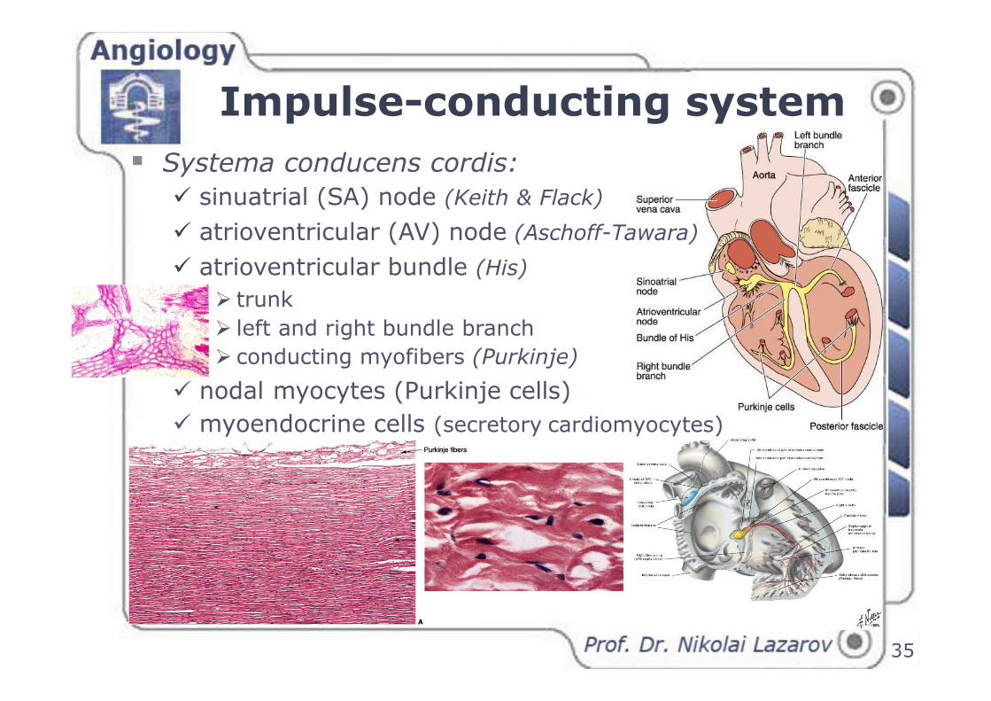
Impulse-conducting system
35
� Systema conducens cordis:
� sinuatrial (SA) node (Keith & Flack)
� atrioventricular (AV) node (Aschoff-Tawara)
� atrioventricular bundle (His)
� trunk
� left and right bundle branch
� conducting myofibers (Purkinje)
� nodal myocytes (Purkinje cells)
� myoendocrine cells (secretory cardiomyocytes)

Pericardium
36
� Pericardium serosum:
� lamina visceralis (epicardium)
� lamina parietalis � pericardium fibrosum
� cavitas pericardii; liquor pericardii
� porta arteriosa et porta venosa
� sinus transversus pericardii
� sinus obliquus pericardii

Radiological appearance of the heart
� The heart in radiographs:
� antero-posterior view – breastbone and vertebral column
� right border – two arcs (vascular and atrial)
� left border – four arcs (vascular and ventricular)
37

Projection and auscultation areas of the heart valves
38
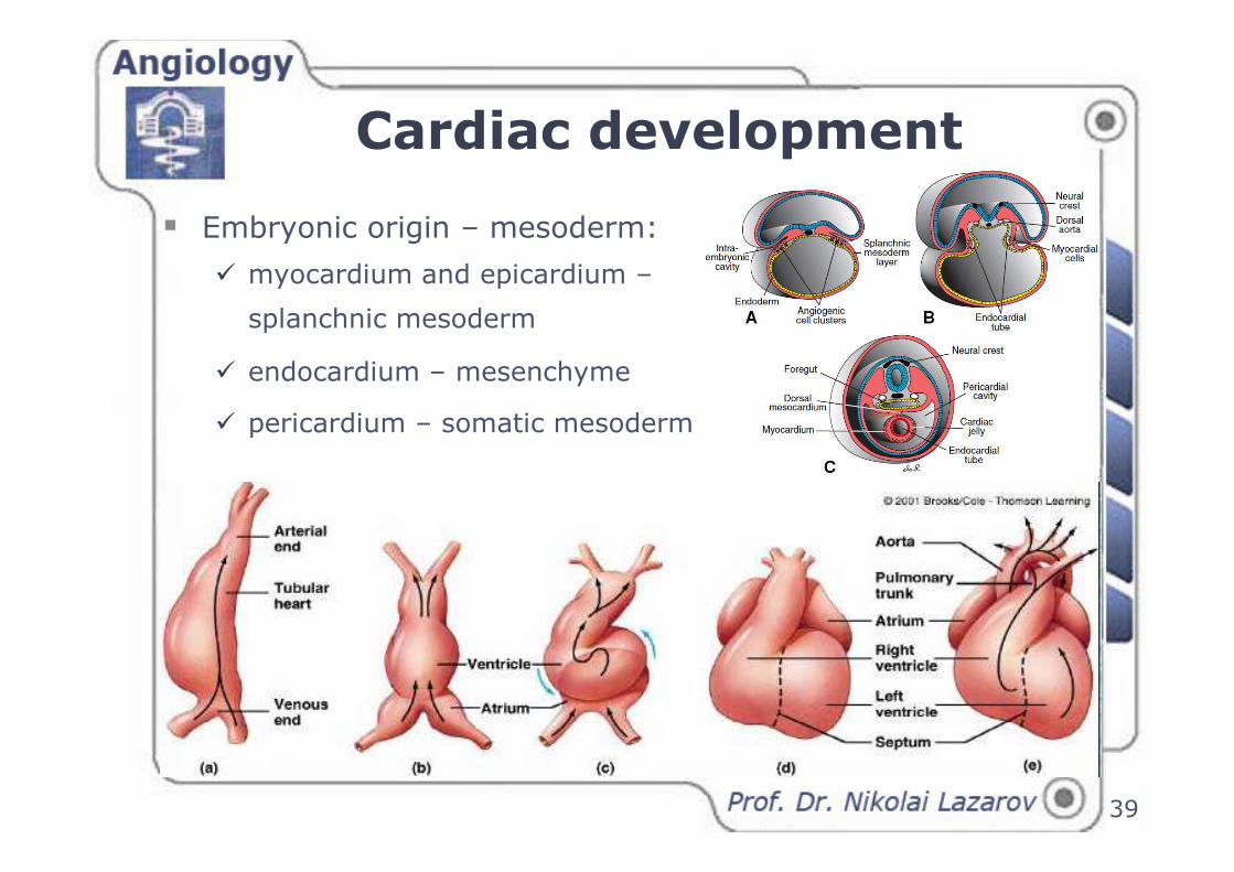
Cardiac development
39
� Embryonic origin – mesoderm:
� myocardium and epicardium –
splanchnic mesoderm
� endocardium – mesenchyme
� pericardium – somatic mesoderm

Cardiac development
40
� Stages:
� angioblasts � cell clusters
� tuft of blood vessels
� cardiogenic area
� cnpaired trunk
� sinus venosus
� truncus arteriosus
� left and right side – end of the 1st mo.
� formation of atria and ventricles
� formation of valves
� formation of three septa:
o interatrial – foramen ovale
o aortopulmonary� aorta and pulmonary trunk
o interventricular, septum inferius –
foramen interventriculare
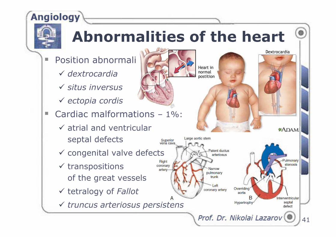
Abnormalities of the heart
� Position abnormalities:
� dextrocardia
� situs inversus
� ectopia cordis
� Cardiac malformations – 1%:
� atrial and ventricular
septal defects
� congenital valve defects
� transpositions
of the great vessels
� tetralogy of Fallot
� truncus arteriosus persistens
41

Mohandas (Mahatma) Gandhi
The heart has no language
…it speaks to the heart!Thank you ...