case analysis low-grade appendiceal mucinous neoplasm: … · case analysis low-grade appendiceal...
TRANSCRIPT

Carcinomas of the appendix are rare and have an
annual age-adjusted incidence of 0.4 cases per
100,000. Ravi Marudanayagam et al. had analyzed
2660 appendectomy specimens, and 0.6% of them
were reported “cystadenoma”.1
Mucinous cystadenoma of the appendix is an un-
common clinical finding. It is generally termed “mu-
cocele” of the appendix, which simply refers to a cys-
J Soc Colon Rectal Surgeon (Taiwan) December 2012
Case Analysis
Low-Grade Appendiceal Mucinous
Neoplasm: A Rare Cause of Acute Abdomen
Kai-Chen Wang
Cheng-Hsien Chen
Cheng-Zhi Chen
Hsuan-Yuan Huang
Jau-Jie You
Tsang-Chi Lin
Hong-Chang Chen
Ting-Ming Huang
Division of Colorectal Surgery, Department
of Surgery, Chang-Hua Christian Hospital,
Changhua, Taiwan
Key Words
Low-grade appendiceal mucinous
neoplasm;
Appendiceal caner;
Acute abdominal pain
Purpose. Low-grade appendiceal mucinous neoplasm (LAMN) is a raretype of appendiceal cancer. Patients with LAMN may initially presentwith acute abdominal pain. Despite the use of CT, ultrasound or colo-noscopy, preoperative diagnosis is difficult. In this study, we retrospectiveanalyze cases of LAMN diagnosed in the past 10 years in Chang-HuaChristian Hospital and review the associated literature.
Materials and Methods. This study retrospectively analyzed 15 patientswho underwent surgery and was histopathologically confirmed LAMN atChang-Hua Christian Hospital between January 2000 to August 2011. Pa-tient charts and data on patient demographics; clinical features; ultra-sonography (US), colonoscopy and computed tomography (CT) findings;pathology reports; pre-operative diagnosis and operative method werereviewed.
Results. The were 15 cases of LAMN at Chang-Hua Christian Hospitalover the past ten years. In our review, there were eight (53.3%) female pa-tients. The median age was 67 years (47-85 years), and the most commonpresentation was abdominal pain (93.3%). On US in six patients, findingswere abdominal cystic mass and cyst wall calcification. The CT findingwas well-encapsulated cystic mass in thirteen patients. Appendectomywas performed in ten patients. Right hemicolectomy was performed in
five patients, and there was one patient found concomitant colon ade-nocarcinoma in the specimen.
Conclusion. LAMN is difficult to diagnose before operation. The actualdiagnosis is usually made intraoperatively or during histopathologic ex-amination of the excised specimen. Although surgical treatment is straight-forward, proper management of the incidentally found lesion requiresunderstanding of the potential complications of widespread peritonealdisease. It should be kept in mind that LAMN may coexist with otherneoplasms, and follow-up colonoscopy and pelvic examination is war-ranted for the high association with other colon and ovarian malignancies.[J Soc Colon Rectal Surgeon (Taiwan) 2012;23:183-189]
Received: November 15, 2011. Accepted: July 2, 2012.
Correspondence to: Dr. Kai-Chen Wang, Division of Colorectal Surgery, Department of Surgery, Chang-Hua Christian Hospital,
Changhua, Taiwan. E-mail: [email protected]
183
tic mass filled with mucin in a dilated appendix.2
However, as it possesses the potential behavior of
malignant widespreading, the term “adenoma” has
been abandoned. The 4th edition of World Health Or-
ganization (WHO) classification had officially intro-
duced “low-grade appendiceal mucinous neoplasm”
(LAMN) as the appropriate name.3
The presenting clinical signs are variable. Pre-
operative colonoscopy, ultrasonography (US), and
computed tomography (CT) are methods in diagnos-
ing LAMN and distinguishing the mucocele from
mimicking diseases. However, the diagnosis is usu-
ally made intraoperatively or postoperatively on his-
topathological examination.4-6
As the presenting illness differs, the treatment is
not always the same. In this report, we analyzed the
cases in our hospital and reviewed related papers for a
better understanding of the disease.
Patients and Methods
In Chang-Hua Christian Hospital, each pathologic
diagnosis has a code. According to the coding system,
we retrospectively searched cases from January 2000
to August 2011. Totally there are fifteen specimens
labeled as LAMN. We review these fifteen charts and
recorded data including patient demographics, clini-
cal features, US, colonoscopy and CT findings, con-
comitant diseases, and conditions for which surgery
was indicated. Pathology reports, operative and post-
operative management, and information on last fol-
low-up were also recorded. In this study, the descrip-
tive variables of standard deviation and median were
used.
Results
A total 15 patients were admitted and received
surgery as a result of LAMN. The median age was 67
years (47-85 years). Regarding to the clinical signs,
14 of our patients (93.3%) experienced abdominal
pain. The pain was mainly located over right-lower
quadrant area (n = 10; 71.4%); one patient had para-
umbilical pain and two complained of diffuse abdomi-
nal pain with no specific location. Other presenting
signs included abdominal distention (n = 3), nausea/
vomiting (n = 4), and weight loss (n = 1) (Table 1).
During the physical examination, rebound tender-
ness over the pain site was noted in 9 patients (60.0%).
A palpable mass was perceived in one patient. Before
operation, abdominal ultrasonography (US) was per-
formed in 6 patients, which revealed a cystic mass
with variable internal echogenicity, layered wall, and
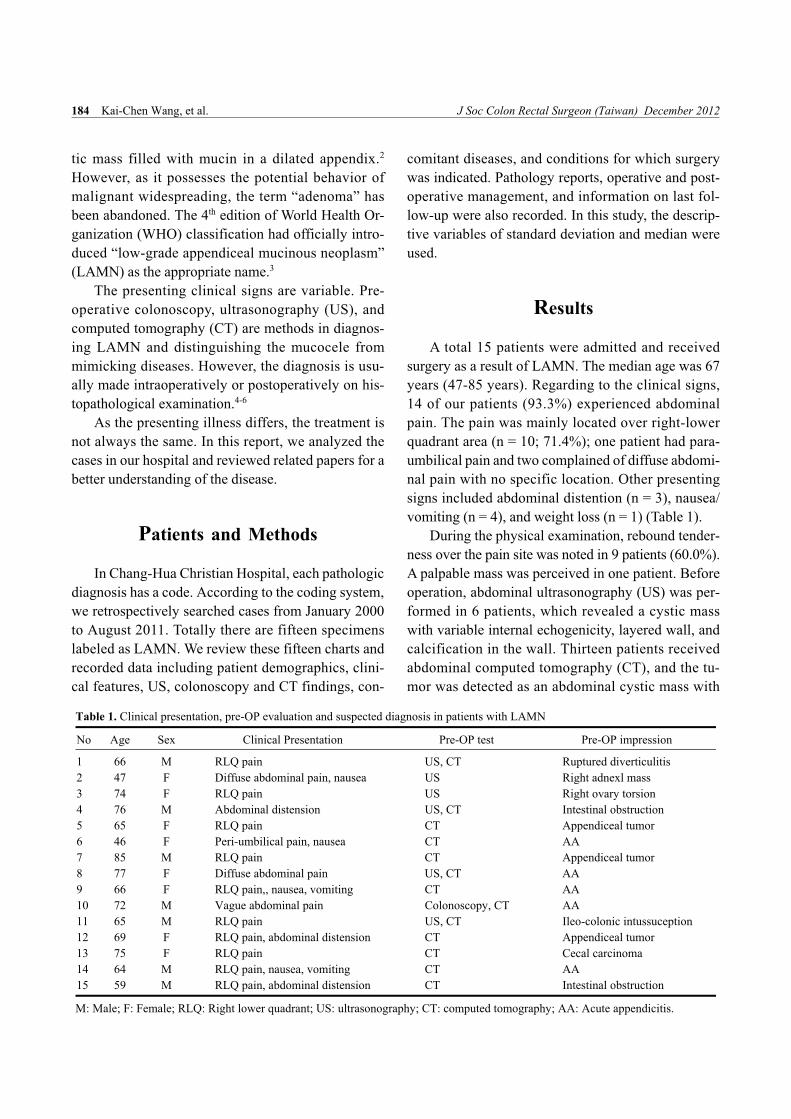
calcification in the wall. Thirteen patients received
abdominal computed tomography (CT), and the tu-
mor was detected as an abdominal cystic mass with
184 Kai-Chen Wang, et al. J Soc Colon Rectal Surgeon (Taiwan) December 2012
Table 1. Clinical presentation, pre-OP evaluation and suspected diagnosis in patients with LAMN
No Age Sex Clinical Presentation Pre-OP test Pre-OP impression
1 66 M RLQ pain US, CT Ruptured diverticulitis
2 47 F Diffuse abdominal pain, nausea US Right adnexl mass
3 74 F RLQ pain US Right ovary torsion
4 76 M Abdominal distension US, CT Intestinal obstruction
5 65 F RLQ pain CT Appendiceal tumor
6 46 F Peri-umbilical pain, nausea CT AA
7 85 M RLQ pain CT Appendiceal tumor
8 77 F Diffuse abdominal pain US, CT AA
9 66 F RLQ pain,, nausea, vomiting CT AA
10 72 M Vague abdominal pain Colonoscopy, CT AA
11 65 M RLQ pain US, CT Ileo-colonic intussuception
12 69 F RLQ pain, abdominal distension CT Appendiceal tumor
13 75 F RLQ pain CT Cecal carcinoma
14 64 M RLQ pain, nausea, vomiting CT AA
15 59 M RLQ pain, abdominal distension CT Intestinal obstruction
M: Male; F: Female; RLQ: Right lower quadrant; US: ultrasonography; CT: computed tomography; AA: Acute appendicitis.

wall calcification situated in the right lower quadrant
(Fig. 1).
Eight patients underwent emergent surgery under
the impression of acute abdominal conditions (acute
appendicitis in 5 cases; right ovarian torsion in 1 pa-
tient; ruptured diverticulitis in 1 patient; ileo-colonic
intussuception in 1 patient) (Table 2). In three pa-
tients, surgery was indicated for appendiceal tumor
detected by image pre-operatively. Four of our cases
were operated for other indications. One was explor-
atory laparotomy for suspective adnexal mass; an-
other one was for suspective cecalc cancer; the other
two were for adhesiolysis.
Ten patients received appendectomy; one of them
also had cecum removed due to gross tumor invasion.
Right hemicolectomy was performed in 5 patients. In
all patients, histopathological examination of speci-
mens found low-grade appendiceal mucinous neo-
plasm.
Two patients expired at the 37th and 60th postoper-
ative day respectively. There were no gastrointestinal
tract complications, and the enteral feeding could be
fully achieved through nasogastric tube. The main
cause of death were pneumonia induced by long-term
ventilation use. The other 13 patients recovered un-
eventfully with a median hospital stay of 6.4 days
(range: 4-60 days). The 13 survived patients had no
evidence of recurrence or metastasis at their last fol-
low-up (median: 50.6 months; range: 6-108 months).
Discussion
Low-grade appendiceal mucinous neoplasm
(LAMN) is a rare tumor of the appendix associated
with cystic dilatation, to which the more general term
of mucocele has been applied (Fig. 2). Mucocele of
the appendix denotes an obstructive dilatation of the
appendiceal lumen due to abnormal accumulation of
mucus, which may be caused by a retention cyst,
mucosal hyperplasia, cystadenoma and cystadeno-
carcinoma.7,8 Mucocele of the appendix is more fre-
quent in women and is usually observed in patients
older than 50 years. In this study, the patients we re-
Vol. 23, No. 4 Low-Grade Appendiceal Mucinous Neoplasm 185
Table 2. Treatment, accompanying disease and follow-up interval in patients with LAMN
No Surgery Accompanying disease Follow-up (months)
1 Right hemicolectomy AA, ascending colon diverticula 108
2 Appendectomy + bilateral salpingectomy Right hydrosalpinx 54
3 Right hemicolectomy Ischemic colitis 72
4 Open appendectomy AA 37 days
5 LA AA 48
6 Appendectomy + cecectomy Colonic diverticulitis 31
7 LA � 39
8 Right hemicolectomy Terminal ileum necrosis 60 days
9 LA AA 13
10 Right hemicolectomy Adenocarcinoma of the ascending colon 85
11 Right hemicolectomy AA 6
12 LA AA 54
13 Open appendectomy AA 57
14 LA AA 46
15 Open appendectomy AA 41
LA: laparoscopic appendectomy; AA: Acute appendicitis.
Fig. 1. CT scan shows a cystic mass with mural thickeningin the cecum.

viewed included 8 women and 7 men; the youngest
age is 46-year-old.
The most common presenting symptom associ-
ated with LAMN has been abdominal pain; however,
one-fourth of patients are asymptomatic and are found
incidentally. Other symptoms such as bloody stool,
intussusception have also been reported.9
Without specific symptoms, image studies are
usually needed for differential diagnosis. By ultra-
sound, cystic masses with varying internal echo-
genicity and a layered wall with calcification may be
seen; we found this in 3 patients. The typical findings
by computed tomography are well-encapsulated cys-
tic masses with low attenuation. However, if the
mucocele ruptures, it may be misdiagnosed as rup-
tured appendicitis or diverticulitis. Colonoscopy is
usually nondiagnostic, as mucosal biopsies will often
be normal.4,5,10
LAMN generally grow slowly, and tend to pro-
duce the clinical picture of low-grade pseudomyxoma
peritonei in which spread beyond the peritoneum or
nodal metastasis is unusual. Histologically, LAMN
may have villous, serrated or undulating morphology,
but unlike adenomas, they rest on fibrous tissue rather
than lamina propria (Fig. 3). They tend to involve the
appendix in a circumferential fashion with atrophy of
the underlying lymphoid tissue.11 Because LAMN can
proliferate outside the appendix in a malignant way,
producing pseudomyxoma peritonei and even distant
metastasis,12,13 it is inappropriate to regard LAMN as
“one kind of adenoma”. The term “mucinous cysta-
denocarcinoma” has been used for well-differentiated
mucinous tumor with cystic structures; however, such
a diagnosis should be avoided because this neoplasm
dose not constitute a separate disease entity.14 The 4th
edition of World Health Organization (WHO) classifi-
cation asserted “low-grade appendiceal mucinous
neoplasm” the appropriate name.
Recently, serrated polyps in the colon have gener-
ated interest as the possible precursor lesions of mi-
crosatellite instable carcinomas.19-22 Jass et al.22 noted
that the serration in serrated polyps is the result of
accommodation of an enlarged cytoplasmic compart-
ment due to increased secretory mucins. LAMNs of-
ten have tall, mucinous epithelium in villous areas
and, perhaps because of this, occasionally have ser-
rated glands.23 Therefore, distinguishing between vil-
lous adenomas, serrated adenomas, and even circum-
ferential mucosal hyperplasia in the appendix can
sometimes pose a considerable challenge. Others have
commented on the morphologic similarity between
villous adenomas of the colon and serrated adenomas.24
In 1999, Szych et al.25 found frequent K-ras mutations
and loss of heterozygosity of chromosome 5q in
LAMNs, a pattern similar to colorectal adenomas. To-
gether with their findings, LAMNs arising via the
chromosomalinstability pathway of colorectal car-
cinogenesis was suggested. For this reason, colono-
scopy examination was recommended to rule out syn-
chronous colon cancer.
The morbidity/mortality associated with LAMN
186 Kai-Chen Wang, et al. J Soc Colon Rectal Surgeon (Taiwan) December 2012
Fig. 2. The specimen was diagnosed as low-grade appen-diceal mucinous neoplasm after pathologic exam.
Fig. 3. Low-grade appendiceal mucinous neoplasm. Thismorphology is smilialr to adenomas, but there is nolamina propia and the neoplastic epithelium rests onfibrous stroma.
stems from rupture and intraperitoneal spread of
mucin-producing epithelium, which may cause pseu-
domyxoma peritonei. As a result, gentle tissue han-
dling during operation cannot be overemphasized.15
Since there are no evidence in regard to lymphatic or
hematogenous spreading of LAMN, if the mass con-
fines in the appendix body without local invasion or
cecal involvement, simple appendectomy and meso-
appendix excision is considered sufficient treatment.16
Successful removal of appendiceal mucoceles laparo-
scopically has been reported.26,27 In our practice, there
were 5 patients (33.3%) had their LAMN removed by
laparoscopic appendectomy. For mass involving the
cecum or adjacent organs, right hemicolectomy is of-
ten required. LAMN is associated with colon and
ovarian malignancy.15-17 In our review, 1 patient had
concomitant ascending colon cancer (6.6%). There-
fore, intraoperative exploration of the entire gastroin-
testinal tract and ovaries in females is warranted. All
gross peritoneal implants should undergo a biopsy
and be removed with grading of the degree of epithe-
lial atypia for prognostic purposes.18
Rarity of the tumor and absence of randomized
clinical trials preclude the compilation of guidelines
for the follow-up practice. CT, US, pelvic exam and
colonoscopy are recommended by some authors to
detect other colon and ovarian malignancies. During
the follow-up period, there was no evidence of disease
recurrence or metastasis in our group.8,11
Conclusion
Low-grade appendiceal mucinous neoplasm is
more frequent in elderly patients and may cause acute
abdomen. US, CT and sometimes colonoscopy are
helpful in diagnosis. However, actual diagnosis is
usually made intraoperatively or during histopatho-
logic examination of the excised specimen. Appen-
dectomy with removal of the mesoappendix or right
hemicolectomy is the treatment of choice, depending
on the degree of tumor invasion. It may coexist with
other neoplasms. Thorough examination of the ab-
dominal and pelvic cavity during surgery is war-
ranted. Follow-up CT, US, or colonoscopy are also
recommended.
References
1. Marudanayagam R, Williams GT, Rees BI. Review of the
pathological results of 2660 appendicectomy specimens. J
Gastroenterol 2006;41:745-9.
2. Rosai J. Gastrointestinal tract. In: Rosai J, editor. Rosai and
Ackerman’s surgical pathology. 9th ed. New York: Mosby;
2004:761-64.
3. Bradleya RF, Cortinaa G, Geisingerb KR. Pseudomyxoma
peritonei: review of the controversy. Current Diagnostic Pa-
thology Volume 13, Issue 5:410-16.
4. Kim SH, Lim HK, Lee WJ, Lim JH, Byun JY. Mucocele of
the appendix: ultrasonographic and CT findings. Abdom Im-
aging 1998;23:292-6.
5. Madwed D, Mindelzun R, Jeffrey RB Jr. Mucocele of the
appendix: imaging findings. AJR Am J Roentgenol 1992;
159:69-72.
6. Emmi S, Galasso MG, Ursino V, Scala R, Guastella T.
Appendiceal mucoceles. A case report. Minerva Chir 1998;
53:807-10.
7. Qizilbash AH. Mucoceles of the appendix. Their relationship
to hyperplastic polyps, mucinous cystadenomas, and cysta-
denocarcinomas. Arch Pathol 1975;99:548-55.
8. Aho AJ, Heinonen R, Laurén P. Benign and malignant
mucocele of the appendix. Histological types and prognosis.
Acta Chir Scand 1973;139:392-400.
9. Higa E, Rosai J, Pizzimbono CA, Wise L. Mucosal hyper-
plasia, mucinous cystadenoma, and mucinous cystadeno-
carcinoma of the appendix. A re-evaluation of appendiceal
“mucocele”. Cancer 1973;32:1525-41.
10. Kahn M, Friedman IH. Mucocele of the appendix: diagnosis
and surgical management. Dis Colon Rectum 1979;22:267-9.
11. Misdraji J, Yantiss RK. Appendiceal mucinous neoplasms a
clinicopathologic analysis of 107 cases. The American Jour-
nal of Surgical Pathology 27:1089-103.
12. Carr NJ, Sobin LH. Unusual tumors of the appendix and
pseudomyxoma peritonei. Semin Diagn Pathol 1996;13:
314-25.
13. Seidman JD, Elsayed AM, Sobin LH, Tavassoli FA. Associa-
tion of mucinous tumors of the ovary and appendix: a clinico-
pathologic study of 25 cases. Am J Surg Pathol 1993;17:
22-34.
14. Ronnett BM, Yan H, Kurman RJ, Shmookler BM, Wu L,
Sugarbaker PH. Patients with pseudomyxoma peritonei asso-
ciated with disseminated peritoneal adenomucinosis have a
significantly more favorable prognosis than patients with
peritoneal mucinous carcinomatosis. Cancer 2001;92:85-91.
15. Kahn M, Friedman IH. Mucocele of the appendix: diagnosis
and surgical management. Dis Colon Rectum 1979;22:267-9.
16. Shayani V. Mucinous cystadenoma of the cecum missed at
laparoscopic appendectomy. Surg Endosc 1999;13:1236-7.
17. Machado NO, Chopra P, Pande G. Appendiceal tumour re-
trospective clinicopathological analysis. Trop Gastroenterol
2004;25:36-9.
Vol. 23, No. 4 Low-Grade Appendiceal Mucinous Neoplasm 187

18. Rampone B, Roviello F, Marrelli D, Pinto E. Giant ap-
pendiceal mucocele: report of a case and brief review. World
J Gastroenterol 2005;11:4761-3.
19. Iino H, Jass JR, Simms LA, Young J, Leggett B. DNA
microsatellite instability in hyperplastic polyps, serrated
adenomas, and mixed polyps: a mild mutator pathway for
colorectal cancer? J Clin Pathol 1999;52:5-9.
20. Hawkins NJ, Ward RL. Sporadic colorectal cancers with
microsatellite instability and their possible origin in hyper-
plastic polyps and serrated adenomas. J Natl Cancer Inst
2001;93:1307-13.
21. Jass JR. Hyperplastic polyps of the colorectum-innocent or
guilty? Dis Colon Rectum 2001;44:163-6.
22. Jass JR, Young J, Leggett BA. Hyperplastic polyps and DNA
microsatellite unstable cancers of the colorectum. Histo-
pathology 2000;37:295-301.
23. Misdraji J, Yantiss RK, Graeme-Cook FM, Balis UJ, Young
RH. Appendiceal mucinous neoplasms: a clinicopathologic
analysis of 107 cases. Am J Surg Pathol 2003;27:1089-103.
24. Hawkins NJ, Bariol C, Ward RL. The serrated neoplasia path-
way. Pathology (Phila) 2002;34:548-55.
25. Szych C, Staebler A, Connolly DC, Wu R, Cho KR, Ronnett
BM. Molecular genetic evidence supporting the clonality and
appendiceal origin of Pseudomyxoma peritonei in women.
Am J Pathol 1999;154: 1849-55.
26. Lau H, Yuen WK, Loong F, Lee F. Laparoscopic resection of
an appendiceal mucocele. Surg Laparosc Endosc Percutan
Tech 2002;12:367-70.
27. Navarra G, Asopa V, Basaglia E, Jones M, Jiao LR, Habib
NA. Mucous cystadenoma of the appendix: is it safe to re-
move it by a laparoscopic approach? Surg Endosc 2003;
17:833-4.
188 Kai-Chen Wang, et al. J Soc Colon Rectal Surgeon (Taiwan) December 2012

王愷晟等 J Soc Colon Rectal Surgeon (Taiwan) 2012;23:182-189 189
病例分析
闌尾低度惡性黏液瘤 ⎯ 罕見之急性腹痛原因
王愷晟 陳成賢 陳志誠 黃玄遠 尤昭傑 林倉祺 陳宏彰 黃燈明
彰化基督教醫院 外科部 大腸直腸外科
目的 「闌尾低度惡性黏液瘤」是一種十分罕見的闌尾腫瘤。患者可能以急性腹痛為臨床表現。雖然配合電腦斷層、腹部超音波與大腸境的檢查,術前診斷仍有其困難性。在
本篇報告中,我們收集彰化基督教醫院近十年來的案例進行分析,並回顧相關之研究文
獻。
方法 本研究是採取回溯性分析。案例來自於本院 2000年 1月至 2011年 8月間經病理檢驗證明為「闌尾低度惡性黏液瘤」之 15 位患者。我們記錄並分析其臨床表現、腹部超音波檢查報告、大腸鏡報告、電腦斷層之發現、病理報告、術前診斷與手術方法。
結果 在十年間本院總共有 15 患者。其中 8 位 (53.3%) 是女性。平均確定診斷之年齡為 67歲 (47-85歲)。最常見之臨床表現為腹痛 (93.3%)。其中 6位接受腹部超音波檢查的患者,發現到腹內有囊狀腫塊,與囊壁鈣化的現象。有 13位患者接受電腦斷層檢查,影像顯示外膜完整之囊腫。有 10 位患者接受闌尾切除術。有 5 位接受右側結腸切除手術,其中一位患者檢體中同時發現大腸腺癌。
結論 「闌尾低度惡性黏液瘤」在術前不易診斷,其確診往往有賴於手術中直接觀察與手術後之檢體病理分析。儘管手術切除是最直接的治療方法,但仍須小心保持腫瘤之完
整性,以避免腫瘤破裂造成黏液瘤細胞擴散。因其可能同時與其他種類的癌症同時存在
(如大腸癌或卵巢癌),術後的檢查如大腸鏡與骨盆檢查是必需的。
關鍵詞 闌尾低度惡性黏液瘤、闌尾腫瘤、急性腹痛。