case report churg-strauss syndrome-associated eosinophil-rich cholangitis · ·...
TRANSCRIPT
Am J Digest Dis 2017;4(4):34-39www.ajdd.us /ISSN:2329-6992/AJDD0059553
Case Report Churg-Strauss syndrome-associated eosinophil-rich cholangitis
Zhaohai Yang1, Michael G Davis2, Steven M Rowe2
1Department of Pathology, Penn State College of Medicine and Penn State Health Milton S. Hershey Medical Cen-ter, Hershey, PA, USA; 2Department of Medicine, University of Alabama at Birmingham, Birmingham, AL
Received June 15, 2017; Accepted July 8, 2017; Epub July 25, 2017; Published July 30, 2017
Abstract: Churg-Strauss syndrome (aka eosinophilic granulomatosis with polyangiitis) is characterized by asthma, peripheral eosinophilia and systemic vasculitis. Liver involvement is very rare. We report a case of Churg-Strauss syndrome who presented with asthma, peripheral eosinophilia, cholestatic hepatitis and mononeuropathy multi-plex. Paraaortic lymph node biopsy showed marked eosinophilic infiltrate with fibrinoid necrosis of the small vessels and eosinophilic necrotizing granuloma, confirming the diagnosis of Churg-Strauss syndrome. Liver biopsy showed portal inflammation with eosinophil-rich cholangitis and non-necrotizing venulitis. We conclude that this case repre-sents a form of Churg-Strauss syndrome-associated eosinophil-rich cholangitis presenting with cholestatic hepatitis. Churg-Strauss syndrome should be considered in the differential diagnosis in patients presenting with cholestatic hepatitis.
Keywords: Churg-Strauss syndrome, liver, jaundice, cholangitis, vasculitis
Churg-Strauss syndrome (CSS, aka eosinophil-ic granulomatosis with polyangiitis) was initially described in 1951. It is a small vessel-predom-inant, systemic necrotizing vasculitis character-ized by asthma, hypereosinophilia and necrotiz-ing vasculitis with extravascular eosinophilic granulomas [1]. Although asthma is the defin-ing symptom, extrapulmonary involvement is also common in CSS. Based on the study of the largest CSS series, the common extrapulmo-nary manifestations include mononeuropathy multiplex (78%), weight loss (71%), paranasal sinusitis (61%), fever (57%), myalgia (54%), skin involvement (51%) and arthralgia (42%) [2].Gastrointestinal tract involvement occurred in 33% of the patients, with the overwhelming presentation being as abdominal pain, proba-bly due to ischemia secondary to mesenteric vasculitis. Liver involvement in CSS is rare and the most common clinical presentation is abnormal liver function tests, found in only 7% of patients [2]. The histological finding of liver biopsy in CSS patients varies from unremark-able [2, 3] to hepatitis [4] and fibrosis [2]. Vasculitis in the liver is exceedingly rare [2, 3, 5], and there are only a handful cases of bile duct involvement reported in the English litera-
ture [6, 7]. We report a case of CSS presenting with cholestatic hepatitis and eosinophil-rich cholangitis.
Report of a case
A 53-year-old African-American man with a past medical history of hypertension, hyperlipid-emia, and gastroesophageal reflux disease ini-tially presented with three months of progres-sive shortness of breath and was diagnosed with adult-onset asthma. His symptoms impro- ved temporarily following treatment with taper-ing corticosteroids and montelukast. Two months later he was admitted for worsening shortness of breath. At that time, the patient exhibited acute renal failure, elevated erythro-cyte sedimentation rate (ESR) and serum IgE level, marked peripheral eosinophilia, markedly elevated total bilirubin (predominantly direct) and alkaline phosphatase (Table 1). He also developed vague abdominal pain, skin rash on the chest and upper extremities, and right hand weakness. An abdominal computed tomogra-phy (CT) scan revealed paraaortic lymphade-nopathy. At the referring hospital, he underwent a laparotomy with lymph node and liver biop-

Churg-Strauss syndrome-associated eosinophil-rich cholangitis
35 Am J Digest Dis 2017;4(4):34-39
sies and prednisone were resumed. One week prior to admission the dyspnea recurred upon reduction of prednisone and he was referred to our hospital. He also had occasional subjective fevers and chills, night sweats, a 50-pound weight loss, and cough with mild blood-streaked sputum production. He denied use of tobacco or recent sick contacts. There was no history of allergies. Medications on admission included irbesartan, prednisone, inhaled fluticasone, and albuterol rescue inhaler.
Upon admission, physical examination revealed an acutely ill-appearing man in moderate respi-ratory distress, with an O2 saturation of 90% on room air. He was icteric, with significant erythe-ma and injection of the tonsillar pillars. Auscultation of the chest revealed inspiratory and expiratory wheeze with dry crackles in the
apices and a prominent P2. Abdominal exam was significant for hepatomegaly and tender-ness to palpation in the right upper quadrant. There was diminished grip strength in the right median nerve distribution, accompanied by paresthesia. His skin had diffuse palpable pur-pura on the upper thorax and bilateral upper extremities.
The chest radiograph was unremarkable. Abdominal ultrasound showed hepatomegaly with normal caliber of the common bile duct. High resolution CT scan (without contrast) of the chest revealed bilateral patchy ground glass opacities with areas of nodularity, some-what more predominant in the apex (Figure 1). Pulmonary function test showed mild restric-tion and moderate reduction in DLCO (diffuse capacity of the lung for carbon monoxide). Transthoracic echocardiogram showed a nor-mal left ventricle ejection fraction (65%) with moderate right ventricular enlargement.
In addition to the key laboratory results listed in Table 1, the patient was found to have mild anemia. Blood and sputum cultures including acid fast stain were all negative for bacteria. Serology tests for viral hepatitis, syphilis, lepto-spirosis, strongyloides, human immunodefi-ciency virus, and multiple autoantibodies were negative. Complements (C3 and C4) were slightly reduced. Serum protein electrophoresis was positive for a small M-spike, while urine protein electrophoresis was negative. Immuno- fixation electrophoresis (IFE) showed two mono-clonal IgM lambda and one free kappa chain. Galactomanin (for fungal infection) was nega-tive. Stool was negative for ova, parasite and fecal leukocytes.
Table 1. Results of key laboratory tests
Laboratory test At Presentation On immuno-suppression Reference range and unit
Peripheral eosinophils 33%4652
3%417
0-5%0-600 × 106/L
ALT 25 47 15-58 IU/LAST 24 30 15-40 IU/LTotal bilirubin 26 1.8 0.4-1.4 mg/dL (to convert to µmol/L, multiply by 17.1)ALP 1800 376 39-117 IU/LGGT ND 340 0-65 IU/LESR 100 58 < 20 mm/hourSerum IgE 2456 182 ≤ 87 kIU/LALT: Alanine transaminase. AST: Aspartate transaminase. ALP: Alkaline phosphatase. GGT: γ-Glutamyl transferase. ND: No data. ESR: Erythrocyte sedimentation rate.
Figure 1. High resolution CT scan of the lungs no-table for bilateral patchy ground glass opacities and focal nodularity (arrow).
Churg-Strauss syndrome-associated eosinophil-rich cholangitis
36 Am J Digest Dis 2017;4(4):34-39
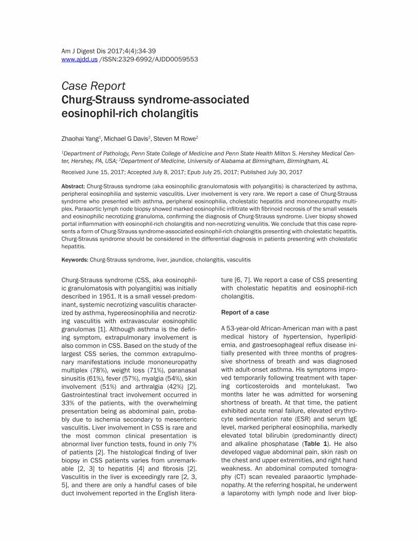
Review of the lymph node biopsy showed marked eosinophilic infiltration. There was geo-graphic necrotizing granuloma with rare multi-nucleated giant cells. The center of the necro-sis was deeply eosinophilic, and there were small vessels with fibrinoid necrosis (Figure 2). The liver biopsy showed expansion of the portal tract with mixed inflammatory infiltrate includ-ing many eosinophils and some lymphocytes and plasma cells. The inflammation extended to the bile duct with bile duct damage; however, it did not appear to be duct-centric nor was bile duct destruction identified. Focal eosinophilic non-necrotizing vasculitis of the portal vein was noted. The inflammation was largely limited to the portal tract, with only focal lobular inflam-mation and minimal interface hepatitis. Focal cholestasis was present at the site of lobular inflammation. Granulomas were not identified (Figure 3). A sural nerve biopsy showed perivas-
cular eosinophilic infiltration (figure not shown). The diagnosis of CSS was rendered, and the patient responded well to prednisone and cyclophosphamide, with resolution of most symptoms except for mild residual paresthesia of the hand. At one-year follow-up, he had nor-mal ESR and metabolic profile without anemia or jaundice, though hepatomegaly and intermit-tent wheezing persisted.
Discussion
CSS is a clinicopathological entity. The Ame- rican College of Rheumatology proposed six diagnostic criteria (asthma, peripheral eosino-philia, mononeuropathy or polyneuropathy, non-fixed pulmonary infiltrate on radiograph, paranasal sinus abnormality, and biopsy con-taining a blood vessel with extravascular eosin-ophils); the presence of four of the six criteria is considered consistent with CSS, with a diag-
Figure 2. Lymph node biopsy. A. Marked eosinophilic infiltrate in the lymph node (H&E, original magnification × 200). B. Geographic necrotizing granuloma (H&E, original magnification × 40). C. The center of the necrosis is deeply eosinophilic (right upper portion). Fibrinoid necrosis of small vessel is present (arrow) (H&E, original magnification × 200). D. Multinucleated giant cells are present at the edge of the necrotizing granuloma (arrow) (H&E, original magnification × 200).

Churg-Strauss syndrome-associated eosinophil-rich cholangitis
37 Am J Digest Dis 2017;4(4):34-39
nostic sensitivity of 85% and specificity of 99.7% [8]. The present case meets five of the six proposed criteria, thus compatible with a diagnostic of CSS. Although polyarteritis nodo-sa, Wegener’s granulomatosis and microscopic polyangiitis are often considered as differential diagnoses of CSS, the presence of asthma and
marked eosinophilia makes those three enti-ties very unlikely [9]. Other causes of peripheral eosinophilia such as parasite or fungal infec-tion, allergy, drug sensitivity, and endocrine dis-order [10] had been essentially ruled out by the combination of clinical history, physical exami-nation, radiological and laboratory findings.
Figure 3. Liver biopsy. A. Mixed portal inflammation without significant interface activity (H&E, original magnification × 100). B. Eosinophil-rich cholangitis (H&E, original magnification × 400). C. Focal lobular inflammation and cholate stasis (H&E, original magnification × 200). D. Eosinophil-rich non-necrotizing vasculitis of the portal vein (H&E, original magnification × 400).
Table 2. CSS cases with obstructive liver disease reported in the literature
# Author Year Icteric ALT (U/L)
AST (U/L)
Bilirubin (mg/dL)
ALP (U/L)
GGT (U/L) Antibody Liver biopsy
1 Conn 1982 [7] Yes ND 136 TB: 3.4DB: 2.4
1486 ND AMA Periportal hepatitis and granuloma: PBC
2 Brooklyn 2000 [6] No ND 23 TB: 0.5 491 191 ASMAp-ANCA
Bile duct-centric inflammation: granuloma-associted cholangiopathy
3 Bennett 2005 [11] No N N N 400 400 No Necrotizing granuloma without vasculitis
4 Yuksel 2008 [4] No 51 48 TB: 5DB: 3.7
565 119 RF Active interface hepatitis
5 Present case Yes 25 24 TB: 26 1800 340* No Portal inflammation with eosinophil-rich cholangitis, and non-necrotizing vasculitis
AMA: anti-mitochondria antibody. ASMA: anti-smooth muscle antibody. ANCA: anti-neutrophil cytoplasmic antibody. RF: rheumatoid factor. TB: total bilirubin. DB: direct bilirubin. N: normal. *After initiation of treatment.

Churg-Strauss syndrome-associated eosinophil-rich cholangitis
38 Am J Digest Dis 2017;4(4):34-39
The presence of peripheral eosinophilia and enlarged lymph node in the present case may suggest a diagnosis of hematologic malignan-cy, which was initially considered at the refer-ring institution. However, the lymph node biop-sy showed no evidence of lymphoma. Rather, there was marked eosinophilic infiltrate with necrotizing small vessel vasculitis and eosino-philic necrotizing granulomas, which are the typical findings in the vasculitic phase of CSS. CSS vasculitis in the liver is very rare, with only one such case noted in the largest CSS series and the type of the vasculitis was not specified in that report [2]. Necrotizing vasculitis was reported in the liver in only two single-case reports [3, 5], and necrotizing granulomas with-out vasculitis was reported in another [11]. The classic histological findings of CSS are not always present in modern materials [12, 13]. It is now recognized that CSS manifestation depends on the natural history and is also affected by treatment (e.g. corticosteroid thera-py); eosinophilic infiltrate and non-necrotizing vasculitis are more common in modern-era CSS cases [12, 14]. The findings in the liver and nerve biopsies of the present case are fully consistent with that notion.
It is intriguing that this case presented with jaundice with obstructive cholestasis on labo-ratory studies (elevated GGT and ALP). The presence of portal inflammation with bile duct damage in the liver biopsy correlates well with the clinical presentation. Similar laboratory abnormalities have been reported in four other cases in the English literature (Table 2). Case #1 had jaundice and positive anti-mitochondria antibody (1:80); in addition, ALP level was mark-edly elevated to 1486 IU/L. Liver biopsy showed prominent periportal hepatitis with eosinophilic infiltrate and granuloma adjacent to a portal vein, which was interpreted as primary biliary cirrhosis (PBC) [7]. The liver biopsy in case #2 showed portal inflammation with bile duct involvement, and the authors interpreted this as cholangiopathy, possibly associated with granulomatous reaction [6]. Liver biopsy in case #3 showed necrotizing granuloma without vasculitis [11]. Case #4 demonstrated active interface hepatitis [4]. Of note, the GGT/ALP levels were only moderately elevated and no jaundice was mentioned in the three latter cases. Consistent with normal transaminase values, the present case showed no significant
interface hepatitis, and no granulomas were identified in the liver. The portal infiltrate was quite dense and diffuse, in contrast to the mild and patchy nature characteristic of PBC. Although the presence of a few eosinophils is compatible with a diagnosis of PBC, the degree of eosinophilic infiltrate in this case would be very unusual for that entity. The clinical presen-tation of cholestatic hepatitis in our case is similar to case #1; while the histologic features are more consistent with case #2 in that the bile duct is infiltrated by inflammatory cells accompanied by bile duct damage. The inflam-matory cells in our case were not centered on the bile duct as described in case #2 [6], and there was no evidence of associated granulo-ma in the liver. Although the present case also showed focal lobular hepatitis and some plas-ma cells, the lack of significant interface he- patitis along with normal transaminases and negative autoimmune serology strongly argue against a diagnosis of autoimmune hepatitis. Taken together, CSS-associated eosinophil-rich cholangitis is the most likely underlying cause in our case.
Low level of M-protein on IFE was present in this case. Various autoantibodies have been identified in CSS patient [2]. Monoclonal gam-mopathy was previously reported which disap-peared upon successful treatment [5]. Whether those M-spikes represent true monoclonal or oligoclonal immunoglobulin is unknown.
In summary, we report a case of CSS with a clinical presentation of cholestatic hepatitis and an unusual histological pattern of bile duct inflammation. We conclude that the eosinophil-rich cholangitis is the underlying cause of cho-lestatic hepatitis in this case, which was res- ponsive to appropriate immunosuppressive therapy. CSS-associated eosinophil-rich chol-angitis should be considered in the differential diagnosis when patients present with hepatic abnormalities.
Disclosure of conflict of interest
None.
Address correspondence to: Dr. Zhaohai Yang, Divi- sion of Anatomic Pathology, Mail Code H179, Penn State Health Hershey Medical Center, Department of Pathology, Penn State College of Medicine and Penn State Health Milton S. Hershey Medical Cen-

Churg-Strauss syndrome-associated eosinophil-rich cholangitis
39 Am J Digest Dis 2017;4(4):34-39
ter, 500 University Drive, Hershey, PA 17033, USA. Tel: 717-531-8739; Fax: 717-531-7741; E-mail: zy- [email protected]
References
[1] Churg J, Strauss L. Allergic granulomatosis, al-lergic angiitis, and periarteritis nodosa. Am J Pathol 1951; 27: 277-301.
[2] Guillevin L, Cohen P, Gayraud M, Lhote F, Jar-rousse B, Casassus P. Churg-strauss syn-drome. Clinical study and long-term follow-up of 96 patients. Medicine (Baltimore) 1999; 78: 26-37.
[3] Della Rossa A, Baldini C, Tavoni A, Tognetti A, Neglia D, Sambuceti G, Puccini R, Colangelo C, Bombardieri S. Churg-strauss syndrome: clini-cal and serological features of 19 patients from a single Italian centre. Rheumatology (Ox-ford) 2002; 41: 1286-94.
[4] Yuksel I, Ataseven H, Basar O, Köklü S, Ertuğrul I, Ibiş M, Temuçin T, Saşmaz N. Churg-strauss syndrome associated with acalculous chole-cystitis and liver involvement. Acta Gastroen-terol Belg 2008; 71: 330-2.
[5] Gambari PF, Ostuni PA, Lazzarin P, Fassina A, Todesco S. Eosinophilic granuloma and necro-tizing vasculitis (Churg-strauss syndrome?) in-volving a parotid gland, lymph nodes, liver and spleen. Scand J Rheumatol 1989; 18: 171-5.
[6] Brooklyn TN, Prouse P, Portmann B, Ramage JK. Churg-strauss syndrome and granuloma-tous cholangiopathy. Eur J Gastroenterol Hepa-tol 2000; 12: 809-11.
[7] Conn DL, Dickson ER, Carpenter HA. The as-sociation of Churg-strauss vasculitis with tem-poral artery involvement, primary biliary cirrho-sis, and polychondritis in a single patient. J Rheumatol 1982; 9: 744-8.
[8] Masi AT, Hunder GG, Lie JT, Michel BA, Bloch DA, Arend WP, Calabrese LH, Edworthy SM, Fauci AS, Leavitt RY, et al. The American Col-lege of Rheumatology 1990 criteria for the classification of Churg-strauss syndrome (aller-gic granulomatosis and angiitis). Arthritis Rheum 1990; 33: 1094-100.
[9] Lhote F, Cohen P, Guillevin L. Polyarteritis no-dosa, microscopic polyangiitis and Churg-strauss syndrome. Lupus 1998; 7: 238-58.
[10] Weller PF. Approach to the patient with eosino-philia. UpToDate; 2010.
[11] Bennett AN, Sangle SR, Jan W, Jenner M, Cav-enagh J, Hughes G, D’Cruz DP. Hepatomegaly as a rare presentation of Churg-strauss syn-drome. Rheumatology (Oxford) 2005; 44: 1458-9.
[12] Churg A. Recent advances in the diagnosis of Churg-strauss syndrome. Mod Pathol 2001; 14: 1284-93.
[13] Reid AJ, Harrison BD, Watts RA, Watkin SW, McCann BG, Scott DG. Churg-strauss syn-drome in a district hospital. QJM 1998; 91: 219-29.
[14] Lanham JG, Elkon KB, Pusey CD, Hughes GR. Systemic vasculitis with asthma and eosino-philia: a clinical approach to the Churg-strauss syndrome. Medicine (Baltimore) 1984; 63: 65-81.