case report developmentally delayed male with mincer blade …downloads.hindawi.com › journals ›...
TRANSCRIPT
Case ReportDevelopmentally Delayed Male with Mincer Blade Obstructingthe Oesophagus for a Period of Time Suspected to Be 6 Months
Christian Grønhøj Larsen and Birgitte Charabi
Department of Otorhinolaryngology, Head and Neck Surgery and Audiology, Copenhagen University Hospital (Rigshospitalet),2100 Copenhagen, Denmark
Correspondence should be addressed to Christian Grønhøj Larsen; [email protected]
Received 13 March 2015; Revised 24 June 2015; Accepted 28 June 2015
Academic Editor: Marcello Picchio
Copyright © 2015 C. Grønhøj Larsen and B. Charabi. This is an open access article distributed under the Creative CommonsAttribution License, which permits unrestricted use, distribution, and reproduction in any medium, provided the original work isproperly cited.
Introduction. Sharp, retained foreign bodies in the oesophagus are associated with severe complications. Developmentally delayedpatients are especially subject to foreign objects. We describe a 37-year-old, developmentally delayed male with a mincer bladeobstructing the oesophagus. Six months prior to surgical intervention, the patient was hospitalized in a condition of sepsis andpneumonia where the thoracic X-ray reveals a foreign body in the proximal oesophagus. When rehospitalized 6 months later, amincer blade of the type used in immersion blenders was surgically removed. During these 6 months the patient’s main symptomswere dysphagia, weight loss, and diarrhoea.When developmentally delayed patients present with dysphagia, we strongly encouragethe awareness of the possible presence of foreign bodies. To our knowledge this is the first reported case of a mincer blade in theoesophagus.
1. Introduction
Chronically retained foreign bodies (FBs) are common inchildren but rare in adults [1, 2]. For developmentally delayedadults, especially those in the subgroupwith pica, FBs can notonly be the cause of severe morbidity such as obstruction,bleeding, and perforation but also be lethal because thepatient’s inability to communicate the symptoms makes itdifficult to arrive at an accurate diagnosis. Ingestion of a sharpFB is associated with a high risk of morbidity as a result ofthe possible perforation of the gastrointestinal tract. Earlydiagnosis and adequate management are imperative for theprevention of serious complications.
2. Case Report
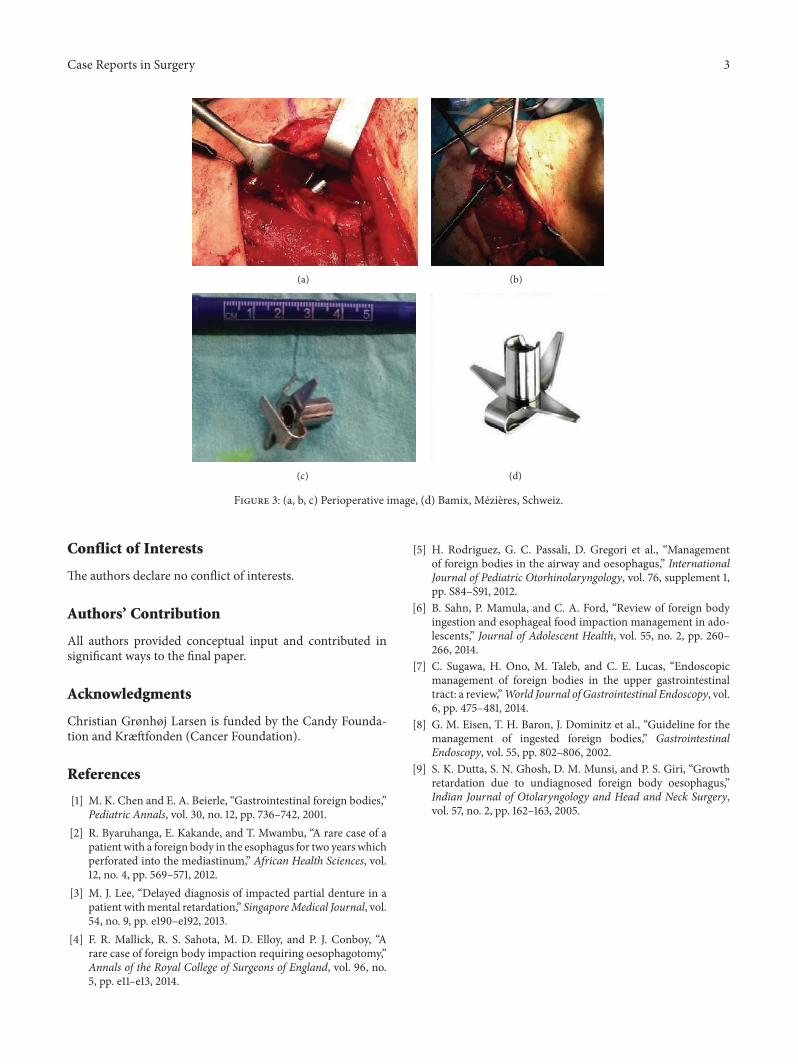
A 37-year-old male with the mental age of a 1-year-old wasreferred to our department. Six months earlier, the patienthad been admitted to a neighbouring hospital with aspirationpneumonia in a condition of sepsis and complaining of dys-phagia. A chest X-ray was performed (Figure 1). The patientwas treated with antibiotics and discharged on the 23th day,
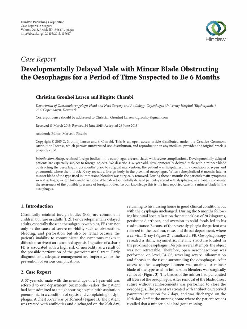
returning to his nursing home in good clinical condition, butwith the dysphagia unchanged. During the 6 months follow-ing his initial hospitalization the patient’s loss of 20 kilograms,persistent diarrhoea, and aversion to solid foods led to hisreadmittance. Because of the severe dysphagia the patient wasreferred to the local ear, nose, and throat department, wherea cervical X-ray (Figure 2) visualized a FB. Oesophagoscopyrevealed a shiny, asymmetric, metallic structure located inthe proximal oesophagus. Despite several attempts, the objectwas not retractable. Therefore, open oesophagotomy wasperformed on level C4-C5, revealing severe inflammationand fibrosis in the tissue surrounding the oesophagus. Afteraccess to the oesophageal lumen was attained, a mincerblade of the type used in immersion blenders was surgicallyremoved (Figure 3). The blades of the mincer had penetratedall layers of the oesophagus. After removal of the blade, directsuture without reinforcements was performed to close theoesophagus.The patient was treatedwith antibiotics, receivedparenteral nutrition for 7 days, and was discharged on the10th day. Staff at the nursing home where the patient residesrecalled that a mincer blade had gone missing.
Hindawi Publishing CorporationCase Reports in SurgeryVolume 2015, Article ID 139647, 3 pageshttp://dx.doi.org/10.1155/2015/139647

2 Case Reports in Surgery
Figure 1: Chest X-ray 6 months prior to admittance to our depart-ment. Severe right-sided pneumonia and a suspected foreign objectin the oesophagus.
Figure 2: Chest X-ray.
3. Discussion
Developmentally delayed persons with pica are especiallysubject to FBs in the oesophagus [3]. Objects often impacthere because of the passive, distensible, and accommodatingnature of the organ. Our report documents the case of adevelopmentally delayed male with a mincer blade lodgedin the proximal oesophagus. When assessing the thoracicX-ray 6 months prior to admittance it is likely that theobject was visible in the top edge of the picture with thecharacteristic asymmetric sharp edges of themincer pointingout (Figure 1). This might explain the patient’s symptoms ofprolonged dysphagia, weight loss, and diarrhoea.
Similar cases of chronically retained FBs have beenreported with successful outcomes, [1, 4, 5] although mostobjects usually pass spontaneously, and less than 1% needsurgical removal [4]. Early diagnosis and management mightprevent the serious complications of penetration, infection,and necrosis [6]. When FBs are suspected, the appropriatediagnostic approach should be biplane X-rays, which reveal
the location, size, and shape of possible objects, alternativelya CT scan of the esophagus [5]. Direct vision such as oesoph-agoscopy or laryngoscopic-aided views are highly usefulmeans of obtaining additional diagnostic information.More-over, endoscopy may also provide the treatment as manyobjects can be extracted endoscopically [7]. Migratory oeso-phageal FBs are particularly rare [1].
Sharp objects most commonly lodge at the upper levelof the oesophagus due to penetration into the upper oeso-phageal sphincter. Whenever possible, FBs in the oesoph-agus should be sought and removed endoscopically [5];however, if surgical removal is unavoidable, it must beperformed without delay, in order to avoid potentially severecomplications, such as mediastinitis, fistula, pneumothorax,respiratory distress, retropharyngeal abscess, and stricture.Nonsharp objects lodged in the oesophagus for more than24 hours should also be removed endoscopically. However,if they remain for more than a week, there is significant riskof erosion into surrounding structures, and surgical backupshould always be available [1, 5].
When dealing with the severely mentally retarded groupof patients, common symptoms such as dysphagia, odynoph-agia, coughing, choking, and haematemesis may be com-promised. For this reason, information about the patient’sgeneral condition, appetite, and bowel habits is vital. If thegeneral condition of a patient with these symptoms worsensand persists over a long period of time, FBs should always beconsidered and biplane X-rays of the upper oesophagus mustbe performed without delay [8].
Earlier cases of oesophageal FBs involving developmen-tally challenged individuals have included impacted dentures[3], coins [9], and food items, but this is the first reported caseof a mincer blade from an immersion blender lodging in theoesophagus.
A thorough elucidation of developmentally delayedpatients with dysphagia is imperative, and the presence offoreign objects in the pharynx, oesophagus, or intestine mustalways be considered when such patients present with adeclining general condition, reduced appetite, abnormalbowel movements, or dysphagia. Biplane X-rays of the oeso-phagus and endoscopy should be performed initially and,particularly in cases involving sharp FBs, surgery must notbe delayed.
4. Conclusion
We know the following:
(i) The elucidation of developmentally delayed patientsis difficult.
(ii) This specific patient-population is especially subjectto foreign bodies in the oesophagus.
This study adds the following:
(i) When developmentally delayed patients present withdysphagia, altered bowel movements, and declininggeneral condition, foreign bodiesmust be considered.
(ii) Biplane X-rays and endoscopy are vital initial steps.
Case Reports in Surgery 3
(a) (b)
(c) (d)
Figure 3: (a, b, c) Perioperative image, (d) Bamix, Mezieres, Schweiz.
Conflict of Interests
The authors declare no conflict of interests.
Authors’ Contribution
All authors provided conceptual input and contributed insignificant ways to the final paper.
Acknowledgments
Christian Grønhøj Larsen is funded by the Candy Founda-tion and Kræftfonden (Cancer Foundation).
References
[1] M. K. Chen and E. A. Beierle, “Gastrointestinal foreign bodies,”Pediatric Annals, vol. 30, no. 12, pp. 736–742, 2001.
[2] R. Byaruhanga, E. Kakande, and T. Mwambu, “A rare case of apatientwith a foreign body in the esophagus for two yearswhichperforated into the mediastinum,” African Health Sciences, vol.12, no. 4, pp. 569–571, 2012.
[3] M. J. Lee, “Delayed diagnosis of impacted partial denture in apatient withmental retardation,” SingaporeMedical Journal, vol.54, no. 9, pp. e190–e192, 2013.
[4] F. R. Mallick, R. S. Sahota, M. D. Elloy, and P. J. Conboy, “Arare case of foreign body impaction requiring oesophagotomy,”Annals of the Royal College of Surgeons of England, vol. 96, no.5, pp. e11–e13, 2014.
[5] H. Rodriguez, G. C. Passali, D. Gregori et al., “Managementof foreign bodies in the airway and oesophagus,” InternationalJournal of Pediatric Otorhinolaryngology, vol. 76, supplement 1,pp. S84–S91, 2012.
[6] B. Sahn, P. Mamula, and C. A. Ford, “Review of foreign bodyingestion and esophageal food impaction management in ado-lescents,” Journal of Adolescent Health, vol. 55, no. 2, pp. 260–266, 2014.
[7] C. Sugawa, H. Ono, M. Taleb, and C. E. Lucas, “Endoscopicmanagement of foreign bodies in the upper gastrointestinaltract: a review,”World Journal of Gastrointestinal Endoscopy, vol.6, pp. 475–481, 2014.
[8] G. M. Eisen, T. H. Baron, J. Dominitz et al., “Guideline for themanagement of ingested foreign bodies,” GastrointestinalEndoscopy, vol. 55, pp. 802–806, 2002.
[9] S. K. Dutta, S. N. Ghosh, D. M. Munsi, and P. S. Giri, “Growthretardation due to undiagnosed foreign body oesophagus,”Indian Journal of Otolaryngology and Head and Neck Surgery,vol. 57, no. 2, pp. 162–163, 2005.

Submit your manuscripts athttp://www.hindawi.com
Stem CellsInternational
Hindawi Publishing Corporationhttp://www.hindawi.com Volume 2014
Hindawi Publishing Corporationhttp://www.hindawi.com Volume 2014
MEDIATORSINFLAMMATION
of
Hindawi Publishing Corporationhttp://www.hindawi.com Volume 2014
Behavioural Neurology
EndocrinologyInternational Journal of
Hindawi Publishing Corporationhttp://www.hindawi.com Volume 2014
Hindawi Publishing Corporationhttp://www.hindawi.com Volume 2014
Disease Markers
Hindawi Publishing Corporationhttp://www.hindawi.com Volume 2014
BioMed Research International
OncologyJournal of
Hindawi Publishing Corporationhttp://www.hindawi.com Volume 2014
Hindawi Publishing Corporationhttp://www.hindawi.com Volume 2014
Oxidative Medicine and Cellular Longevity
Hindawi Publishing Corporationhttp://www.hindawi.com Volume 2014
PPAR Research
The Scientific World JournalHindawi Publishing Corporation http://www.hindawi.com Volume 2014
Immunology ResearchHindawi Publishing Corporationhttp://www.hindawi.com Volume 2014
Journal of
ObesityJournal of
Hindawi Publishing Corporationhttp://www.hindawi.com Volume 2014
Hindawi Publishing Corporationhttp://www.hindawi.com Volume 2014
Computational and Mathematical Methods in Medicine
OphthalmologyJournal of
Hindawi Publishing Corporationhttp://www.hindawi.com Volume 2014
Diabetes ResearchJournal of
Hindawi Publishing Corporationhttp://www.hindawi.com Volume 2014
Hindawi Publishing Corporationhttp://www.hindawi.com Volume 2014
Research and TreatmentAIDS
Hindawi Publishing Corporationhttp://www.hindawi.com Volume 2014
Gastroenterology Research and Practice
Hindawi Publishing Corporationhttp://www.hindawi.com Volume 2014
Parkinson’s Disease
Evidence-Based Complementary and Alternative Medicine
Volume 2014Hindawi Publishing Corporationhttp://www.hindawi.com