case report open access klebsiella pneumoniae bacteremia
TRANSCRIPT

Huang et al. BMC Gastroenterology 2013, 13:139http://www.biomedcentral.com/1471-230X/13/139
CASE REPORT Open Access
Klebsiella pneumoniae bacteremia and renosplenicabscesses without intestinal symptoms as theinitial manifestations of non-steroidalanti-inflammatory drug-induced colitis: a rarecase reportHung-Ling Huang3, Po-Liang Lu1,4, Chun-Yu Lin1,4, Yen-Hsu Chen1,3,4,5, Chao-Hung Kuo2 and Wei-Ru Lin1*
Abstract
Background: Non-steroidal anti-inflammatory drugs (NSAIDs), the most widely prescribed drugs in the world, cancause gastrointestinal damage, including colitis. However, the prevalence of NSAID-induced colitis is unknownbecause the disease is often asymptomatic.
Case presentation: We report the case of a 64-year-old female patient with a history of long-term NSAID use, whowas hospitalized with septic shock caused by Klebsiella pneumoniae bacteremia. Computed tomography revealedmultiple renal and splenic abscesses with diffuse colon wall thickening. A colonoscopy confirmed colitis withdiffuse ulcers. NSAIDs were discontinued after this hospitalization. The abscesses improved after antibiotictreatment. A short course of balsalazide treatment was given under the suspicion of ulcerative colitis. Balsalazidewas discontinued four months later due to a non-compatible clinical course. A follow-up colonoscopy two yearslater revealed a normal colon mucosa, and NSAID-induced colitis was diagnosed.
Conclusion: This is the first reported case of combined bacterial splenic and renal abscesses without intestinalmanifestations as the initial presentation of NSAID-induced colitis. In contrast to cases of K. pneumoniae bacteremiawith primary liver abscesses in patients with diabetes mellitus in Taiwan, we presented the first case with abscessescaused by community-acquired K. pneumoniae in the kidneys and spleen without liver invasion. In conclusion, ourcase report alerts clinicians to the possibility that K. pneumoniae bacteremia combined with multiple abscesses canbe associated with severe NSAID-induced colitis.
Keywords: Non-steroidal anti-inflammatory drugs, Colitis, Bacteremia, Klebsiella pneumoniae, Renosplenic abscesses
BackgroundNon-steroidal anti-inflammatory drugs (NSAIDs), despitetheir well-known adverse effects on the gastrointestinal(GI) tract, are widely prescribed worldwide. NSAIDs dam-age different regions of the GI tract, including the distalsmall bowel and colon can be the target of NSAIDs. Astudy using fecal calprotectin (a non-degraded neutro-phil cytosolic protein) to diagnose NSAID enteropathy
* Correspondence: [email protected] of Infectious Diseases, Department of Internal Medicine, KaohsiungMedical University Hospital, Kaohsiung Medical University, 100, Tzyou 1stRoad, Kaohsiung 807, TaiwanFull list of author information is available at the end of the article
© 2013 Huang et al.; licensee BioMed CentralCommons Attribution License (http://creativecreproduction in any medium, provided the or
found that the prevalence of NSAID enteropathy wasaround 44% [1]. Studies evaluating the colonic sideeffects of NSAIDs have suggested that NSAID-inducedcolitis is common; however, symptomatic NSAID-induced colitis is rare [2,3]. People taking NSAIDs aretwo to five times more likely to develop colonic inflam-mation than the general population. NSAIDs affect thelarge intestine by causing colonic ulceration and stric-ture formation. Approximately 10% of newly diagnosedcolitis cases may be related to NSAID administration[4,5]. However, the prevalence of NSAID-induced colitisis unknown because the disease is often asymptomatic.
Ltd. This is an Open Access article distributed under the terms of the Creativeommons.org/licenses/by/2.0), which permits unrestricted use, distribution, andiginal work is properly cited.
Huang et al. BMC Gastroenterology 2013, 13:139 Page 2 of 6http://www.biomedcentral.com/1471-230X/13/139
NSAID-induced colitis usually has nonspecific histo-logical findings. The diagnosis of NSAID-induced co-lonic ulceration has been made based on a history ofNSAID use and the exclusion of other causes [2]. Thetemporal relationship between NSAID use and symp-toms that resolve after cessation confirm the diagnosisof NSAID-induced colitis.Intra-abdominal abscesses as the initial presentation of
NSAID-induced colitis has not been reported previ-ously. Herein, we report the unusual case of a patientwith renal and splenic abscesses but without intestinalsymptom as the initial manifestations of NSAID-induced colitis.
Case presentationA 64-year-old Taiwanese woman presented with a his-tory of well-controlled type 2 diabetes mellitus andhypertension. She had been self-treating regularly withNSAIDs from pharmacy (diclofenac, 50 mg three timesdaily) for more than two years because of chronic lowback pain caused by intervertebral disc herniation. Gen-eral malaise, poor appetite, intermittent postprandialnausea, urinary urgency and frequency were present onemonth before admission. She visited local clinics and re-ceived no specific diagnosis. Due to progressive shortnessof breath and drowsiness for one week, she was sent to amedical center. She appeared acutely ill but afebrile, witha blood pressure of 74/34 mmHg, a pulse rate of 86/min,a respiratory rate of 22/min, and an oxygen saturationlevel of 93% in ambient air. Physical examination revealedno specific local findings. A blood assay revealed thefollowing findings: leukocyte count, 44,400/μL with 85%neutrophils; hemoglobin, 12.3 g/dL; platelet count, 466,000/μL; C-reactive protein, 235.76 mg/L; total bilirubin,1.17 mg/dL; direct bilirubin, 0.17 mg/dL; aspartate amino-transferase, 38 IU/L; alanine aminotransferase, 21 IU/L;blood urea nitrogen, 96.77 mg/dL; creatinine, 7.8 mg/dL;sodium, 140 mmol/L; potassium, 8.6 mmol/L; and lactate,20.3 mmol/L. The arterial blood gas analysis revealed se-vere metabolic acidosis (pH, 7.02; HCO3, 7.1 mmol/L). Achest radiograph revealed no pulmonary lesions. Urineanalysis via catheterization revealed only mild hematuria.She was admitted to the intensive care unit due to
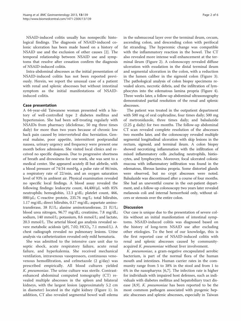
septic shock, acute respiratory failure, acute renalfailure, and hyperkalemia. She received mechanicalventilation, intravenous vasopressors, continuous veno-venous hemofiltration, and cefmetazole (2 g/day) wasprescribed empirically. All blood cultures yieldedK. pneumoniae. The urine culture was sterile. Contrast-enhanced abdominal computed tomography (CT) re-vealed multiple abscesses in the spleen and bilateralkidneys, with the largest lesion (approximately 5.2 cmin diameter) located in the right kidney (Figure 1). Inaddition, CT also revealed segmental bowel wall edema
in the submucosal layer over the terminal ileum, cecum,ascending colon, and descending colon with perifocalfat stranding. The hyperemic change was compatiblewith the inflammatory reaction in the bowel. The CTalso revealed more intense wall enhancement at the ter-minal ileum (Figure 2). A colonoscopy revealed diffuseulceration with exudation in the distal terminal ileumand segmental ulceration in the colon, with a reductionin the lumen caliber in the sigmoid colon (Figure 3).The pathological analysis of colon biopsy specimens re-vealed ulcers, necrotic debris, and the infiltration of lym-phocytes into the edematous lamina propria (Figure 4).Three weeks later, a follow-up abdominal ultrasonographydemonstrated partial resolution of the renal and splenicabscesses.The patient was treated in the outpatient department
with 500 mg of oral cephradine, four times daily; 500 mgof metronidazole, three times daily; and balsalazide(2.25 g daily) for two months. The follow-up abdominalCT scan revealed complete resolution of the abscessestwo months later, and the colonoscopy revealed multiplesegmental longitudinal ulceration with skip lesions in therectum, sigmoid, and terminal ileum. A colon biopsyshowed necrotizing inflammation with the infiltration ofmixed inflammatory cells ,including neutrophils, histio-cytes, and lymphocytes. Moreover, focal ulcerated colonicmucosa with inflammatory infiltration was found in theedematous, fibrous lamina propria. Some distorted glandswere observed, but no crypt abscesses were noted.Balsalazide was discontinued after a course of four months.She had an uneventful course in the out-patient depart-ment, and a follow-up colonoscopy two years later revealedmelanosis coli and internal hemorrhoid only, without ul-cers or stenosis over the entire colon.
DiscussionOur case is unique due to the presentation of severe col-itis without an initial manifestation of intestinal symp-toms. NSAID-induced colitis was diagnosed based onthe history of long-term NSAID use after excludingother etiologies. To the best of our knowledge, this isthe first reported case of NSAID-induced colitis withrenal and splenic abscesses caused by community-acquired K. pneumoniae without liver involvement.K. pneumoniae, a gram-negative encapsulated aerobic
bacterium, is part of the normal flora of the humanmouth and intestines. Human carrier rates in the com-munity range from 5 to 38% in the stool and from 1 to6% in the nasopharynx [6,7]. The infection rate is higherfor individuals with impaired host defenses, such as indi-viduals with diabetes mellitus and hepatobiliary tract dis-ease [8,9]. K. pneumoniae has been reported to be themost common pathogen associated with pyogenic hep-atic abscesses and splenic abscesses, especially in Taiwan

Figure 1 Computed tomography scans of the abdomen revealing several rim-enhancing hypodense lesions in bilateral kidneys andthe spleen.
Huang et al. BMC Gastroenterology 2013, 13:139 Page 3 of 6http://www.biomedcentral.com/1471-230X/13/139
and countries in Eastern and Southeast Asia [10-12],and this bacterium accounts for 10–16% of cases ofsplenic abscess and approximately 25% of cases of renalabscess [13,14].Intra-abdominal abscesses have diverse symptoms.
Fever and abdominal pain are the most frequent symp-toms, but a few cases have vague symptoms, as in thecase presented herein. Imaging analysis, including ultra-sonography and computed tomography (CT), are neces-sary to diagnose intra-abdominal infections [15].Splenic abscesses are rare, with a reported frequency
of 0.14–0.7% in case reports [16]. Chang et al. analyzed67 splenic abscess cases which occurred over a period of
Figure 2 Computed axial tomography scans of the abdomen showingascending colon, descending colon, and rectum with perifocal fat strathe colonoscopy.
19 years and found that K. pneumoniae was the mostfrequently encountered pathogen in blood cultures orabscess cultures, similar to observations from otherAsian countries [11]. Ferraioli G. et al. found a differentresult; they observed that the most common pathogenswere polymicrobial pathogens in pus cultures andStaphylococcus species in blood cultures [16].The pathogenesis of intra-abdominal abscesses caused
by K. pneumoniae remains unclear. One hypothesis forthe pathogenesis of pyogenic intra-abdominal abscessesinvolves hematogenous bacterial seeding from the GItract. An animal study demonstrated that K. pneumoniaestrains with genetic regulatory networks for translocation
marked thickening of the wall of the terminal ileum, cecum,nding. These finding are compatible with the findings of

Figure 3 A colonoscopy revealed extensive segmental longitudinal ulceration of the mucosa extending over the rectum, sigmoidcolon, and terminal ileum.
Huang et al. BMC Gastroenterology 2013, 13:139 Page 4 of 6http://www.biomedcentral.com/1471-230X/13/139
have the ability to cross the intestinal barrier [17]. A care-ful search for the source of K. pneumoniae bacteremiaother than primary bacteremia should be considered,and which should include colon lesions survey [18]. Inour case, the finding of thickened colonic wall promptedus to consider the colon as the possible source of K.pneumoniae bacteremia.Prostaglandins represent one of the most important
components of mucosal defense in the colon and are
Figure 4 Photomicrograph of a sigmoid colon biopsy specimen displthe edematous lamina propria (hematoxylin and eosin, original magn
involved in the maintenance of microcirculation andblood flow to modulate the mucosal immune system.NSAIDs inhibit colonic prostaglandin synthesis, leadingto the development of colitis and the aggravation ofpreexisting intestinal diseases.The ileocecal region is the most common site of
NSAID-induced colonic injury. Pathological examinationof colon specimens from patients with NSAID-inducedcolitis usually reveals sharply demarcated, semilunar or
aying an ulcer, necrotic debris, and lymphocyte infiltration intoification ×100).

Huang et al. BMC Gastroenterology 2013, 13:139 Page 5 of 6http://www.biomedcentral.com/1471-230X/13/139
circumferential, superficial ulcerations with normal sur-rounding mucosa. There are no specific histopatho-logical findings for NSAID-induced colitis except forthe diaphragm-like stricture. Cellular infiltration intothe lamina propria and epithelium may involve predom-inantly neutrophils or lymphocytes, depending on thechronicity of the lesion [19]. Symptomatic patients mayhave watery or bloody diarrhea, weight loss, fatigue,abdominal pain, anorexia, iron-deficiency anemia, andhypoalbuminemia.There have been no studies to determine the risk of
colitis related to different types of NSAIDs; however, manyreports have stated that diclofenac, mefenemic acid,piroxicam, and ibuprofen could cause colorectal ulcers orcolitis [4,20-22]. According to one case-controlled study,the long-term use of NSAIDs increased the risk of colonicmucosal lesions, suggesting that NSAIDs may contributeto the pathogenesis of colonic ulcers or colitis [23]. Inaddition, naproxen has been implicated in eosinophiliccolitis [24], and diclofenac may be associated with pseudo-membranous colitis [25].In this case, the colonoscopy findings and pathological
findings were not able to determine the etiology of thecolitis. Due to evidence of a possible association betweenthe use of NSAIDs and the exacerbation or relapse of in-flammatory bowel diseases [26,27], NSAID-exacerbatedinflammatory bowel disease was initially suspected. Thispatient was initially treated with balsalazide, but she be-came stable despite the discontinuation of balsalazide.The discontinuation of NSAIDs is the cornerstone of
the treatment of NSAID-induced colitis. Symptoms usu-ally resolve within days to weeks of the withdrawal ofNSAIDs, with the restoration of normal histology. Ourpatient received a colonoscopy two years later that re-vealed complete restoration of the colon mucosa; thus,this patient was diagnosed with NSAID-induced colitis.
ConclusionsWe present a unique case of NSAID-induced colitis with-out any GI symptoms at initial presentation, that was as-sociated with septic shock caused by K. pneumoniaebacteremia with multiple renal and splenic abscesses.The colitis was found incidentally by CT and then iden-tified as NSAID-induced colitis. This is also the firstreported case of disseminated abscess formation causedby community-acquired K. pneumoniae in the kidneysand spleen without liver involvement in an NSAID-induced colitis patient.This case is a reminder that hematogenous intra-
abdominal abscesses caused by K. pneumoniae necessi-tate a thorough investigation of the GI tract. Assessingthe presence of colitis should be considered even in theabsence of preceding gastrointestinal symptoms, espe-cially for those who have previously used NSAIDs.
ConsentWritten informed consent was obtained from the patientfor publication of this case report and any accompanyingimages. A copy of the written consent is available for re-view by the Editor of this journal.
AbbreviationsNSAIDs: Non-steroidal anti-inflammatory drugs; GI: Gastrointestinal;CT: Computed tomography.
Competing interestsThe authors declared that they have no competing interests.
Authors’ contributionsHLH, WRL, and PLL prepared the manuscript. YHC and CHK providedlaboratory support. WRL and CHK cared for the patient and provided adviceon the clinical aspects of the case report. All authors read and approved thefinal version of the manuscript.
Author details1Division of Infectious Diseases, Department of Internal Medicine, KaohsiungMedical University Hospital, Kaohsiung Medical University, 100, Tzyou 1stRoad, Kaohsiung 807, Taiwan. 2Division of Gastroenterology, Department ofInternal Medicine, Kaohsiung Medical University Hospital, Kaohsiung MedicalUniversity, Kaohsiung, Taiwan. 3Department of Internal Medicine, KaohsiungMedical University Hospital, Kaohsiung Medical University, Kaohsiung, Taiwan.4School of Medicine, College of Medicine, Kaohsiung Medical University,Kaohsiung, Taiwan. 5Graduate Institute of Medicine, College of Medicine,Kaohsiung Medical University, Kaohsiung, Taiwan.
Received: 26 April 2013 Accepted: 20 September 2013Published: 22 September 2013
References1. Deshpande V, Hsu M, Kumarasinghe MP, Lauwers GY: The clinical
significance of incidental chronic colitis: a study of 17 cases. Am J SurgPathol 2010, 34:463–469.
2. Kurahara K, Matsumoto T, Iida M, Honda K, Yao T, Fujishima M: Clinical andendoscopic features of nonsteroidal anti-inflammatory drug-inducedcolonic ulcerations. Am J Gastroenterol 2001, 96:473–480.
3. Puspok A, Kiener HP, Oberhuber G: Clinical, endoscopic, and histologicspectrum of nonsteroidal anti-inflammatory drug-induced lesions in thecolon. Dis Colon Rectum 2000, 43:685–691.
4. Tanner AR, Raghunath AS: Colonic inflammation and nonsteroidal anti-inflammatory drug administration. An assessment of the frequency ofthe problem. Digestion 1988, 41:116–120.
5. Katsinelos P, Christodoulou K, Pilpilidis I, Xiarchos P, Papagiannis A,Dimiropoulos S, Amperiadis P, Vasiliadis T, Tarpagos A, Katsos I, Eugenidis N:Colopathy associated with the systemic use of nonsteroidalantiinflammatory medications. An underestimated entity.Hepatogastroenterology 2002, 49:345–348.
6. Fung CP, Lin YT, Lin JC, Chen TL, Yeh KM, Chang FY, Chuang HC, Wu HS,Tseng CP, Siu LK: Klebsiella pneumoniae in gastrointestinal tract andpyogenic liver abscess. Emerg Infect Dis 2012, 18:1322–1325.
7. Podschun R, Ullmann U: Klebsiella spp. as nosocomial pathogens:epidemiology, taxonomy, typing methods, and pathogenicity factors.Clin Microbiol Rev 1998, 11:589–603.
8. Kang CI, Kim SH, Bang JW, Kim HB, Kim NJ, Kim EC, Oh MD, Choe KW:Community-acquired versus nosocomial Klebsiella pneumoniaebacteremia: clinical features, treatment outcomes, and clinicalimplication of antimicrobial resistance. J Korean Med Sci 2006, 21:816–822.
9. Wu HS, Wang FD, Tseng CP, Wu TH, Lin YT, Fung CP: Characteristics ofhealthcare-associated and community-acquired Klebsiella pneumoniaebacteremia in Taiwan. J Infect 2012, 64:162–168.
10. Lederman ER, Crum NF: Pyogenic liver abscess with a focus on Klebsiellapneumoniae as a primary pathogen: an emerging disease with uniqueclinical characteristics. Am J Gastroenterol 2005, 100:322–331.
11. Chang KC, Chuah SK, Changchien CS, Tsai TL, Lu SN, Chiu YC, Chen YS,Wang CC, Lin JW, Lee CM, Hu TH: Clinical characteristics and prognostic

Huang et al. BMC Gastroenterology 2013, 13:139 Page 6 of 6http://www.biomedcentral.com/1471-230X/13/139
factors of splenic abscess: a review of 67 cases in a single medicalcenter of Taiwan. World J Gastroenterol 2006, 12:460–464.
12. Siu LK, Yeh KM, Lin JC, Fung CP, Chang FY: Klebsiella pneumoniae liverabscess: a new invasive syndrome. Lancet Infect Dis 2012, 12:881–887.
13. Lee CH, Leu HS, Hu TH, Liu JW: Splenic abscess in southern Taiwan.J Microbiol Immunol Infect 2004, 37:39–44.
14. Lee BE, Seol HY, Kim TK, Seong EY, Song SH, Lee DW, Lee SB, Kwak IS:Recent clinical overview of renal and perirenal abscesses in 56consecutive cases. Korean J Intern Med 2008, 23:140–148.
15. Solomkin JS, Mazuski JE, Bradley JS, Rodvold KA, Goldstein EJ, Baron EJ,O’Neill PJ, Chow AW, Dellinger EP, Eachempati SR, et al: Diagnosis andmanagement of complicated intra-abdominal infection in adults andchildren: guidelines by the Surgical Infection Society and the InfectiousDiseases Society of America. Clin Infect Dis 2010, 50:133–164.
16. Ferraioli G, Brunetti E, Gulizia R, Mariani G, Marone P, Filice C: Managementof splenic abscess: report on 16 cases from a single center. Int J Infect Dis2009, 13:524–530.
17. Tu YC, Lu MC, Chiang MK, Huang SP, Peng HL, Chang HY, Jan MS, Lai YC:Genetic requirements for Klebsiella pneumoniae-induced liver abscess inan oral infection model. Infect Immun 2009, 77:2657–2671.
18. Jeong SW, Jang JY, Lee TH, Kim HG, Hong SW, Park SH, Kim SG, Cheon YK,Kim YS, Cho YD, et al: Cryptogenic pyogenic liver abscess as the herald ofcolon cancer. J Gastroenterol Hepatol 2012, 27:248–255.
19. Goldstein NS, Cinenza AN: The histopathology of nonsteroidal anti-inflammatory drug-associated colitis. Am J Clin Pathol 1998, 110:622–628.
20. Ravi S, Keat A, Keat E: Colitis caused by non-steroidal anti-inflammatorydrugs. Postgrad Med J 1986, 62:773–776.
21. Witham R: Voltaren (diclofenac sodium)-induced ileocolitis.Am J Gastroenterol 1991, 86:246–247.
22. Pan YS, Chen LT, Tseng CA, Su YC, Jan CM, Wang WM, Tsai KB: Clinical andendoscopic features of non-steroidal anti-inflammatory drug-inducedcolorectal ulcerations. J Formos Med Assoc 2005, 104:804–810.
23. Shibuya T, Ohkusa T, Yokoyama T, Harada A, Beppu K, Sakamoto N, Osada T,Nagahara A, Terai T, Otaka M: Colonic mucosal lesions associated withlong‐term or short‐term administration of nonsteroidal anti‐inflammatory drugs. Colorectal Dis 2010, 12:1113–1121.
24. Alfadda AA, Storr MA, Shaffer EA: Eosinophilic colitis: epidemiology,clinical features, and current management. Therap Adv Gastroenterol 2011,4:301–309.
25. Gentric A, Pennec Y: Diclofenac-induced pseudomembranous colitis.Lancet 1992, 340:126–127.
26. Ballinger A: Adverse effects of nonsteroidal anti-inflammatory drugs onthe colon. Curr Gastroenterol Rep 2008, 10:485–489.
27. Kefalakes H, Stylianides TJ, Amanakis G, Kolios G: Exacerbation ofinflammatory bowel diseases associated with the use of nonsteroidalanti-inflammatory drugs: myth or reality? Eur J Clin Pharmacol 2009,65:963–970.
doi:10.1186/1471-230X-13-139Cite this article as: Huang et al.: Klebsiella pneumoniae bacteremia andrenosplenic abscesses without intestinal symptoms as the initialmanifestations of non-steroidalanti-inflammatory drug-induced colitis: a rare case report. BMCGastroenterology 2013 13:139.
Submit your next manuscript to BioMed Centraland take full advantage of:
• Convenient online submission
• Thorough peer review
• No space constraints or color figure charges
• Immediate publication on acceptance
• Inclusion in PubMed, CAS, Scopus and Google Scholar
• Research which is freely available for redistribution
Submit your manuscript at www.biomedcentral.com/submit