case study open access submerged goiter proven to be ... · case study open access submerged goiter...
TRANSCRIPT
a SpringerOpen Journal
Tsoukalas et al. SpringerPlus 2014, 3:46http://www.springerplus.com/content/3/1/46
CASE STUDY Open Access
Submerged goiter proven to be metastaticinfiltration of a neuro-endocrine Merkel cellcarcinomaNikolaos Tsoukalas1*, Menelaos Zoulamoglou2, Maria Tolia2, Evangelos Bournakis3, Elin Ronne4
and Vasileios Barbounis2
Abstract
Background: Merkel cell carcinoma (MCC) is an uncommon neuroendocrine cutaneous carcinoma. Metastases tothe thyroid gland are rare and may present diagnostic difficulties.
Case presentation: A 73-year-old woman presented with a hard mass in the adipose tissue of the right inguinalarea. This mass was surgically excised and the histology examination showed the existence of a MCC. CT scansrevealed a sizable lesion with imaging features of a submerged goiter, invasive to the upper mediastinum. Thepatient received chemotherapy following by locoregional radiotherapy at the bed of the excised lesion. During thenext 10 months the patient was asymptomatic, serum markers values were normal and CT scans findings werestable. However, afterwards NSE and chromogranin values raised and CT scans revealed an enlargement of thesubmerged goiter. The patient became symptomatic, mainly experiencing respiratory inconvenience. Surgicalexcision of the right lobe of the thyroid gland was decided and performed without any complications. Thehistopathology examination showed infiltration of the thyroid gland by a neuroendocrine carcinoma withcharacteristics compatible with MCC.
Conclusions: The rare case of metastatic infiltration of the thyroid gland by a MCC based on histological andimmunohistochemical findings was described. This case report is of clinical significance indicating that by anyabnormal finding in the thyroid gland in patients with a malignant disease, the diagnostic approach should alwayscontain consideration of metastasis from the primary tumor.
Keywords: Metastasis; Neuroendocrine tumours; Thyroid; Merkel cell carcinoma; Submerged goiter
BackgroundMerkel Cell Carcinoma (MCC) is an uncommon neuro-endocrine cutaneous carcinoma which is characterizedby high incidence of early loco-regional relapse and distantmetastases (Poulsen 2004).
Case presentationA 73-year-old woman with no prior medical history,presented with a hard mass (diameter = 46 mm) in theadipose tissue of the right inguinal area which was con-firmed by CT scan (Nov 2009). A month later, this mass
* Correspondence: [email protected] of Medical Oncology, “401” General Military Hospital,Gennimata N. 10-12, 11524 Ampelokipi, Athens, GreeceFull list of author information is available at the end of the article
© 2014 Tsoukalas et al.; licensee Springer. ThisAttribution License (http://creativecommons.orin any medium, provided the original work is p
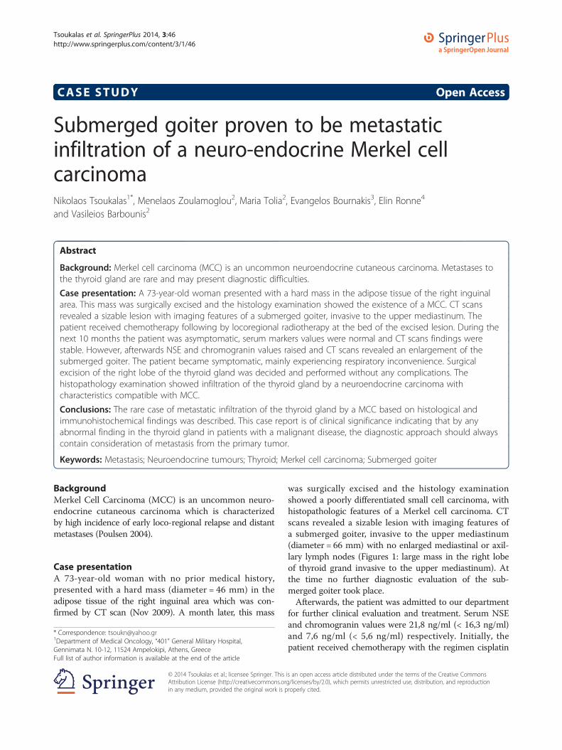
was surgically excised and the histology examinationshowed a poorly differentiated small cell carcinoma, withhistopathologic features of a Merkel cell carcinoma. CTscans revealed a sizable lesion with imaging features ofa submerged goiter, invasive to the upper mediastinum(diameter = 66 mm) with no enlarged mediastinal or axil-lary lymph nodes (Figures 1: large mass in the right lobeof thyroid grand invasive to the upper mediastinum). Atthe time no further diagnostic evaluation of the sub-merged goiter took place.Afterwards, the patient was admitted to our department
for further clinical evaluation and treatment. Serum NSEand chromogranin values were 21,8 ng/ml (< 16,3 ng/ml)and 7,6 ng/ml (< 5,6 ng/ml) respectively. Initially, thepatient received chemotherapy with the regimen cisplatin
is an open access article distributed under the terms of the Creative Commonsg/licenses/by/2.0), which permits unrestricted use, distribution, and reproductionroperly cited.
Figure 1 CT scan large mass in the right lobe of thyroid grand invasive to the upper mediastinum.
Tsoukalas et al. SpringerPlus 2014, 3:46 Page 2 of 5http://www.springerplus.com/content/3/1/46
75 mg/m2 day1 and etoposide 100 mg/m2 day1-3 (6 cyclesevery 3 weeks) following by locoregional radiotherapyat the bed of the excised lesion (total dose 45 Gy). Bothof these treatments were well tolerated. Three monthslater (Jan 2011) serum NSE and chromogranin values werewithin normal limits and the subsequent CT scans revealedstable disease. During the next 10 months the patient wasasymptomatic, serum markers values were normal and CTscans findings were still stable.However, in November 2011 follow up NSE and chromo-
granin values raised to 26 ng/ml and 9,2 ng/ml respectively.
In addition to that, CT scans revealed an enlarge-ment of the submerged goiter which was invasiveto the upper mediastinum, displacing trachea tothe left, still without enlarged mediastinal or axillarylymph nodes again. Moreover, an ultrasound of thethyroid gland confirmed a significant increase of thesize of the submerged right lobe. The patient becamesymptomatic, mainly experiencing respiratory incon-venience. Surgical excision of the right lobe of thethyroid gland was decided and performed withoutany complications.

Tsoukalas et al. SpringerPlus 2014, 3:46 Page 3 of 5http://www.springerplus.com/content/3/1/46
The histopathology examination showed infiltration ofthe thyroid gland by a neuroendocrine carcinoma withcharacteristics compatible with Merkel cell carcinoma.The tumor consisted of uniform, small to mediumsized cells with a round nucleus, finely dispersed chro-matin, inconspicuous nucleoli and scant cytoplasm(Figure 2: hematoxylin-eosin-staining H-E showing boththyroid follicles in the down left part and tumor cells inthe upper right part, original magnification ×100). Therewere numerous mitoses and areas with necrosis. The tumorwas characterized of positive immunohistochemical reac-tion for antibodies to cytokeratin CAM 5.2, cytokeratin20 CK20 (Figure 3: immunoperoxidase staining for CK20showing positive tumor cells, original magnification ×400),neurofilaments NF (Figure 4: immunoperoxidase stainingfor NF showing positive tumor cells, original magnifica-tion ×400) and neuroendocrine markers chromogranin,synaptophysin and CD56. Immunohistochemical reactionfor antibodies to TTF-1 was negative.After the surgery the patient was asymptomatic. The
serum NSE and chromogranin values were within normallimits and the subsequent CT scans were normal withoutany evidence of remaining disease. Consequently, despitethe aggressive nature of this metastatic neoplasm, thepatient remained in complete remission following amultidisciplinary approach for a long period of time.
DiscussionMerkel cell carcinoma is a rare neuroendocrine tumorof the skin, accounting for less than 1% of cutaneousmalignancies. The origin of this cutaneous neuroendocrine
Figure 2 H-E staining showing both thyroid follicles in the down leftmagnification ×100.
tumor is thought to be the Merkel cells or the skin-pressure receptors (Poulsen 2004). Merkel cell carcin-oma tends to grow fast and metastasize to other partsof the body. Usually it spreads to nearby lymph nodesinitially and then may spread to liver, bone, lungs orbrain, where it can interfere with the functioning of thisorgans (Tai et al. 2000a). Even under treatment, this typeof carcinoma commonly metastasizes beyond skin. Thetreatment of this tumor should be based on a multidisciplin-ary approach with surgery, chemotherapy and radiotherapy(Eng et al. 2007; Ott et al. 1999; Eng et al. 2004a; Eng et al.2004b). First of all the surgical remove of the tumor is veryimportant and should take place when it is feasible. Add-itionally, chemotherapy regimens like cisplatin-etoposide(or carboplatin-etoposide, topotecan, CAV) can be usedand radiotherapy can be administered in the specificinvolved fields (Tai et al. 2000b; Fenig et al. 1997).Metastases to the thyroid gland are rare and may
present diagnostic difficulties not only in the cytologicalspecimens but also in the histological specimens. Themost common primary tumors that metastasise to thethyroid gland are kidney cancers, colorectal cancers, lungcancers, breast cancers and sarcomas (Chung et al. 2012).In fact, metastasis from Merkel cell carcinoma to thethyroid gland is an exceptionally rare clinical condi-tion. A rigorous search of the literature disclosed onlyone similar case in which the confirmation of the thy-roid gland infiltration was based on the findings of fineneedle aspiration FNA (Stoll et al. 2010).Therefore, the present case is very interesting and unique
because the diagnosis of the metastatic infiltration of
part and tumor cells in the upper right part, original
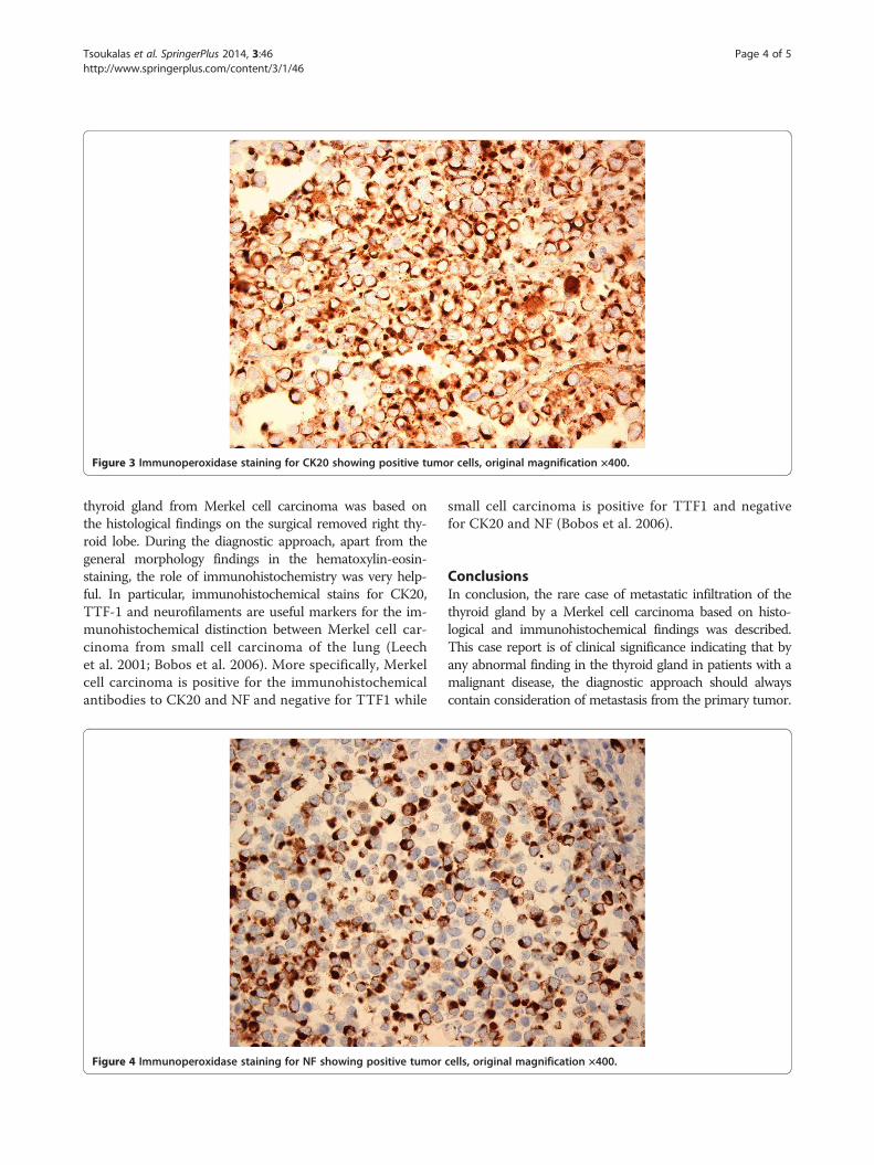
Figure 3 Immunoperoxidase staining for CK20 showing positive tumor cells, original magnification ×400.
Tsoukalas et al. SpringerPlus 2014, 3:46 Page 4 of 5http://www.springerplus.com/content/3/1/46
thyroid gland from Merkel cell carcinoma was based onthe histological findings on the surgical removed right thy-roid lobe. During the diagnostic approach, apart from thegeneral morphology findings in the hematoxylin-eosin-staining, the role of immunohistochemistry was very help-ful. In particular, immunohistochemical stains for CK20,TTF-1 and neurofilaments are useful markers for the im-munohistochemical distinction between Merkel cell car-cinoma from small cell carcinoma of the lung (Leechet al. 2001; Bobos et al. 2006). More specifically, Merkelcell carcinoma is positive for the immunohistochemicalantibodies to CK20 and NF and negative for TTF1 while
Figure 4 Immunoperoxidase staining for NF showing positive tumor
small cell carcinoma is positive for TTF1 and negativefor CK20 and NF (Bobos et al. 2006).
ConclusionsIn conclusion, the rare case of metastatic infiltration of thethyroid gland by a Merkel cell carcinoma based on histo-logical and immunohistochemical findings was described.This case report is of clinical significance indicating that byany abnormal finding in the thyroid gland in patients with amalignant disease, the diagnostic approach should alwayscontain consideration of metastasis from the primary tumor.
cells, original magnification ×400.

Tsoukalas et al. SpringerPlus 2014, 3:46 Page 5 of 5http://www.springerplus.com/content/3/1/46
Competing interestsAuthors state that they have not any financial relationship with anyorganization or any other competing interests.
Authors’ contributionsNT: Conception and design, Collection and assembly of data, Data analysisand interpretation, Provision of study materials or patients, Manuscriptwriting, Final approval of manuscript. MZ: Collection and assembly of data,Data analysis and interpretation, Manuscript writing, Final approval ofmanuscript. MT: Collection and assembly of data, Administrative support,Final approval of manuscript. EB: Collection and assembly of data,Administrative support, Final approval of manuscript. ER: Collection andassembly of data, Data analysis and interpretation, Final approval ofmanuscript. VB: Conception and design, Provision of study materials orpatients, Final approval of manuscript.
Author details1Department of Medical Oncology, “401” General Military Hospital,Gennimata N. 10-12, 11524 Ampelokipi, Athens, Greece. 2Department ofMedical Oncology, “Hippocration” General Hospital, Athens, Greece.3Department of Clinical Therapeutics, "Alexandra" Hospital, University ofAthens School of Medicine, Athens, Greece. 4Department of Pathology,“Evaggelismos” General Hospital, Athens, Greece.
Received: 30 June 2013 Accepted: 10 October 2013Published: 24 January 2014
ReferencesPoulsen M (2004) Merkel-cell carcinoma of the skin. Lancet Oncol 5:593–9Tai PT, Yu E, Tonita J et al (2000a) Merkel cell carcinoma of the skin. J Cutan Med
Surg 4:186–95Eng TY, Boersma MG, Fuller CD et al (2007) A comprehensive review of the
treatment of Merkel cell carcinoma. Am J Clin Oncol 30:624–36Ott MJ, Tanabe KK, Gadd MA et al (1999) Multimodality management of Merkel
cell carcinoma. Arch Surg 134:388–92Eng TY, Boersma MG, Fuller CD et al (2004a) Treatment of merkel cell carcinoma.
Am J Clin Oncol 27:510–5Eng TY, Naguib M, Fuller CD et al (2004b) Treatment of recurrent Merkel cell
carcinoma: an analysis of 46 cases. Am J Clin Oncol 27:576–83Tai PT, Yu E, Winquist E et al (2000b) Chemotherapy in neuroendocrine/Merkel
cell carcinoma of the skin: case series and review of 204 cases. J Clin Oncol18:2493–9
Fenig E, Brenner B, Katz A et al (1997) The role of radiation therapy andchemotherapy in the treatment of Merkel cell carcinoma. Cancer 80:881–5
Chung AY, Tran TB, Brumund KT et al (2012) Metastases to the thyroid: a reviewof the literature from the last decade. Thyroid 22:258–68
Stoll L, Mudali S, Ali SZ (2010) Merkel cell carcinoma metastatic to the thyroid gland:aspiration findings and differential diagnosis. Diagn Cytopathol 38:754–7
Leech SN, Kolar AJ, Barrett PD et al (2001) Merkel cell carcinoma can bedistinguished from metastatic small cell carcinoma using antibodies tocytokeratin 20 and thyroid transcription factor 1. J Clin Pathol 54:727–9
Bobos M, Hytiroglou P, Kostopoulos I et al (2006) Immunohistochemicaldistinction between merkel cell carcinoma and small cell carcinoma of thelung. Am J Dermatopathol 28:99–104
doi:10.1186/2193-1801-3-46Cite this article as: Tsoukalas et al.: Submerged goiter proven to bemetastatic infiltration of a neuro-endocrine Merkel cell carcinoma.SpringerPlus 2014 3:46.
Submit your manuscript to ajournal and benefi t from:
7 Convenient online submission
7 Rigorous peer review
7 Immediate publication on acceptance
7 Open access: articles freely available online
7 High visibility within the fi eld
7 Retaining the copyright to your article
Submit your next manuscript at 7 springeropen.com