catechol oxidase mimetic activity of copper(i) complexes
TRANSCRIPT

Polyhedron 113 (2016) 5–15
Contents lists available at ScienceDirect
Polyhedron
journal homepage: www.elsevier .com/locate /poly
Catechol oxidase mimetic activity of copper(I) complexesof 3,5-dimethyl pyrazole derivatives: Coordination behavior,X-ray crystallography and electrochemical study
http://dx.doi.org/10.1016/j.poly.2016.03.0550277-5387/� 2016 Elsevier Ltd. All rights reserved.
⇑ Corresponding author.E-mail address: [email protected] (P. Bera).
1 Ananyakumari Santra and Gopinath Mondal equally contribute to the work.
Ananyakumari Santra a,1, Gopinath Mondal a,1, Moumita Acharjya a, Pradip Bera a, Anangamohan Panja a,Tarun K. Mandal b, Partha Mitra c, Pulakesh Bera a,⇑aDepartment of Chemistry, Panskura Banamali College, Vidyasagar University, Midnapore (E), West Bengal 721152, IndiabDepartment of Biotechnology, Haldia Institute of Technology, Hatiberia, Haldia, West Bengal 721 657, IndiacDepartment of Inorganic Chemistry, Indian Association for the Cultivation of Science, Kolkata, West Bengal 700032, India
a r t i c l e i n f o
Article history:Received 19 February 2016Accepted 29 March 2016Available online 13 April 2016
Keywords:Copper(I) pyrazolyl complexCatecholase activityX-ray CrystallographyDFTCyclovoltammetry
a b s t r a c t
N1-substituted 3,5-dimethylpyrazole (dmpz) namely, methyl-3,5-dimethyl pyrazole-1-dithioate (L1) andbenzyl-3,5-dimethyl-pyrazole-1-dithioate (L2) were reacted with zinc(II), copper(II) and cadmium(II)chloride to study their coordination behavior. Ligands L1 and L2 produced isostructural complexes withcomposition [CuI(L1)2][CuICl2] (1) and [CuI(L2)2][CuICl2] (2), respectively when reacted withCuCl2�2H2O. Copper (I) (d10 system) analogues, 1 and 2, exhibit prominent catechol oxidase activity inwhich a nice correlation-the easily oxidizable copper(I) center favoring the oxidation of 3,5-DTBC(3,5-ditertiary butyl catechol) is observed. Most importantly, these two compounds represent the classof copper(I) compounds that are rarely employed for the study of catecholase activity. The kinetics studyexhibits a deuterium kinetic isotope effect in the catalytic oxidation of 3,5-DTBC by O2 as evidenced byabout 1.9 times rate retardation in the deuterated solvent, suggesting the hydrogen atom transfer in therate-determining step from the substrate hydroxy group to the metal-bound superoxo species. Withother d10 cations, zinc(II) and cadmium(II) the ligand L1and/or L2 undergoes dissociation into parent pyra-zole unit i.e., 3,5-dimethylpyrazole (dmpz) during formation of a monomeric tetrahedral Zn(dmpz)2Cl2(3) complex and a helical 1-D polymeric chain, [Cd(dmpz)2Cl2]n(4). The possible role of intramolecularhydrogen-bonding induced helicity has been explored in 4.
� 2016 Elsevier Ltd. All rights reserved.
1. Introduction
Several di- and polynuclear copper complexes have beenreported in order to understand the active site geometric and elec-tronic structures and to mimic the catalytic reactivity [1–9]. As themet form of catechol oxidase contains CuII, thus most of the modelsare reported to be CuII-based complexes [10–18]. It is now wellknown that in the catalytic cycle CuII is reduced to CuI with con-comitant oxidation of catechol and reoxidation of CuI by molecularoxygen regenerates CuII [13]. However, literature survey revealsthat CuI-complexes have rarely been employed for mimicking thefunction of catechol oxidase [18]. The reason may be due to lackof proper structural organization between metal and donor ligandwith respect to Cu(II)–Cu(I) redox behavior during alcohol
oxidation. In this context pyrazole derived ligands might be apotential chelate to form stable Cu(I) complexes with mimetic cat-echolate oxidation. Pyrazole being isomeric with imidazole, whichis an integral part of biological system, is considered to be the mostflexible ligand in coordination chemistry due to its diatopic natureand the coordination flexibility of pyrazolido ion [19,20]. The easeof synthesis of pyrazolyl ligands, varieties of coordination modeswith fascinating chemistry, and isomeric structure to imidazolethat present in proteins, are the reasons for broad and intensivestudies of pyrazole derived compounds. The rich chemistry ofpyrazole and their derivatives is attributed to the several chemicalmerit points such as the presence of localized lone pair of electronon an in-plane orbital and the presence of low energy anti bondingvacant p⁄ orbitals of the aromatic system guaranteed their p-acid-ity. The five member pyrazole ring is an important example ofp-acid ligand showing r-donor ability along with p-acceptorcapacity. Pyrazole is a weak acid comparable to aliphatic alcohols,and is also weaker base than pyridine or simple aromatic amines.

6 A. Santra et al. / Polyhedron 113 (2016) 5–15
The alkylation or arylation of the pyrazole ring changes these val-ues by no more than ±2 pKa units (the acid pKa = 14.2 and basicpKa = 2.5) [21,22]. Further, planarity of the heterocycles ascer-tained their fewer encumbrances to the coordination sphere duringcomplexation. Besides these the presence of substituent(s) in thepyrazole ring in N1 or 3(5) positions may vary the steric profileand redox environment of the coordination system. The increasingdemands of huge pyrazole based pharmaceutical drugs and cat-alytic promiscuity of its metal complexes [23] spirited us to con-tinue the research on pyrazole based compounds. The presentstudy includes the interaction of two pyrazole based ligands withd10 metal ions e.g., Cu(I), Zn(II) and Cd(II) emphasizing the stereochemical changes in the formation of coordination compounds.Among the essential elements, copper plays crucial role to allorganisms living strictly in aerobic environments. It is present inthe active sites of several metalloenzymes and metalloproteins[24,25]. Many copper proteins/enzymes activate molecular oxygen,and the functions of these proteins are of oxygen carriers (hemo-cyanin) [26] or as catalysts in oxygenation (oxygenases) or oxida-tion (oxidases) [27] reactions. One of the major enzymes thatutilizes molecular dioxygen is catechol oxidase [28], a member oftype-3 copper proteins, which catalyzes exclusively the oxidationof a wide range of o-diphenols (catechols) to the correspondingo-quinones through the four-electron reduction of molecular oxy-gen to water in a process known as catecholase activity [29–31].Quinones are highly reactive intermediates that undergo auto-polymerization to produce a brown pigment namely melanin, anatural antiseptic, which helps to protect damaged plants againstboth bacterial and fungal diseases [32]. The met state of catecholoxidase consists of two CuII centers, each copper is coordinatedto three imidazole groups from histidyl residues and joined by abridging OH [28,33]. Isoelectronic metal systems e.g., Cd(II),Zn(II) are also studied simultaneously with the same set of ligandsto compare the structure and activity. The present work emphasizeon the effect of substitution on N1 of 3,5-dimethyl pyrazole (dmpz)in the solid state isolation of coordination compounds with d10
metal ions to look insight the structure and activity relationship.The ligands methyl-3,5-dimethyl pyrazole-1-dithioate (L1),benzyl-3,5-dimethyl-pyrazole-1-dithioate (L2) and complex 1,[CuI(L1)2][CuICl2], were obtained following the methods describedin the previous works [34,36]. Herein, we report for the first timethe isolation and structural characterization of three d10 complexes[CuI(L2)2][CuICl2] (2), Zn(dmpz)2Cl2 (3) and [Cd(dmpz)2Cl2]n (4) andthe catecholase activity of 2 along with analogous complex 1 withdetailed kinetics investigations has also been explored.
2. Experimental
2.1. Materials and methods
CuCl2�2H2O, CdCl2�H2O, ZnCl2, DMSO and CS2 were reagentgrade, commercial products of MERCK and were used without fur-ther purification. Reagent grade benzyl chloride and methyl iodidewere purchased from Spectrochem chemical company. Analyticalgrade, methanol, ethylene diamine and ethylene glycol were pur-chased from Himedia chemical company and used without furtherpurification. Solvent ethanol (Changshu Yangyuan Chemical,China) was dried and distilled before use. Ligand L1, L2 and com-plex 1 were synthesized following earlier methods [34–36].
2.2. Synthesis of [Cu(L2)2] [CuCl2] (2)
CuCl2�2H2O (10 mmol, 1.70 g) dissolved in 20 mL of ethanol isadded drop wise to the acetonitrile solution of L2 (10 mmol,2.62 g) with constant stirring. Stirring is continued for another half
an hour where a deep blue colored compound separated out. Theprecipitate formed was filtered, washed with excess ethanol anddried in an oven at 90 �C. Yield: 80%, Mass spectrum m/z value:587 (molecular ion peak for CuI(L2)2+). Anal. Calc. for C26H28Cl2Cu2
N4S4: C, 43.20; H, 3.90; N, 7.75; S, 17.75. Observed: C, 43.30; H,3.87; N, 7.70; S, 17.78.
2.3. Synthesis of Zn(dmpz)2Cl2 (3)
0.136 g (2.0 mmol) ZnCl2 in 5 mL dry methanol is added to theethanolic solution of L1 (1 mmol, 0.18 g) and reflux for 3 h whereupon a white compound is obtained. Recrystallization of the crudeproduct from acetonitrile produces good quality crystals of 4 suit-able for X-ray crystallography. Yield: 78%. Anal. Calc. for C10H16Cl2N4Zn: C, 40.42; H, 5.65; N, 15.71. Observed: C, 40.35; H, 5.70; N,15.75. IR/cm�1: m(C@N) 1573, m(C–N) 1472. Mass spectrum m/z value:354 (molecular ion peak).
2.4. Synthesis of [Cd(dmpz)2Cl2]n(4)
0.209 g (1.0 mmol) CdCl2�H2O in 10 mL dry ethanol is added tothe ethanolic solution of L1 (2 mmol, 0.36 g) and reflux for 3 hwhere upon a bright yellow mass is obtained. Light yellow singlecrystals of 3 suitable for X-ray structure determination wereobtained by the evaporation of aqueous ethanol (v/v 1:1) over along period of time at room temperature. Yield 70%, Anal. Calc.for C30H48Cd3Cl6N12: C, 33.18; H, 4.54; N, 13.27. Observed: C,33.14; H, 4.52; N, 13.35. IR/cm�1: m(C@N)1585, m(H2O) 3417, m(C–N)1405.
2.5. Characterization
The elemental analysis (C, H, N, and S) of the ligand and com-plexes were performed using FISONS EA-1108 CHN analyzer. Cyclicvoltammetric experiments were performed at room temperature inmethanol solvent using tetra butyl ammonium perchlorate as asupporting electrolyte on a CH Instrument electrochemical work-stationmodel CHI630E. The conventional three-electrode assemblyis comprised of a platinumworking electrode, a platinumwire aux-iliary electrode and a Ag/AgCl reference electrode. The IR spectra(4000–500 cm�1) were recorded on a Perkin Elmer Spectrum TwoFT-IR Spectrophotometer with sample prepare as KBr pellets.UV–Vis absorption spectra of the samples were recorded on a Per-kin Elmer Lambda 35 spectrophotometer in the wavelength rangeregion 200–800 nm at room temperature. Mass spectra of ligandswere obtained using a Waters HRMS XEVO-G2QTOF#YCA351. Allthe quantum chemical calculations were performed using densityfunctional theory (DFT) implemented in GAUSSIAN 09. The optimiza-tion was done using split basis set. The B3LYP hybrid functionaland 6-31 g (d,p) basis set for hydrogen, carbon, nitrogen, sulfurand LANL2DZ basis set for copper are used for the optimization ofall the geometries. The single crystal X-ray diffraction of 2, 3 and4 was carried out on a Bruker SMART APEX II X-ray diffractometerequipped with graphite-monochromated Mo Ka radiation(k = 0.71073 Å) and 16 CCD area detector. The intensity data werecollected in the p and x scan mode, operating at 50 kV, 30 mA at296 K [37]. The data reduction was performed using the SAINT and
SADABS programs [38]. All calculations in the structural solutionand refinement were performed using the Bruker SHELXTL program[39]. The structure was solved by the heavy atom method andrefined by full-matrix least-squares methods. All the non-hydrogenatoms were refined anisotropically; the hydrogen atoms were geo-metrically positioned and fixed with isotropic thermal parameters.The final electron density maps showed no significant difference.

A. Santra et al. / Polyhedron 113 (2016) 5–15 7
The catecholase activity of 1 and 2 was examined by the reac-tion of 100 equivalents of 3,5-di-tert-butylcatechol (3,5-DTBCH2)with 1 � 10�5 M solutions of the complexes under aerobicconditions at ambient temperature in methanol. The reactionwas followed spectro photometrically by monitoring growth ofthe absorbance as a function of time at ca. 400 nm which is charac-teristic of o-quinone chromophore. To determine the dependenceof rate of the reaction on substrate concentration and to evaluatevarious kinetic parameters, 1 � 10�5 M solutions of the complexeswere mixed with at least 10 equivalents of substrate to maintainpseudo-first order condition. To check the rate dependency on cat-alyst concentrations similar set of experiments were performed ata fixed concentration of substrate with various catalyst concentra-tions. The rate of a reaction was determined by the initial ratemethod, and the average initial rate over three independentmeasurements was recorded.
3. Result and discussion
3.1. Complex formation reaction
In an objective to develop a model system for mimicking cate-chol oxidase, coordination compounds derived from N(1) substi-tuted 3,5-dimethylpyrazolyl (dmpz) ligands, e.g., L1 and L2
(Scheme 1) and d10 metals (e.g., CuI, CdII and ZnII) were synthesizedand structurally characterized. Extension of the ligand 3,5-dimethyl pyrazole (dmpz) in N(1) position with substitution –C(S)SR group (R = –Me or –CH2Ph) imparts one additional bondingsite (S) in the ligand which forms chelate changing significantlythe coordination properties. In order to study the coordinationproperties of the pyrazolyl NS-donor ligands, L1 and L2 werereacted with MIICl2 salts (M = Cu, Cd and Zn). Both chelates L1
and L2 act as neutral bidentate ligands with pyrazolyl ternary ringN(2) and thione S atom in [Cu(L1)2][CuCl] (1) and [Cu(L2)2][CuCl](2). These sulfur containing ligands have sufficient reducing prop-erties to reduce CuII to CuI in the reaction condition and attemptsto prepare copper(II) chelates with L1 and L2 therefore result theformation of complexes 1 and 2, respectively. The thermodynamicstability of these complex species resulted mainly from the chelateeffect as the CFSE contribution is zero for Cu(I) (d10 system).Reduction of metal during complexation with sulfur containing
NN
S SCH3
CH3
H3C NN
S SCH2Ph
CH3
H3C
L1 L2
Scheme 1. Ligand structures.
H3C
O O
CH3
H2N NH2NH
H3C
1. Formation of L1/L2:
2. Dissociation mechanism of L1/L2:
NN
CH3
H3C
S SROR'H
NN
CH3
H3C
S OR'
OH
Scheme 2. Dissociation mechanism
N-heterocyclic chelate was also reported earlier though the reasonis not clearly explained there [40].
Unlike reaction with CuCl2�2H2O, we observe that the reactionsof ligand L1 and L2 with other d10 species e.g., ZnCl2 and CdCl2�H2O,take place following a dissociation of the ligands into dmpz beforethe formation of corresponding complexes. A monomeric Zn(dmpz)2Cl2 (3) and a stable Cl-bridged polymer with composition[Cd(dmpz)2Cl2]n (4) were formed, respectively. The dissociationof L1 and/or L2 into parent ligand (dmpz) is attributed to the metalcatalyzed dissociation leading to formation of basic ligand (dmpz)and O,O0-dialkyl ester of thiocarbonic acid with the evolution ofRSH (alkyl thiol) for which a rational mechanism is proposed andshown in Scheme 2. Preparations of complexes with zinc(II) andcadmium(II) keeping the structural identity of L1 and L2 in theproducts were unsuccessful. The existence of dmpz in the coordi-nation sphere reveals the dissociation of L1 and/or L2 into dmpzoccurs is the presence of metal ion during complex formation.The reasons are by no means certain but no doubt involve severalfactors, of which size of the ligand is probably a major one. Being asmaller donor (N-donor) than ligand L1 and/or L2 (NS-donor), dmpzprefers to stabilized the bridged Cd(II)-dmpz complex than Cd(II)chelates with L1 and L2.There is a delicate balance between forma-tion of chelate complex with undissociated ligand molecules(L1/L2) and formation of polymeric chain with dissociated fragmentdmpz. High polymerization effect that includes the hydrogenbonding in the system outweighs the combined effects of chelateformation and ligand dissociation leading to formation of a rela-tively stable chain structure. The strong intermolecular interac-tions also give the thermodynamic stability of the Cl-bridgedcomplex 4.
On the other hand, ZnCl2�6H2O with L1/L2crystallizes as mono-meric Zn(dmpz)2Cl2 (3). The presence of dmpz in the coordinationsphere once again proves that the chelating ligand (L1/L2) has thetendency to dissociate into basic unit of pyrazole in presence ofmetal before crystallization into a stable product. Here also thechelate effect is not prominent in the non-transition ZnII ion sys-tem. Since the d10 configuration (e.g., ZnII and CdII) have no CFSE,then the stereochemistry of a particular compound depends onthe size, polarizing power of MII ions and the steric requirementof the ligand(s). The reason for the formation of monomeric Zn-complex instead of polymeric structure as in the case in [Cd(dmpz)2Cl2]n (4) may be attributed to the smaller size of Zn2+
(74 pm) than Cd2+ (95 pm). The smaller size of Zn(II) would havegiven much more strain to the bridged complex. In fact, in litera-ture, the polymeric complexes of pyrazole based ligands withCd2+ ion are numerous than Zn2+ ions [41]. Dissociation of substi-tuted 3,5-dimethyl pyrazole ligand into dmpz is reported earlierin the literature [42,43]. 1-carboxamide-3,5-dimethyl pyrazoleformed complexes with CuCl2 and CuCBr2 with a coordination todissociated dmpz. It was supposed that Bronsted basicity of Cl
N
CH3 1. CS2/KOH2. RI/Cl N
N
CH3
H3C
S SR
R'N
NH
CH3
H3C
S
OR' OR'HS R
R=methyl/benzyl
R'=methyl/ethyl
of ligand L1 and L2 to dmpz.

8 A. Santra et al. / Polyhedron 113 (2016) 5–15
and Br ions low to promote the carboximide group for fair coordi-nation. But the decarboxamidation of 1-carboxamide-3,5-dimethylpyrazole to dmpz leads to formation of Cu-dmpz complex [44].
Free energy calculation for 3 and 4 (as proposed in theScheme 2) give the values �39.67 and �32.84 kcal/mole, respec-tively (Table S1) attesting the dissociation of parent ligand intodmpz. It is also worth noting that the free energy value of the reac-tion of CuCl2 and L1 according to the proposed reaction scheme is�35.62 kcal/mol. However, the stabilization of [Cu(L1)2][CuCl] (1)and [Cu(L2)2][CuCl] (2) with retention of original ligand structuremay be explained with strong chelate effect of the system withsmaller copper ion than cadmium or zinc ion. It can be concludedthat N1 substituted 3,5-dimethyl pyrazolyl ligands having sizelarger than 3,5-dimethyl pyrazole (dmpz) have a tendency todissociate into mono dentate dmpz with sufficient thermodynamicstability during coordination with relatively larger d10 ion
Table 1Crystal data and structure refinements for compounds 1–4.
1* 2
Formula C14H20Cl2Cu2N4S4 C26
Mr 570.62 72Crystal system triclinic tricSpace group P�1 P�1T (K) 100 29a (Å) 10.5271(9) 7.7b (Å) 10.7784(9) 12c (Å) 10.9396(9) 16a (�) 68.855(4) 71b (�) 65.795(4) 86c (�) 78.787(5) 81V (Å3), Z 2 2Dcalc (g cm–3) 1.797 1.5Absorption coefficient (mm–1) 2.674 1.8F(000) 576.0 73F(0000) 578.93 73h, k, l (max) 14, 14, 14 9,R1 0.0526(4419) 0.1wR2 0.1420(5169) 0.32h(maximum) 56.60 49
* Ref. [34].
Fig. 1. Molecular structure of 1 showing the atom numbering
until unless sufficient chelate effect come into force in thesystem.
3.2. Description of crystal structures
The structures of [CuI(L2)2][CuICl2] (2), Zn(dmpz)2Cl2 (3) and [Cd(dmpz)2Cl2]n (4) have been determined by single crystal X-raydiffraction method. Structural characterization of the complex 1is reported earlier by us [34]. To compare the structural featureswith isostructural complex 2, the details of complex 1 (ORTEP andcrystal data) is reproduced along with other complexes. The detailsof the refinement of all compounds reported are given in Table 1.The perspective views of molecular structures of 1 and 2 aredepicted in Figs. 1 and 2, respectively, and the selected bondlengths and angles are given in Table 2. Both 1 and 2 crystallizedin same fashion with similar geometry. The coordination geometry
3 4
H28Cl2Cu2N4S4 C30H48Cd3Cl6N12 C10H16Cl2N4Zn2.74 1126.70 328.56linic triclinic monoclinic
P�1 C 2/c3 293 29375(3) 9.5338(13) 15.0045(17).790(4) 12.4107(17) 8.2983(7).256(6) 20.436(3) 23.957(2).425(11) 84.995(3) 90.823(11) 79.306(3) 95.790(4).026(12) 68.158(3) 90
2 886 1.697 1.47181 1.834 2.0006.0 1116.0 1344.08.98 1113.14 1348.8315, 19 11, 14, 24 17, 9, 28143(2016) 0.0407(6503) 0.0285(2262)094(5028) 0.1078(7746) 0.0772(2612).44 50.00 50.00
scheme. Ellipsoids are drawn at the 30% probability level.

Fig. 2. Crystal structure of 2 showing the atom numbering scheme. Ellipsoids are drawn at the 30% probability level.
Table 2Selected bond lengths (Å) and bond angles (�) of 1–4.
1* 2 3 4
Bond Angle (�) Bond Angle (�) Bond Angle (�) Bond Angle (�)S1–Cu1–N1 84.53(8) S1–Cu1–S4 134.9(2) N1–Zn1–N3 103.50(9) Cl3–Cd1–Cl6 90.29(5)S1–Cu1–S11 121.61(4) S1–Cu1–N1 84.2(4) N1–Zn1–Cl1 101.98(7) Cl3–Cd1–Cl4 79.41(4)S1–Cu1–N12 130.53(9) S1–Cu1–N3 115.3(4) N1–Zn1–Cl2 114.88(7) Cl3–Cd1–Cl5 85.20(4)N2–Cu1–S11 133.22(9) S4–Cu1–N1 120.0(4) N3–Zn1–Cl1 114.41(7) Cl3–Cd1–N3 92.9(1)N2–Cu1–N12 108.3(1) S4–Cu1–N3 85.7(4) N3–Zn1–Cl2 102.04(7) Cl3–Cd1–N7 167.6(1)S11–Cu1–N12 84.38(9) N1–Cu1–N3 121.2(5) Cl1–Zn1–Cl2 119.36(3) Cl6–Cd1–Cl4 167.1(5)Cu1–S1–C1 99.5(1) Cu1–S1–C6 98.4(6) Zn1–N1–N2 123.6(2) Cl6–Cd1–Cl5 80.04(5)C1–S2–C5 102.5(2) Cu1–S4–C19 98.3(6) Zn1–N1–C1 130.8(2) Cl6–Cd1–N3 91.4(1)N2–N1–C1 117.2(2) Cu1–N1–N2 117.0(9) N2–N1–C1 105.3(2) Cl6–Cd1–N7 99.4(1)N2–N1–C2 109.9(2) Cu1–N1–C1 136(1) Zn1–N3–N4 120.7(2) Cl4–Cd1–Cl5 91.28(4)C1–N1–C2 132.8(2) Cu1–N1–N2 117.0(9) Zn1–N3–C5 134.3(2) Cl4–Cd1–N3 96.8(1)Cu1–N2–N1 116.9(2) Cu1–N1–C1 136(1) N3–N4–H4 124.0 Cl4–Cd1–N7 89.9(1)Cu1–N2–C4 135.6(2) N2–N1–C1 107(1) N3–N4–C7 112.1(2) Cl5–Cd1–N3 171.2(1)N1–N2–C4 106.2(3) H3–C3–C1 126 H4–N4–C7 123.9 Cl5–Cd1–N7 88.8(1)S1–C1–S2 l 122.9(2) H3–C3–C4 126 N3–Cd1–N7 94.7(2)
Bond Length (Å) Bond Length (Å) Bond Length (Å) Bond Length (Å)Cu1–S1 2.2640 Cu1–S1 2.287 Zn1–N1 2.013 Cd1–Cl3 2.722Cu1–N2 2.042 Cu1–S4 2.274 Zn1–N3 2.016 Cd1–Cl6 2.621Cu1–S11 2.269 Cu1–N1 2.04 Zn1–Cl1 2.2259 Cd1–Cl4 2.638Cu1–N12 2.025 Cu1–N3 2.01 Zn1–Cl2 2.2196 Cd1–Cl5 2.667S1–C1 1.663 S1–C6 1.63 N1–N2 1.354 Cd1–N3 2.278S2–C1 1.730 S2–C6 1.75 N1–C1 1.336 Cd1–N7 2.317S2–C5 1.794 S2–C7 1.82 N3–N4 1.357 Cd2–Cl1 2.679N1–N2 1.396 S3–C19 1.72 N3–C5 1.328 Cd2–Cl3 2.695N1–C1 1.389 S3–C20 1.80 N4–H4 1 0.861 Cd2–Cl4 2.647N1–C2 1.407 S4–C19 1.64 N4–C7 1.334 Cd2–Cl2 2.596
* Ref. [34].
A. Santra et al. / Polyhedron 113 (2016) 5–15 9
of both the complexes around the Cu-atom is tetrahedral with anN2S2 chromophore with the participation of two ligand moieties.Cu(I) is coordinated by the ternary N-atom of pyrazole ring andthione sulfur of each ligand and a linear CuICl2 unit is presentoutside the coordination sphere to satisfy the residual charge. Incomplex 1, the chelate bite angle of N(1)–Cu(1)–S(1) and N(12)–Cu(1)–S(11) are 84.53(8)� and 84.38(9)�, respectively. The corre-sponding chelate bite angles in complex 2 i.e., N(1)–Cu(1)–S(1)and N(3)–Cu(1)–S(4) are 84.2(4)� and 85.7(4)�, respectively. Thesebite angles are much smaller than the ideal tetrahedral angle(109.28�) as well as the other two tetrahedral angles N(2)–Cu(1)–N(12) [108.3(10)�]and S(1)–Cu(1)–S(11) [121.61(3)�] in 1, and S(1)–Cu(1)–N(3) [115.3(4)] and S(4)–Cu(1)–N(1) [120.0(4)] in 2.The lowering of the biting angles is due to the closeness of thecoordinating N and S atoms in the ligand with a view to facilitate
the formation of 5-membered chelate rings. The angle N(2)–Cu(1)–N(12) [108.3(10)�] in 1 reveals that the out plane position ofthe two adjacent pyrazole rings giving less steric crowding amongmethyl groups at 3(5) position. In complex 2, the pyrazole rings arealso situated in out plane position to minimize the crowding in 3(5) position with angle N(1)–Cu(1)–N(3) [121.2(5)�].
Zn(dmpz)2Cl2 (3) crystallized in the monoclinic C2/c spacegroup with all atoms in general positions and the whole moleculegiven by the formulae is present in the asymmetric unit (Fig. 3). In3, the coordination sphere around the Zn atom adopts a distortedtetrahedral environment. The coordination sphere consists of twonitrogen atoms of 3,5-dimethyl pyrazole (dmpz) and two chlorineatoms. The coordination angles vary in the range 101.98(7)–119.36(3). The Cl1–Zn1–Cl2 angle is very high [119.36(3)] from theidealized tetrahedral bond angle proving that the Cl–Cl crowding

Fig. 3. Molecular structure of 3 showing the atom numbering scheme. Ellipsoidsare drawn at the 30% probability level.
10 A. Santra et al. / Polyhedron 113 (2016) 5–15
is relatively greater than the methyl–methyl repulsion in thedmpz. The two dmpz ring planes are nearly perpendicular witheach other as suggested by the dihedral angle between two planesof 84.62�. Crystal packing of 3 is mainly stabilized by significantlystrong hydrogen bonding interaction (Table 3) involving chlorideions and pyrazolyl protons of the adjacent molecules, leading tothe formation of 1D supramolecular hydrogen bonded chain struc-ture as shown in Fig. 4.
The crystal structures of 4 constructed 1D polymers consistingof doubly bridged metal centers with Cl atoms (Fig. 5). In thechains, Cd� � �Cd separation is found to be 4.111 Å. The six coordi-nated geometry of the complex 4 is somewhat distorted octahe-dron as reflected from the significant deviation of the cisoid andtransoid angles from the ideal values. In addition to the four
Table 3Hydrogen bond parameters (Å, �) for 3 and 4.
Complex D–H� � �A d(D–H) d(H� � �A) d(D� � �A) <D–H� � �A3 N4–H4� � �Cl2a 0.86 2.82 3.329(3) 128
N2–H5� � �Cl1a 0.86 2.66 3.475(2) 160
4 N8–H8� � �Cl1b 0.86 2.35 3.175(5) 160N9–H9� � �Cl5c 0.86 2.33 3.163(5) 165N10–H13� � �Cl1 0.86 2.33 3.159(5) 162N11–H5� � �Cl3c 0.86 2.36 3.206(5) 167N12–H20� � �Cl3 0.86 2.36 3.189(5) 161N15–H4� � �Cl5 0.86 2.32 3.140(5) 160
Symmetry code: a = 2 � x, y. 3/2 � z; b = 1 + x, y, z; c = �1 + x, y, z.
Fig. 4. 1D hydrogen bonded s
bridging Cl atoms, the coordination number of Cd is completedby the two neutral monodentate dmpz ligand which are cis coordi-nated. The axial positions are occupied by a bridging Cl atom and aternary pyrazolyl nitrogen in 4, with Cl3–Cd1–N7 angle of 167.6(1)�. The adjacent Cd2Cl2 planes are almost perpendicular to eachother in which dihedral angles between the adjacent Cd2Cl2 coresvary in the range 84.32–85.22�. Such an arrangement increasesthe possibility of the intra-chain hydrogen bonding interactionsbetween bridging chlorides and pyrazole protons with donor-accepter distances in the range 3.140–3.189 Å (Table 2). Carefulinspection of crystal packing further reveals that Cd and one bridg-ing Cl construct a helical chain along the crystallographic a-axisand pitch of the helix is just equal to one unit cell length of thea-axis (9.5338(13) Å). However, two adjacent helices with alternat-ing right- and left-handed chiralities are packed together along thea-axis leading to racemic helical polymers which is consistent withthe centro symmetric space group P�1 in the triclinic unit cell.Another interesting feature of compound 4 is that similarhomochiral chains are stacked along the b-axis (Fig. 6) with thehelix-to-helix separation of 12.4107(17) Å, which is equal to thelength of the b-axis. Therefore, the features of the chain structuresin the complex arise from the intra molecular hydrogen bondinginteractions; pyrazolyl ligand able to participate in hydrogen bond-ing interaction with bridging chlorides there by induce helicity inthe chain structure in 4. The present example clearly justifies thatthe non-covalent interactions like hydrogen-bonding is involved tostabilize folded protein like structures instead of linear chain in thebiological systems.
3.3. Electrochemical studies of 1 and 2
The experiment were performed to assess the redox behavior ofcomplex 1 and 2 in solution in order to check the feasibility of cat-echolase activity of the relevant synthesized species. In the biolog-ical systems, redox potential of metallo-enzymes plays crucial rolefor redox transformation in the biochemical reactions. The studiesof the electrochemical behaviors of the complexes have been per-formed in the identical experimental conditions as that of kineticstudies (vide infra). The electrochemical data of the complex 1and 2 have been recorded in methanol in presence of tetra butylammonium perchlorate (0.1 M) as a supporting electrolyte at aplatinum working electrode and using a Ag/AgCl reference elec-trode. Electrochemical responses of both complexes are similarand cyclic voltammograms of 1 and 2 are depicted in Fig. 7. Scan-ning in the anodic direction reveals an oxidative response at 0.50 Vfor 1 and 0.47 V for 2 versus Ag/AgCl electrode. Upon reversal ofscan direction a reduction process is appeared at 0.33 V for 1 and
upramolecular chain of 3.

Fig. 5. Double chloride-bridged cadmium polymer of 4 showing intramolecular hydrogen bonding interactions.
Fig. 6. Molecular packing of 4, viewed along the a-axis, showing the alternating stack of left and right-handed helices propagated along the b axis.
A. Santra et al. / Polyhedron 113 (2016) 5–15 11
0.32 V for 2. These quasi reversible redox processes are responsiblefor copper(II)/copper(I) redox couple. From the electrochemicaldata (DEp = 0.17 V, E1/2 = (Epa + Epc)/2 = 0.42 for 1 and DEp = 0.15 V,E1/2 = 0.40 for 2), it is clear that the electrochemical responses ofboth complexes are very similar which is consistent with bothidentical geometry and coordination environment around themetal center. Slightly different electrode potentials may be dueto the effect of substitution at the ligand backbone along with dif-ferent extent of geometrical distortion present in the complexes.
3.4. Catechol oxidase mimetic activity
Catechol oxidase is a dinuclear copper enzyme that catalyzesthe two electron transfer reaction during the oxidation of a widerange of catechols to the corresponding o-quinones by O2. Weintroduced analogous Cu(I)-pyrazolyl complexes (1 and 2) to checkthe catecholase activity in this report. Catecholase activity is usu-ally studied with 3,5-di-tert-butylcatechol (3,5-DTBC) as substratebecause of its lower redox potential that facilitates ease of
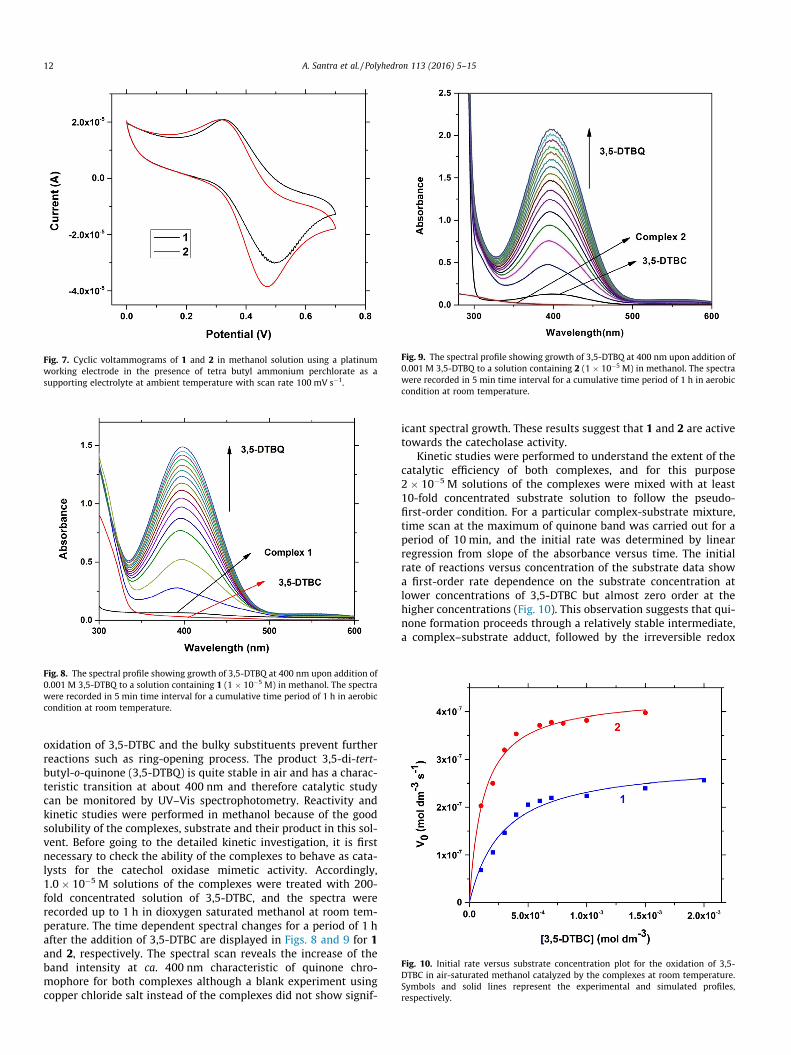
Fig. 7. Cyclic voltammograms of 1 and 2 in methanol solution using a platinumworking electrode in the presence of tetra butyl ammonium perchlorate as asupporting electrolyte at ambient temperature with scan rate 100 mV s�1.
Fig. 8. The spectral profile showing growth of 3,5-DTBQ at 400 nm upon addition of0.001 M 3,5-DTBQ to a solution containing 1 (1 � 10�5 M) in methanol. The spectrawere recorded in 5 min time interval for a cumulative time period of 1 h in aerobiccondition at room temperature.
Fig. 9. The spectral profile showing growth of 3,5-DTBQ at 400 nm upon addition of0.001 M 3,5-DTBQ to a solution containing 2 (1 � 10�5 M) in methanol. The spectrawere recorded in 5 min time interval for a cumulative time period of 1 h in aerobiccondition at room temperature.
Fig. 10. Initial rate versus substrate concentration plot for the oxidation of 3,5-DTBC in air-saturated methanol catalyzed by the complexes at room temperature.Symbols and solid lines represent the experimental and simulated profiles,respectively.
12 A. Santra et al. / Polyhedron 113 (2016) 5–15
oxidation of 3,5-DTBC and the bulky substituents prevent furtherreactions such as ring-opening process. The product 3,5-di-tert-butyl-o-quinone (3,5-DTBQ) is quite stable in air and has a charac-teristic transition at about 400 nm and therefore catalytic studycan be monitored by UV–Vis spectrophotometry. Reactivity andkinetic studies were performed in methanol because of the goodsolubility of the complexes, substrate and their product in this sol-vent. Before going to the detailed kinetic investigation, it is firstnecessary to check the ability of the complexes to behave as cata-lysts for the catechol oxidase mimetic activity. Accordingly,1.0 � 10�5 M solutions of the complexes were treated with 200-fold concentrated solution of 3,5-DTBC, and the spectra wererecorded up to 1 h in dioxygen saturated methanol at room tem-perature. The time dependent spectral changes for a period of 1 hafter the addition of 3,5-DTBC are displayed in Figs. 8 and 9 for 1and 2, respectively. The spectral scan reveals the increase of theband intensity at ca. 400 nm characteristic of quinone chro-mophore for both complexes although a blank experiment usingcopper chloride salt instead of the complexes did not show signif-
icant spectral growth. These results suggest that 1 and 2 are activetowards the catecholase activity.
Kinetic studies were performed to understand the extent of thecatalytic efficiency of both complexes, and for this purpose2 � 10�5 M solutions of the complexes were mixed with at least10-fold concentrated substrate solution to follow the pseudo-first-order condition. For a particular complex-substrate mixture,time scan at the maximum of quinone band was carried out for aperiod of 10 min, and the initial rate was determined by linearregression from slope of the absorbance versus time. The initialrate of reactions versus concentration of the substrate data showa first-order rate dependence on the substrate concentration atlower concentrations of 3,5-DTBC but almost zero order at thehigher concentrations (Fig. 10). This observation suggests that qui-none formation proceeds through a relatively stable intermediate,a complex–substrate adduct, followed by the irreversible redox

A. Santra et al. / Polyhedron 113 (2016) 5–15 13
transformation of the intermediate at the rate determining step.This type of rate saturation kinetics on the concentration of thesubstrate may be explained by considering the Michaelis–Mentenmodel, originally developed for the enzyme kinetics, which giveslinear double reciprocal Lineweaver–Burk plot to analyze valuesof the parameters Vmax, KM, and Kcat. The observed and simulatedinitial rates versus substrate concentration plot and the Linewea-ver�Burk plot for both complexes are shown in Fig. 11. Analysisof the experimental data yielded Michaelis binding constant (KM)values of 2.84 (±0.38) � 10�4 M for 1 and 1.18 (±0.12) � 10�4 for
Fig. 11. Lineweaver–Burk plots for the oxidation of 3,5-DTBC catalyzed by 1 and 2in methanol. Symbols and solid lines represent the experimental and simulatedprofiles, respectively.
Fig. 12. Electrospray ionization mass spectrum (ESI-MS positive) of a 1:50 m
2 and Vmax values of 2.97 (±0.12) � 10�7 M s�1 and 4.35(±0.09) � 10�7 M s�1 for 1 and 2, respectively. The turnover num-ber (Kcat) value is obtained by dividing the Vmax by the concentra-tion of the catalyst used, and is found to be 106.9 and 156.6 h�1 for1 and 2, respectively. Moreover, for a particular substrate concen-tration, varying the complex concentration, a first-order depen-dency on the catalyst concentration was observed. The turnoverrates are slightly higher than other copper(I) model systems andeven also higher than some copper(II) model complexes [17], butsignificantly lower than those recently reported for the binuclearcopper complexes found in the recent review on catechol oxidasemodel systems [45].
3.5. Electrospray ionization mass spectral study
As both 1 and 2 have similar structural features, the electro-spray ionization mass spectrum (ESI-MS positive) of the more reac-tive representative compound 2was recorded in methanol solutionand is shown in Fig. S1. The base peak at m/z = 325.05 with line-to-line separation of unity is assigned to [CuI(L2)]+ (calculatedm/z = 324.99). The peak at m/z = 587.12 with line-to-line separa-tion of 1.0 is consistent with the whole molecular species offormula [CuI(L2)2]+ (calculatedm/z = 587.05). In order to get furtherinsight into the nature of possible complex–substrate intermedi-ates, ESI-MS positive spectrum of a mixture of complex 2 and3,5-DTBC in 1:50 molar ratio was recorded after 5 min of mixingin methanol. The observed spectrum as depicted in Fig. 12 is signif-icantly different from the mass spectrum of complex 2 alone. Ascan be seen from the Fig. 12, the most abundant species found atm/z = 325.06 in the mass spectrum corresponds to the originalcompound 2 which is assigned to the mass of [CuI(L2)]+. Anotherpeak at m/z = 587.10 is again found in the mass spectrum of origi-nal compound which corresponds to the undissociated cationic
ixture of 2 and 3,5-DTBCH2 in methanol, recorded after 5 min of mixing.

14 A. Santra et al. / Polyhedron 113 (2016) 5–15
part of 2 i.e., [CuI(L2)2]+. In addition to the masses related to theoriginal complex species, the mass at m/z = 259.08 is assignableto potassium adduct of product 3,5-DTBQ of unipositive species[K + 3,5-DTBQ]+ (calculated m/z = 259.11). Another product relatedpeak is found at m/z = 755.39 which can be assigned as a monoca-tionic species of formula [Na(3,5-DTBQ)3 + 4H2O]+ (calculatedm/z = 755.47). Most importantly, the peak at m/z = 1031.34 is quiteinteresting because the peak position and line-to-line separation ofunity clearly indicate that the peak arises from the complex-substrate aggregate of unipositive species [CuI(L2)2-(3,5-DTBC)2]+
(calculated m/z = 1031.37). Another minor peak at m/z = 655.09(Fig. S2) is also need to mention as that matches with dioxygencontaining species of unipositive charge of formula [CuI(L2)2 +O2 + 2H2O]+ (calculated m/z = 655.06) although the same peak ismuch more prominent in the mass spectrum of the complex alone(found at m/z = 655.09) (Fig. S1).
3.6. Comparative catecholase activity and proposed mechanism
The reaction kinetics shows that both complexes are very reac-tive towards the catechol oxidase mimicking activity, and the ratesaturation kinetics clearly indicates that the reaction proceedsthrough a stable complex substrate intermediate formation. Theexistence of [CuI(L2)2-(3,5-DTBC)2]+ species in mass spectrum of2 in presence of excess 3,5-DTBC strongly supports the affinity ofthe complex cation towards the incoming substrate. Althoughmass spectroscopy does not necessarily reflect the actual speciespresent in the solution state because some stable aggregates maybe formed in situ (because of their relative stability) when com-pared to the other cationic species on the time scale of the ESI-MS spectrum. Nevertheless, the aggregates found in the ESI-MSspectra deserve importance because they offer clear evidence ofcoordination of the substrate to the complexes. In the mass spec-trum, it can be noticed that m/z peak for the adduct of metal com-plex with the bulky benzyl substitution in 2 compared to methylgroup in 1 does not inhibit the coordination of substrate to metalcenter. Therefore, the difference in catecholase activity between1 and 2 can be explained by the redox potential of the metal cen-ters in these systems-the ease of conversion of Cu(I) to Cu(II) in 2
O
Ot-Bu
t-Bu
HO
HO
H2O2
NN
S S
R1
CuI N NSSR1
NN
S S
R1
CuII
N N SS R1
O
O
t-But-Bu
O
OH.
Cataly
R1 = be
H+
Scheme 3. Plausible mechanistic pathway for the oxidation
(as suggested by the electrochemical data) favors the oxidation of3,5-DTBC which is reflected from the higher Kcat value for 2.
In order to justify the involvement of molecular dioxygen in thecatalytic cycle, UV–Vis spectra of a mixture of 1 � 10�5 M solutionof complex 1 with 100 fold excess of 3,5-DTBC were recorded innitrogen atmosphere, and no spectral growth at 400 nm wasobserved. But upon exposure to air gradual increase of quinoneband intensity at 400 nm is noticed. This observation unambigu-ously proves that molecular dioxygen oxidizes 3,5-DTBC to the cor-responding 3,5-DTBQ in the catalytic cycle in the presence ofcopper(I) complex as a catalyst. In our recent study, we proposedthat catecholate bound copper(I) complex react with moleculardioxygen to form a superoxo intermediate in which copper(I) cen-ter is oxidized to copper(II) [18]. The intermediate involved in theintramolecular proton transfer process from oxygen atom of cate-chol moiety to the superoxide oxygen atom is associated with elec-tron transfer from catecholate to superoxide, leading to theformation of peroxo-intermediate at the rate determining step.That mechanistic pathway for the oxidation of catechol by molec-ular dioxygen was further supported by means of DFT study [18].In the present system, mass spectral data suggests the interactionof both molecular dioxygen and substrate to the metal centers andthus one can expect that the catalytic oxidation of 3,5-DTBC mayproceed thorough the similar mechanistic pathway [18]. It is note-worthy that if the rate determining step involves in breaking of the–O–H bond, a kinetic isotope effect (KIE) should be observed uponchanging the solvent from CH3OH to CH3OD. In order to probe suchmechanistic pathway, comparative kinetic studies were performedunder a given set of conditions both in methanol and deuteratedmethanol, and the results indicate that about 1.9 times rate retar-dation is observed when the kinetic study is conducted in CH3OD.Based on the above results, a plausible mechanism is temptedinvolving stepwise pathways, keeping in mind that it is impossibleto prove any single mechanism (Scheme 3). The catalytic cycle isexpected to start with the formation of complex–substrate aggre-gate which in turn produces the superoxide intermediate by thereaction with aerial oxygen in which copper(I) center undergoesan oxidation to a copper(II). The next step in the catalytic cycle isthe rate determining one which involves the intramolecular protontransfer process from oxygen atom of catechol moiety to the
OO
t-Bu
t-Bu
NN
S S
R1
CuI
N N SS R1
O
HO
t-But-Bu
NN
S S
R1
CuII
N N SS R1
O
HO
t-But-Bu
O
O.
tic Cycle
nzyl or methyl
RDS
- H+
of 3,5-DTBC by dioxygenin presence of catalyst 1 or 2.

A. Santra et al. / Polyhedron 113 (2016) 5–15 15
superoxide along with the intra molecular electron transfer to gen-erate a peroxo-intermediate. Finally, the last step involves therapid breakdown of the peroxo-intermediate to the products(3,5-DTBQ and H2O2) with concomitant regeneration of the activecatalyst to complete the catalytic cycle.
4. Conclusion
Ligational behavior of 3,5-dimethyl pyrazole (dmpz) derivedligands (L1 and L2) were compared with d10 system e.g., Zn(II),Cd(II) and Cu(I). L1 and L2 behave as neutral bidentate NS chelateand stabilized the Cu(I) complex with composition [CuI(L1/L2)2][CuICl2] (1 for L1 and 2 for L2). Whereas L1 and L2 dissociate intobasic 3,5-dimethyl pyrazole before formation of monomeric Zn(dmpz)2Cl2 (3) and polymeric [Cd(dmpz)2Cl2]n (4) from the reac-tion of L1 with ZnCl2 and CdCl2, respectively. Structural diversityof the d10 metal system with pyrazolyl ligands is attributed tothe steric profile of the ligands. The most distinguished feature ofbis(chloro) bridged chain structure of 4 arises from the intramolec-ular hydrogen bonding interactions. Pyrazolyl ligands able to par-ticipate in hydrogen bonding interaction with bridging chloridesthereby induce helicity in the chain structure in 4. Copper ana-logues (1 and 2) exhibits efficient catecholase activity in which anice correlation, the easily oxidizable copper(I) center favoringthe oxidation of 3,5-DTBC is observed. The kinetics study exhibiteda deuterium kinetic isotope effect in the catalytic oxidation of3,5-DTBC by O2, suggesting the hydrogen atom transfer in therate-determining step from the substrate hydroxy group to themetal-bound superoxo species. Thus copper(I) complexes withpyrazolyl NS chelate with sufficient stability may thus be a classicexample in mimicking copper catalytic reactivity and need to beexplored further.
Acknowledgements
We gratefully acknowledge to the University Grants Commis-sion (UGC), Government of India for financial support [Ref. GrantNo. F 42-280/2013(SR)]. We are also thankful to Dr. Sang Il Seokof Korea Research Institute of Chemical Technology (KRICT), SouthKorea for X-ray crystallographic data collection.
Appendix A. Supplementary data
CCDC 1431421–1431423 contains the supplementary crystallo-graphic data for 2–4. These data can be obtained free of charge viahttp://www.ccdc.cam.ac.uk/conts/retrieving.html, or from theCambridge Crystallographic Data Centre, 12 Union Road, Cam-bridge CB2 1EZ, UK; fax: (+44) 1223-336-033; or e-mail:[email protected]. Supplementary data associated with thisarticle can be found, in the online version, at http://dx.doi.org/10.1016/j.poly.2016.03.055.
References
[1] N.A. Rey, A. Neves, A.J. Bortoluzzi, C.T. Pich, H. Terenzi, Inorg. Chem. 46 (2007)348.
[2] C. Belle, C. Beguin, I. Gautier-Luneau, S. Hamman, C. Philouze, J.L. Pierre, F.Thomas, S. Torelli, Inorg. Chem. 41 (2002) 479.
[3] J. Ackermann, F. Meyer, E. Kaifer, H. Pritzkow, Chem. Eur. J. 8 (2002) 247.[4] M. Merkel, N. Mçller, M. Piacenza, S. Grimme, A. Rompel, B. Krebs, Chem. Eur. J.
11 (2005) 1201.[5] A. Majumder, S. Goswami, S.R. Batten, M.S. El Fallah, J. Ribas, S. Mitra, Inorg.
Chim. Acta 359 (2006) 2375.[6] M.K. Panda, M.M. Shaikh, R.J. Butcher, P. Ghosh, Inorg. Chim. Acta 372 (2011)
145.[7] A. Panja, S. Goswami, N. Shaikh, P. Roy, M. Manassero, R.J. Butcher, P. Banerjee,
Polyhedron 24 (2005) 2921.[8] S. Majumder, S. Sarkar, S. Sasmal, E.C. Sañudo, S. Mohanta, Inorg. Chem. 50
(2011) 7540.[9] S.Y. Shaban, A.E.-M.M. Ramadan, M.M. Ibrahim, M.A. Mohamed, R.V. Eldik,
Dalton Trans. 44 (2015) 14110.[10] Y. Thio, X. Yang, J.J. Vittal, Dalton Trans. 43 (2014) 3545.[11] M.R. Mendoza-Quijano, G. Ferrer-Sueta, M. Flores-Álamo, N. Aliaga-Alcalde, V.
Gómez-Vidales, V.M. Ugalde-Saldívar, L. Gasque, Dalton Trans. 41 (2012) 4985.[12] M.J. Gajewska, W.-M. Ching, Y.-S. Wen, C.-H. Hung, Dalton Trans. 43 (2014)
14726.[13] P. Comba, B. Martin, A. Muruganantham, J. Straub, Inorg. Chem. 51 (2012)
9214.[14] K.S. Banu, T. Chattopadhyay, A. Banerjee, S. Bhattacharya, E. Suresh, M. Nethaji,
E. Zangrando, D. Das, Inorg. Chem. 47 (2008) 7083.[15] M. Rolff, J. Schottenheim, H. Decker, F. Tuczek, Chem. Soc. Rev. 40 (2011) 4077.[16] G. Grigoropoulou, K.C. Christoforidis, M. Louloudiand, Y. Deligiannakis,
Langmuir 23 (2007) 10407.[17] Á. Kupán, J. Kaizer, G. Speier, M. Giorgi, M. Réglier, F. Pollreisz, J. Inorg.
Biochem. 103 (2009) 389.[18] M. Shyamal, T.K. Mandal, A. Panja, A. Saha, RSC Adv. 4 (2014) 53520.[19] M.A. Halcrow, Dalton Trans. (2009) 2059.[20] R. Mukherjee, Coord. Chem. Rev. 203 (2000) 151.[21] J. Pérez, L. Riera, Eur. J. Inorg. Chem. (2009) 4913.[22] M.G. Cowan, S. Brooker, Coord. Chem. Rev. 256 (2012) 2944.[23] T.P. Camergo, F.F. Maia, C. Chaves, B. Souza, A.J. Bortoluzzi, N. Castilho, T.
Bortotto, H. Terenzi, E.E. Castellano, W. Haase, Z. Tomkowicz, R.A. Peralta, A.Neves, J. Inorg. Biochem. 146 (2015) 77.
[24] M. Fontecave, J.L. Pierre, Coord. Chem. Rev. 170 (1998) 125.[25] E.A. Lewis, W.B. Tolman, Chem. Rev. 104 (2004) 1047.[26] K.A. Magnus, H. Ton, J.E. Carpenter, Chem. Rev. 94 (1994) 727.[27] H. Sigel, Metal Ions in Proteins, Marcel Dekker, New York, 1981.[28] T. Klabunde, C. Eicken, J.C. Sacchettini, B. Krebs, Nat. Struct. Biol. 5 (1998) 1084.[29] A. Koval, P. Gamez, C. Belle, K. Selmeczi, J. Reedijk, Chem. Soc. Rev. 35 (2006)
814.[30] K. Selmeczi, M. Reglier, M. Giorgi, G. Speier, Coord. Chem. Rev. 245 (2003) 191.[31] R. Than, A.A. Feldmann, B. Krebs, Coord. Chem. Rev. 182 (1999) 211.[32] B.J. Dervall, Nature 189 (1961) 311.[33] A. Rompel, H. Fischer, D. Meiwes, K. Büldt-Karentzopoulos, R. Dillinger, F.
Tuczek, H. Witzel, B. Krebs, J. Biol. Inorg. Chem. 4 (1999) 56.[34] G. Mondal, P. Bera, A. Santra, S. Jana, T.N. Mandal, A. Mondal, S.I. Seok, P. Bera,
New J. Chem. 38 (2014) 4774.[35] F. de Sousa Gerimário, C. Gatto Claudia, S. ResckInes, M. Deflon Victor, J. Chem.
Crystallogr. 41 (2011) 401.[36] P. Bera, I.C. Baek, S.I. Seok, N. Saha, Russ. J. Coord. Chem. 35 (2009) 526.[37] Bruker, SMART (Version 5.625) Data Collection Program, Bruker AXS Inc.,
Madison, Wisconsin, USA, 2001.[38] Bruker, SAINT (Version 6.28a) and SADABS (Version 2.03) Data Reduction and
Absorption Correction Program, Bruker AXS Inc., Madison, Wisconsin, USA,2001.
[39] G.M. Sheldrick, SHELXTL (Version 6.12) Structure Analysis Program, Bruker AXSInc., Madison, Wisconsin, USA, 2001.
[40] T.N. Mandal, S. Roy, A.K. Barik, S. Gupta, R.J. Butcher, S.K. Kar, Inorg. Chim. Acta362 (2009) 1315.
[41] W.G. Haanstra, W.L. Driessen, R.A.G. Graff, J. Reedijk, Y.F. Wang, C.H. Stam,Inorg. Chim. Acta 186 (1991) 215.
[42] I.R. Evans, K.M. Szécsényi, V.M. Leovac, Acta Crystallogr., Sect. E 61 (2005)m641.
[43] B. Hollo, Z.D. Tomic, P. Pogany, A. Kovacs, V.M. Leovaca, K.M. Szecsenyi,Polyhedron 28 (2009) 3881.
[44] K.M. Szécsényi, V.M. Leovac, V.I. Cešljevic, A. Kovács, G. Pokol, G. Argay, A.Kálmán, G.A. Bogdanovic, Z.K. Jacimovic, A.S. de Biré, Inorg. Chim. Acta 353(2003) 253.
[45] S.K. Dey, A. Mukherjee, Coord. Chem. Rev. 310 (2016) 80.