ccr focus - home | clinical cancer...
TRANSCRIPT

CCR FOCUS
Resistance to Trastuzumab in Breast CancerPaula R. Pohlmann,1 Ingrid A. Mayer,1,2 and Ray Mernaugh3
Abstract HER2 is a transmembrane oncoprotein encoded by the HER2/neu gene and is overex-
AuthProg3Dep
Nash
Rece
12/15
Gran(SPO
Breas
RequCente
paula
F
do
ww
pressed in approximately 20 to 25% of invasive breast cancers. It can be therapeutically
targeted by trastuzumab, a humanized IgG1 kappa light chain monoclonal antibody.
Although trastuzumab is currently considered one of the most effective treatments in
oncology, a significant number of patients with HER2-overexpressing breast cancer do
not benefit from it. Understanding the mechanisms of action and resistance to trastu-
zumab is therefore crucial for the development of new therapeutic strategies. This
review discusses proposed trastuzumab mode of action as well as proposed mechan-
isms for resistance. Mechanisms for resistance are grouped into four main categories:
(1) obstacles preventing trastuzumab binding to HER2; (2) upregulation of HER2 down-
stream signaling pathways; (3) signaling through alternate pathways; and (4) failure to
trigger an immune-mediated mechanism to destroy tumor cells. These potential me-
chanisms through which trastuzumab resistance may arise have been used as a guide
to develop drugs, presently in clinical trials, to overcome resistance. The mechanisms
conferring trastuzumab resistance, when completely understood, will provide insight
on how best to treat HER2-overexpressing breast cancer. The understanding of each
mechanism of resistance is therefore critical for the educated development of strate-
gies to overcome it, as well as for the development of tools that would allow definitive
and efficient patient selection for each therapy. (Clin Cancer Res 2009;15(24):7479–91)
In the past four decades the development of strategies for thetreatment of breast cancer has focused on understanding the ex-pression, regulation, and function of critical signaling pathwaysinvolved in cancer initiation and progression. This process al-lowed the identification of breast cancer subsets with distinctbiology (1–4), as well as the development of targeted therapies.Notable examples are the successful use of hormonal therapyfor women with hormone-sensitive tumors (5), and the useof anti-human epidermal growth factor receptor 2 (HER2) ther-apy for women with HER2-overexpressing tumors (6).HER2 is a 185-kDa transmembrane oncoprotein (p185)
encoded by the HER2/neu gene and overexpressed in approxi-mately 20 to 25% of invasive breast cancers (7, 8). HER2/neuwas initially identified in a rat glioblastoma model (7, 9), andthen linked to an aggressive biological behavior in breast cancer,
ors' Affiliations: 1Department of Medicine, 2Breast Cancer Research
ram, Vanderbilt-Ingram Comprehensive Cancer Center, and
artment of Biochemistry, Vanderbilt University School of Medicine,
ville, Tennessee
ived 7/8/09; revised 10/19/09; accepted 10/23/09; published online
/09.
t support: Breast Cancer Specialized Program of Research Excellence
RE) P50 CA98131; BCRF-AACR Grant for Translational Research in
t Cancer and the Breast Cancer.
ests for reprints: Paula R. Pohlmann, D-3100 Vanderbilt Medical
r North, 1161 21st Avenue South, Nashville, TN 37232-2358. E-mail:
2009 American Association for Cancer Research.
i:10.1158/1078-0432.CCR-09-0636
7479w.aacrjournals.org
Researon Julclincancerres.aacrjournals.org Downloaded from
which translated into shorter disease-free interval and overallsurvival in patients with early and advanced disease (10).HER2, also known as ErbB2, is a tyrosine kinase (TK) recep-
tor. It is a member of the HER (or ErbB) growth factor receptorfamily. This family of receptors comprise four distinct receptors,the epidermal growth factor receptor (EGFR) or ErbB1, HER2 (orErbB2), HER3 (or ErbB3), and HER4 (or ErbB4; ref. 8). Homo-or heterodimerization of these receptors results in phosphoryla-tion of residues from the intracellular domain of the receptor.This results in the recruitment of signaling molecules from thecytoplasm and initiation of several signaling pathways. Themoststudied HER2 downstream signaling pathways are the RAS/Raf/Mitogen-activated protein kinase (MAPK) and the phosphoino-sitide 3-kinase (PI3K)/Akt cascades. Figure 1 illustrates some in-tracellular effects of homo- and heterodimerization of HER2.HER2 dimerization triggers a number of processes in the cell,culminating in increased cell motility, survival and proliferation,as well as resistance to apoptosis (11).One of the most successful strategies in the development of
targeted therapy in oncology has involved the production ofmonoclonal antibodies (mAb) directed against epitopes pres-ent on tumor cells. Likewise, antibody-based therapy targetingHER2 is based on the development of mAbs against epitopespresent in the HER2 extracellular domain. Upon binding totheir cognate epitopes, these antibodies exert their antitumoreffects by a variety of proposed mechanisms. The clinical useof anti-HER2 extracellular domain mAbs contrasts with anothertherapeutic approach involving TK inhibitors, small moleculesthat target the ErbB receptor kinases from intracellular domainsto prevent downstream signaling through the receptor.
Clin Cancer Res 2009;15(24) December 15, 2009
ch. y 13, 2018. © 2009 American Association for Cancer

CCR FOCUS
7480Clin Cancer Res 2009;15(24) December 15, 2009
Research. on July 13, 2018. © 2009 Amclincancerres.aacrjournals.org Downloaded from
Fig. 1. HER2 activation. A, receptor dimerizationis required for HER2 function (8). In the absence ofa ligand, EGFR (represented in blue), HER3, andHER4 assume a tethered conformation. Intethered receptors the dimerization site fromextracellular domain II is hidden byintramolecular interactions between domains IIand IV. Growth factors alter the conformation ofthese receptors by binding simultaneously to twosites on extracellular domain domains: I and III(122). HER2 (represented in purple) occurs in anopen position and is naturally ready fordimerization. Although no ligand has beenidentified for HER2 (123), the receptor maybecome activated by homodimerization (HER2/HER2 pair) or heterodimerization (represented infigure by EGFR/HER2 and HER2/HER3 dimers).The role of HER4 in oncogenesis is less defined(HER2/HER4 dimer not represented). Theactivation of the HER2 receptor triggers a numberof downstream signaling steps throughcytoplasm and nucleus, culminating withincreased cell growth, survival, and motility (forreviews see refs. 68, 124–127). B, activation ofPI3K/Akt pathway is one of the most studiedprocesses involved with HER2 activation. PI3K iscomposed of an 85-kDa regulatory subunit and a110-kDa catalytic subunit, stably bound to eachother and inactive in quiescent cells. Uponactivation by the TKs from HER2, p85 binding tothe receptor TKs relieves p110α catalytic unit torelocate at the plasma membrane. There p110αphosphorylates and convertsphosphatidylinositol (4,5)-bisphosphate (PIP2)into phosphatidylinositol (3,4,5)-triphosphate(PIP3). PIP3 acts as a docking site for pleckstrinhomology (PH)-containing proteins, such as Aktand phosphatidylinositol-dependent kinase 1(PDK1). At the membrane, Akt bound to PIP3
becomes phosphorylated at threonine 308 andserine 473. PDK1 also contributes with Aktactivation. Activated Akt activates mTOR and hasseveral other intracellular effects, includinginteraction with transcription factors, metabolicpathways, apoptosis, and angiogenesis, resultingin cell proliferation, invasion, and survival (128).PIP3 in turn is dephosphorylated back to PIP2 byPTEN. PTEN is therefore a negative regulator ofPI3K/Akt signaling and functions as a tumorsuppressor. C, the RAS/Raf/MAPK signalingcascade is also triggered by HER2 activation.Growth Factor Receptor-Bound Protein 2 (GRB2)is an adaptor protein that contains one SrcHomology 2 (SH2) domain, which recognizes thetyrosine-phosphorylated sites on the activatedreceptor for binding. GRB2 binds to the guaninenucleotide exchange factor Son of Sevenless(SOS) by one of its SH3 domains. When the GRB2/SOS complex docks to the activated receptor,SOS becomes activated and removes guanosinediphosphate (GDP) from inactive RAS. Free RAScan then bind guanosine-5′-triphosphate (GTP)and become active. RAS/GTP binds efficiently toRaf-1 (MAP3K), which becomes activated. Raf-1can then activate MEK1 (MAP2K1) and MEK2(MAP2K2), which are essential signaling nodesdownstream RAS and Raf-1. MEK phosphorylatesand activates the extracellular signal-regulatedkinase (ERK) 1 and ERK2. Activation of ERK resultsin a broad spectrum of effects relevant to the cellphysiology, including cell cycle control,differentiation, and migration, as well asapoptosis and angiogenesis.
www.aacrjournals.org
erican Association for Cancer

Trastuzumab Resistance
Trastuzumab (Herceptin) is a humanized IgG1 kappa light chainmAb inwhich the complementary-determining regions (CDR) of aHER2-specific mouse mAb were joined to human antibody frame-work regions through genetic engineering (12, 13). Trastuzumabhas been approved by the U.S. Food and Drug Administration(FDA) for the treatment of HER2-overexpressing breast cancer inadjuvant and metastatic settings (6, 14–16). Nevertheless, a sig-nificant number of patients with HER2-overexpressing breast can-cer will be initially or eventually resistant to anti-HER2-basedtherapy with trastuzumab (14, 17). Understanding the mechan-isms of resistance to trastuzumab is therefore crucial for the devel-opment of new therapeutic strategies.This CCR Focus will discuss targeted therapy resistance in dif-
ferent settings, including the treatment of non-small cell lungcancer (18), colorectal cancer (19), gastrointestinal stromal tu-
7481www.aacrjournals.org
Researon Julclincancerres.aacrjournals.org Downloaded from
mors (20), chronic myeloid leukemia (21), and here, trastuzu-mab resistance in HER2-positive breast cancer. Resistance toanti-VEGF will also be discussed (22).
Trastuzumab
Trastuzumab complementary-determining region aminoacids complement and bind to amino acids present on domainIV of theHER2 ectodomain (13, 23). Themechanismof action oftrastuzumab is not fully understood and it is likely multifaceted.Being an IgG1, its proposed functions may be divided into thosemediated by Fab (fragment, antigen binding) or Fc (fragment,crystallizable) regions, as represented in Fig. 2. The Fab containsthe antigen-binding sites of the antibody, whereas the Fccontains the binding sites for Fc receptors present on immune
Fig. 2. Trastuzumab schematic structure. The structure of HER2 ectodomain in complex with trastuzumab Fab was described by Cho et al. (23). A, schematicof trastuzumab (IgG1 kappa). Brackets indicate the Fab and the Fc portions of IgG1. CH1 to CH3 indicate the heavy chain constant domains 1 to 3, whereasCL indicates light chain constant domain. VH and VL denote variable heavy chain and variable light chain respectively. B, structural model of humanIgG1 VL adapted from Edmundson et al. (129). Complementarity-determining regions 1 to 3 represented in black, yellow, and red are also known ashypervariable regions. Complementarity-determining regions from variable heavy chain and from variable light chains are aligned and form a surface thatcomplements the tridimensional antigen structure. The two sets of six complementarity-determining region loops in the antigen-binding sites are theonly murine components of a humanized antibody such as trastuzumab. C, trastuzumab Fab-related function results from its binding to domain IV of HER2.HER2 indicates the human EGFR 2 (in purple). Pertuzumab, another anti-HER2 humanized mAb, binds to an epitope present on domain II of HER2.D, trastuzumab Fc-related functions result from the binding of its Fc portion to other cells that express Fc receptors, such as immune cells, hepatocytes, andendothelial cells. The Fc region of trastuzumab can bind to Fcγ receptor III (RIII) present on the surface of effector cells from the immune system and triggertumor cell death via ADCC (29). WBC indicates white blood cell.
Clin Cancer Res 2009;15(24) December 15, 2009
ch. y 13, 2018. © 2009 American Association for Cancer

CCR FOCUS
cells, platelets, hepatocytes, and endothelial cells (24, 25). TheFab portion is responsible for most of the studied pharmacody-namics effects of trastuzumab (26–28), whereas its Fc is relevantfor both pharmacodynamics' (29, 30) and pharmacokinetics'(31–33) properties.Trastuzumab is used in clinical doses considered capable ofproducing saturation of the receptor. When the conventional4 mg/kg loading dose followed by 2 mg/kg weekly doses weregiven to 22 patients in a phase I trial (34), the mean maximumconcentration of trastuzumab in plasma was approximately70 μg/mL (95% confidence interval 64.7-79.1).Trastuzumab Fab-related functions. Trastuzumab binds with
high affinity to HER2 extracellular domain, producing a cyto-static effect that is associated with G1 arrest via upregulationof the cyclin-dependent kinase (Cdk) inhibitor p27 (28, 35–38). It blocks intracellular signaling via the PI3K/Akt pathway(28) and inhibits PI3K signaling by increasing the phosphataseand tensin homolog deleted on chromosome 10 (PTEN) mem-brane localization and phosphatase activity (39).In preclinical models the recruitment of HER3 to HER2 is
critical formaintaining cell proliferation ofHER2-overexpressingcell lines (40), and depends on the presence of a HER3 ligand(41). When ligand is present, trastuzumabmay disrupt the inter-action between EGFR and HER2, but it does not interfere withthe interaction of HER2/HER3 (42). When HER2-amplifiedcells are cultured in the absence of ligand, trastuzumab leads todownregulation of proximal and distal Akt signaling regardlessof the presence of HER3 (26, 43).HER2 may undergo proteolytic cleavage that results in shed-
ding of the extracellular domain and production of a truncatedand phosphorylated (active) membrane-bound fragment,p95HER2. Trastuzumab inhibits HER2 cleavage and the gener-ation of the active truncated p95HER2 in vitro, probably block-ing the cleavage site of the receptor (27, 44, 45).Trastuzumab modulates the effects of different pro- and
anti-angiogenic factors as well, inducing normalization andregression of the vasculature in an animal model of HER2-overexpressing tumors (46). This includes inhibition of theproduction of the vascular endothelial growth factor (VEGF;refs. 36, 47).Downregulation of HER2 through endocytosis has also been
proposed as a mechanism of HER2 action, however, the avail-able preclinical data are conflicting. Hommelgaard and collea-gues (48) described the preferential association of HER2 withplasma membrane protrusions in the SKBR3 breast cancer cellline, which makes HER2 an internalization-resistant receptor.On the other hand, Yarden and colleagues (49, 50) proposeda mechanism by which antibodies may form large antigen-antibody lattices at the cell surface, which then collapse intothe cytoplasm and undergo degradation in lysosomes. In thiscase aggregation and internalization would be increased withthe concomitant use of antibodies targeting different epitopeson HER2.Trastuzumab Fc-related functions. The importance of anti-
body-dependent cellular cytotoxicity (ADCC) function and ofan operational Fc receptor for trastuzumab antitumoral effectwas shown in several xenograft models (29, 51, 52). Gennariand colleagues (53) studied 11 patients treated with trastuzu-mab only in the neoadjuvant setting for 4 weeks, revealing high
7482Clin Cancer Res 2009;15(24) December 15, 2009
Researcon July clincancerres.aacrjournals.org Downloaded from
levels of trastuzumab in patient serum, and saturating concen-trations of trastuzumab in tumor tissue. Moreover, an infiltra-tion by lymphoid cells was observed in all cases, and amongpatients presenting clinical responses, a higher capability to me-diate in vitro ADCC was present. In addition, Musolino and col-leagues (30) showed that certain FcγR polymorphisms inpatients treated with trastuzumab are significantly associatedwith ADCC capacity in addition to such clinical endpoints astumor response rates and progression-free survival.The clinical importance of complement activation mediated
by trastuzumab Fc is less clear. Trastuzumab has been shown tofix complement and to cause destruction of the HER2-positivecell line BT474 in vitro (52). On the other hand, in a modelof glioblastoma multiforme treated with trastuzumab, com-plement-dependent cellular cytotoxicity (CDCC or simplyCDC) was inefficient, possibly due to a strong expressionof complement inhibitory factors (CD55 and CD59) on theHER2-positive glioblastoma cell lines used (A172 andU251MG; ref. 54).Apart from the Fc-mediated cytotoxicity, the Fc portion of
human IgG1 such as trastuzumab is important for maintain-ing the serum levels of the antibody. Intact IgG has been longrecognized as more stable in serum and having longer half-lifethan Fab fragments (55, 56). Intact human IgG1 Fc binds toFcRn receptors on endothelial cells and on phagocytes, be-comes internalized and recycled back to the blood stream toenhance its half-life within the body (25, 32, 57). This prop-erty of intact IgG1 molecules is critical for their stability andpersistence in the circulation, and relevant to the process oftherapeutic mAb design (33, 58). Using a humanized FcRnxenograft model, Petkova and colleagues (59) evaluated thein vivo half-life of trastuzumab and of several trastuzumab Fcmutants, demonstrating shorter half-life of the mutant anti-body containing a critical substitution of isoleucine at position253 to alanine when compared with wild-type trastuzumab, aspredicted by the mutant's failure to bind human FcRn (60). Ina population pharmacokinetics study evaluating data from476 patients from phase I, II, and III studies (61), a significantinterpatient variation in trastuzumab clearance was found.Trastuzumab terminal half-life was estimated at 28.5 days(similar to that of endogenous IgG1), but the clinical pharma-cokinetics of trastuzumab according to FcRn polymorphismshas not been described.
Mechanisms of Resistance to Trastuzumab
The most intensively studied general mechanisms of trastu-zumab resistance are: (1) obstacles for trastuzumab bindingto HER2; (2) upregulation of HER2 downstream signalingpathways; (3) signaling through alternate pathways; and (4)failure to trigger immune-mediated mechanisms to destroytumor cells. It should be noted that most of the mechanismsof trastuzumab resistance have been identified in preclinicalmodels and have not yet been validated in clinical samples.One important goal for this field is to determine which ofthe mechanisms outlined are clinically relevant. It is likelythat, as with other anticancer agents, clinical resistance willbe multifactorial.
www.aacrjournals.org
h. 13, 2018. © 2009 American Association for Cancer

Trastuzumab Resistance
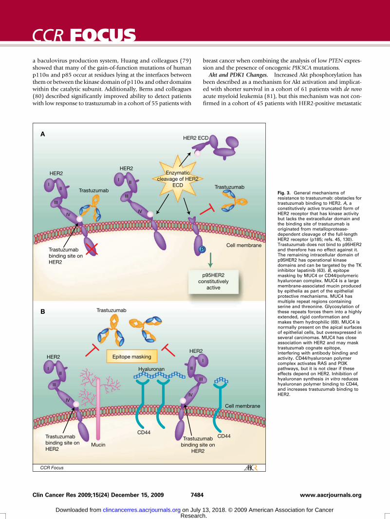
Obstacles for trastuzumab binding to HER2p95HER2. A constitutively active, truncated form of HER2
receptor, p95HER2, has kinase activity but lacks the extracellu-lar domain and the binding site of trastuzumab (Fig. 3A). Theexpression of full-length (p185) and truncated (p95) HER2 wasevaluated on 337 primary breast cancer samples and 81 sam-ples from metastatic lymph nodes using Western blot (62).Truncated p95HER2 was present in 2.1% of the p185HER2-negative tumors, in 19.5% of low, in 31.8% of moderate, andin 60% of tumors that were highly positive for p185HER2 (P <0.001). A higher proportion of node-positive patients thannode-negative patients express p95HER2 (44, 62).Although in vitro data suggested trastuzumab could block the
cleavage of p185HER2 and consequent production of constitu-tively active p95HER2 (27), a retrospective study revealed astrong association between the presence of p95HER2 and clin-ical resistance to trastuzumab treatment (63). In that study, sen-sitivity to trastuzumab was evaluated according to p95HER2expression measured by immunofluorescence in 46 patientswith metastatic breast cancer. Only 1 out of 9 patients(11.1%) expressing p95HER2 responded to trastuzumab,whereas 19 of the 37 patients (51.4%) with tumors expressingp185HER2 achieved clinical response.On the other hand, trastuzumab binds to the extracellular
domain and forms a trastuzumab-extracellular domain complexthat was found to have faster clearance than free trastuzumab inpreclinical models and in a population pharmacokinetics study(61). In a phase II trial evaluating trastuzumab single agent inHER2-overexpressing metastatic breast cancer, extracellulardomain HER2 plasma concentrations greater than 500 ng/mLwere associated with shorter serum half-life and subtherapeutictrough levels of trastuzumab (64). Extracellular domain HER2level has since been evaluated as potential predictor for treat-ment response, but a pooled analysis of four clinical trials inwhich trastuzumab was used in metastatic breast cancer (65) re-vealed that both baseline extracellular domain levels and extra-cellular domain drop following therapy had low predictivevalues for clinical benefit with trastuzumab therapy.Lapatinib, a small molecule that inhibits both HER2 and
EGFR kinases, was tested by Scaltriti and colleagues (63) inp95HER2 preclinical models as a strategy to prevent HER2 sig-naling despite loss of the trastuzumab binding site. Treatmentof p95HER2-positive MCF-7 cells with lapatinib-inhibitedp95HER2 phosphorylation, reduced downstream phosphoryla-tion of Akt and MAPK, and inhibited cell growth. It also inhib-ited growth of MCF-7p95HER2 xenograft tumors. By contrast,trastuzumab had no effect on this model. The addition of lapa-tinib to chemotherapy was then evaluated for treatment of pa-tients with stage IV HER-overexpressing breast cancer that wasprogressive after chemotherapy and trastuzumab. In a phase IIItrial (66), the addition of lapatinib to capecitabine produced animprovement in the median time to progression, when com-pared with the use of chemotherapy alone, resulting in FDA ap-proval of this combination for the treatment of patients withadvanced or metastatic breast cancer whose tumors overexpressHER2 and who have received prior therapy including an anthra-cycline, a taxane, and trastuzumab. Lapatinib has been also test-ed alone or in combination with trastuzumab for patients with
7483www.aacrjournals.org
Researon Julclincancerres.aacrjournals.org Downloaded from
HER2-overexpressing metastatic breast cancer after progressionon trastuzumab, with improved progression-free survival forthe combination arm (67). The combination trastuzumab-lapatinib is currently being tested in the neoadjuvant and adju-vant settings (68).Mucin 4. Epitope masking has also been investigated as a
mechanism of resistance to trastuzumab. Mucin 4 (MUC4) islarge, highly O-glycosylated membrane-associated glycoprotein(69), which may interfere with trastuzumab binding to theHER2 receptor (Fig. 3B). In a preclinical model using the hu-man HER2 positive JIMT-1 cell line, which is primarily resistantto trastuzumab, Nagy and colleagues (70) observed that thepresence of MUC4 was associated with trastuzumab epitopemasking and decreased antibody-binding capacity. In that mod-el, JIMT-1 resistance to trastuzumab could be reversed usingRNA interference to knockdown MUC4.CD44/hyaluronan polymer complex. CD44 is a transmem-
brane receptor for hyaluronan. Binding of polymeric hyaluro-nan activates CD44-mediated signal transduction pathwaysincluding RAS and PI3K in ovary cancer cells (71). CD44 andhyaluronan may also hinder the access of trastuzumab to HER2receptor by masking its cognate epitope, and lead to treatmentresistance (Fig. 3B). Palyi-Krekk and colleagues (72) showedthat treating JIMT-1 cells with an inhibitor of the hyaluronansynthesis significantly reduces the hyaluronan levels of JIMT-1cells both in vivo and in vitro, leading to increased binding oftrastuzumab to HER2 and its subsequent antitumoral effects.Ghatak and colleagues (73) showed that the binding of endog-enous hyaluronan polymer to CD44 contributes to PI3K/Aktactivation, but it was not clear if the described Akt activationwas dependent on HER2. Blockage of hyaluronan polymer-CD44 binding by anti-CD44 antibodies or by hyaluronan oli-gomers caused suppression of the PI3K/Akt pathway withconsequent inhibition of anchorage-independent growth ofcells in culture and of tumors in vivo.
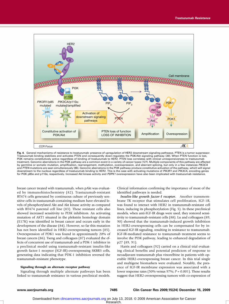
Upregulation of HER2 downstream signaling pathwaysPTEN. The loss of function of PTEN caused by mutation of
PTEN itself, or by transcriptional regulation, has been describedin several tumors and in up to 50% of breast cancers (74).PTEN normally inhibits the activation of PI3K. Therefore PTENloss results in the constitutive upregulation of PI3K/Akt (Fig. 4;ref. 75). Nagata and colleagues (39) showed that decreased le-vels of the PTEN phosphatase resulted in increased PI3K/Aktphosphorylation and signaling, preventing trastuzumab-medi-ated growth arrest of HER2-overexpressing breast cancer cells.In PTEN-deficient models however, PI3K inhibitors rescuedtrastuzumab resistance in vitro and in vivo. Patients withPTEN-deficient HER2-overexpressing metastatic breast cancerhad significantly poorer responses to trastuzumab-based thera-py than those with tumors expressing normal PTEN (39, 74).PI3K. PI3Kmutations have also been implicated in trastuzu-
mab resistance throughPI3K/Akt pathway activation (Fig. 4). ThePIK3R1 gene encodes the PI3K regulatory subunit p85α. PIK3R1mutations affect p85 function, inducing constitutive activation ofthe PI3K/Akt pathway (76, 77). In addition, PIK3CA, the genethat encodes the catalytic subunit of p100α of PI3K, is also fre-quently mutated or overexpressed in human cancer (78). Using
Clin Cancer Res 2009;15(24) December 15, 2009
ch. y 13, 2018. © 2009 American Association for Cancer
CCR FOCUS
a baculovirus production system, Huang and colleagues (79)showed that many of the gain-of-function mutations of humanp110α and p85 occur at residues lying at the interfaces betweenthemor between the kinase domain of p110α andother domainswithin the catalytic subunit. Additionally, Berns and colleagues(80) described significantly improved ability to detect patientswith low response to trastuzumab in a cohort of 55 patients with7484Clin Cancer Res 2009;15(24) December 15, 2009
Researcon July clincancerres.aacrjournals.org Downloaded from
breast cancer when combining the analysis of low PTEN expres-sion and the presence of oncogenic PIK3CA mutations.Akt and PDK1 Changes. Increased Akt phosphorylation has
been described as a mechanism for Akt activation and implicat-ed with shorter survival in a cohort of 61 patients with de novoacute myeloid leukemia (81), but this mechanism was not con-firmed in a cohort of 45 patients with HER2-positive metastatic
h. 13, 2018. © 2009 American A
Fig. 3. General mechanisms ofresistance to trastuzumab: obstacles fortrastuzumab binding to HER2. A, aconstitutively active truncated form ofHER2 receptor that has kinase activitybut lacks the extracellular domain andthe binding site of trastuzumab isoriginated from metalloprotease-dependent cleavage of the full-lengthHER2 receptor (p185; refs. 45, 130).Trastuzumab does not bind to p95HER2and therefore has no effect against it.The remaining intracellular domain ofp95HER2 has operational kinasedomains and can be targeted by the TKinhibitor lapatinib (63). B, epitopemasking by MUC4 or CD44/polymerichyaluronan complex. MUC4 is a largemembrane-associated mucin producedby epithelia as part of the epithelialprotective mechanisms. MUC4 hasmultiple repeat regions containingserine and threonine. Glycosylation ofthese repeats forces them into a highlyextended, rigid conformation andmakes them hydrophilic (69). MUC4 isnormally present on the apical surfacesof epithelial cells, but overexpressed inseveral carcinomas. MUC4 has closeassociation with HER2 and may masktrastuzumab cognate epitope,interfering with antibody binding andactivity. CD44/hyaluronan polymercomplex activates RAS and PI3Kpathways, but it is not clear if theseeffects depend on HER2. Inhibition ofhyaluronan synthesis in vitro reduceshyaluronan polymer binding to CD44,and increases trastuzumab binding toHER2.
www.aacrjournals.org
ssociation for Cancer
Trastuzumab Resistance
breast cancer treated with trastuzumab, when pAkt was evaluat-ed by immunohistochemistry (82). Trastuzumab-resistantBT474 cells generated by continuous culture of previously sen-sitive cells in trastuzumab-containing medium have elevated le-vels of phosphorylated Akt and Akt kinase activity as comparedwith BT474 parental cell line (83). These resistant cells alsoshowed increased sensitivity to PI3K inhibitors. An activatingmutation of AKT1 situated in the plekstrin homology domain(E17K) was identified in breast cancer and occurs early in thedevelopment of the disease (84). However, so far this mutationhas not been identified in HER2-overexpressing tumors (85).Overexpression of PDK1 was found in approximately 20% ofbreast cancers (86). Tseng and colleagues (87) evaluated the ef-fects of concurrent use of trastuzumab and a PDK-1 inhibitor ina preclinical model using trastuzumab-resistant insulin-likegrowth factor-1 receptor (IGF-IR)-overexpressing SKBR3 cells,generating data indicating that PDK-1 inhibition reversed thetrastuzumab-resistant phenotype.
Signaling through an alternate receptor pathway
Signaling through multiple alternate pathways has beenlinked to trastuzumab resistance in various preclinical models.
7485www.aacrjournals.org
Researon Julclincancerres.aacrjournals.org Downloaded from
Clinical information confirming the importance of most of theidentified pathways is needed.Insulin-like growth factor-I receptor. Another transmem-
brane TK receptor that stimulates cell proliferation, IGF-1R,was found to interact with HER2 in trastuzumab-resistant celllines, inducing its phosphorylation (Fig. 5). In these preclinicalmodels, when anti-IGF-IR drugs were used, they restored sensi-tivity to trastuzumab-resistant cells (88). Lu and colleagues (89,90) showed that the trastuzumab-induced growth inhibitionin HER2-overexpressing cells can be compensated for by in-creased IGF-IR signaling, resulting in resistance to trastuzumab.IGF-IR-mediated resistance to trastuzumab treatment seems toinvolve the PI3K pathway, leading to enhanced degradation ofp27 (89, 91).Harris and colleagues (92) carried on a clinical trial evaluat-
ing clinical benefits and potential predictors of response toneoadjuvant trastuzumab plus vinorelbine in patients with op-erable HER2-overexpressing breast cancer. In this trial singleand multigene biomarkers were evaluated. Notably, the pres-ence of IGF-IR membrane expression was associated with alower response rates (50% versus 97%; P = 0.001). These resultssuggest that HER2-overexpressing tumors with co-expression of
Fig. 4. General mechanisms of resistance to trastuzumab: presence of upregulation of HER2 downstream signaling pathways. PTEN is a tumor suppressor.Trastuzumab binding stabilizes and activates PTEN and consequently down-regulates the PI3K/Akt signaling pathway (39). When PTEN function is lost,PI3K remains constitutively active regardless of binding of trastuzumab to HER2. PTEN loss correlates with clinical unresponsiveness to trastuzumabtreatment. Genomic aberrations in the PI3K pathway are a common event in a variety of cancer types (127). Multiple components of this pathway are affectedby germline or somatic mutation, amplification, rearrangement, methylation, overexpression, and aberrant splicing, but only in a few instances PIK3CAand PTENmutations are seen simultaneously (86). Genomic aberrations in the PI3K pathway produce constitutive activation of the pathway, which will signaldownstream to the nucleus regardless of trastuzumab binding to HER2. This is the case with activating mutations of PIK3R1 and PIK3CA, encoding genesfor PI3K p85α and p110α, respectively. Increased Akt kinase activity and PDPK1 overexpression have also been implicated with trastuzumab resistance.
Clin Cancer Res 2009;15(24) December 15, 2009
ch. y 13, 2018. © 2009 American Association for Cancer

CCR FOCUS
IGF-IR are more likely to be resistant to this trastuzumab-containing regimen.p27. As a Cdk inhibitor involved in regulating cell prolifer-ation, p27 is a distal downstream regulator of multiple converg-ing growth factor receptor pathways including EGFR, HER2,and IGF-IR. Nahta and colleagues (93) showed that HER2-overexpressing trastuzumab-resistant cells have low p27 levels,low p27/Cdk2 complexes, and, thus, increased Cdk2 activityand proliferation rate. When p27 expression was increased bytransfection or pharmacological induction with a proteasomeinhibitor, trastuzumab sensitivity was restored. Consistent withdata that implicates Akt in p27 regulation, Yakes and colleagues(26) showed that Akt inhibition is at least partially responsiblefor the changes in cell cycle- and apoptosis-regulatory mole-cules after HER2 blockade with trastuzumab.EGFR and HER3. EGFR homodimers and EGFR/HER3 het-
erodimers, lacking HER2, could potentially bypass trastuzumabblockade. However, these dimers are less likely to be the drivingforce in HER2-overexpressing breast cancer, because HER2 isthe preferred heterodimer partner of this family of receptors(94). In a recent study examining trastuzumab-resistant breastcancer cells to gain insights into trastuzumab resistance me-chanisms, both EGFR and HER3 expression were increased afterlong-term trastuzumab exposure in culture (95). Interestingly,chronic trastuzumab exposure of trastuzumab-resistant celllines induced sensitivity to anti-EGFR agents. Unfortunately, aphase I-II clinical trial designed to evaluate safety and efficacy ofthe EGFR TK inhibitor gefitinib with weekly trastuzumab sug-gested that the combination was unlikely to result in clinical
7486Clin Cancer Res 2009;15(24) December 15, 2009
Researcon July clincancerres.aacrjournals.org Downloaded from
benefit when compared with trastuzumab alone (96). On theother hand, as previously mentioned, lapatinib, an EGFR/HER2 TK inhibitor, has clinical activity in the treatment of pa-tients who progressed on trastuzumab (66), but this effect mayresult from its ability to inhibit HER2 TK.HER3 is the favored receptor for dimerization with HER2
(94), and growing evidence supports HER3 as being a requiredpartner for HER2 in HER2-overexpressing breast cancer (40,97). Investigating the oligomerization properties of the ErbB re-ceptors, Wehrman and colleagues (42) confirmed that interac-tions between EGFR/HER2 and HER2/HER3 are detected in thepresence of ligand. In addition, they found that trastuzumab isineffective in blocking HER2/HER3 dimerization.Pertuzumab, an anti-HER2 humanized mAb that binds to an
epitope located in domain II of HER2, sterically blocks a bind-ing pocket necessary for receptor dimerization and signaling. Ittherefore prevents HER2 dimerization, including HER2/HER3heterodimerization. Pertuzumab is active against HER2-overex-pressing cell lines and xenografts, and preclinical data suggestpotential synergism with trastuzumab (98). Preliminary resultsfrom a phase II trial of pertuzumab in combination with tras-tuzumab in patients with HER2-overexpressing metastaticbreast cancer with disease progression on trastuzumab havesuggested good tolerance and clinical activity of the combina-tion (99). Clinical trials testing pertuzumab in combinationwith trastuzumab in different settings, as well as pertuzumabwith chemotherapy are ongoing (68).c-Met. Frequently co-expressed with HER2 in cell lines, the
c-Met receptor may contribute to trastuzumab resistance
Fig. 5. General mechanisms of resistance to trastuzumab: presence of signaling through an alternate receptor and/or pathway. Signaling may continueregardless of trastuzumab binding to HER2 when other receptors remain active on the tumor cell. The activity of certain receptors may also increase as aresult of trastuzumab blockage of HER2, as a cell survival mechanism (see text for details). Trastuzumab-induced growth inhibition in HER2-overexpressingcells can be compensated for by increased IGF-IR signaling, resulting in resistance to trastuzumab. In preclinical models in which HER2-overexpressingtumor cells are cultured in the presence of ligand, resembling what is likely to happen in vivo, trastuzumab does not interfere with HER2/HER3heterodimerization and therefore does not block signaling from these heterodimers (42). c-Met is frequently co-expressed with HER2 in cell lines andcontributes to trastuzumab resistance through sustained Akt activation.
www.aacrjournals.org
h. 13, 2018. © 2009 American Association for Cancer
Trastuzumab Resistance
through sustained Akt activation (Fig. 5). HER2-overexpressingbreast cancer cells respond to trastuzumab with a rapid upregu-lation of c-Met receptor expression, and c-Met activation pro-tects cells against trastuzumab (100). Loss of c-Met functionproduced through RNA interference improves the response ofthese cell lines to trastuzumab.CXCR4, α6β1, and α6β4 integrins. Acquired resistance to
trastuzumab was associated with CXCR4 upregulation andnuclear redistribution. Inhibition of CXCR4 reversed acquiredtrastuzumab resistance in vitro (101). In vitro experiments sug-gest both α6β1 and α6β4 integrins may be involved inde novo and/or acquired resistance to targeted therapy againstHER2 (102).
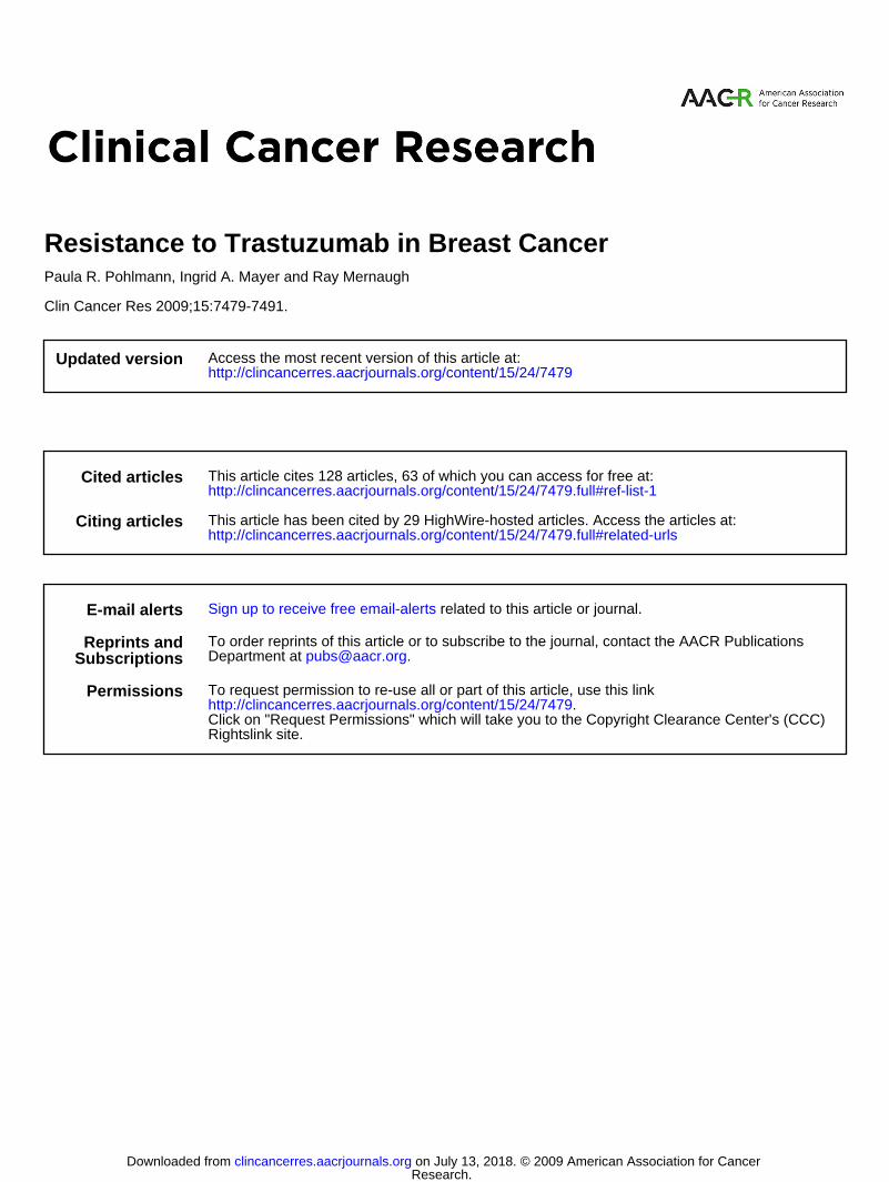
Failure to trigger immune-mediated mechanisms to destroytumor cellsFc, Fc receptor, and ADCC. A genomic polymorphism pro-
ducing the phenotype expression of valine (V) or phenylala-nine (F) at amino acid 158 on the FcγRIIIa significantlyinfluences the affinity of IgG1 to the Fcγ receptor (Fig. 6;ref. 103). Immune effector cells carrying the FcγRIIIa V/V al-leles mediate ADCC of trastuzumab and anti-HER2/neu IgG1variants better than cells bearing the F allele (60). In theclinic, the FcγRIII 158V/F polymorphism interfere with theability to generate ADCC responses in vitro during trastuzu-mab treatment and significantly impair clinical response
7487www.aacrjournals.org
Researon Julclincancerres.aacrjournals.org Downloaded from
rates and progression-free survival of patients treated withtrastuzumab in the metastatic setting (30). In an attemptto improve FcγIIIR binding, Lazar and colleagues (104) useda combination of computational structure-based protein de-sign methods coupled with high-throughput protein screen-ing to optimize the FcγR binding capacity of therapeuticantibodies. The described engineered Fc variants with im-proved FcγR affinity and specificity had enhanced cytotoxi-city in vitro, with improved affinity and ADCC for boththe V158 and F158 forms of FcγRIIIa. The authors concludethat this progress has promising implications for therapeuticmAb design, with potential future expanding of the popula-tion of patients responsive to anticancer antibody-basedtreatment (104).Recent additional evidence for the importance of immune-
mediated mechanisms in antibody-based therapy of HER2-overexpressing tumors came from a lapatinib preclinical studyby Scaltriti and colleagues (105). The authors treated SKBR3and MCF7 cells with lapatinib alone or in combination withtrastuzumab, producing inhibition of HER2 phosphorylationand preventing receptor ubiquitination, which resulted in amarked accumulation of inactive HER2 receptors at the cellsurface. Trastuzumab alone caused degradation of the receptor.Lapatinib-induced accumulation of HER2 in tumors was alsoobserved in BT474 xenografts. In mice trastuzumab-lapatinibcombination produced complete tumor remissions in all cases
Fig. 6. General mechanisms of resistance to trastuzumab: failure to trigger immune-mediated mechanisms to destroy tumor cells. ADCC is a process inwhich the Fab region of an antibody binds to its cognate antigen present on a target cell (for instance cancer cell), whereas its Fc region engages with the Fcreceptor present on an effector cell from immune system. This process triggers degranulation of cytotoxic granules from effector cell toward the target celland culminates with target cell apoptosis (131). In humans, the Fc receptor family comprises FcγRI (CD64); FcγRII (CD32), with three isoforms FcγRIIa, FcγRIIb(inhibitory), and FcγRIIc; and FcγRIII (CD16), including two isoforms FcγRIIIa and FcγRIIIb (132). There is a correlation between the clinical efficacy oftherapeutic antibodies in humans and their allotype of high-affinity (V158) or low-affinity (F158) polymorphic forms of FcγRIIIa. Epitope masking previouslydiscussed in the obstacles for trastuzumab binding to HER2 section would also play a role by preventing antibody-based cell destruction.
Clin Cancer Res 2009;15(24) December 15, 2009
ch. y 13, 2018. © 2009 American Association for Cancer

CCR FOCUS
after 10 days of treatment. Accumulation of HER2 at the cellsurface by lapatinib was shown to enhance immune-mediatedtrastuzumab-dependent cytotoxicity.Further evidence on the importance of ADCC was providedby Barok and colleagues (29), who showed that trastuzumabcan trigger ADCC and destroy trastuzumab-resistant HER2 pos-itive cell lines and xenografts. Taken together, these observa-tions point to ADCC as a major player in trastuzumabantitumoral effect, at least when the antibody binds to HER2.Thus far the ADCC-triggered pathways on target cells have notreceived much attention, but it would be very interesting toknow how ADCC affects cells that are prone to enhancePI3K/Akt and RAS/Raf/MAPK pathways to an extreme, in orderto survive.Trastuzumab clearance mechanisms are not well described.
Multiple proteases involved with infection, inflammation, andtumor environments have been shown to be able to cleave IgGmolecules on site, generating monovalent or bivalent antibodyfragments, Fab′ or F(ab′)2, which lack Fc-mediated effectorfunction (106). These antibody fragments can still bind to theircognate antigen, but lack binding sites for complement and forFc receptors, and can no longer trigger Fc-mediated functionssuch as ADCC. Although cleavage of HER2 is well documented,it is uncertain if trastuzumab undergoes cleavage on tumor siteas well.
Discussion
Trastuzumab is an essential and effective targeted anticanceragent. It has been moved into first-line treatment of HER2-overexpressing breast cancer both in the adjuvant and meta-static settings. As an antibody, trastuzumab has several properties,
7488Clin Cancer Res 2009;15(24) December 15, 2009
Researcon July clincancerres.aacrjournals.org Downloaded from
either related to its Fab and/or Fc function. Unfortunately, a signif-icant number of patients develop progressive disease after initialtreatment with trastuzumab-containing regimens, requiring ad-ditional therapy.At the diagnosis of progressive disease after trastuzumab
treatment, reasonable options for patients are: (1) participationon clinical trials containing various combinations of trastuzu-mab and/or lapatinib with PI3K/Akt pathway inhibitors, otherTK inhibitors, IGF-1 inhibitors, cell cycle regulators, mammali-an target of rapamycin (mTOR) inhibitors, anti-angiogenictherapy or chemotherapy; (2) participation on clinical trialsevaluating vaccines, other anti-HER2 antibodies such as pertu-zumab, or anti-HER2 immunotoxins; (3) continuing anti-HER2therapy including trastuzumab beyond progression with a dif-ferent chemotherapy regimen (107); or (4) switching to a lapa-tinib containing regimen (66, 68). The observation of clinicalbenefit of the addition of trastuzumab to second-line chemo-therapy suggests HER2 blockade is still important for some pa-tients. This can be currently achieved in clinical practice withtrastuzumab or lapatinib.Based on emerging knowledge of the HER2-signaling path-
ways and of signaling through alternate pathways, several drugsdirected to HER2-overexpressing breast cancer are in differentphases of clinical development, whereas others are still strug-gling to leave the bench (Table 1). In the case of anti-HER2therapy, except for the presence of the HER2 itself, there areno validated biomarkers reliably predicting benefit from trastu-zumab that can be used for either clinical trial development orindividual therapeutic decisions, because it is unclear which ofthe mechanisms of action or resistance to trastuzumab are moreimportant in each patient subset. In addition, resistant cellspresent deletions, insertions, and missense point mutationsthat influence the function of receptors, adaptor proteins, and
Table 1. New anti-HER2 agents
General strategy
Identificationh. 13, 201
Target
www.aacr
8. © 2009 American Association for Canc
Reference
Inhibiting downstream signalingpathways of HER2
Perifosine
PI3-kinase/Akt 108 Everolimus mTor 109 GDC-0941 PI3K 43HER family TK inhibitors
Lapatinib Dual TK inhibitor of EGFR andHER2 approved for clinical use110, 111
Neratinib (HKI-272)
TK inhibitor: HER1/HER2 110 BIBW2992 Irreversible dualHER1/HER2 inhibitor
112Inhibiting the activity of othergrowth factor receptors
Pertuzumab
HER2/HER3 41 rhIGFBP-3 IGF-1R 113 IMC-A12 IGF-1R 114 BMS-554417 IGF-1R 115 SU11274 MET 100Decreasing the expression of proteinprocessing of HER2 and othersignaling components
Tanespimycin (17-AAG)
HSP90 inhibitor 116 LAQ824 Histone deacetylase (HDAC) inhibitors 117 Bortezomib Proteasome inhibitor 118Increasing toxicity of trastuzumab toHER2 positive cells
Trastuzumab-DM1immunotoxin
HER2
119Increasing immunity againstHER2 positive cells
HER2 vaccines
Anti-HER2 vaccine 120 Ertumaxomab Trifunctional bispecific antibodytargeting HER2 and CD3
121Genetically engineeredFc of mAb
Enhanced FcγRIII receptorbinding on effector cells
104
journals.org
er

Trastuzumab Resistance
second messengers, allowing for tumor cell survival in the pres-ence of trastuzumab. Tumor cells seem to be highly wired andactivate or up-regulate alternative survival pathways upon treat-ment with a single form of targeted therapy. For this reason theinhibition of one deregulated protein is unlikely to be enoughto solve the trastuzumab resistance problem. To add to thiscomplex scenario, cellular pathways from HER2 activation areredundant, and activation of escape mechanisms by resistantcells may also involve constantly switching host-tumor environ-mental characteristics.The challenge is to prioritize strategies for faster development.
Several compounds have been tested in preclinical models, butonly a few are currently moving on into the clinical developmentplatforms. It is critical that this field begin to move from thein vitro descriptions of mechanism to clinical validation. Thestudy of the incidence of abnormalities on the analyzed path-ways could help with selecting priorities for development.Brugge and colleagues (86) produced a list of aberrations inthe PI3K pathway or in its interacting pathways that have beendescribed in cancer, with estimations of number of cases in-volved with each aberration per year in the United States.Another strategy would include advancing clinical assay
development, permitting accurate diagnosis of the implicatedresistance mechanism in each subset of patients. The develop-ment of such assays and of the corresponding targeted therapyshould preferably occur simultaneously. Once the main trastu-zumab resistance mechanism is identified for a subgroup ofpatients, this information can be used for the design of subse-quent clinical trials. On the other hand, the clinical trials wouldaid on clinical assay study and validation.
7489www.aacrjournals.org
Researon Julclincancerres.aacrjournals.org Downloaded from
In this scenario, for patients who present with trastuzumabresistance owing to interference with trastuzumab binding, astrategy to overcome resistance may involve the use of treat-ments that do not depend on molecular binding to the HER2extracellular domain, including the use of TK inhibitors such aslapatinib (63), or of inhibitors of the downstream signalingpathways of HER2. If p95HER2 is thought to be the drivingforce for resistance to trastuzumab in a subgroup of patients,pertuzumab, trastuzumab-DM1, trastuzumab-mediatedADCC, and HER2-extracellular domain-based vaccines maynot be an option. Other patients may present with tumorscontaining PTEN loss, PIK3R1/p85α, PIK3CA/p110α muta-tions or amplification, as well as activation of alternate path-ways. If combined, these alterations may complicate thetargeting of intracellular molecules, as they would requirethe simultaneous use of different inhibitors. In this case, rea-sonable options would be to increase tumor cell destructionthrough extracellular mechanisms by increasing or enablingADCC, or by using immunotoxins such as the antibody-drugconjugate trastuzumab-DM1, or the bispecific anti-HER2/CD3antibody ertumaxomab.Thus, understanding trastuzumab resistance mechanisms is
critical for the educated development of strategies to overcomeit, as well as for the development of tools that would allow de-finitive and efficient patient selection for therapies that couldmitigate that resistance.
Disclosure of Potential Conflicts of Interest
The authors have no potential conflicts of interest to disclose.
References
1. Perou CM, Sorlie T, Eisen MB, et al. Molecularportraits of human breast tumours. Nature 2000;406:747–52.2. Hu Z, Fan C, Oh DS, et al. The molecular por-traits of breast tumors are conserved across mi-croarray platforms. BMC Genomics 2006;7:96.
3. Sorlie T, Perou CM, Tibshirani R, et al. Gene ex-pressionpatterns of breast carcinomas distinguishtumor subclasses with clinical implications.Proc Natl Acad Sci U S A 2001;98:10869–74.
4. Sorlie T, Tibshirani R, Parker J, et al. Repeatedobservation of breast tumor subtypes in inde-pendent gene expression data sets. Proc NatlAcad Sci U S A 2003;100:8418–23.
5. Osborne CK, Yochmowitz MG, Knight WA 3rd,McGuire WL. The value of estrogen and pro-gesterone receptors in the treatment of breastcancer. Cancer 1980;46:2884–8.
6. SlamonDJ, Leyland-Jones B, Shak S, et al. Use ofchemotherapyplusamonoclonal antibodyagainstHER2 for metastatic breast cancer that over-expresses HER2. N Engl J Med 2001;344:783–92.
7. Schechter AL, Stern DF, Vaidyanathan L, et al. Theneu oncogene: an erb-B-related gene encoding a185,000-Mr tumourantigen.Nature1984;312:513–6.
8. Olayioye MA, Neve RM, Lane HA, Hynes NE.The ErbB signaling network: receptor hetero-dimerization in development and cancer. EMBOJ 2000;19:3159–67.
9. Shih C, Padhy LC, Murray M, Weinberg RA.Transforming genes of carcinomas and neuro-blastomas introduced into mouse fibroblasts.Nature 1981;290:261–4.
10. Slamon DJ, Clark GM, Wong SG, Levin WJ,
Ullrich A, McGuire WL. Human breast cancer:correlation of relapse and survival with amplifi-cation of the HER-2/neu oncogene. Science 1987;235:177–82.
11. Moasser MM. The oncogene HER2: itssignaling and transforming functions and its rolein human cancer pathogenesis. Oncogene 2007;26:6469–87.
12. Carter P, Presta L, Gorman CM, et al. Human-ization of an anti-p185HER2 antibody for humancancer therapy. Proc Natl Acad Sci U S A 1992;89:4285–9.
13. Fendly BM, Winget M, Hudziak RM, Lipari MT,Napier MA, Ullrich A. Characterization of murinemonoclonal antibodies reactive to either the humanepidermal growth factor receptor orHER2/neugeneproduct. Cancer Res 1990;50:1550–8.
14. Cobleigh MA, Vogel CL, Tripathy D, et al. Mul-tinational study of the efficacy and safety ofhumanized Anti-HER2 monoclonal antibody inwomen who have HER2-overexpressing meta-static breast cancer that has progressed afterchemotherapy for metastatic disease. J ClinOncol 1999;17:2639–48.
15. Piccart-Gebhart MJ, Procter M, Leyland-JonesB, et al. Trastuzumab after adjuvant chemother-apy in HER2-positive breast cancer. N EnglJ Med 2005;353:1659–72.
16. Romond EH, Perez EA, Bryant J, et al. Trastu-zumab plus adjuvant chemotherapy for operableHER2-positive breast cancer. N Engl J Med 2005;353:1673–84.
17. Vogel CL, CobleighMA, TripathyD, et al. Efficacyand safety of trastuzumab as a single agent in first-
Clin Canc
ch. y 13, 2018. © 2009 A
line treatment of her2-overexpressing meta-static breast cancer. J Clin Oncol 2002;20:719–26.
18. Hammerman PS, Jänne PA, Johnson BE.Resistance to epidermal growth factor receptortyrosine kinase inhibitors in non-small cell lungcancer. Clin Cancer Res 2009;15:7502–9.
19. Banck MS, Grothey A. Biomarkers of resistanceto epidermal growth factor receptor monoclonalantibodies in patients with metastatic colorectalcancer. Clin Cancer Res 2009;15:7492–501.
20. Gramza AW, Corless CL, Heinrich MC. Resis-tance to tyrosine kinase inhibitors in gastrointes-tinal stromal tumors. Clin Cancer Res 2009;15:7510–8.
21. Milojkovic D, Apperley J. Mechanisms ofresistance to imatinib and second-generationtyrosine inhibitors in chronic myeloid leukemia.Clin Cancer Res 2009;15:7519–27.
22. Ellis LM, Hicklin DJ. Resistance to targetedtherapies: refining anticancer therapy in the eraof molecular oncology. Clin Cancer Res 2009;15:7471–8.
23. Cho HS, Mason K, Ramyar KX, et al. Structureof the extracellular region of HER2 alone and incomplex with the Herceptin Fab. Nature 2003;421:756–60.
24. Akilesh S, Christianson GJ, Roopenian DC,Shaw AS. Neonata l FcR express ion inbone marrow-derived cells functions to protectserum IgG from catabolism. J Immunol 2007;179:4580–8.
25. Roopenian DC, Akilesh S. FcRn: the neonatalFc receptor comes of age. Nat Rev Immunol2007;7:715–25.
er Res 2009;15(24) December 15, 2009
merican Association for Cancer

CCR FOCUS
26. Yakes FM, Chinratanalab W, Ritter CA, King W,Seelig S, Arteaga CL. Herceptin-induced inhibitionof phosphatidylinositol-3 kinase and Akt is requiredfor antibody-mediated effects onp27, cyclinD1, andantitumor action. Cancer Res 2002;62:4132–41.27. Molina MA, Codony-Servat J, Albanell J, RojoF, Arribas J, Baselga J. Trastuzumab (herceptin),a humanized anti-Her2 receptor monoclonal an-tibody, inhibits basal and activated Her2 ectodo-main cleavage in breast cancer cells. Cancer Res2001;61:4744–9.
28. Baselga J, Albanell J, Molina MA, Arribas J.Mechanism of action of trastuzumab and scien-tific update. Semin Oncol 2001;28:4–11.
29. BarokM, Isola J, Palyi-Krekk Z, et al. Trastuzumabcauses antibody-dependent cellular cytotoxicity-mediated growth inhibition of submacroscopicJIMT-1 breast cancer xenografts despite intrinsicdrug resistance. Mol Cancer Ther 2007;6:2065–72.
30. Musolino A, Naldi N, Bortesi B, et al. Immuno-globulin G fragment C receptor polymorphismsand clinical efficacy of trastuzumab-based therapyin patients with HER-2/neu-positive metastaticbreast cancer. J Clin Oncol 2008;26:1789–96.
31. RoopenianDC, ChristiansonGJ, Sproule TJ, et al.TheMHC class I-like IgG receptor controls perinatalIgG transport, IgG homeostasis, and fate of IgG-Fc-coupled drugs. J Immunol 2003;170:3528–33.
32. Ghetie V, Ward ES. Multiple roles for themajor histocompatibility complex class I- relatedreceptor FcRn. Annu Rev Immunol 2000;18:739–66.
33. Carter P, McDonagh CF. Designer antibody-based therapeutics for oncology. AACR Educa-tion Book 2005:147–54.
34. Storniolo AM, Pegram MD, Overmoyer B,et al. Phase I dose escalation and pharmacokine-tic study of lapatinib in combination with trastu-zumab in patients with advanced ErbB2-positivebreast cancer. J Clin Oncol 2008;26:3317–23.
35. Arteaga CL, Chinratanalab W, Carter MB. Inhi-bitors of HER2/neu (erbB-2) signal transduction.Semin Oncol 2001;28:30–5.
36. Baselga J, Albanell J. Mechanism of action ofanti-HER2 monoclonal antibodies. Ann Oncol2001;12:S35–41.
37. Lane HA, Motoyama AB, Beuvink I, Hynes NE.Modulation of p27/Cdk2 complex formationthrough 4D5-mediated inhibition of HER2 recep-tor signaling. Ann Oncol 2001;12:S21–2.
38. Sliwkowski MX, Lofgren JA, Lewis GD,Hotaling TE, Fendly BM, Fox JA. Nonclinicalstudies addressing the mechanism of action oftrastuzumab (Herceptin). Semin Oncol 1999;26:60–70.
39. NagataY, Lan K-H, ZhouX, et al. PTENactivationcontributes to tumor inhibition by trastuzumab,and loss of PTEN predicts trastuzumab resis-tance in patients. Cancer Cell 2004;6:117–27.
40. Lee-Hoeflich ST, Crocker L, Yao E, et al. A cen-tral role for HER3 in HER2-amplified breast can-cer: implications for targeted therapy. CancerRes 2008;68:5878–87.
41. Agus DB, Akita RW, Fox WD, et al. Targetingligand-activated ErbB2 signaling inhibits breastand prostate tumor growth. Cancer Cell 2002;2:127–37.
42. Wehrman TS, Raab WJ, Casipit CL, DoyonnasR, Pomerantz JH, Blau HM. A system for quanti-fying dynamic protein interactions defines a rolefor Herceptin in modulating ErbB2 interactions.Proc Natl Acad Sci U S A 2006;103:19063–8.
43. Junttila TT, Akita RW, Parsons K, et al. Ligand-Independent HER2/HER3/PI3K Complex IsDisrupted by Trastuzumab and Is Effectively In-hibited by the PI3K Inhibitor GDC-0941. CancerCell 2009;15:429–40.
44. Christianson TA, Doherty JK, Lin YJ, et al.NH2-terminally truncated HER-2/neu protein:
Clin Cancer Res 2009;15(24) December 1
clincancerres.aaDownloaded from
relationship with shedding of the extracellulardomain and with prognostic factors in breastcancer. Cancer Res 1998;58:5123–9.
45. Codony-Servat J, Albanell J, Lopez-TalaveraJC, Arribas J, Baselga J. Cleavage of the HER2ectodomain is a pervanadate-activable processthat is inhibited by the tissue inhibitor of metal-loproteases-1 in breast cancer cells. Cancer Res1999;59:1196–201.
46. Izumi Y, Xu L, di Tomaso E, Fukumura D, JainRK. Tumour biology: herceptin acts as an anti-angiogenic cocktail. Nature 2002;416:279–80.
47. Petit AM, Rak J, Hung MC, et al. Neutralizingantibodies against epidermal growth factor andErbB-2/neu receptor tyrosine kinases down-reg-ulate vascular endothelial growth factor productionby tumor cells in vitro and in vivo: angiogenic impli-cations for signal transduction therapy of solidtumors. Am J Pathol 1997;151:1523–30.
48. Hommelgaard AM, Lerdrup M, van Deurs B.Association with membrane protrusionsmakes ErbB2 an internalization-resistant recep-tor. Mol Biol Cell 2004;15:1557–67.
49. Ben-Kasus T, Schechter B, Lavi S, Yarden Y,Sela M. Persistent elimination of ErbB-2/HER2-overexpressing tumors using combinations ofmonoclonal antibodies: Relevance of receptorendocytosis. Proc Natl Acad Sci U S A 2009;106:3294–9.
50. Friedman LM, Rinon A, Schechter B, et al.Synergistic down-regulation of receptor tyrosinekinases by combinations of mAbs: Implicationsfor cancer immunotherapy. Proc Natl Acad SciU S A 2005;102:1915–20.
51. Clynes RA, Towers TL, Presta LG, RavetchJV. Inhibitory Fc receptors modulate in vivocytoxicity against tumor targets. Nat Med2000;6:443–6.
52. Spiridon CI, Guinn S, Vitetta ES. A comparisonof the in vitro and in vivo activities of IgG and F(ab')2 fragments of a mixture of three monoclo-nal anti-Her-2 antibodies. Clin Cancer Res 2004;10:3542–51.
53. Gennari R, Menard S, Fagnoni F, et al. Pilotstudy of the mechanism of action of preopera-tive trastuzumab in patients with primary opera-ble breast tumors overexpressing HER2. ClinCancer Res 2004;10:5650–5.
54. Mineo JF, Bordron A, Quintin-Roue I, et al. Re-combinant humanised anti-HER2/neu antibody(Herceptin) induces cellular death of glioblasto-mas. Br J Cancer 2004;91:1195–9.
55. Spiegelberg HL, Grey HM. Catabolism of hu-man {γ}g immunoglobulins of different heavychain subclasses: ii. catabolism of {γ}g myelomaproteins in heterologous species. J Immunol1968;101:711–6.
56. Fahey JL, Robinson AG. Factors controllingserum γ-globulin concentration. J Exp Med1963;118:845–68.
57. Brambell FW. The transmission of immunityfrom mother to young and the catabolism ofimmunoglobulins. Lancet 1966;2:1087–93.
58. Presta LG, Shields RL, Namenuk AK, Hong K,Meng YG. Engineering therapeutic antibodiesfor improved function. Biochem Soc Trans2002;30:487–90.
59. Petkova SB, Akilesh S, Sproule TJ, et al.Enhanced half-life of genetically engineeredhuman IgG1 antibodies in a humanized FcRnmouse model: potential application in humorallymediated autoimmune disease. Int Immunol2006;18:1759–69.
60. ShieldsRL,NamenukAK,HongK, et al. High res-olutionmappingof thebindingsiteonhuman IgG1for Fc γRI, Fc γRII, FcγRIII, FcRnanddesignof IgG1variants with improved binding to the Fc γR. J BiolChem 2001;276:6591–604.
74905, 2009
Research. on July 13, 2018. © 2009 Amcrjournals.org
61. Bruno R, Washington CB, Lu J-F, Lieberman G,Banken L, Klein P. Population pharmacokineticsof trastuzumab in patients With HER2+ meta-static breast cancer. Cancer Chemother Pharma-col 2005;56:361–9.
62. Molina MA, Saez R, Ramsey EE, et al. NH(2)-terminal truncated HER-2 protein but not full-lengthreceptor is associated with nodal metastasis in hu-man breast cancer. Clin Cancer Res 2002;8:347–53.
63. Scaltriti M, Rojo F, Ocana A, et al. Expressionof p95HER2, a Truncated Form of the HER2 re-ceptor, and response to anti-HER2 therapies inbreast cancer. J Natl Cancer Inst 2007;99:628–38.
64. Baselga J, Tripathy D, Mendelsohn J, et al.Phase II studyofweekly intravenous recombinanthumanized anti- p185HER2monoclonal antibodyin patients with HER2/neu-overexpressing meta-static breast cancer. J Clin Oncol 1996;14:737–44.
65. Lennon S, Barton C, Banken L, et al. Utility ofserum HER2 extracellular domain assessment inclinical decision making: pooled analysis of fourtrials of trastuzumab in metastatic breast cancer.J Clin Oncol 2009;27:1685–93.
66. Geyer CE, Forster J, Lindquist D, et al. Lapatinibplus capecitabine for HER2-positive advancedbreast cancer. N Engl JMed 2006;355:2733–43.
67. O'Shaughnessy J, Blackwell KL, Burstein H,et al. A randomized study of lapatinib alone orin combination with trastuzumab in heavily pre-treated HER2+ metastatic breast cancer progres-sing on trastuzumab therapy [meeting abstracts].J Clin Oncol 2008;26:1015.
68. Baselga J, Swain SM. Novel anticancer tar-gets: revisiting ERBB2 and discovering ERBB3.Nat Rev Cancer 2009;9:463–75.
69. Carraway KL, Price-Schiavi SA, Komatsu M,JepsonS, PerezA,CarrawayCA.Muc4/sialomucincomplex in themammarygland andbreast cancer.J Mammary Gland Biol Neoplasia 2001;6:323–37.
70. Nagy P, Friedlander E, TannerM, et al. Decreasedaccessibility and lack of activation of ErbB2 inJIMT-1, a herceptin-resistant, MUC4-expressingbreast cancer cell line. Cancer Res 2005;65:473–82.
71. Bourguignon LY, Zhu H, Zhou B, Diedrich F,Singleton PA, Hung MC. Hyaluronan promotesCD44v3–2 interaction with Grb2–185(HER2) andinduces Rac1 and Ras signaling during ovariantumor cell migration and growth. J Biol Chem2001;276:48679–92.
72. Palyi-Krekk Z, Barok M, Isola J, Tammi M,Szollosi J, Nagy P. Hyaluronan-induced maskingof ErbB2 and CD44-enhanced trastuzumab inter-nalisation in trastuzumab resistant breast can-cer. Eur J Cancer 2007;43:2423–33.
73. Ghatak S, Misra S, Toole BP. Hyaluronan oli-gosaccharides inhibit anchorage-independentgrowth of tumor cells by suppressing the phos-phoinositide 3-kinase/Akt cell survival pathway.J Biol Chem 2002;277:38013–20.
74. Pandolfi PP. Breast cancer-loss of PTENpredicts resistance to treatment. N Engl J Med2004;351:2337–8.
75. Simpson L, Parsons R. PTEN: life as atumor suppressor. Exp Cell Res 2001;264:29–41.
76. Jimenez C, Jones DR, Rodriguez-Viciana P,et al. Identification and characterization ofa new oncogene derived from the regulatorysubunit of phosphoinositide 3-kinase. EMBO J1998;17:743–53.
77. Philp AJ, Campbell IG, Leet C, et al. The phos-phatidylinositol 3′-kinase p85{α} gene is an onco-gene in human ovarian and colon tumors.Cancer Res 2001;61:7426–9.
78. Saal LH, Holm K, Maurer M, et al. PIK3CA mu-tations correlate with hormone receptors, nodemetastasis, and ERBB2, and are mutually exclu-sive with PTEN loss in human breast carcinoma.Cancer Res 2005;65:2554–9.
www.aacrjournals.org
erican Association for Cancer

Trastuzumab Resistance
79. Huang C-H, Mandelker D, Schmidt-Kittler O,et al. The structure of a human p110{α}/p85{α}complex elucidates the effects of oncogenicPI3K{α} mutations. Science 2007;318:1744–8.
80. Berns K, Horlings HM, Hennessy BT, et al. Afunctional genetic approach identifies thePI3Kpath-way as a major determinant of trastuzumab resis-tance inbreast cancer. CancerCell 2007;12:395–402.
81.Min YH, Eom JI, Cheong JW, et al. Constitutivephosphorylation of Akt/PKB protein in acute my-eloid leukemia: its significance as a prognosticvariable. Leukemia 2003;17:995–7.
82. Gori S, Sidoni A, Colozza M, et al. EGFR,pMAPK, pAkt and PTEN status by immunohisto-chemistry: correlationwithclinicaloutcome inHER2-positive metastatic breast cancer patients treatedwith trastuzumab. Ann Oncol 2009;20:648–54.
83. Chan CT, Metz MZ, Kane SE. Differentialsensitivities of trastuzumab (Herceptin)-resistant human breast cancer cells to phos-phoinositide-3 kinase (PI-3K) and epidermalgrowth factor receptor (EGFR) kinase inhibi-tors. Breast Cancer Res Treat 2005;91:187–201.
84. Dunlap J, Le C, Shukla A, et al. Phosphatidyli-nositol-3-kinase and AKT1 mutations occur earlyin breast carcinoma. Breast Cancer Res Treat.Epub 2009 May 6.
85. Stemke-Hale K, Gonzalez-Angulo AM, Lluch A,etal.An integrativegenomicandproteomicanalysisof PIK3CA, PTEN, AKT mutations in breast can-cer. Cancer Res 2008;68:6084–91.
86. Brugge J, Hung M-C, Mills GB. A new muta-tional activation in the PI3K Pathway. Cancer Cell2007;12:104–7.
87. Tseng P-H, Wang Y-C, Weng S-C, et al. Over-coming trastuzumab resistance in HER2-overexpressing breast cancer cells by using anovel celecoxib-derived phosphoinositide-de-pendent kinase-1 inhibitor. Mol Pharmacol2006;70:1534–41.
88. Nahta R, Yuan LX, Zhang B, Kobayashi R,Esteva FJ. Insulin-like growth factor-I receptor/human epidermal growth factor receptor 2 het-erodimerization contributes to trastuzumab re-sistance of breast cancer cells. Cancer Res2005;65:11118–28.
89. Lu Y, Zi X, Pollak M. Molecular mechanismsunderlying IGF-I-induced attenuation of thegrowth-inhibitory activity of trastuzumab (Her-ceptin) on SKBR3 breast cancer cells. Int J Can-cer 2004;108:334–41.
90. Lu Y, Zi X, Zhao Y, Mascarenhas D, Pollak M.Insulin-like growth factor-I receptor signaling andresistance to trastuzumab (Herceptin). J NatlCancer Inst 2001;93:1852–7.
91. Nahta R, Yu D, Hung MC, Hortobagyi GN,Esteva FJ. Mechanisms of disease: understandingresistance to HER2-targeted therapy in humanbreast cancer. Nat Clin Pract Oncol 2006;3:269–80.
92. Harris LN, You F, Schnitt SJ, et al. Predictorsof resistance to preoperative trastuzumab andvinorelbine for HER2-positive early breast can-cer. Clin Cancer Res 2007;13:1198–207.
93. Nahta R, Takahashi T, Ueno NT, Hung MC,Esteva FJ. P27(kip1) down-regulation is associ-ated with trastuzumab resistance in breast can-cer cells. Cancer Res 2004;64:3981–6.
94. Tzahar E, Waterman H, Chen X, et al. A hierar-chical network of interreceptor interactions deter-mines signal transduction by Neu differentiationfactor/neuregulin and epidermal growth factor.Mol Cell Biol 1996;16:5276–87.
95. Narayan M, Wilken JA, Harris LN, Baron AT,Kimbler KD, Maihle NJ. Trastuzumab-inducedHER reprogramming in “resistant” breast carci-noma cells. Cancer Res 2009;69:2191–4.
96. Arteaga CL, O'Neill A, Moulder SL, et al. Aphase I-II study of combined blockade of theErbB receptor network with trastuzumab and
www.aacrjournals.org
clincancerres.Downloaded from
gefitinib in patients with HER2 (ErbB2)-overex-pressing metastatic breast cancer. Clin CancerRes 2008;14:6277–83.
97. Holbro T, Beerli RR, Maurer F, Koziczak M,Barbas CF 3rd, Hynes NE. The ErbB2/ErbB3 het-erodimer functions as an oncogenic unit: ErbB2requires ErbB3 to drive breast tumor cell prolifer-ation. Proc Natl Acad Sci U S A 2003;100:8933–8.
98. Nahta R, Hung M-C, Esteva FJ. The HER-2-targeting antibodies trastuzumab and pertu-zumab synergistically inhibit the survival of breastcancer cells. Cancer Res 2004;64:2343–6.
99. Baselga J, Cameron D, Miles D, et al. Objectiveresponse rate in a phase II multicenter trial ofpertuzumab (P), a HER2 dimerization inhibitingmonoclonal antibody, in combination with tras-tuzumab (T) in patients (pts) with HER2-positivemetastatic breast cancer (MBC) which has pro-gressed during treatment with T. ASCO MeetingAbstracts25. 2007, p. 1004.
100. Shattuck DL, Miller JK, Carraway KL 3rd,Sweeney C. Met receptor contributes totrastuzumab resistance of Her2-overexpressingbreast cancer cells. Cancer Res 2008;68:1471–7.
101. Tripathy D, Mukhopadhyay P, Verma U, et al.Targeting of the chemokine receptor CXCR4 inacquired trastuzumab resistance [Abstract 306].Breast Cancer Res Treat 2007;106:S32.
102. Huang C, Gee J, Nicholson R, Osborne K,Schiff R. {α }6{β }1 and {α }6{β }4 integrinsand their critical role in promoting resistance tomultiple treatment strategies for breast cancer.AACR Meeting Abstracts2008. 2008, p. 1974.
103. Koene HR, Kleijer M, Algra J, Roos D, von demBorne AE, de Haas M. Fc γRIIIa-158V/F polymor-phism influences the binding of IgG by naturalkiller cell Fc γRIIIa, independently of the FcγRIIIa-48L/R/H phenotype. Blood 1997;90:1109–14.
104. Lazar GA, Dang W, Karki S, et al. Engineeredantibody Fc variants with enhanced effector func-tion. Proc Natl Acad Sci U S A 2006;103:4005–10.
105. Scaltriti M, Verma C, Guzman M, et al. Lapa-tinib, a HER2 tyrosine kinase inhibitor, inducesstabilization and accumulation of HER2 and po-tentiates trastuzumab-dependent cell cytotoxi-city. Oncogene 2009;28:803–14.
106. Gearing AJH, Thorpe SJ, Miller K, et al. Selec-tive cleavage of human IgG by the matrix metal-loproteinases, matrilysin and stromelysin.Immunol Lett 2002;81:41–8.
107. von Minckwitz G, du Bois A, Schmidt M, et al.Trastuzumab beyond progression in humanepidermal growth factor receptor 2-positive ad-vanced breast cancer: a german breast group26/breast international group 03–05 study. J ClinOncol 2009;27:1999–2006.
108. Leighl N, Dent S, Clemons M, et al. A Phase 2study of perifosine in advancedormetastatic breastcancer. Breast Cancer Res Treat 2008;108:87–92.
109. O'Regan R, Andre F, Campone M, et al.RAD001 (everolimus) in combination with weeklypaclitaxel and trastuzumab in patients with HER-2-overexpressingmetastatic breast cancerwithpriorresistance to trastuzumab: a multicenter phase Iclinical trial. Cancer Res 2009;69:3119.
110. Wong KK, Fracasso PM, Bukowski RM, et al.HKI-272, an irreversible pan erbB receptor tyrosinekinase inhibitor: Preliminary phase 1 results in pa-tientswith solid tumors. J ClinOncol 2006;24:125s.
111. Di Leo A, Gomez HL, Aziz Z, et al. Phase III,double-blind, randomized study comparing lapa-tinib plus paclitaxel with placebo plus paclitaxelas first-line treatment for metastatic breast can-cer. J Clin Oncol 2008;26:5544–52.
112. Hickish T, Wheatley D, Lin N, et al. Use ofBIBW 2992, a novel irreversible EGFR/HER2 tyro-sine kinase inhibitor (TKI), to treat patients withHER2-positive metastatic breast cancer after fail-ure of treatment with trastuzumab. J Clin Oncol2009;27:1023.
7491 Clin Canc
Research. on July 13, 2018. © 2009 Aaacrjournals.org
113. Jerome L, Alami N, Belanger S, et al. Recom-binant human insulin-like growth factor bindingprotein 3 inhibits growth of human epidermalgrowth factor receptor-2-overexpressing breasttumors and potentiates herceptin activity in vivo.Cancer Res 2006;66:7245–52.
114. McKian KP, Haluska P. Cixutumumab. ExpertOpin Investig Drugs 2009;18:1025–33.
115. Haluska P, Carboni JM, Loegering DA, et al.In vitro and in vivo antitumor effects of the dualinsulin-like growth factor-i/insulin receptor inhib-itor, BMS-554417. Cancer Res 2006;66:362–71.
116. Zsebik B, Citri A, Isola J, Yarden Y, Szollosi J,Vereb G. Hsp90 inhibitor 17-AAG reducesErbB2 levels and inhibits proliferation of the tras-tuzumab resistant breast tumor cell line JIMT-1.Immunol Lett 2006;104:146–55.
117. Fuino L, Bali P, Wittmann S, et al. Histone dea-cetylase inhibitor LAQ824 down-regulates Her-2and sensitizes human breast cancer cells to tras-tuzumab, taxotere, gemcitabine, and epothiloneB. Mol Cancer Ther 2003;2:971–84.
118. Cardoso F, Durbecq V, Laes J-Fo, et al. Borte-zomib (PS-341, Velcade) increases the efficacy oftrastuzumab (Herceptin) in HER-2-positive breastcancer cells in a synergistic manner. Mol CancerTher 2006;5:3042–51.
119. Vukelja S, Rugo H, Vogel C, et al. A phase IIstudy of trastuzumab-DM1, a first-in-class HER2antibody-drug conjugate, in patients with HER2+metastatic breast cancer. Cancer Res 2009;69:33.
120. DisisML, Goodell V, Schiffman K, Knutson KL.Humoral epitope-spreading following immuniza-tion with a HER-2/neu peptide based vaccine incancer patients. J Clin Immunol 2004;24:571–8.
121. Kiewe P, Hasmuller S, Kahlert S, et al. Phase Itrial of the trifunctional anti-HER2 × anti-CD3antibody ertumaxomab in metastatic breast can-cer. Clin Cancer Res 2006;12:3085–91.
122. Li S, Schmitz KR, Jeffrey PD, Wiltzius JJ,Kussie P, Ferguson KM. Structural basis for inhi-bition of the epidermal growth factor receptor bycetuximab. Cancer Cell 2005;7:301–11.
123. Hynes NE, Lane HA. ERBB receptors and can-cer: the complexity of targeted inhibitors. NatRev Cancer 2005;5:341–54.
124. Valabrega G, Montemurro F, Aglietta M. Tras-tuzumab: mechanism of action, resistance andfuture perspectives in HER2-overexpressingbreast cancer. Ann Oncol 2007;18:977–84.
125. CitriA,YardenY.EGF-ERBBsignalling: toward thesystems level.NatRevMolCell Biol 2006;7:505–16.
126. Bader AG, Kang S, Zhao L, Vogt PK. Oncogen-ic PI3K deregulates transcription and translation.Nat Rev Cancer 2005;5:921–9.
127. Liu P, Cheng H, Roberts TM, Zhao JJ. Target-ing the phosphoinositide 3-kinase pathway incancer. Nat Rev Drug Discov 2009;8:627–44.
128. HennessyBT,SmithDL,RamPT, LuY,MillsGB.Exploiting the PI3K/AKT pathway for cancer drugdiscovery. Nat Rev Drug Discov 2005;4:988–1004.
129. Edmundson AB, Ely KR, Abola EE, Schiffer M,Panagiotopoulos N. Rotational allomerism anddivergent evolution of domains in immunoglobu-lin light chains. Biochemistry 1975;14:3953–61.
130. Anido J, Scaltriti M, Bech Serra JJ, et al.Biosynthesis of tumorigenic HER2 C-terminalfragments by alternative initiation of translation.EMBO J 2006;25:3234–44.
131. Blink EJ, Trapani JA, Jans DA. Perforin-dependent nuclear targeting of granzymes:A central role in the nuclear events of granule-exocytosis-mediated apoptosis? Immunol CellBiol 1999;77:206–15.
132. Jefferis R, Lund J. Interaction sites on humanIgG-Fc for FcγR: current models. Immunol Lett2002;82:57–65.
er Res 2009;15(24) December 15, 2009
merican Association for Cancer
2009;15:7479-7491. Clin Cancer Res Paula R. Pohlmann, Ingrid A. Mayer and Ray Mernaugh Resistance to Trastuzumab in Breast Cancer
Updated version
http://clincancerres.aacrjournals.org/content/15/24/7479
Access the most recent version of this article at:
Cited articles
http://clincancerres.aacrjournals.org/content/15/24/7479.full#ref-list-1
This article cites 128 articles, 63 of which you can access for free at:
Citing articles
http://clincancerres.aacrjournals.org/content/15/24/7479.full#related-urls
This article has been cited by 29 HighWire-hosted articles. Access the articles at:
E-mail alerts related to this article or journal.Sign up to receive free email-alerts
Subscriptions
Reprints and
To order reprints of this article or to subscribe to the journal, contact the AACR Publications
Permissions
Rightslink site. Click on "Request Permissions" which will take you to the Copyright Clearance Center's (CCC)
.http://clincancerres.aacrjournals.org/content/15/24/7479To request permission to re-use all or part of this article, use this link
Research. on July 13, 2018. © 2009 American Association for Cancerclincancerres.aacrjournals.org Downloaded from