cd28-dependent rac1 activation is the molecular target of...
TRANSCRIPT

IntroductionAzathioprine and its metabolite 6-mercaptopurine (6-MP) are widely used as immunosuppressive and
anti-inflammatory agents in organ transplantationand the treatment of chronic inflammatory diseases.For instance, azathioprine has been therapeuticallyused in kidney (1) and heart (2) transplantation andvarious autoimmune and chronic inflammatory dis-eases, such as multiple sclerosis (3), rheumatoid arthri-tis (4), systemic lupus erythematosus (5), primary bil-iary cirrhosis (6), and inflammatory bowel diseases(IBDs) (7–11). Although azathioprine has been in clin-ical use for about four decades (12), its precise mecha-nisms of action are still unknown. However, inhibitionof purine nucleotide biosynthesis with suppression ofDNA and RNA synthesis and downregulation of B andT cell function have been suggested as major therapeu-tic mechanisms (13–17).
The azathioprine molecule is composed of two moi-eties: mercaptopurine and an imidazole derivative (14,18). After oral administration and absorption, the pro-drug azathioprine undergoes approximately 90% con-version to 6-MP by nonenzymatic attack by sulphydryl-containing compounds such as glutathione or cysteinethat are present in every mammalian cell (14, 19). 6-MP
The Journal of Clinical Investigation | April 2003 | Volume 111 | Number 8 1133
CD28-dependent Rac1 activation isthe molecular target of azathioprine in primary human CD4+ T lymphocytes
Imke Tiede,1 Gerhard Fritz,2 Susanne Strand,1 Daniela Poppe,1 Radovan Dvorsky,3
Dennis Strand,1 Hans Anton Lehr,4 Stefan Wirtz,1 Christoph Becker,1 Raja Atreya,1
Jonas Mudter,1 Kai Hildner,1 Brigitte Bartsch,1 Martin Holtmann,1 Richard Blumberg,5
Henning Walczak,6 Heiko Iven,7 Peter R. Galle,1 Mohammad Reza Ahmadian,3
and Markus F. Neurath1
1Laboratory of Immunology, Department of Medicine, and2Department of Toxicology, University of Mainz, Mainz, Germany3Max Planck Institute of Molecular Physiology, Dortmund, Germany4Department of Pathology, University of Mainz, Mainz, Germany5Brigham and Women’s Hospital, Gastroenterology Division, Harvard Medical School, Boston, Massachusetts, USA6Division of Apoptosis Regulation, Tumor Immunology Program, German Cancer Research Center, Heidelberg, Germany7Department of Pharmacology, University of Lübeck, Lübeck, Germany
Azathioprine and its metabolite 6-mercaptopurine (6-MP) are immunosuppressive drugs that areused in organ transplantation and autoimmune and chronic inflammatory diseases such as Crohndisease. However, their molecular mechanism of action is unknown. In the present study, we haveidentified a unique and unexpected role for azathioprine and its metabolites in the control of T cellapoptosis by modulation of Rac1 activation upon CD28 costimulation. We found that azathioprineand its metabolites induced apoptosis of T cells from patients with Crohn disease and controlpatients. Apoptosis induction required costimulation with CD28 and was mediated by specific block-ade of Rac1 activation through binding of azathioprine-generated 6-thioguanine triphosphate (6-Thio-GTP) to Rac1 instead of GTP. The activation of Rac1 target genes such as mitogen-activatedprotein kinase kinase (MEK), NF-κB, and bcl-xL was suppressed by azathioprine, leading to a mito-chondrial pathway of apoptosis. Azathioprine thus converts a costimulatory signal into an apoptot-ic signal by modulating Rac1 activity. These findings explain the immunosuppressive effects of aza-thioprine and suggest that 6-Thio-GTP derivates may be useful as potent immunosuppressive agentsin autoimmune diseases and organ transplantation.
J. Clin. Invest. 111:1133–1145 (2003). doi:10.1172/JCI200316432.
Received for publication July 16, 2002, and accepted in revised formJanuary 28, 2003.
Address correspondence to: Markus F. Neurath, Laboratory ofImmunology, Department of Medicine, University of Mainz,Langenbeckstrasse 1, 55101 Mainz, Germany. Phone: 49-6131-172489; Fax: 49-6131-175508; E-mail: [email protected] of interest: The authors have declared that no conflict ofinterest exists.Nonstandard abbreviations used: 6-mercaptopurine (6-MP); 6-thioguanine triphosphate (6-Thio-GTP); inflammatory boweldisease (IBD); thiopurine S-methyltransferase (TPMT);hypoxanthine phosphoribosyl transferase (HPRT); 6-thioguanine(6-TG); peripheral blood mononuclear cell (PBMC); laminapropria mononuclear cell (LPMC); calcium- and magnesium-freeHBSS (HBSS-CMF); T cell receptor (TCR); mitogen-activatedprotein kinase kinase (MEK); signal transducer and activator oftranscription–3 (STAT-3); electrophoretic mobility shift assay(EMSA); mitochondrial membrane potential (∆Ψm);carbonylcyanide-4-(trifluoromethoxy)-phenylhydrazone (FCCP);MEK kinase (MEKK); recombinant IL-2 (rIL-2).
See the related Commentary beginning on page 1122.

is then enzymatically converted by xanthine oxidase to6-thiouric acid, by thiopurine S-methyltransferase(TPMT) to 6-methyl-MP, and by hypoxanthine phos-phoribosyl transferase (HPRT) to 6-thioguanine (6-TG).Whereas the TPMT pathway appears to be importantfor azathioprine-mediated side effects such as myelo-suppression, the 6-TG generated by the HPRT pathwaymost likely mediates the immunosuppressive propertiesof 6-MP (14). In particular, lymphocytes have beenshown to enzymatically convert 6-MP to 6-TG (20).
Crohn disease is a major form of IBD leading to achronic granulomatous inflammation that can occuranywhere in the alimentary tract (21). Although theprecise pathogenesis is still unknown, recent data sug-gest that genetic factors (e.g., NOD2 mutations), envi-ronmental factors (e.g., bacterial antigens), and an acti-vation of the mucosal immune system play a majorpathogenic role (21–29). In the chronic phase of thisdisease, cytokines produced by macrophages and Tlymphocytes in the lamina propria have been shown toplay a key pathogenic role (30–32). In particular, it hasrecently been demonstrated that proinflammatorycytokines such as IL-6 and IL-12 may cause T cell resist-ance against apoptosis in Crohn disease that leads toinappropriate lymphocyte accumulation in the gut anddisease perpetuation (30, 33).
Azathioprine is considered the gold standard ofimmunosuppressive therapy for Crohn disease (11, 34).Interestingly, it has been shown that patients withCrohn disease who were treated with azathioprine foronly a few weeks did not respond to therapy, suggest-ing that this drug requires prolonged periods of timeto achieve clinical responses (35, 36). Furthermore, arecent meta-analysis demonstrated that the odds ratioof response to azathioprine in this disease increasedwith the cumulative dose administered (35). Althoughazathioprine has been in clinical use for IBD for aboutfour decades (12), its precise mechanism of action isstill unknown (14).
In the present study, we focused on the potentialmechanism of action of azathioprine in T lymphocytes.We observed that the small GTPase Rac1 is the molec-ular target for the azathioprine metabolite 6-thiogua-nine triphosphate (6-Thio-GTP). Azathioprine thusappears to induce immunosuppression by inhibitingRac1 activation in T cells.
MethodsIsolation of primary CD4+ T lymphocytes and lamina pro-pria cells. Human peripheral blood mononuclear cells(PBMCs) from healthy volunteers (25–42 years of age)and patients with Crohn disease (31–65 years of age)were isolated using Ficoll-Hypaque gradients. PBMCswere further purified using CD4 monoclonal anti-bodies attached to immunomagnetic beads accordingto the manufacturer’s protocol, followed by treatmentwith Detachabead (Dynal, Oslo, Norway). In somecases, CD4+ T cells were further separated by negativeselection techniques using CD45RO or CD45RA
monoclonal antibodies (PharMingen, Heidelberg,Germany) and immunomagnetic beads (Dynal).Reanalysis of sorted populations revealed a purity ofover 96%. Lamina propria mononuclear cells (LPMCs)from patients with IBD were isolated from gut speci-mens using a short-term (5-minute) digestion proto-col with collagenase V, hyaluronidase (Sigma-Aldrich,St. Louis, Missouri, USA), and DNase (Roche,Mannheim, Germany), as recently described in detailelsewhere (37). Briefly, specimens were washed in cal-cium- and magnesium-free HBSS (HBSS-CMF), cutinto 0.5-cm pieces, and incubated twice in HBSS con-taining EDTA (0.37 mg/ml) and DTT (0.145 mg/ml)at 37°C for 15 minutes, followed by subsequent diges-tion for 5 minutes with collagenase V 400 (1 mg/ml),deoxyribonuclease (0.1 mg/ml DNase), and hyaloro-nidase (0.1 mg/ml) in a shaking incubator at 37°C.The digested tissue was washed twice in RPMI medi-um containing 10% FCS and centrifuged. The tissuewas finally mechanically homogenized (DAKOhomogenizer, Hamburg, Germany) for 4 minutes, andcells were filtered through a sterile 40-µm cell strain-er (Falcon, San Diego, California, USA). The cells werethen cultured in the presence or absence of azathio-prine, as specified below.
Cell culture and stimulation conditions. Cell cultures of T cells were performed in complete medium consistingof RPMI-1640 (Biochrom KG, Berlin, Germany) sup-plemented with 3 mM L-glutamine (Whittaker, Walk-ersville, Maryland, USA), 100 U/ml each of penicillinand streptomycin (Whittaker), and 10% heat-inactivat-ed FCS (PAA Laboratories, Linz, Austria).
T cell stimulation for the analysis of azathioprine- and 6-MP–induced apoptosis. T lymphocytes were stimulatedin cell culture for 3–5 days (as specified in Results) withcoated antibodies to CD3 (0.04 µg/ml HIT3a,PharMingen) and soluble CD28 antibodies (1 µg/ml,PharMingen). Such CD28 costimulation is known toprevent apoptosis of primary T cells during T cellreceptor (TCR) stimulation through activation of bcl-xL and NF-κB (38–40). In our experimental system,we observed no further increase in T cell proliferationupon administration of 10- or even 100-fold increaseddosages of anti-CD3 antibodies (mean values of threeexperiments: no anti-CD, 138,667 cpm; 0.004 µg/ml,296,500 cpm; 0.4 µg/ml, 306,333 cpm; 4 µg/ml,348,500 cpm), which strongly suggests that the anti-CD3 dosage that we used is optimal and that there is atrue CD28-dependent effect on Rac1 and NF-κB acti-vation. In addition, recombinant IL-2 (R&D Systems,Wiesbaden, Germany) was used at a final concentrationof 40 U/ml in all experiments. Lamina propria T lym-phocytes were stimulated with antibodies to CD2 (4 µg/ml; Immunotech, Marseille, France) and CD28 (1 µg/ml). Azathioprine, 6-MP, and 6-TG (Sigma-Aldrich) were added to the T cell cultures at indicatedconcentrations and time points. To block CD95L-mediated (FasL-mediated) apoptosis, the NOK-1 anti-body (4 µg/ml, PharMingen) was used whose Fab frag-
1134 The Journal of Clinical Investigation | April 2003 | Volume 111 | Number 8
ments bind to human CD95L (FasL) (41). Recombi-nant human FasL (Alexis Biochemicals, Heidelberg,Germany) was used as positive control for the NOK-1antibody (10 µg/ml) (data not shown).
Analysis of T cell vitality and clonal expansion. To induceT cell expansion, purified T lymphocytes were stimu-lated with coated antibodies to CD3 (0.04 µg/ml), sol-uble antibodies to CD28 (1 µg/ml), and recombinantIL-2 (40 U/ml) for the indicated periods of time. Stim-ulated T lymphocytes were cultured in the presence orabsence of azathioprine or 6- MP (Sigma-Aldrich), asspecified in the Results. Cell viability was assessed bytrypan blue (Sigma-Aldrich) staining at the beginningand at the end of the cell culture. The clonal expan-sion of T cells during cell culture was calculated by theratio between cell numbers at the beginning and atthe end of the cell culture.
FACS analysis. Analysis of peripheral blood T cells andlamina propria T lymphocytes by FACS (FACSscan,Becton Dickinson, Berlin, Germany) was performedusing FITC-labeled murine antibodies to human CD4(PharMingen), anti-mouse IgG1 isotype control anti-bodies (PharMingen), antibodies to bcl-xL and p mito-gen-activated protein kinase kinase (pMEK) (SantaCruz Biotech, Santa Cruz, California, USA), and goatanti-mouse Fab-FITC (DAKO). For intracellular stain-ing analysis, T cells were fixed in PBS/4% paraformalde-hyde (Merck, Munich, Germany) for 10 minutes at4°C, washed twice in PBS, permeabilized in PBS/0.1%Triton X-100 (Sigma-Aldrich, Steinheim, Germany),and treated with mouse anti-human bcl-xL or MEKantibodies (2 µg per 5 × 10⁵ cells) for 30 minutes on ice.T cells were washed twice in PBS/0.1% Triton X-100before incubation with goat anti-mouse Fab-FITC (2 µlper 105 cells). Finally, T cells were washed twice in PBSbefore FACS analysis.
Analysis of cell apoptosis by annexin V staining and bystaining according to Nicoletti. To determine induction ofapoptosis in primary T lymphocytes, cells were coin-cubated with the indicated amounts of azathioprine,6-MP, or 6-TG as specified in Results and then ana-lyzed by FACS. For FACS analysis, apoptotic cells weredetected by staining with annexin V and propidiumiodide using the Annexin V FITC Apoptosis DetectionKit I (PharMingen) (30). In brief, T cells were washedtwice in PBS, and the pellet was resuspended in annex-in V binding buffer (PharMingen) at a concentrationof 106 cells per milliliter. Annexin V FITC and pro-pidium iodide were added (5 µl of each per 105 cells).Samples were gently mixed and incubated for 15 min-utes at room temperature in the dark before FACSanalysis. To verify the results obtained with the annex-in V/propidium iodide staining, analysis of T cells dis-playing DNA fragmentation was also determined bythe Nicoletti technique, as previously described indetail (42). 106 T cells were fixed in 70% ethanol,washed once in HBSS, resuspended in 1 ml of HBSSand 1 ml of phosphate citric acid buffer (0.2 mol/lNa2PO4 and 0.004 mol/l citric acid [pH 7.8]), and
incubated at room temperature for 5 minutes. Aftercentrifugation, cells were resuspended in 1 ml ofHBSS containing 20 µg/ml propidium iodide and 10U DNase-free RNase A and incubated at room tem-perature for 30 minutes. Finally, cells were analyzedon a FACScan flow cytometer. Because of the frag-mentation of DNA, apoptotic cells appear ashypodiploid in the DNA histogram.
Analysis of cellular apoptosis by transmission electronmicroscopy. For transmission electron microscopy, cellswere coincubated with 6-MP (5 µM), washed in PBS,and fixed in 2.5% glutaraldehyde (pH 7.2) overnight.Cells were postfixed in 1% osmium tetroxide for 2hours and dehydrated in graded ethanol. After trans-fer to propylene oxide, the cells were finally embeddedin agar 100 resin (Plano, Wetzlar, Germany) followedby polymerization at 60°C for 24 hours. Semithin and80-nm ultrathin sections were made with an ultrami-crotome (Ultracut, Reichert-Jung, Vienna, Austria) andplaced on grids. Ultrastructural analysis and pho-tomicroscopy were performed with a transmissionelectron microscope (model EM 410, Philips, Eind-hoven, the Netherlands).
Measurement of 6-TG levels in primary CD4+ T lympho-cytes. T lymphocytes were stimulated with antibodies toCD3 and to CD28 as described above and treated withthe indicated amounts of azathioprine for 5 days. Cellswere washed in PBS, and the pellet was lysed in 200 µlof dH2O followed by addition of 200 µl of 2N H2SO4.The samples were boiled at 95°C for 15 minutes torelease 6-TG. Samples were analyzed for 6-TG contentusing high-performance liquid chromatography, aspreviously described (19).
Isolation of mRNA from primary T lymphocytes. T lym-phocytes were stimulated with antibodies to CD3 andCD28 in the presence or absence of azathioprine or 6-MP. The mRNA of T cells was extracted at the indi-cated time points using the Tri-reagent (Sigma-Aldrich) according to the manufacturer’s protocol. Inbrief, the cell pellet was lysed in 1 ml of Tri-reagentbefore addition of 200 µl of chloroform (Sigma-Aldrich, Steinheim, Germany). Each sample was gentlymixed before incubation (for 5 minutes at 20°C) andcentrifugation (12,000 g for 15 minutes at 4°C). Theaqueous phase was transferred to a fresh tube, and 0.5ml of isopropanol (Sigma-Aldrich) was added. Sampleswere allowed to stand for 5 minutes at room tempera-ture before centrifugation at 12,000 g (for 10 minutesat 4°C). The supernatants were removed, and the RNApellet was washed in 75% ethanol.
Gene arrays. mRNA from T lymphocytes was analyzedby the human apoptosis expression array (R&D) as fol-lows. To generate radiolabeled cDNA, 4 µl of humanapoptosis-specific primers (R&D) were added to 4 µgof the isolated mRNA, heated in a thermal cycler at90°C for 2 minutes, and incubated at 42°C for 180minutes in the presence of reverse transcriptase buffer(50 mM Tris-HCl, 8 mM MgCl2, 30 mM KCl, 1 mMDTT), 333 µM dATP, 333 µM dGTP, 333 µM dTTP,
The Journal of Clinical Investigation | April 2003 | Volume 111 | Number 8 1135
1.67 µM dCTP, 20 U of RNasin, 20 µCi of 32P-labeleddCTP (Amersham, Freiburg, Germany), and 50 U ofAMV reverse transcriptase. For hybridization, dena-tured salmon testis DNA (Sigma-Aldrich) was pre-warmed to 65°C in hybridization solution (0.9 MNaCl, 0.05 M sodium phosphate, 5 mM EDTA, 2%SDS, 0.1% PVP, 0.1% BSA, 0.1% Ficoll). The cDNAexpression array was prehybridized in 5 ml of the pre-warmed solution for 1 hour at 65°C, followed by addi-tion of the heat-denatured labeled cDNA. Afterovernight hybridization at 65°C, the cDNA expressionarray was washed in wash solution (90 mM NaCl, 5mM sodium phosphate, 0.5 mM EDTA, 1% SDS), airdried, and subjected to autoradiography overnight at 80°C using Kodak BioMax MR films (Kodak,Rochester, New York, USA).
Preparation of cellular extracts. Cells were treated withazathioprine and 6-MP for the indicated periods oftime before extraction of cellular proteins. The cellswere then washed in PBS, resuspended in 1 ml ofT40E10N150 buffer (40 mM Tris-HCl [pH 7.9], 10 mMEDTA [pH 8.0], 150 mM NaCl), put on ice for 5 min-utes, and centrifuged for 4 minutes at 4°C at 1,000rpm. The cell pellet was then resuspended on ice in 70µl of buffer (10 mM HEPES [pH 8.0], 400 mM NaCl,0.1 mM EDTA, 5% glycerol, 1 mM DTT, 1 mM PMSF)and centrifuged at 15,000 rpm for 30 minutes at 4°C.The protein concentrations in supernatants weredetermined with the Bio-Rad Protein Assay (Bio-Rad,Munich, Germany).
Isolation of nuclear proteins. T lymphocytes werewashed twice in cold PBS and resuspended in 500 µl ofbuffer A (10 mM HEPES, 1.5 mM MgCl2, 19 mM KCl,0.5 mM PMSF, 1 mM DTT) followed by addition of 20µl of Triton X-100 (Sigma-Aldrich) and incubation onice for 5 minutes. Cells were centrifuged for 10 min-utes at 4°C, followed by the resuspension of thenuclear pellet in buffer C (20 mM HEPES, 1.5 mMMgCl2, 0.2 mM EDTA, 25% glycerol, 0.5 mM PMSF, 1mM DTT). Finally, the nuclei were homogenized usingmini stir bars for 1 hour at 4°C followed by centrifu-gation at 15,000 rpm for 15 minutes at 4°C. The con-centration of nuclear proteins in supernatants wasassessed using the Bio-Rad Protein Assay.
Western blots. Equal amounts of extract (30 µg) wereadded to 8 µl of electrophoresis sample buffer (FirmaRoth, Karlsruhe, Germany). After boiling, the extractswere loaded on 15% SDS-PAGE gels and electrophoreti-cally separated. Proteins were transferred to nitrocellu-lose membranes, and detection was performed with theECL Western blotting analysis system (Amersham).Western blots were made using the following antibodies:anti-human bcl-xL antibody (1:1000 dilution; Oncogene,Cambridge, Massachusetts, USA), anti-human signaltransducer and activator of transcription–3 (STAT-3),anti-human β-actin, anti-human ERK2 (Dianova, Ham-burg, Germany), anti-human Rac1, anti-human vav,anti-human MEK, anti-human STAT3 (1:500 dilution,Santa Cruz Biotech), anti–phospho-IκB, anti–phospho-
MEK (1:1000 dilution, PharMingen and R&D), andHRP-linked anti-mouse, anti-rabbit, or anti-goat Ig(1:5000 dilution; Amersham). Densitometry of Westernblots was performed using the ChemiImager 5500 soft-ware (Alpha Innotech, San Leandro, California, USA).
Electrophoretic mobility shift assays. Electrophoreticmobility shift assays (EMSAs) were performed as pre-viously described (30). Oligonucleotides for EMSA wereobtained from Santa Cruz Biotech (Heidelberg, Ger-many). The sequences were as follows: NF-κB, 5′-AGTTGAGGGGACTTTCCCAGGC-3′ and 3′-TCAACTCCC-CTGAAAGGGTCCG-5′; and SP-1, 5′-ATTCGATCGGGG-CGGGGCGAGC-3′ and 3′-TAAGCTAGCCCCGCCCCGCT-CG-5′. Oligonucleotides were end labeled with [32P]γ-ATP (>5000 Ci/mmol; Amersham, Arlington Heights,Illinois, USA) using T4 polynucleotide kinase (NewEngland Biolabs, Beverly, Massachusetts, USA). Radio-labeled DNA (25,000 cpm) were added to the bindingreaction that also contained 1 µg of synthetic DNAduplex of poly(dIdC) (Pharmacia, Piscataway, New Jer-sey, USA), 15 µg of nuclear proteins, and binding buffer(25 mM HEPES [pH 7.5], 150 mM KCl, 5 mM DTT,10% glycerol). For supershift assays, 1 µg of antibodiesspecific for the p50 or p65 subunit of NF-κB (bothobtained from Santa Cruz Biotech) were used. Com-plex formation was allowed to proceed for 30 minutesat room temperature. Finally, the complexes were sep-arated from unbound DNA by native polyacrylamidegel electrophoresis on 5% gels. The gels were exposed toKodak MS films on intensifying screens at –80°C.
Analysis of proteolytic activity of caspase-3, caspase-8, andcaspase-9. T lymphocytes were incubated with the indi-cated amounts of azathioprine or 6-MP as specified inthe Results. Caspase-3 proteolytic activity was deter-mined at day 5 by the caspase-3/CPP 32 fluorometricprotease assay (Bio Source, Worcester, Massachusetts,USA) according to the manufacturer’s protocol. Cas-pase-8 and caspase-9 proteolytic activity were assessedin parallel by the corresponding fluorometric proteaseassays (Bio Source).
Measurement of the mitochondrial membrane potential.Changes in the mitochondrial membrane potential(∆Ψm) were estimated in primary CD4+ T lymphocytesloaded with the ∆Ψm-sensitive fluorescent dye JC-1(Molecular Probes, Eugene, Oregon, USA). Cells wereloaded with JC-1 (10 µg/ml) for 20 minutes at 37°C,rinsed with dye-free PBS, and analyzed by FACS. Thefluorescence emission wavelength of JC-1 depends onthe aggregation of the JC-1 molecules that in turndepend on the ∆Ψm (the greater the ∆Ψm, the greaterthe aggregation). Changes in ∆Ψm were measured bymonitoring JC-1 fluorescence at 590 nm. Carbonyl-cyanide-4-(trifluoromethoxy)-phenylhydrazone (FCCP;Biomol, Plymouth Meeting, Pennsylvania, USA), a sub-stance that induces depolarization of the mitochondr-ial membrane potential, was used as a positive control.Cells were treated with FCCP (50 µM) for 18 hours.
Blocking of caspase-8 and caspase-9 activity. PrimaryCD4+ T lymphocytes from peripheral blood were iso-
1136 The Journal of Clinical Investigation | April 2003 | Volume 111 | Number 8

lated as described above and stimulated with antibod-ies to CD3 and CD28 in the presence or absence ofindicated amounts of N-acetyl-Leu-Glu-His-Asp-alde-hyde (Biomol), a reversible inhibitor of caspase-9, orAc-Ile-Glu-Thr-Asp-aldehyde (Bachem, Heidelberg, Ger-many), an inhibitor of caspase-8. Apoptosis was deter-mined by FACS analysis after 5 days of cell culture.
Rac and Ras activation assays. We determined the levelsof Rac1- and Ras-bound GTP by immunoblot tech-niques (Upstate Biotech, London, United Kindom). Inbrief, T cells were stimulated for indicated periods oftime in the presence or absence of azathioprine, 6-MP,or 6-TG. Cell lysates were incubated with 10 µg/ml PAK-1 agarose (Rac assays) or 10 µg/mg Raf RBDagarose (Ras assay) for 60 minutes (Rac assays) or 30minutes (Ras assays) at 4°C. Agarose beads were collect-ed by centrifugation followed by denaturation, boilingof the samples, and SDS-PAGE analysis. Proteins weretransferred to nitrocellulose membranes and incubatedwith 1 µg/ml murine anti-human Rac1 or anti-human
Ras (clone Ras10) antibodies overnight at 4°C, followedby detection with goat anti-mouse HRP-conjugated IgG(1:1000 dilution) and the ECL detection system. To ana-lyze the capacity of 6-Thio-GTP to bind to Rac1 or Ras,chemically synthesized 6-Thio-GTP (obtained from JenaBioscience, Jena, Germany) was used. Recombinant Rac1or Ras was incubated with radiolabeled GTP ([3H]GTP,Amersham) and increasing amounts of 6-Thio-GTP(0–500 µM) followed by Rac1 and Ras pull-downthrough PAK and Raf RGD and analysis of [3H]GTP-bound Rac1 or Ras by scintillation counting.
Immunocytochemical analysis. Immunocytochemistrywas performed as described previously (30, 43). Briefly,T cells were stimulated as above with IL-2 and anti-bodies to CD3 and CD28 and cultured in Lab-Tecchambered coverglass (Nunc, Wiesbaden, Germany)for 3–5 days, as specified in the Results. The cells werethen fixed in 4% paraformaldehyde/ PBS for 15 min-utes and washed twice in 0.01M PBS/0.1% Tween. Cellswere then pretreated with 3% BSA in PBS/0.1% Tween
The Journal of Clinical Investigation | April 2003 | Volume 111 | Number 8 1137
Figure 1Azathioprine and its metabolites induce T cell apoptosis. CD45RA (a) and CD45RO (b) T cell subsets were isolated from peripheral blood ofhealthy volunteers and stimulated with antibodies to CD3 and CD28 and recombinant IL-2 in the presence or absence of indicated concentra-tions of azathioprine and 6-MP. T cell apoptosis was assessed by FACS analysis after 5 days of cell culture. Azathioprine and 6-MP led to aninduction of annexin-positive, propidium iodide–negative T cells, suggesting that they induced T cell apoptosis. The FACS data is representativeof 6–10 independent experiments per group. The average induction of specific apoptosis from 6–10 patients per group (induction of annexin-positive, propidium iodide–negative cells compared with untreated cells indicated by black sections of bars, induction of annexin-positive, pro-pidium iodide–positive cells compared with untreated cells indicated by white sections) by azathioprine and 6-MP ± SEM is shown in the lowerpanels. Statistically significant changes are indicated (*P < 0.01). (c) Determination of apoptosis and cell cycle distribution by the Nicoletti tech-nique. Peripheral blood CD4+ T cells from healthy volunteers were stimulated as above for 5 days, followed by analysis of DNA content by theNicoletti technique. The percentage of apoptotic cells in the subdiploid peak is indicated by M1. (d) Azathioprine and 6-MP induce morpho-logic changes of CD45RA CD4+ T cells (upper panels) and CD45RO CD4+ T cells (lower panels) indicative of apoptosis, as assessed by trans-mission electron microscopy. Peripheral blood CD4+ T cells from healthy volunteers were stimulated as above for 5 days. Upon 6-MP treatment,T cells exhibited typical signs of apoptosis (arrowheads), with dense nuclear condensation and degeneration of organelles. Magnification, ×7200.

Figure 2(a) Peripheral blood CD4+ T cells from healthy volunteers were stimu-lated with antibodies to CD3 and CD28 and recombinant IL-2 and cul-tured in the presence or absence of 6-MP and 6-TG for 4–5 days. T cellapoptosis was assessed by FACS analysis (upper panels). The averagelevel of 6-MP– and 6-TG–specific apoptosis (induction of annexin-pos-itive, propidium iodide–negative T cells compared with untreated cells)± SEM from four independent experiments is shown in the lower panel.(b) Kinetics of azathioprine-induced apoptosis in primary CD4+ T lym-phocytes. CD4+ T cells were cultured in the presence or absence of aza-thioprine for 2–5 days as indicated. T cell apoptosis was assessed byFACS analysis at the indicated time points. (c) 6-MP suppresses clon-al expansion of activated primary CD4+ T lymphocytes in cell culture.CD4+ T cells were cultured in the presence (+) or absence (–) of 6-MPfor 3–5 days. The clonal expansion of T cells during cell culture was cal-culated as specified in Methods. (d) CD4+ T cells were cultured in theabsence of azathioprine or 6-MP for 5 days, followed by addition ofazathioprine or 6-MP to the cell culture for an additional 5 days. Thepercentage of annexin-positive, propidium iodide–negative cells wasthen determined at day 10 by FACS analysis. The average percentageof azathioprine- and 6-MP–specific apoptosis (induction of annexin-positive, propidium iodide–negative cells compared with untreatedcells indicated by black sections of bars, induction of annexin-positive,propidium iodide–positive cells compared with untreated cells indi-cated by white sections) ± SEM is shown in the lower panel.
for 30 minutes at room temperature and incubated for1 hour at room temperature with the primary anti-body (1:200 dilution of rabbit anti-human Rac1 orrabbit anti-human vav; Santa Cruz Biotech, Hercules,California, USA, or PharMingen) in PBS/0.1% Tween.Samples without primary antibody or isotope controlantibodies served as negative control and resulted inno staining (data not shown). Next, samples wererinsed in PBS and incubated with Cy3-labeled goatanti-rabbit IgG antibodies (1:200 dilution; JacksonImmuno Research, Hamburg, Germany) for 1 hour atroom temperature. Cells were rinsed with PBS, andnuclei were counterstained with Hoechst33342 bluefor fluorescence (Molecular Probes, Burlingame, Cali-fornia, USA). Finally, cells were washed in PBS andanalyzed with an Olympus Microscope. Six to tenhigh-power fields were finally analyzed in all patientsper condition, and selected high-power fields were fur-ther analyzed by confocal laser microscopy.
Statistical analysis. Tests for significance of differenceswere made by Student’s t test using the program Stat-Works (Macintosh, Wiesbaden, Germany).
ResultsAzathioprine and its metabolites induce apoptosis of activat-ed and preactivated CD4+ T cells from peripheral blood. Aza-thioprine and its metabolite 6-MP are immunosup-pressive drugs that are frequently used for treatmentof autoimmune and chronic inflammatory diseases
such as Crohn disease (11, 34). Since the regulation ofT cell apoptosis plays a major role in these diseases (44,45), we wanted to determine whether azathioprineinduces apoptosis of primary naive or memory CD4+
T lymphocytes upon activation through the TCR/CD3complex and CD28. Accordingly, peripheral bloodCD45RA and CD45RO CD4+ T cell subsets fromhealthy volunteers were isolated and stimulated withrecombinant IL-2 and anti-CD3 plus anti-CD28 anti-bodies in the presence or absence of azathioprine and6-MP. As shown in Figure 1a, we found that both aza-thioprine and 6-MP caused a significant induction ofannexin V–positive, propidium iodide–negative naiveCD45RA T cells after 5 days, suggesting that thesedrugs are able to induce T cell apoptosis. Similar toCD45RA T cells, azathioprine and 6-MP inducedapoptosis of CD45RO memory T cells (Figure 1b), andthis effect on average was even more pronounced thanin CD45RA T cells. Azathioprine and 6-MP alsoinduced a marked increase in the number of apoptot-ic CD4+ T cells, as assessed by Nicoletti staining andtransmission electron microscopy (Figure 1, c and d).
T cell apoptosis occurred at low concentrations (5µM) of azathioprine (Figure 1, a and b). This concen-tration of azathioprine resulted in intracellular 6-TGlevels at day 5 (31.5 pmol/mg DNA at 0.5 µM azathio-prine and 168 pmol/mg DNA at 5 µM azathioprine ver-sus 0 pmol/mg DNA at 0 µM azathioprine) that werecomparable to those reported in leukocytes of patientswith Crohn disease receiving long-term azathioprine or6-MP treatment (100–2,305 pmol/mg DNA) (46), sug-gesting that this concentration is relevant for therapywith azathioprine in vivo. Since 6-TG is a key metabo-lite with immune modifier activity of both azathioprine
1138 The Journal of Clinical Investigation | April 2003 | Volume 111 | Number 8

Figure 3Azathioprine treatment induces T cell apoptosis in IBD. (a) LPMCs were isolated and stimulated with anti-CD2/CD28 plus IL-2 in the pres-ence or absence of 6-MP for 5 days followed by FACS analysis. (b) Peripheral blood CD4+ T cells were isolated from patients with IBD receiv-ing azathioprine/6-MP and patients with IBD before therapy and stimulated with anti-CD3/CD28 plus IL-2 in the presence or absence of 6-MP for 5 days. The number of apoptotic cells (annexin-positive, propidium iodide–negative cells) was determined by FACS analysis after5 days and stratified according to clinical data on azathioprine therapy. Data represent mean values ± SEM from eight patients with IBD pergroup. (c–h) TUNEL assays for the detection of apoptotic lamina propria cells in patients with IBD under azathioprine therapy. Successfulazathioprine treatment associated with low-level gut inflammation was characterized by a high number of apoptotic cells (shown at low [d]and high [g] magnification) as compared with control patients with low-level gut inflammation not receiving azathioprine (c and f). In con-trast, unsuccessful azathioprine treatment associated with high-level gut inflammation was characterized by a very low percentage of apop-totic cells (e and h). Data are representative of 5–13 patients per group. Magnification, ×20 (bar = 100 µm) (c–e) and ×40 (bar = 50 µm)(f–h). (i) Quantification of the percentage of apoptotic, TUNEL-positive LPMCs in patients with IBD receiving 6-MP as compared withuntreated control patients. Clinical 6-MP responsiveness was associated with a marked, significant (P < 0.01) increase in the percentage ofapoptotic LPMCs as compared with control patients with IBD not receiving 6-MP. Data are mean values ± SEM of 5–13 patients per group.
and 6-MP (14), these data were also consistent with thehypothesis that therapeutically relevant 6-TG concen-trations directly induced apoptosis in azathioprine-treated primary T cells. In subsequent studies, we therefore tested whether 6-TG was able to induce apop-tosis. As shown in Figure 2a, 6-TG induced a markedapoptosis of primary blood CD4+ T lymphocytes thatwas at least comparable to the effects of 6-MP, consis-tent with the idea that 6-TG mediates the immunosup-pressive properties of azathioprine and 6-MP.
Kinetic studies showed that azathioprine and 6-MPinduced T cell apoptosis after 4–5 days of cell culture only(Figure 2b), whereas no apoptosis induction was notedduring the first 2 days after administration. This data isconsistent with the well-known delayed onset of clinicalactivity of this drug (14). Azathioprine-induced inductionof apoptosis was accompanied by a reduction in the clon-al expansion of T cells at day 5, whereas no significantchanges were seen at earlier time points (Figure 2c).
The above data suggested that azathioprine and itsmetabolites induce apoptosis of T lymphocytes upon
stimulation with recombinant IL-2 (rIL-2) and anti-bodies to CD3 plus CD28. To clarify whether azathio-prine can also induce apoptosis of already preactivat-ed T lymphocytes, we performed experiments in whichazathioprine or 6-MP was added after 5 days to the Tcell culture. Accordingly, azathioprine or 6-MP wasadded at day 5 to the T cell cultures, and apoptosis wasdetermined by annexin V/propidium iodide stainingat day 10. Interestingly, azathioprine also inducedapoptosis when added at this time point, suggestingthat this drug is capable of inducing apoptosis inalready preactivated T lymphocytes (Figure 2d).
Azathioprine treatment induces apoptosis of blood and lam-ina propria T cells in patients with inflammatory bowel dis-eases. Next, we wanted to determine whether azathio-prine is capable of inducing T cell apoptosis inautoimmune or chronic inflammatory diseases invivo. In these studies, we focused on patients withIBDs such as ulcerative colitis and Crohn disease, forwhom azathioprine is considered the gold standard ofimmunosuppressive therapy (11, 21). It was found that
The Journal of Clinical Investigation | April 2003 | Volume 111 | Number 8 1139
patients treated with azathioprine or 6-MP showed asignificant increase (P < 0.05) in the number of apop-totic blood T cells when the cells were stimulated withCD28 in cell culture as compared with untreatedpatients before therapy (Figure 3b). An additionaltreatment of T cells from 6-MP–treated patients with6-MP in vitro did not cause a further significantincrease in the percentage of apoptotic cells, suggest-ing that the patients were treated with sufficientdosages of 6-MP in vivo. Furthermore, we observedthat 6-MP is capable of inducing apoptosis in anti-CD2 plus anti-CD28 activated lamina propria lym-phocytes in ulcerative colitis (Figure 3a), suggestingthat the beneficial effect of this drug in IBD could bedue to the induction of local T cell apoptosis in areasof intestinal inflammation. Consistent with thishypothesis, we found that successful 6-MP treatmentin patients with IBD led to a significant increase (P < 0.01) in the number of apoptotic lamina propriamononuclear cells as compared with untreatedpatients with IBD (Figure 3, c–i). In contrast, 6-MP–resistant patients with IBD showed no increase in the
number of apoptotic cells, suggesting that 6-MPresponsiveness in patients with IBD correlates with thepresence of apoptotic lamina propria cells.
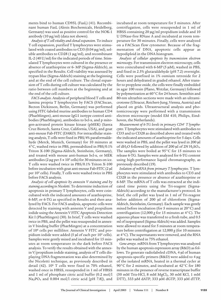
Azathioprine and its metabolites induce a mitochondrialpathway of apoptosis that is related to CD28 signaling. In ini-tial functional studies focusing on the mechanism ofazathioprine-induced apoptosis, we next assessed acti-vation of caspases in primary blood T lymphocytes by6-MP. Accordingly, CD4+ T lymphocytes were stimu-lated with rIL-2 and anti-CD3 plus anti-CD28 anti-bodies in the presence or absence of 6-MP, and caspaseactivity was determined after 5 days. As shown in Fig-ure 4a, 6-MP led to a marked induction of both cas-pase-9 and caspase-3, whereas the induction of cas-pase-8, on average, was less pronounced. Since thesedata suggested that 6-MP–induced apoptosis mightinvolve activation of a mitochondrial pathway ofapoptosis involving caspase-9, we focused in consecu-tive studies on the role of caspase-9 in 6-MP–inducedapoptosis. It was found that acetyl-LEHD-CHO, a sub-stance that (at the concentrations used) specificallyblocks caspase-9 activation (Figure 4b), suppressed 6-MP–induced apoptosis of primary T lymphocytes,whereas acetyl-IETD-CHO, a specific inhibitor of cas-pase-8, had lesser effects. These results strongly sug-gest that 6-MP utilizes a caspase-9–sensitive mito-chondrial pathway to induce T cell apoptosis uponCD28 costimulation. Consistent with this hypothesis,both azathioprine and 6-MP led to a loss of ∆Ψm inprimary T cells (Figure 4c) that is known to occur dur-ing mitochondrial pathways of apoptosis (47).
In subsequent studies on the mechanism of aza-thioprine-induced apoptosis, we determined therequirement of T cell costimulation with CD28 for
1140 The Journal of Clinical Investigation | April 2003 | Volume 111 | Number 8
Figure 4Azathioprine induces a mitochondrial pathway of apoptosis. (a)Activity of caspase-3, -8, and -9 upon treatment of T cells with 6-MP.CD45RA and CD45RO T cell subsets were stimulated with antibod-ies to CD3 and CD28 and recombinant IL-2 for 5 days in the presenceor absence of 6-MP, as indicated. There was a marked induction ofcaspase-9 activity upon azathioprine treatment. A second independ-ent experiment showed similar results (data not shown). Data on cas-pase-9 activity from three independent healthy blood donors areshown in the right lower panel. (b) Specific blockade of caspase-9 byacetyl-LEHD-CHO (Ac-LEHD-CHO) suppresses 6-MP–induced apop-tosis. CD4+ T lymphocytes from the peripheral blood of healthy vol-unteers were stimulated with antibodies to CD3 and CD28 in thepresence or absence of 6-MP, 10 µM acetyl-LEHD-CHO, and 10 µmacetyl-IETD-CHO. Although acetyl-IETD-CHO had little effect, 6-MP–induced T cell apoptosis could be suppressed by acetyl-LEHD-CHO.(c) Measurement of ∆Ψm in primary CD4+ T lymphocytes upon treat-ment with azathioprine, 6-MP, and FCCP (positive control). Periph-eral blood CD4+ T cells from healthy volunteers were stimulated withantibodies to CD3 and CD28 and recombinant IL-2 and cultured inthe presence or absence of azathioprine or 6-MP for 5 days as indi-cated. Cells were then loaded with JC-1 for 20 minutes followed byFACS analysis to determine ∆Ψm. Both azathioprine and 6-MP as wellas FCCP led to a marked reduction of ∆Ψm as compared with untreat-ed primary CD4+ T cells. One representative experiment of two isshown. Aza, azathioprine; UT, untreated.
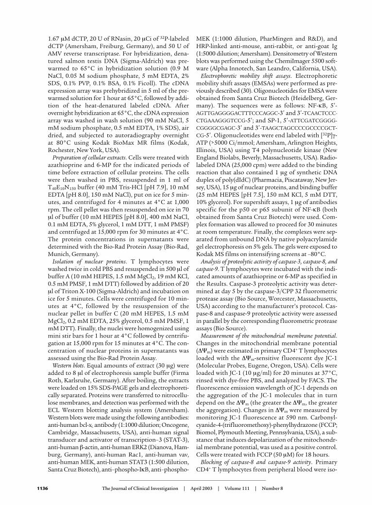
Figure 5(a) Azathioprine-induced apoptosis is critically dependent on costim-ulation with CD28. CD4+ T lymphocytes were stimulated in the pres-ence or absence of azathioprine and 6-MP, as indicated. T cell apop-tosis was assessed by FACS analysis using annexin V/propidium iodidestaining at day 5 of cell culture. (b) Azathioprine-induced apoptosisis independent of the CD95/CD95L system. Primary CD4+ T lym-phocytes were stimulated as above in the presence or absence of aza-thioprine and a neutralizing CD95L antibody. T cell apoptosis wasassessed by FACS analysis at day 5 of cell culture. (c) The left panelshows a gene array for apoptosis-related genes in T lymphocytes.CD4+ T lymphocytes were stimulated as above in the presence orabsence of azathioprine. The right panel shows that 6-MP suppress-es bcl-xL protein expression. Cellular proteins were isolated after 3 daysof cell culture and assessed for bcl-xL or cellular NF-κB expression byWestern blot analysis. (d) FACS analysis for intracellular bcl-xL expres-sion in permeabilized lymphocytes upon 6-MP treatment. PurifiedCD4+ T lymphocytes were stimulated in the presence or absence of 6-MP. FACS analysis for bcl-xL in permeabilized cells was performedafter 5 days of cell culture. (e) 6-MP suppresses nuclear NF-κB activa-tion. CD4+ T lymphocytes were stimulated in the presence or absenceof 6-MP, as indicated. Nuclear proteins were isolated after 3 days andanalyzed for NF-κB (upper panel) or SP-1 (middle panel) activity by gelretardation assays (EMSAs). Nuclear extracts from PMA-stimulatedJurkat T cells served as positive controls. The lower panel represents asupershift analysis of the upper complex using extracts from anti-CD3–plus anti-CD28–stimulated primary T cells. The addition of antibod-ies to p50 or p65 to the EMSA reaction is indicated. 6-MP treatmentled to downregulation of the NF-κB p50/p65 complex.
azathioprine- and 6-MP–induced apoptosis (Figure5a). It was found that azathioprine and 6-MP inducedapoptosis when primary blood CD4+ T lymphocyteswere stimulated through the TCR/CD3 complex plusCD28, whereas no apoptosis was seen when cells werestimulated with anti-CD3 antibodies alone (Figure5a), demonstrating a selectivity of this drug for co-stimulated T lymphocytes. This requirement forCD28 costimulation led us to further investigate theCD28-related signaling events involved in azathio-prine- and 6-MP–induced apoptosis.
CD28 costimulation has been shown to affect boththe death receptor and the mitochondrial pathways ofapoptosis induction (48). Given its implication in acti-vation-induced cell death of T cells, we first studied thepossible contribution of the CD95L/CD95 interactionto the apoptotic effect of azathioprine. Interestingly, wefound that specific blockade of the CD95L/CD95 sys-tem by CD95L-neutralizing antibodies did not affectazathioprine-induced apoptosis of anti-CD3– plus anti-CD28–stimulated primary blood CD4+ T lymphocytes(Figure 5b), suggesting that azathioprine induces T cellapoptosis through a CD95-independent pathway.
To identify candidate genes that may be responsi-ble for azathioprine-induced apoptosis, we next char-acterized the gene products that are induced or suppressed by azathioprine during T cell apoptosis. Inthese studies, we used gene arrays that included vari-ous genes involved in apoptosis (Figure 5c). It wasfound that azathioprine strongly suppressed bcl-xL
mRNA expression in anti-CD3– plus anti-CD28–acti-vated T lymphocytes, whereas mRNA expression ofvarious other genes such as IL-2R, TACI, and GAPDHwas not affected. Furthermore, bcl-xL protein expres-sion in activated T cells was strongly suppressed byazathioprine and 6-MP (Figure 5, c and d), stronglysuggesting that azathioprine induces suppression ofbcl-xL mRNA and protein levels in activated primaryCD4+ T lymphocytes. Since bcl-xL expression in T lym-phocytes is known to be controlled by the transcrip-tion factor NF-κB (38, 40, 49), we next determinedwhether CD28-induced activation of NF-κB wasaltered in primary T lymphocytes upon 6-MP treat-ment. As shown in Figure 5e, 6-MP suppressed CD28-induced activation of NF-κB p50/p65. In particular,6-MP downregulated the nuclear expression of thep65 subunit of NF-κB that has been implicated inantagonizing T cell apoptosis.
Azathioprine causes a specific blockade of the Rac1 activa-tion pathway in primary T lymphocytes. Since activationof NF-κB upon CD28 costimulation is mediated byphosphorylation and degradation of IκB (49), we nextdetermined IκB phosphorylation in 6-MP–treatedcells. It was found that IκB phosphorylation was sup-
The Journal of Clinical Investigation | April 2003 | Volume 111 | Number 8 1141

Figure 6Azathioprine blocks the Rac1/MEK kinase pathway. (a) Analysis ofphosphorylation of MEK, a MAP kinase kinase that can be activatedby MEKK (see Figure 8). Purified CD4+ T lymphocytes were stimu-lated in the presence or absence of 6-TG. Intracellular staining forphospho-MEK by FACS analysis was made after 3 days. (b) 6-MPsuppresses CD28-induced MEK and IκB phosphorylation. PurifiedCD4+ T lymphocytes were stimulated in the presence or absence of6-MP. Cellular proteins were isolated after 3 days and analyzed byWestern blotting. The upper left panels show phospho-IκB (p-IκB)or phospho-MEK (p-MEK) activity upon 6-MP treatment. Bandintensity was quantified by densitometry and normalized to actin lev-els. The lower left panels show IκB or MEK protein expression uponazathioprine and 6-MP treatment. Band intensity was normalized toERK2 levels. The right panels show Rac1 and vav protein expressionupon azathioprine and 6-MP treatment. Azathioprine and 6-MPtreatment had little effect on Rac1 protein levels, whereas vav levelswere increased in cellular extracts. Band intensity was quantified bydensitometry and normalized to ERK2 levels. Den, densitometry. (c)6-MP induces vav accumulation. CD4+ T lymphocytes were stimu-lated in the presence or absence of azathioprine, 6-MP, or 6-TG for5 days. Cells were immunostained with Rac1-specific antibodies andvav-specific antibodies and Cy3-labeled secondary antibodies (red).Nuclei were counterstained with Hoechst blue. Confocal microscopyshowed that the expression of the Rac1-associated guanosineexchange factor vav was increased upon treatment with azathioprineand its metabolites, whereas Rac1 levels were nearly unchanged.
pressed by 6-MP treatment of T cells, whereas IκB pro-tein expression levels remained unchanged (Figure6b), strongly suggesting that CD28-mediated NF-κBactivation through phosphorylation and degradationof IκB is a target of this drug. Since IκB phosphoryla-tion in CD28-stimulated primary T cells is mediatedby the vav/Rac1/MEK kinase (MEKK) activation path-way (49), we next assessed potential modulation ofthis pathway by azathioprine and 6-MP. Interesting-ly, we observed that azathioprine and its metabolitessuppressed MEK phosphorylation, but the expressionlevels of MEK were nearly unchanged (Figure 6, a andb). Furthermore, the expression of Rac1 was not sig-nificantly suppressed by azathioprine and 6-MP,whereas both drugs induced the expression of theRac1-activating guanosine exchange factor vav (Fig-ure 6, b and c), a regulatory protein previously shownto be a key target for CD28 signaling (50–52).
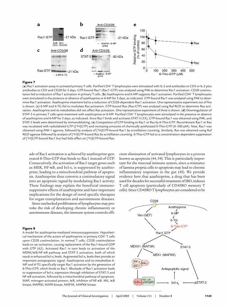
CD28 costimulation induced Rac1 activation in pri-mary T cells (Figure 7a) was strongly suppressed byazathioprine, 6-MP, or 6-TG treatment, as shown by areduction of GTP-bound Rac1 (Figure 7b). Since Rac1is known to bind and activate STAT-3 (53), a key fac-tor for regulating bcl-xL expression in T cells (30), wealso assessed Rac1-bound STAT-3 and observed thatRac-1 bound STAT-3 was reduced in azathioprine-treated T cells (Figure 7d). Rac1 activation uponCD28 costimulation thus appears to control both theNF-κB and STAT-3 activation pathways that are
known to prevent T cell apoptosis upon CD28 co-stimulation, and azathioprine and its metabolitessuppress this activation pathway.
To exclude a general inhibitory effect of azathioprineand its metabolites on the activation of GTPases, wenext determined the activation of Ras that is known tooccur upon CD3/TCR stimulation of T cells. It wasfound that the activation of Ras was virtually unaffect-ed (Figure 7c), suggesting a specific inhibition of Rac1activation by azathioprine. This marked specificitypointed to the potential direct binding of an azathio-prine metabolite to Rac1. Indeed, we were able to detect6-TG in Rac1 pull-downs from primary T cells uponazathioprine treatment (approximately 120 pg of 6-TGper microgram), suggesting that azathioprine-generat-ed 6-Thio-GTP can directly bind to Rac1 in vivo. Con-sistently, 6-Thio-GTP was able to bind to recombinantRac1 under in vitro conditions (although with loweraffinity than GTP) (Figure 7e). Furthermore, 6-Thio-GTP did not significantly compete with GTP forrecombinant Ras, suggesting that 6-Thio-GTP maybind to selected GTPases only.
Taken together, these data suggest that azathioprineand its metabolites target CD28-mediated signaling inprimary T cells by specifically suppressing the activa-tion of the GTPase Rac1 through 6-Thio-GTP, leadingto a mitochondrial pathway of apoptosis (Figure 8).
DiscussionIn the present study, we have identified a unique andunexpected role for azathioprine and its metabolites inthe control of T cell apoptosis by modulation of Rac1activation upon CD28 costimulation. Specific block-
1142 The Journal of Clinical Investigation | April 2003 | Volume 111 | Number 8
ade of Rac1 activation is achieved by azathioprine-gen-erated 6-Thio-GTP that binds to Rac1 instead of GTP.Consecutively, the activation of Rac1 target genes suchas MEK, NF-κB, and bcl-xL is suppressed by azathio-prine, leading to a mitochondrial pathway of apopto-sis. Azathioprine thus converts a costimulatory signalinto an apoptotic signal by modulating Rac1 activity.These findings may explain the beneficial immuno-suppressive effects of azathioprine and have importantimplications for the design of novel specific therapiesfor organ transplantation and autoimmune diseases.
Since unchecked proliferation of lymphocytes may pro-voke the risk of developing chronic inflammatory orautoimmune diseases, the immune system controls effi-
cient elimination of activated lymphocytes in a processknown as apoptosis (44, 54). This is particularly impor-tant for the mucosal immune system, since a resistanceof lamina propria cells to apoptosis may lead to chronicinflammatory responses in the gut (45). We provide evidence here that azathioprine, a drug that has beenused for decades for successful treatment of IBD, inducesT cell apoptosis (particularly of CD45RO memory Tcells). Since CD45RO T lymphocytes are considered to be
The Journal of Clinical Investigation | April 2003 | Volume 111 | Number 8 1143
Figure 8A model for azathioprine-mediated immunosuppression. Hypotheti-cal mechanism of the action of azathioprine in primary CD4+ T cellsupon CD28 costimulation. In normal T cells, CD28 costimulationleads to vav activation, causing replacement of the Rac1-bound GDPwith GTP (62). Activated Rac1 in turn leads to activation of theMEKK/IκB/NF-κB pathway and STAT-3 activation, both of whichresult in enhanced bcl-xL levels. Augmented bcl-xL levels then provide animportant antiapoptotic signal. Azathioprine and its metabolites 6-MP and 6-TG specifically target Rac1 activation by the generation of6-Thio-GTP, which binds to Rac1. Blockade of Rac1 activation leadsto suppression of bcl-xL expression through inhibition of STAT-3 andNF-κB activation, followed by a mitochondrial pathway of apoptosis.MAP, mitrogen-activated protein; IκB, inhibitor of NF-κB. IKK, IκBkinase; MAPKK; MAPK kinase; MAP3K, MAPKK kinase.
Figure 7(a) Rac1 activation assay in activated primary T cells. Purified CD4+ T lymphocytes were stimulated with IL-2 and antibodies to CD3 or IL-2 plusantibodies to CD3 and CD28 for 3 days. GTP-bound Rac1 (Rac1-GTP) was analyzed using PAK to determine Rac1 activation. CD28 costimu-lation led to induction of Rac1 activation in primary T cells. (b) Azathioprine and 6-MP suppress Rac1 activation. Purified CD4+ T lymphocyteswere stimulated in the presence or absence of azathioprine or 6-MP for 3 days, as indicated. GTP-bound Rac1 was analyzed using PAK to deter-mine Rac1 activation. Azathioprine treatment led to a reduction of CD28-dependent Rac1 activation. One representative experiment out of fiveis shown. (c) 6-MP and 6-TG fail to modulate Ras activation. GTP-bound Ras (Ras-GTP) was analyzed using Raf RGD to determine Ras acti-vation. Azathioprine and its metabolites did not affect Ras activation. One representative experiment of three is shown. (d) Downregulation ofSTAT-3 in primary T cells upon treatment with azathioprine or 6-MP. Purified CD4+ T lymphocytes were stimulated in the presence or absenceof azathioprine and 6-MP for 3 days, as indicated. Since Rac1 binds and activates STAT-3 (53), GTP-bound Rac1 was obtained using PAK, andSTAT-3 levels were determined by immunoblotting. (e) Competition of GTP binding to Rac1 or Ras by 6-Thio-GTP. Recombinant Rac1 or Raswas incubated with radiolabeled GTP ([3H]GTP) and increasing amounts of chemically synthesized 6-Thio-GTP (0–500 µM). Next, Rac1 wasobtained using PAK-1 agarose, followed by analysis of [3H]GTP-bound Rac1 by scintillation counting. Similarly, Ras was obtained using RafRGD agarose followed by analysis of [3H]GTP-bound Ras by scintillation counting. 6-Thio-GTP led to a concentration-dependent suppressionof [3H]GTP-bound Rac1 but had little effect on [3H]GTP-bound Ras.

key effector cells in IBD, one may speculate that the excel-lent therapeutic efficacy of azathioprine in these diseasescould be due to the induction of local T cell apoptosis.This hypothesis is supported by the finding that success-ful azathioprine treatment in patients with IBD leads toan increased number of apoptotic T cells in the peripher-al blood and the lamina propria. This mechanism ofaction based on apoptotis induction of activated T cellswould also explain why azathioprine is effective in bothCrohn disease and ulcerative colitis, although both dis-eases seem to have a different pathogenesis and are asso-ciated with a different T cell cytokine profile (31, 32, 45).
The induction of apoptosis by azathioprine was criti-cally dependent on T cell costimulation with CD28,which is known to inhibit TCR-induced apoptosis dur-ing a primary T cell response by activation of the anti-apoptotic bcl-xL protein (55, 56). Interestingly, azathio-prine downregulates bcl-xL expression at the mRNA andprotein levels, strongly suggesting that this drug blocksa key regulatory pathway in CD28 signaling. The factthat azathioprine regulates bcl-xL expression would sug-gest that azathioprine regulates a mitochondrial path-way of apoptosis. Indeed, we observed that azathio-prine-mediated apoptosis led to downregulation of themitochondrial membrane potential and could be sup-pressed by a specific inhibitor of caspase-9.
We next analyzed the MEK/NF-κB signaling pathwaythat is known to induce bcl-xL expression upon CD28costimulation in primary T cells (49). It was found thatazathioprine and its metabolites suppressed MEK andNF-κB activation through IκB phosphorylation,although IκB and MEK protein levels were nearly unaf-fected. These data suggested that the suppression of theMEK/NF-κB signaling pathway by azathioprine is medi-ated by an unexpected specific mechanism involving anazathioprine metabolite rather than by azathioprine-induced suppression of protein production. Indeed, weobserved that the azathioprine metabolite 6-Thio-GTPdirectly binds to Rac1, a Rac GTPase that is known toplay a key role in CD28 signaling and MEK/NF-κB acti-vation in T cells (57–59). Furthermore, the levels of vav1,a CD28-responsive guanosine exchange factor for Rac1whose activity is suppressed by the adapter protein Cb1-b (50–52), were upregulated by azathioprine treat-ment. This finding is consistent with a compensatoryupstream mechanism to achieve Rac1 activation in pri-mary T cells upon administration of azathioprine.
Rac proteins play a major role in T cell development,differentiation, and proliferation (58–60). Whereas dom-inant positive Rac mutations have been associated withincreased cell proliferation and tumors, functionallyinactive Rac mutations are associated with immunode-ficiencies in humans (61). Azathioprine-mediated sup-pression of Rac1 activation in T cells was mediated bydirect binding of an azathioprine metabolite (6-Thio-GTP) to Rac1 instead of GTP. Although the precisemechanism by which binding of 6-Thio-GTP suppress-es Rac1 activation in T cells remains to be determined,the function of another GTPase (Ras) was not inhibited
by azathioprine and its metabolites. Our data are thusconsistent with a model in which the specificity of the 6-Thio-GTP–induced blockade of Rac1 function is relat-ed to the structure of the Rac1 protein. In any case, aza-thioprine specifically blocked the Rac1-mediated activation of NF-κB and STAT-3 in primary T cells thatmediate bcl-xL activation upon CD28 costimulation andtherefore provide an important antiapoptotic signal inanti-CD28–stimulated T lymphocytes (Figure 8). TheCD28 signaling pathway, however, is not only importantfor the initial activation of T cells but also for maintain-ing their viability and responsiveness during a persistentimmune response (38, 56). Azathioprine-induced sup-pression of CD28 signaling events may be particularlyimportant for the mechanism of action of this drug,since it is frequently used in chronic inflammatory dis-eases in which repeated antigen-specific stimulation ofeffector T cells occurs and in which elimination of effec-tor T cells is needed (33, 45). Consistent with this idea,we observed that azathioprine can also induce apoptosisof T lymphocytes that were preactivated with CD28.
Taken together, our data identify a unique and novelmolecular mechanism of action of azathioprine on thebasis of the suppression of Rac1 activation. Further stud-ies showed that azathioprine-induced suppression ofRac1 activation leads to suppression of bcl-xL expression(through blockade of NF-κB and STAT-3 activation) anda mitochondrial pathway of T cell apoptosis. Our datathus suggest that azathioprine-induced immunosup-pression is mediated by suppression of Rac1 activationand the consecutive induction of T cell apoptosis. Itshould be noted, however, that azathioprine-inducedapoptosis affects mainly CD45RO effector T cells uponcostimulation with CD28, suggesting that azathioprinemay be particularly effective in eliminating pathogenicmemory T cells in autoimmune and chronic inflamma-tory diseases. These data have important implicationsfor the design of novel and more specific therapies forautoimmune diseases. In particular, 6-Thio-GTPderivates with higher affinity to Rac1 may be useful toachieve more powerful immunosuppression in autoim-mune diseases and organ transplantation.
AcknowledgmentsM.F. Neurath was supported by grants from theDeutsche Forschungsgemeinschaft (Ne 490/4-1 andSonderforschungsbereich 548, 432, 490) and the Ger-hard Hess program of the Deutsche Forschungsge-meinschaft (Ne 490/3-1) and is a recipient of a Full-bright scholarship. R. Blumberg was supported by NIHgrants DK44319, DK51362, and DK53056 and theHarvard Digestive Diseases Center. H. Walczak wassupported by the BioFuture Program of the Bun-desministerium für Bildung und Forschung.
1. McGeown, M., et al. 1988. Ten-year results of renal transplantation withazathioprine and prednisolone as only immunosuppression. Lancet.1:983–991.
2. Andreone, P.A., et al. 1986. Reduction of infectious complications follow-ing heart transplantation with triple-drug immunotherapy. J. Heart Trans-plant. 5:13–19.
1144 The Journal of Clinical Investigation | April 2003 | Volume 111 | Number 8

3. British and Dutch Multiple Sclerosis Azathioprine Trial Group. 1988. Dou-ble-masked trial of azathioprine in multiple sclerosis. Lancet. 2:179–186.
4. DeSilva, M., and Hazleman, B.L. 1981. Long-term azathioprine in rheuma-toid arthritis. A double-blind study. Ann. Rheum. Dis. 40:560–568.
5. Ginzler, E., Sharon, E., Diamond, H., and Kaplan, D. 1975. Long term main-tenance therapy with azathioprine in systemic lupus erythematosus. Arthri-tis Rheum. 18:27–35.
6. Christensen, E., et al. 1985. Beneficial effect of azathioprine and predictionof prognosis in primary biliary cirrhosis. Final results of an internationaltrial. Gastroenterology. 89:1084–1091.
7. Candy, S., et al. 1995. A controlled double blind study of azathioprine in themanagement of Crohn’s disease. Gut. 37:674–678.
8. Bouhnik, Y., et al. 1996. Long-term follow-up of patients with Crohn’s dis-ease treated with azathioprine or 6-mercaptopurine. Lancet. 347:215–219.
9. D’Haens, G., Geboes, K., Ponette, E., Penninckx, F., and Rutgeerts, P. 1997.Healing of severe recurrent ileitis with azathioprine therapy in patients withCrohn’s disease. Gastroenterology. 112:1475–1481.
10. Lewis, J.D., Schwartz, J.S., and Lichtenstein, G.R. 2000. Azathioprine formaintenance of remission in Crohn’s disease: benefits outweigh the risk oflymphoma. Gastroenterology. 118:1018–1024.
11. Present, D.H., et al. 1980. Treatment of Crohn’s disease with 6-mercaptop-urine: a long-term randomized, double-blind study. N. Engl. J. Med.302:981–987.
12. Bean, R.H.D. 1962. The treatment of chronic active ulcerative colitis with 6-mercaptopurine. Med. J. Aust. 2:592–593.
13. Dimitriu, A., and Fauci, A.S. 1978. Activation of human B lymphocytes. XI.Differential effects of azathioprine on B lymphocytes and lymphocyte sub-populations regulating B cell function. J. Immunol. 121:2335–2339.
14. Lennard, L. 1992. The clinical pharmacology of 6-mercaptopurine. Eur. J.Clin. Pharmacol. 43:329–335.
15. Röllinghoff, M., Schrader, J., and Wagner, H. 1973. Effect of azathioprineand cytosine arabinoside on humoral and cellular immunity in vitro. Clin.Exp. Immunol. 15:261–269.
16. Abdou, N.I., Zweiman, B., and Casella, S.R. 1973. Effects of azathioprinetherapy on bone marrow-dependent and thymus-dependent cells in man.Clin. Exp. Immunol. 13:55–64.
17. Bach, M.A., and Bach, J.F. 1972. Activities of immunosuppressive agents invitro. II. Different timing of azathioprine and methotrexate in inhibitionand stimulation of mixed lymphocyte reaction. Clin. Exp. Immunol. 11:89–98.
18. Hoffmann, M., Rychlewski, J., Chrzanowska, M., and Hermann, T. 2001.Mechanism of activation of an immunosuppressive drug: azathioprine.Quantum chemical study on the reaction of azathioprine with cysteine. J. Am. Chem. Soc. 123:6404–6409.
19. Kroplin, T., and Iven, H. 2000. Methylation of 6-mercaptopurine and 6-thioguanine by thiopurine S-methyltransferase. A comparison of activityin red blood cell samples of 199 blood donors. Eur. J. Clin. Pharmacol.56:343–345.
20. Van Os, E.C., et al. 1996. Azathioprine pharmacokinetics after intravenous,oral, delayed release oral and rectal foam administration. Gut. 39:63–68.
21. Podolsky, D.K. 1991. Inflammatory bowel disease. N. Engl. J. Med.325:928–937.
22. Beutler, B. 2001. Autoimmunity and apoptosis: the Crohn’s connection.Immunity. 15:5–14.
23. Neurath, M.F., Finotto, S., and Glimcher, L.H. 2002. The role of Th1/Th2polarization in mucosal immunity. Nat. Med. 8:567–573.
24. MacDonald, T.T., Monteleone, G., and Pender, S.L.F. 2000. Recent develop-ments in the immunology of inflammatory bowel disease. Scand. J. Immunol.51:2–9.
25. Shanahan, F. 2002. Crohn’s disease. Lancet. 359:62–69.26. Hugot, J.P., et al. 2001. Association of NOD2 leucine-rich repeat variants
with susceptibility to Crohn’s disease. Nature. 411:599–603.27. Ogura, Y., et al. 2001. A frameshift mutation in NOD2 associated with sus-
ceptibility to Crohn’s disease. Nature. 411:603–606.28. Targan, S.R., et al. 1997. A short-term study of chimeric monoclonal anti-
body cA2 to tumor necrosis factor alpha for Crohn’s disease. N. Engl. J. Med.337:1029–1035.
29. Elson, C.O., Sartor, R.B., Tennyson, G.S., and Riddell, R.H. 1995. Experi-mental models of inflammatory bowel disease. Gastroenterology.109:1344–1367.
30. Atreya, R., et al. 2000. Blockade of IL-6 trans-signaling suppresses T cellresistance against apoptosis in chronic intestinal inflammation: evidence inCrohn’s disease and experimental colitis in vivo. Nat. Med. 6:583–588.
31. Breese, E., Braegger, C.P., Corrigan, C.J., Walker-Smith, J.A., and MacDon-ald, T.T. 1993. Interleukin-2 and interferon-gamma secreting T cells in nor-mal and diseased human intestinal mucosa. Immunology. 78:127–131.
32. Plevy, S.E., et al. 1997. A role for TNF-alpha and mucosal T helper-1cytokines in the pathogenesis of Crohn’s disease. J. Immunol. 159:6276–6282.
33. Boirivant, M., et al. 1999. Lamina propria T cells in Crohn’s disease and othergastrointestinal inflammation show defective CD2 pathway-induced apop-tosis. Gastroenterology. 116:557–565.
34. Sandborn, W.J., et al. 1999. Lack of effect of intravenous administration on
time to respond to azathioprine for steroid-treated Crohn’s disease. Gas-troenterology. 117:527–535.
35. Pearson, D.C., May, G.R., Fick, G.H., and Sutherland, L.R. 1995. Azathio-prine and 6-mercaptopurine in Crohn disease. A meta-analysis. Ann. Intern.Med. 123:132–142.
36. Ewe, K., et al. 1993. Azathioprine combined with prednisolone or monother-apy with prednisone in active Crohn’s disease. Gastroenterology. 105:367–376.
37. Holtmann, M.H., et al. 2002. Tumor necrosis factor-receptor 2 is upregu-lated on lamina propria T cells in Crohn’s disease and promotes experi-mental colitis in vivo. Eur. J. Immunol. 32:3142–3151.
38. Noel, P.J., Boise, L.H., Green, J.M., and Thompson, C.B. 1996. CD28 co-stim-ulation prevents cell death during primary T cell activation. J. Immunol. 157:636–647.
39. Radvanyi, L.G., et al. 1996. CD28 costimulation inhibits TCR-induced apop-tosis during a primary T cell response. J. Immunol. 156:1788–1798.
40. Khoshnan, A., et al. 2000. The NF-κB cascade is important in Bcl-xL expres-sion and for the antiapoptotic effects of the CD28 receptor in primaryhuman CD4+ lymphocytes. J. Immunol. 165:1743–1754.
41. Jodo, S., et al. 2000. CD95 (Fas) ligand-expressing vesicles display antibody-mediated, FcR-dependent enhancement of cytotoxicity. J. Immunol.165:5487–5494.
42. Nicoletti, I., Migliorati, G., Pagliacci, M.C., Grignani, F., and Riccardi, C.1991. A rapid and simple method for measuring thymocyte apoptosis bypropidium iodide staining and flow cytometry. J. Immunol. Methods.139:271–279.
43. Finotto, S., et al. 2001. Treatment of allergic airway inflammation and hyper-responsiveness by local antisense-induced blockade of GATA-3 expression.J. Exp. Med. 193:1247–1260.
44. Scaffidi, C., Kirchhoff, S., Krammer, P.H., and Peter, M.E. 1999. Apoptosissignaling in lymphocytes. Curr. Opin. Immunol. 11:277–285.
45. Neurath, M.F., et al. 2001. Regulation of T-cell apoptosis in inflammatorybowel disease: to die or not to die, that is the mucosal question. TrendsImmunol. 22:21–26.
46. Cuffari, C., Seidman, E.G., Latour, S., and Theoret, Y. 1996. Quantificationof 6-thioguanine in peripheral blood leukocyte DNA in Crohn’s diseasepatients on maintenance 6-mercaptopurine therapy. Can. J. Physiol. Pharma-col. 74:580–585.
47. Zamzami, N., et al. 1995. Reduction in mitochondrial potential constitutesan early irreversible step of programmed lymphocyte death in vivo. J. Exp.Med. 181:1661–1672.
48. Kirchhoff, S., Müller, W.W., Li-Weber, M., and Krammer, P.H. 2000. Up-reg-ulation of c-FLIPshort and reduction of activation-induced cell death inCD28-co-stimulated human T cells. Eur. J. Immunol. 30:2765–2774.
49. Tuosto, L., et al. 2000. Mitogen-activated kinase kinase kinase 1 regulates T cell receptor and CD28-mediated signaling events which lead to NF-κBactivation. Eur. J. Immunol. 30:2445–2454.
50. Chiang, Y.J., et al. 2000. Cbl-b regulates the CD28 dependence of T-cell acti-vation. Nature. 403:216–220.
51. Krawczyk, C., et al. 2000. Cbl-b is a negative regulator of receptor clusteringand raft aggregation in T cells. Immunity. 13:463–473.
52. Raab, M., Pfister, S., and Rudd, C.E. 2001. CD28 signaling via VAV/SLP-76adaptors: regulation of cytokine transcription independent of TCR ligation.Immunity. 15:921–933.
53. Simon, A.R., et al. 2000. Regulation of STAT3 by direct binding of the Rac1GTPase. Science. 290:144–147.
54. Lenardo, M., et al. 1999. Mature T lymphocyte apoptosis — immune regu-lation in a dynamic and unpredictable antigenic environment. Annu. Rev.Immunol. 17:221–253.
55. Boise, L.H., Noel, P.J., and Thompson, C.B. 1995. CD28 and apoptosis. Curr.Opin. Immunol. 7:620–625.
56. Mueller, D.L., Seiffert, S., Fang, W., and Behrens, T.W. 1996. Differential reg-ulation of bcl-2 and bcl-x by CD3, CD28, and the IL-2 receptor in clonedCD4+ helper T cells. A model for the long-term survival of memory cells. J. Immunol. 156:1764–1771.
57. Marinari, B., et al. 2002. Vav cooperates with CD28 to induce NF-κB activa-tion via a pathway involving Rac-1 and mitogen-activated kinase kinase 1.Eur. J. Immunol. 32:447–456.
58. Gomez, M., Tybulewicz, V., and Cantrell, D.A. 2000. Control of pre T cellproliferation and differentiation by the GTPase Rac1. Nat. Immunol.1:348–352.
59. Joneson, T., and Bar-Sagi, D. 1999. Suppression of Ras-induced apoptosisby the Rac GTPase. Mol. Cell. Biol. 19:5892–5901.
60. Li, B., et al. 2000. Role of the guanosine triphosphate Rac2 in T helper 1 celldifferentiation. Science. 288:2219–2222.
61. Williams, D.A., et al. 2000. Dominant negative mutation of the hematopoi-etic-specific Rho GTPase, Rac2, is associated with a human phagocyteimmunodeficiency. Blood. 96:1646–1654.
62. Kaga, S., Ragg, S., Rogers, K.A., and Ochi, A. 1998. Activation of p21-CDC42/Rac-activated kinases by CD28 signaling: p21-activated kinase(PAK) and MEK kinase 1 (MEKK1) may mediate the interplay between CD3and CD28 signals. J. Immunol. 160:4182–4189.
The Journal of Clinical Investigation | April 2003 | Volume 111 | Number 8 1145