cell adhesion to hydroxyl groups of a monolayer film
TRANSCRIPT

Cell adhesion to hydroxyl groups of a monolayer film
NORMAN F. OWENS, DAVID GINGELL
Department of Anatomy and Developmental Biology, The University College and Middlesex School of Medicine, Cleveland Street, London \\ IP6DB, UK
and ANNETTE TROMMLER
Experimental Stomatology and Biomaterials Research Unit, Schumannstrasse, Charite Medical School, Humboldt University, Berlin, 1040, GDR
Summary
We have studied cells on chemically defined mono-molecular films of the long-chain alcohol docosa-nol. Langmuir-Blodgett films of the alcohol weredeposited on glass cover slips, previously madehydrophobic with octadecyl groups. This givesfilms in which the alcohol headgroups faceoutwards to the water. Molecular orientation andfilm integrity were shown by a fluorescence adsorp-tion test. Cell contacts on the films were observed inmedia without proteins by interference reflectionmicroscopy (IRM) and the mechanics of detach-ment were examined by hydrodynamic shearing ina flow chamber. Cell contact 'with docosanol wascompared with that on an adjacent area of octa-decyl glass without a monolayer.
Dictyostelium amoebae settled and spread onboth docosanol and octadecyl glass, but little or nolocomotion was seen on docosanol. On octadecylglass the amoebae moved actively, formingultrathin cytoplasmic lamellae, which look dark
under IRM, and left distinctive trails of membra-nous debris. Hydrodynamic shearing showed thatthe amoebae stuck strongly to both surfaces andcould not be removed from either at the maximumattainable wall shear stress of 6Nm~2. Red bloodcells also adhered to both surfaces and removalfrom both occurred between 1 and 3Nm"2. IRMand scanning electron microscopy (SEM) studiesindicated that this force leads to a minimal measureof red cell adhesion, since removal often involvedthe breakage of cytoplasmic tethers.
Our results show that alcoholic -OH groups, in atwo-dimensional array, provide a surface that isstrongly adhesive for cells. No other method hasmade it possible to demonstrate cell adhesion pur-ely to -OH groups, in a known orientation anddensity, and in the absence of any other functionalgroups on the interface.
Key words: adhesion, monolayer film, Dictyostelium.
Introduction
Although there have been innumerable studies on cell-to-substratum adhesion, most have been concerned with theroles of macromolecular factors such as fibronectin andlaminin or with the influence of small ions in solution,and few have been directed to the question of thesignificance of particular chemical groups in the process.For this, two approaches are possible. One is analytic,involving the detailed dissection of the molecular interac-tions between components of the cell surface and biologi-cally important macromolecules. This is the rationale ofresearch on cell-fibronectin interactions (review, Hynes,1986). An alternative approach is to design very simplesurfaces bearing one type or a limited number of differentchemical groups that are thought to underlie morecomplex biological interactions. This synthetic approachis arguably more fundamental. It has the additional
Journal of Cell Science 91, 269-279 (1988)Printed in Great Britain © The Company of Biologists Limited 1988
advantage of making it possible to distinguish betweenthe roles of the general forces of attraction and repulsionthat act to some degree between all closely apposed bodies(Parsegian & Gingell, 1973; Parsegian, 1984) and more'biochemical' interactions including hydrogen bondingand stereospecific relationships between particular mol-ecular configurations.
To this end we have chosen to study cell adhesion toflat chemically modified glass surfaces, which are suitablefor optical microscopy. The latter factor is important, asany quantitative study of cell-substratum adhesionshould report any changes in the topography of thecontact zone in response to externally applied forces. Astep in this direction has been to prepare monomolecularfilms of a long-chain alcohol, docosanol (C22 H45 OH), byLangmuir—Blodgett deposition on glass coverslips. Thisis technically difficult, but the film produced has a knownnumber of molecules per unit area in a defined onen-
269

tation, which are not distributed in depth.Docosanol monolayers were deposited on glass, pre-
viously made hydrophobic by covalent derivatizationwith octadecyl dimethylchlorosilane (ODMS), whichattaches octadecyl (Cis) hydrocarbon chains to -OHgroups of the glass (octadecyl glass). This ensured thatthe monolayer formed on the first dip had its -OHheadgroups facing the water, in the required orientationfor testing cell adhesion. Film deposition was limited tohalf the glass coverslip so that the adjacent hydrophobicarea formed a convenient control surface.
In addition to observing cell behaviour, we mademeasurements of the force of cell detachment usinghydrodynamic shearing flow in a calibrated parallel platechamber. The monolayer-coated coverslip formed oneface of the chamber. Cell detachment was recorded usingbright-field or interference reflection microscopy (IRM)optics on videotape using a low-light TV system to avoidover-illumination of adherent cells.
Materials and methods
ChemicalsSurface chemically pure water was obtained by distillation fromalkaline potassium permanganate as described by Owens et al.(19876). AnalaR grade n-heptane and chloroform (BDH Ltd,Poole, UK) were redistilled in an all-glass still and mid-rangefractions were taken in each case. The absence of surfactantcontaminants in these fractions was confirmed by spreading a100,1*1 sample onto a cleaned water surface. No measurablechange in surface pressure (FI < O'OS mN m~ ) could bedetected following a 10-fold compression of the water surfaceafter evaporation of the solvent sample. 1-Docosanol (C22-OH)and 1-eicosanol (C20-OH) were purchased from Sigma Chemi-cal Co., Poole, UK, had a purity of >99-5% and were usedwithout further purification. Concanavalin A (Sigma) conju-gated with tetramethylrhodamine B isothiocyanate (ConA-Rh)was from the same stock solution used by Owens et al. (1987a).The lipid alcohol monolayers were spread from solution inredistilled w-heptane at concentrations not exceeding1 mgml"1. The spreading solutions were stored at 20°C in cleanglass-stoppered bottles under a solvent seal of /i-heptane toreduce evaporation losses. Microscope coverglasses (CM-S glassfrom Chance Propper PLC, Smethwick, UK) were used forWilhelmy plates (22mmX22mmX0-17mm) and also as basesfor monolayer deposition (40mmX22mmX017 mm). The lat-ter were derivatized with octadecyldimethyl chlorosilane(ODMS) (from Sigma) as described by Owens et al. (19876).
Cell cultures and mediaMethods for the culture and isolation of Dictyostelium discoid-eum (Ax2) amoebae from shaken suspension in glucose-sup-plemented axenic medium and the isolation of human red bloodcells have been published (Owens et al. 1987a). For controlprotein adsorption studies, the supernatant from a 100 h cultureof Ax2 amoebae was obtained by centrifugation at 1000 g for10 min. Static and flow studies on amoebae were done in 20 niM-NaCl containing 2-5 mM-phosphate buffer. For red cells PBS atpH7-4 was used. Experiments were done at 20°C.
MicroscopyInterference reflection microscopy was done on the equipmentdescribed by Gingell et al. (1982). Cell counting was normally
done with a Zeiss 25X epi-illumination planachromat objective.Higher-resolution photography utilized a Zeiss 100X lens withiris diaphragm. The same microscope was used for fluorescencework, replacing the HP1 epi-illuminator with a dichroic mirrorand interference filter assembly incorporating a barrier filter(Carl Zeiss). For b/w prints we used a 35 mm Nikon F3 cameraand XP1 400 ASA film (Ilford Ltd, Romford, UK).
Flow experiments were recorded on videotape. The systemincluded a 'Falcon' SIT low-light TV camera (Custom CameraDesigns Ltd, Wells, UK) and a Panasonic video recorder(Stanmore Video Ltd, London, UK). Image processing wasdone on an Arlunya integrating framestore (Agar Aids Ltd,Stansted, UK).
Scanning electron microscopy was done on a Jeol 800 field ionemission instrument (Jeol, Collingdale, London, UK). Cellswere fixed during liquid flow with 3 % glutaraldehyde inphosphate-buffered saline, pH 7-4. Post-fixation in osmiumtetroxide was followed by freezing in liquid propane andcritical-point drying. Carbon/platinum coating was performedon Cressington freeze-fracture equipment (Cressington Scien-tific, Watford, UK).
Contamination of octadecyl glass by adsorbed proteinClean octadecyl glass coverslips immersed for 1 h in cell-freesupernatant (isolated from Ax2 culture) were removed and thenrinsed with distilled water. Cell behaviour was examined underIRM by mounting these putatively contaminated surfaces as thefloor of a polytetrafluoroethylene (PTFE) well (Owens et al.1988) on the microscope stage. Dictyostelium amoebae intro-duced at a density of 8x 10⁶ cells ml""1 in buffered saline wereobserved both during the initial cell-substratum contact periodand over an interval of 60 min after settling.
Monolayers at the air/water intetfaceIsotherms of surface pressure versus area (ITA) for docosanol(C22-OH) and eicosanol (C20-OH) monolayers spread on dis-tilled water (pH6-0) at 20-0 (±0-1 )°C were measured on amodified Langmuir-Adam film balance. Procedures are de-scribed by Owens et al. (19876).
Monolayers deposited on octadecyl glassLong-chain alcohol monolayers were deposited at a rate ofl-32cm2min~' (22mmX22mm) or 2-4cm2min~'(40mmX22mm) by the vertical passage of an octadecyl glasscoverslip through a compressed monolayer at the air/waterinterface. Throughout deposition FI was controlled to±0-1 mN m~ . Monolayers with the polar head groups orientedoutermost (H-monolayers) were obtained by passing the cover-slip through the monolayer until it was half-immersed in water.Then the monolayer remaining at the water surface wascarefully removed by vacuum aspiration, after which thecompression barrier was fully reversed to maximize the watersurface area. At this stage the half-coated coverslip was fullyimmersed and collected in a submerged PTFE pot. The potwith the immersed coverslip was transferred to a large glasstrough of distilled water for assembling on the flow chamber.
Flow studiesThe flow chamber has been described by Owens et al. (1987a).To avoid molecular re-arrangement of the depositedH-monolayer ('heads' outermost) on exposure to air it was keptunder water, from preparation to use in the flow chamber. Carewas taken to ensure that the monolayer did not becomedamaged by contact with the chamber during assembly.Clamped, water-filled, silicone rubber tubes were fixed to theinlet and outlet pipes of the chamber before it was removedfrom the water bath and connected to an elevated reservoir. The
270 N. F. Owens et al.

500 1000Pressure, f'(Nm"2)
Fig. 1. Flow rate Q through the conduit as a function of thedifference in pressure P' between its ends. The theoreticallinear relationship is also shown.
bubble-free system was then flushed with electrolyte. The rateof flow through the conduit was regulated by means of twoprecision micrometer flow valves (Nupro type S and M,purchased from North London Valve & Fitting Co., London,UK) connected in parallel in the Bowline. Excellent correlationbetween the flow rates from these valves was obtained whenused separately. At low flow rates type S gave more accuratemeasurements of wall shear stress in the range 0-1 N m~ . Cellswere injected into the inverted conduit through a side-arm andwere then left to settle for 20min. Sedimentation was completeafter about 6min.
Calibration of flow systemThe equations for laminar flow between parallel surfaces whosewidth (W) greatly exceeds their separation (y = 2b) are wellknown (Eskinazi, 1977). The velocity of the liquid is:
(i)
where ji is the absolute viscosity (= kinematic viscosity X den-sity) and AP is the linear pressure drop along a conduit of lengthL.
Putting y = 0 and integrating equation (1) for the cross-sectional area of the conduit between the limits y = ±b yieldsthe volume efflux rate (Q):
2b WAP(2)
In our system the conduit outlet was at atmospheric pressure sothat the hydrostatic pressure P' = AP.
A plot of Q as a function of applied pressure P' across theoverall flow chamber is shown in Fig. 1. The shear stress r isdefined as:
dvAy
From equations (1) and (3), it follows that:
T = •
(3)
(4)
1-0 2-0 3-0Wall shear stress,
4-0(Nm"2)
50 60
Fig. 2. Wall shear stress TW as a function of the pressuredifference P. (O) Type M valve; ( • ) type S valve; (©) bothvalves fully open.
maximum TW at the wall where y= 194^m = ±6. The wallshear Tw exerted by the flow against the monolayer-coatedsurface was calculated from equation (4) and a linear relation-ship between Tw and the applied pressure P is shown in Fig. 2.The pressure difference across the flow conduit was directlymeasured manometrically.
Equation (1) assumes that laminar flow is fully developed,but it gives no indication of conduit length required to establishit. The establishment length Le for fully developed laminar flowbetween two parallel walls of constant separation 2b wascalculated from the equation (Sparrow, 1955):
Le = 0-026.2bRe (5)
and hence the shear stress is a linear function of y and attains a
in which Re is the characteristic Reynolds flow number. ForR < 200, Lc constitutes less than 3 % of the conduit length so hasa negligible influence over the range of wall shears used in ourstudy. This result is consistent with the findings of VanWagenen & Andrade (1980) for the measurement of streamingpotentials generated by laminar flow between parallel glassplates. Le is calculated on the assumption that the fluid flowentering the parallel channel is non-turbulent. In the regionclose to the wall, where V—*Q, 'creeping flow' is obtained. Alocal Reynolds number R^ (y = 10 ,um) appropriate to this formof boundary flow was calculated and found to be always lessthan 1X10"4.
Results
Lipid alcohols at the air-water interfaceThe FI-A isotherms for docosanol and eicosanol mono-layers spread on distilled water at 2O0°C are shown inFig. 3, curves a and b, respectively. Curve a shows twoessentially linear segments meeting at 19 mN m~ , whichmarks the onset of a distinct change in molecular packingand monolayer compressibility. At pressures belowWmNm" 1 , the monolayer is seen to be more com-pressible by the greatly expanded isotherm. Adam (1941)suggested that this region of an isotherm corresponds to apressure-dependent re-arrangement of the headgroups,which allows closer packing of the chains. In the higherpressure regions, above 19mNm~ , the isotherm isalmost vertical due to chain-chain interaction. The smallnon-ionic headgroup does not sterically or electrostati-
Cell adhesion to a monolayer film 271

0-16 0-18 0-20 0-22Area (nm2 molecule"')
0-24
Fig. 3. Mean area occupied per molecule of lipid alcohol atthe air/water interface, as a function of the applied pressuren . Curve a, docosanol; b, eicosanol.
cally hinder the close apposition of the docosanol mol-ecules. Extrapolation of the near vertical region to Fl = 0gives a value of 0-192 nm2 for the cross-sectional area of asingle chain, which agrees closely with X-ray data(Muller, 1927).
The eicosanol isotherm (Fig. 3 curve b) is similar tothat of docosanol, although it is notable that the former ismore expanded at all pressures. The extrapolated area ofCH94nm2 per molecule for a close-packed eicosanolmonolayer is therefore slightly larger. The prominentshoulder featured in curve b is characteristic of theshorter chain (Cn.ig) alcohols (Brooks & Alexander,1960) and represents a two-dimensional phase transitionbetween the liquid and solid compression states in thesemonolayers. Equilibration of the monolayer in this regionwas observed to be very slow. For this reason docosanolwas preferred to eicosanol, since monolayer depositionwas normally made in the condensed region wheren > 2 0 r n N m ~ ' , and was therefore used in cell exper-iments.
It is of interest that Abraham et al. (1983) obtainedcurves almost identical to ours for eicosanol and docosa-nol, but argue from monolayer shear stress measurementsthat all the linear portions of the FI-A curves for bothalcohols represent distinct solid phases. The kink in thedocosanol curve they ascribe to pressure-induced melt-ing.
Deposited films are coherent and intact after flowAll attempts to make T-films (hydrocarbon chains, 'tails',outermost) on normal glass were unsuccessful. Underwater these showed marked granularity, indicative of
Fig. 4. A. Monolayer of docosanol, deposited in the T-stateon hydrophilic glass (alkyl chains facing outwards in air), andthen re-immersed in water; IRM indicates gross molecularrearrangment. Bar, 20^(m. B. Fluorescence image showingthe boundary between a docosanol monolayer, deposited withheadgroups outermost (H-state, right) and octadecyl glass(left). The latter has stained with adsorbed ConA-Rh; in thisdirect print from a colour transparency the fluorescent arealooks black. Bar, 10 jian.
molecular rearrangement (Fig. 4A). In contrast, H-filmson octadecyl glass were structureless by light microscopyunder the highest obtainable resolution. However, if theywere withdrawn from water, light-microscopic granu-larity indicative of reorganization was seen, and for thisreason all operations with them were done (incon-veniently!) under water. We tested depositedH-monolayers for defects, using ConA-Rh, which wasfound to adsorb strongly to the hydrophobic surface ofstearyl glass, but hardly at all to the monolayer. A lowdensity of pinhole defects were seen as bright points on ablack field. This procedure provided a very convenientmeans of confirming the existence of a deposited mono-layer and allowed us to find the edge of the monolayer(Fig. 4B), which was otherwise invisible under IRM orphase-contrast microscopy. The edge was approximatelyhalf way along the coverslip, giving equal areas of filmand octadecyl glass. The virtual absence of fluorescenceon the side on which the monolayer was depositedconfirms the coherence of the monolayer. When a mono-layer preparation was exposed to maximum liquid flowfor 20 min and then tested by ConA-Rh the same resultwas seen, showing that the film was still intact. Carefulmechanical abrasion of the film under water beforeConA-Rh treatment, resulted in brightly fluorescentlines, showing that the film had been removed, but thatthe octadecyl glass remained intact beneath it.
272 N. F. Owens et al.

Fig. 5. A-D. 1RM images of Dictyostelium amoebae on a docosanol monolayer. No trails or peripheral dark areas are seen(dark spots in B are vacuoles). Active peripheral undulations are particularly evident in D. E,F. Amoebae locomoting onoctadecyl glass (1RM) leaving granular trails. In F the rear of the cell shows a dark dendritic pattern. Bars,
Dictyostelium amoebae stick to docosanol andoctadecyl glass but only form ultrathin lamellae andlocomote on octadecyl glassAmoebae spread rapidly on docosanol headgroups butshowed little inclination to locomote. We saw unusualperipheral undulating activity that produced a shiftingpattern of light and dark under IRM (Fig. 5A-D). The
cells seemed unable to get enough lateral grip to loco-mote. In other experiments we noted similar behaviouron headgroups of synthetic glycolipids (Owens &Gingell, unpublished). However, exposure to fluid flowthat exerted the maximum obtainable shearing force of6 N m~ on the chamber wall failed to displace cells fromthe docosanol monolayer or even distort their contact
Cell adhesion to a monolayer film 273

10 20 30Wall shear stress, TW (Nm~2)
4-0
Fig. 6. Removal of red cells (RBC) from docosanolmonolayers ( • • ) , and octadecyl glass (O O), as afunction of wall shear stress, rw. Each curve is based on twoor three experiments (points).
patterns, showing that the cells adhere strongly to thissurface.
In contrast, after settling on octadecyl glass the cellsdeveloped an asymmetric 'fried-egg' shape; the edgethinned centripetally, giving the characteristic dark per-ipheral fringe seen in IRM optics (Gingell, 1981; Gingell& Vince, 1982). These changes took a few minutes, afterwhich many cells began to locomote, trailing an ultrathin(=100 nm) lamella that broke up into what appeared tobe membrane-bounded cytoplasmic fragments. Thesefragments, seen as a dark granular track under IRM,marked the paths of moving cells (Fig. 5E,F). Undermaximum flow, amoebae on octadecyl glass were notremoved, indicating strong adhesion to the hydrophobicsurface, as found on the hydrophilic docosanol.
Mechanical effect of flow on red cellsMechanical responses to detachment forces measured forred blood cells on octadecyl glass and docosanol mono-layers were found to be indistinguishable. Fig. 6 showsthat removal occurs principally over the small range ofwall shear stress values between 1 and 3Nm~ . IRMimages of red cells on docosanol during flow (Fig. 7A)show elongation in the direction of flow, with theproduction of one or more tethers. Fig. 7D shows cellsunder IRM prior to flow on octadecyl glass; tethersproduced under flow are seen in Fig. 7B,C. Scanningelectron microscopy (SEM) images of red cells onoctadecyl glass fixed during flow show tethers drawnfrom the downstream end of each cell, terminating inrather regularly branching dendritic tubes with a diam-eter of around 70nm (Fig. 7E,F). These can also beclearly resolved under IRM (Fig. 7C). They sometimesshowed enlargements at their tips and at knee bends,which seem to be attachment points (Fig. 7B,E,arrowed). Broken dendrites left behind by detached cellscan be seen in both IRM and SEM micrographs. UnderIRM cells were often seen to detach during flow, leaving
dendrites behind, though it was not always possible todetect these fragments.
Discussion
Behaviour of Dictyostelium amoebae on docosanol andoctadecyl glassAlthough Dictyostelium amoebae did not locomote ondocosanol, the fact that they were well attached is clear,since exposure to maximum liquid flow available in oursystem failed to shift them. Since a cell projects severalmicrometres from the wall, the force exerted on it exceedsthe sheer stress exerted on a wall area equal to that of thecell's apposition. On the basis of approximate calcu-lations, Hubbe (1981) gives a total force equal to threetimes the stress on an equivalent wall area. Using this, wecalculate that a lateral force of 1-9x10" N is exerted onan amoeba having a contact area of —100/.mi2 in a flowwhere TW = 6 N m~2. This represents approximately 540times the force of gravity on the cell.
Amoebae could not be removed from octadecyl glass atmaximum flow, showing that they were strongly adherentto this surface as well. On octadecyl glass a detachmentforce measurement is available from the new directmicromanipulation method of Francis et al. (1987). Thisgave a perpendicular removal force in the range 1X 10~to 7xlO~9N per cell, or 360-2500 times the gravitationalforce. From the application of known forces we thereforeanive at the important conclusion that cells on both thedocosanol monolayer and octadecyl glass are stronglyadherent. However, the dynamic range of our presentflow system is insufficient to remove amoebae from eithersurface, so we cannot compare interaction forces.
Since the hydroxyl surface is adhesive, why do theamoebae not locomote as they do on the adhesiveoctadecyl glass? There are several possibilities: (1) thehydrophobic surface actively stimulates movement,implying a membrane transductive process; (2) cells maymake an extensive area of highly adhesive contact on themonolayer, with the result that peripheral pseudopodalactivity may be unable to shift the cells: total internalreflection fluorescence shows that the cells make largeuniform appositions (Todd et al. 1988); (3) cells ondocosanol suffer directional depolarization of loco-motion; (4) the monolayer may have a lateral viscosity toolow to permit complete spreading and locomotion: how-ever, it is almost certain that the monolayer is 'solid' (seebelow); (5) if significant protein adsorption took placebefore or after initial cell spreading, it might reduce theadhesions of pseudopodia put out later. A test relevant tothis latter hypothesis was performed with octadecyl glasssubstrata deliberately exposed to a protein solution.
In a population of 200 Ax2 amoebae on octadecyl glassin the presence of culture medium supernatant, not onecell could be seen that displayed any evidence of eitherlamella formation or motile response. This finding con-trasts sharply with that obtained on a similar surface but,in putatively protein-free media, where some 150amoebae gave a positive lamella response and showedextensive trail patterns. This result is hardly surprising in
274 N. F. Owens et al.

Fig. 7. Red blood cells; A-D, IRM; E,F, SEM (negative images). A. Under flow on docosanol monolayer (from videotape);B,C, under flow on octadecyl glass; D, prior to flow on octadecyl glass; E,F, on octadecyl glass, fixed during flow, showingdendritic processes. Arrows in B,E show expanded terminal attachment points. Bars: A,B,D, lOfim; E, Zfim.
Cell adhesion to a monolayer film 275
view of the extreme dilution of protein likely to be presentas a consequence of the washing procedure adopted in ourexperimental protocol. This shows unambiguously thatthe level of protein contamination in our nominallyprotein-free media is too low to influence cell behaviourby adsorption onto octadecyl glass. We did not do asimilar test for the effect of protein adsorption onto -OHfilms. However, since the concentration of protein in ourexperimental solution is insignificantly low (with respectto octadecyl glass) and since protein adsorption fromusing very dilute solutions takes hours to reach equilib-rium (MacRitchie & Owens, 1969), it is extremelyunlikely that -OH films were significantly contaminatedafter the 1-2 min that cells took to settle onto thesubstratum. Because the last cells to sediment onto thefilms arrive about 6 min after the first, the first cells mightbe thought to spread on a less-contaminated surface thanthe last cells. Since at 20 min after injecting cells all werestuck and behaving similarly, significant protein contami-nation is practically ruled out.
In striking contrast to a docosanol monolayer, octa-decyl glass promotes cell movement and triggers theformation of ultrathin peripheral lamellae. The sameresponse occurs on polylysine-treated glass (Gingell &Vince, 1982) and glass covalently derivatized with aminogroups (Owens et al. 1988), though the way in whichtriggering works remains an intriguing problem. Theresponse is not seen in 20mM-NaCl on normal cleancoverslip glass and was not seen in earlier experiments onpolystyrene treated with sulphuric acid (Gingell & Vince,1982), a process thought to hydroxylate the plastic(Curtis et al. 1983, 1986; see below). The fact that cellsdid not form ultrathin lamellae on docosanol, or on the-OH-rich headgroups of synthetic glycolipids (Owens &Gingell, unpublished) confirms these previous indi-cations that -OH groups do not promote lamella forma-tion.
Stress causes red cell processes to form and break onboth surfacesConsideration of the stress-induced branching patternshows that it can be explained if attachment to thesubstratum occurs only at the tips of the branches and at'knee' angles, which are often slightly expanded. Sincecells displaced by flow leave branched trails whose lengthexceeds the cell diameter, adhesions form during dis-placement as well as during initial settling. The pattern ofbranching indicates: (1) that adhesion points beneathcells remain stationary with respect to the substratum asthe cell body is displaced; the cell membrane flowsaround the static adhesions, as it must do around focalcontacts during fibroblast locomotion. (2) Adhesions that'emerge' at the rear of the cell pull out cytoplasmicstrands. Flow of cell contents out of the strands leavesY-branching membranous tubes reported to lack spectrin(Borenstein & Brash, 1986).
The fact that dendritic fragments often remained ondocosanol and octadecyl glass surfaces after cells hadbeen removed by liquid flow shows that release involvescellular breakage. This important conclusion is sup-ported by the fact that removal forces for cells on
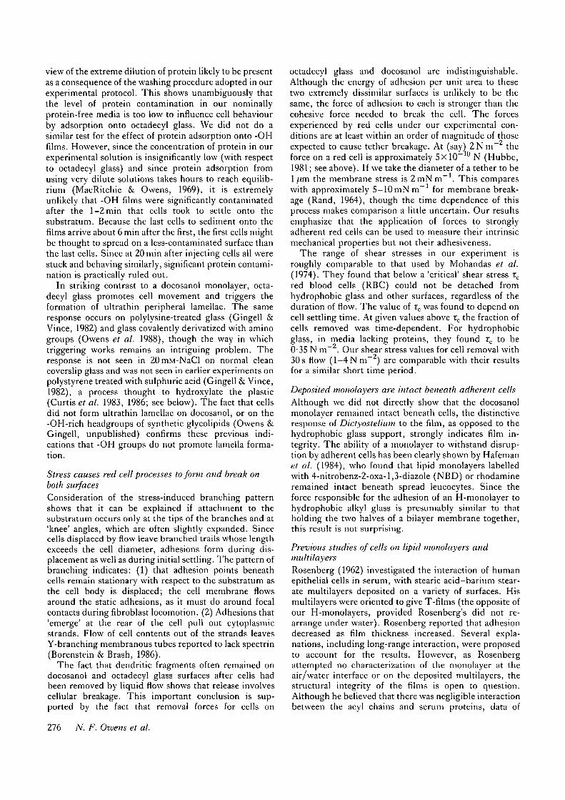
octadecyl glass and docosanol are indistinguishable.Although the energy of adhesion per unit area to thesetwo extremely dissimilar surfaces is unlikely to be thesame, the force of adhesion to each is stronger than thecohesive force needed to break the cell. The forcesexperienced by red cells under our experimental con-ditions are at least within an order of magnitude of thoseexpected to cause tether breakage. At (say) Z N m " theforce on a red cell is approximately 5x 10~10 N (Hubbe,1981; see above). If we take the diameter of a tether to be1 /im the membrane stress is 2 mN m~ . This compareswith approximately 5-10 mNm" 1 for membrane break-age (Rand, 1964), though the time dependence of thisprocess makes comparison a little uncertain. Our resultsemphasize that the application of forces to stronglyadherent red cells can be used to measure their intrinsicmechanical properties but not their adhesiveness.
The range of shear stresses in our experiment isroughly comparable to that used by Mohandas et al.(1974). They found that below a 'critical' shear stress TC
red blood cells (RBC) could not be detached fromhydrophobic glass and other surfaces, regardless of theduration of flow. The value of TC was found to depend oncell settling time. At given values above rc the fraction ofcells removed was time-dependent. For hydrophobicglass, in media lacking proteins, they found rc to be0-35 N m~ . Our shear stress values for cell removal with30 s flow ( l - 4 N m ~ 2 ) are comparable with their resultsfor a similar short time period.
Deposited monolayers are intact beneath adherent cellsAlthough we did not directly show that the docosanolmonolayer remained intact beneath cells, the distinctiveresponse of Dictyostelium to the film, as opposed to thehydrophobic glass support, strongly indicates film in-tegrity. The ability of a monolayer to withstand disrup-tion by adherent cells has been clearly shown by Hafemanet al. (1984), who found that lipid monolayers labelledwith 4-nitrobenz-2-oxa-l,3-diazole (NBD) or rhodamineremained intact beneath spread leucocytes. Since theforce responsible for the adhesion of an H-monolayer tohydrophobic alkyl glass is presumably similar to thatholding the two halves of a bilayer membrane together,this result is not surprising.
Previous studies of cells on lipid monolayers andmultilayersRosenberg (1962) investigated the interaction of humanepithelial cells in serum, with stearic acid-barium stear-ate multilayers deposited on a variety of surfaces. Hismultilayers were oriented to give T-films (the opposite ofour H-monolayers, provided Rosenberg's did not re-arrange under water). Rosenberg reported that adhesiondecreased as film thickness increased. Several expla-nations, including long-range interaction, were proposedto account for the results. However, as Rosenbergattempted no characterization of the monolayer at theair/water interface or on the deposited multilayers, thestructural integrity of the films is open to question.Although he believed that there was negligible interactionbetween the acyl chains and serum proteins, data of
276 N. F. Owens et al.

MacRitchie & Owens (1969) showed high rates of adsorp-tion for bovine serum albumin (BSA) at the air/waterand oil/water interfaces. In addition, an extensive af-finity of ConA for a hydrophobic surface was clearlyshown in the present study.
Since the physical requirements for cell spreadinginclude adhesion and the ability of the substratum toresist the lateral forces exerted during spreading (Harris,1982), the substratum must have a sufficient lateralviscosity. This value may vary with cell type, accordingto the rate at which the stresses are applied. While it isobvious that a cell may spread on a solid, the minimumlateral viscosity of a surface that can just support spread-ing has not to our knowledge been investigated. Althoughthe viscosities of deposited lipid films are not apparentlyavailable, there have been several studies where quan-tities broadly related to film rigidity have been measured.From these we shall also draw conclusions about our owndocosanol films.
Margolis et al. (1978) prepared =100 [im thick layers ofphospholipids by solvent evaporation. Electron spinresonance (e.s.r.) spectra (Margolis et al. 1979) showedthat lipids in thick hydrated films are regularly aligned,presumably as multi-bilayers with their polar headgroupsfacing the water (see Rand, 1981). They found thatfibroblasts cannot spread on lipids in the liquid crystal-line ('fluid') state, but spreading occurred on lipids in thecrystalline gel ('solid') state. Most convincing was thefinding that a phosphatidylethanolamine (PE) film cross-linked with glutaraldehyde was transformed from liquidto solid, and thereby changed from non-adhesive toadhesive. Likewise, fluid non-adhesive dioleoylphospha-tidylcholine (DOPC) and egg lecithin were renderedsolid and adhesive by treatment with osmium tetroxide.Interestingly, in these experiments there was no sign oflipid headgroup recognition.
The results of Smith & McConnell (1978) on hydratedphospholipid multilayers and of Tamm & McConnell(1985) on bilayers on hydrophilic glass show that whenthe phospholipid is in bilayer form Tc corresponds closelywith the temperature at which lateral diffusion measure-ments indicate a solid/liquid transition. Converting Mar-golis's Tc measurements into the language of lateraldiffusion, his experiments show that fibroblasts canspread on multibilayers with the low diffusion coefficientof a solid (often taken rather arbitrarily asD< 10~'°cm2s~1) but do not spread on those with themuch higher values of a liquid. In contrast to Margolis'sresults, numerous reports from McConnell's laboratory(Hafeman et al. 1981, 1982; Weise/a/. 1982; McConnellet al. 1986) show that leucocytes do not adhere or spreadon phosphatidylcholine monolayers deposited on alkyl-derivatized glass, regardless of whether the monolayersare solid or fluid in terms of lateral diffusion. The reasonfor this disagreement is not clear.
From the work of McConnell and his group (Weis et al.1982; Hafeman et al. 1981; Tamm & McConnell, 1985;Subramaniam et al. 1986), it is clear that phospholipidsbelow Tc, which are all solid by fl-A criteria at theair-water interface, show the low lateral diffusion charac-teristics of a solid after deposition, while those that are
liquid at the air-water interface may show either solid orliquid diffusion values of D following deposition. Thusthe support either has no effect or tends to freeze up thealkyl chains of the deposited monolayer molecules. Sincein our work docosanol is solid at the air-water interfaceprior to deposition, these facts strongly indicate thatDictyostelium discoideum and red blood cells adhere to asolid monolayer of docosanol. In relation to McConnell'swork, it would be most interesting if phosphatidylcholine(PC) headgroups are indeed less sticky for cells than -OHgroups, but until fibroblast adhesion is tested on de-posited phospholipid monolayers it is impossible toresolve the issue.
Adhesion of cells to alcoholic -OH groups of oxidizedpolystyreneThe adhesion of cells to strongly hydrophilic hydroxylsurfaces is of particular interest since the discovery byCurtis et al. (1983) that the major difference between(hydrophobic) bacteriological and (hydrophilic) tissue-culture grade polystyrene dishes is almost certainly due tothe greater density of -OH groups on the latter. Maximalbinding of BHK cells was observed at an -OH densitycorresponding to three groups per 1-0 nm2 surface area,whereas the optimal density for leucocytes was found tobe two groups per l-0nm . Our results show that at adensity of five groups per 1*0 nm , marginally higherthan those of Curtis et al. (1986), the surface is stillstrongly adherent. Further comparison is not possible aswe have yet to measure removal force as a function of thesurface density of -OH groups.
Cell adhesion occurs on both hydrophilic andhydrophobic swfacesOur results confirm that cells stick to hydrophilic andhydrophobic surfaces. The high contact angles for water(Mingins & Owens, 1973) measured on octadecyl glass(110° advancing, 105° receding) show that it is stronglyhydrophobic. Two facts support the conclusion that thedocosanol monolayer is hydrophilic under water. First,adsorption of ConA-Rh to hydrophobic octadecyl glassdoes not happen where the monolayer is present; thus thefilm is not hydrophobic and is presumably oriented withthe -OH headgroups towards the water. Second, thisorientation is to be expected for a Langmuir-Blodgettfilm deposited on a hydrophobic base, unless it lateroverturns (Langmuir, 1938). In contrast to our resultswith red cells and amoebae, Curtis et al. (1986) reportedlittle adhesion of BHK cells to hydrophobic polystyrene(in the presence or absence of proteins).
Our conclusion that cells stick to both hydrophilic andhydrophobic surfaces conflicts with attempts to correlateadhesiveness with contact angle (Van Oss et al. 1975). Inprevious work (in the proven absence of protein contami-nants) aldehyde-treated red cells were shown to adhere tothe hydrophobic interface between hexadecane and saltsolution (Gingell & Todd, 1975; Todd & Gingell, 1980)as well as to clean hydrophilic glass (Trommler et al.1985). In the former case adhesion was shown to bequantitatively consistent with the calculated electrodyna-mic (van der Waals') attraction in a secondary minimum,
Cell adhesion to a monolayer film Til

though molecular contact was not ruled out (Parsegian &Gingell, 1980).
The mechanisms of adhesion to the hydroxylated andthe hydrophobic surfaces are not well understood. Whileelectrodynamic attraction is the limit of close contact andforces associated with water structuring at interfaces(Parsegian, 1985) doubtless contribute, it is not knownwhether the hydroxyl headgroups indulge in short-range'biochemical' interactions with the cell surfaces.
We are grateful to Alan Day of this laboratory for makingSEM preparations and Don Clougher at the British Museum(Natural History) for enabling us to use his SEM facilities andfor taking the SEM photographs. The work was supported bygrants from the Science and Engineering Research Council andthe Wellcome Trust to D.G.; A.T. thanks the Ministry ofHigher Education of the G.D.R. for travel grants.
References
ABRAHAM, B. M., MIYANO, K., KETTERSON, J. B. & Xu, S. Q.
(1983). Anomalous melting properties of some classical monolayersystems. Phys. Rev. Lett. 51, 1975-1978.
ADAM, N. K. (1941). Physics and Chemistry of Surfaces, pp. 48-57.London: Oxford University Press.
BORENSTEIN, N. & BRASH, J. L. (1986). Red blood cells depositmembrane components on contacting surfaces. J. Biomed. Mat.Res. 20, 723-730.
BROOKS, J. H. & ALEXANDER, A. E. (1960). Losses by evaporationand solution from monolayers of long-chain aliphatic alcohols.Proc. 3rd. hit. Cong. Surf. Act., Cologne, vol. I I , pp . 196-201.
CURTIS, A. S. G., FORRESTER, J. V. & CLARK, P. (1986). Substrate
hydroxylation and cell adhesion, j ' . Cell Sci. 86, 9-24.CURTIS, A. S. G., FORRESTER, J. V., MCINNES, C. & LAWRIE, F.
(1983). Adhesion of cells to polystyrene surfaces. J . Cell Biol. 97,1500-1506.
ESKINAZI, S. (1977). Principles of Fluid Mechanics. Boston: Allyn &Bacon Inc.
FRANCIS, G. W., FISHER, L. R., GAMBLE, R. A. & GINGELL, D.
(1987). Direct measurement of cell detachment force on single cellsusing a new electromechanical method. J. Cell Sci. 87, 517-523.
GINGELL, D. (1981). The interpretation of interference reflectionimages of spread cells: significant contributions from thinperipheral cytoplasm. J. Cell Sci. 49, 237-247.
GINGELL, D. & TODD, I. E. (1975). Adhesion of red blood cells tocharged interfaces between immiscible liquids. A new method.J. Cell Sci. 18, 227-239.
GINGELL, D., T O D D , 1. E. & HEAVENS, O. S. (1982). Quantitative
interference microscopy: effect of microscope aperture. OpticaActa 29, 901-908.
GINGELL, D. & VINCE, S. M. (1982). Substratum wettability andcharge influence the spreading of Dictyostelium amoebae and theformation of ultrathin cytoplasmic lamellae. J. Cell Sci. 54,255-285.
HAFEMAN, D. G., SEUL, M., CLIFFE, C. M. & MCCONNELL, H. M.(1984). Superoxide enhanced photobleaching during cellularimmune attack against fluorescent lipid monolayer membranes.Biochim. biophys. Acta 772, 20-28.
HAFEMAN, D. G., SMITH, L. M., FEARON, D. T. & MCCONNELL, H.
M. (1982). Lipid monolayer-coated solid surfaces do not perturbthe lateral motion and distribution of C3b receptors onneutrophils. J. Cell Biol. 94, 224-227.
HAFEMAN, D. G., VON TSCHARNER, V. & MCCONNELL, H. M.
(1981). Specific antibody-dependent interaction betweenmacrophages and lipid haptens in polar lipid monolayers. Proc.natn. Acad. Sci. U.S.A. 78, 4552-4556.
HARRIS, A. K. (1982). Traction, and its relations to contraction intissue cell locomotion. In Cell Behaviour (ed. R. Bellairs, A.Curtis & G. Dunn), pp. 109-134. Cambridge: CambridgeUniversity Press.
HUBBE, M. A. (1981). Adhesion and detachment of biological cells invitro. Progr. Surface Sci. 11, 65-138.
HYNES, R. O. (1986). Fibronectins. Scient. Am. June issue, 254,32-41.
LANGMUIR, I. V. (1938). Overturning and anchoring of monolayers.Science LXXXXII, no. 2266, 493-500.
MACRITCHIE, F. & OWENS, N. F. (1969). Coagulation of proteins atinterfaces. J . Colloid Interface Sci. 29, 66-71.
MARGOLIS, L. B. (1984). Cell interaction with model membranes.Probing, modification and simulation of cell surface functions.Biochim. biophys. Acta 779, 161-189.
MARGOLIS, L. B., DYATLOVITSKAYA, E. V. & BERGELSON, L. D.
(1978). Cell—lipid interactions. Cell attachment to lipid substrates.Expl Cell Res. I l l , 454-457.
MARGOLIS, L. B., VASILIEVA, E. J., VASILIEV, J. M. & GELFAND, I.
M. (1979). Upper surfaces of epithelial sheets and fluid lipid filmsare nonadhesive for platelets. Proc. natn. Acad. Sci. U.S.A. 76,2303-2305.
MCCONNELL, H. M., WATTS, T. H., WEIS, R. M. & BRIAN, A. A.
(1986). Supported planar membranes in studies of cell-cellrecognition in the immune system. Biochim. biophvs. Acta 864,95-106.
MINGINS, J. & OWENS, N. F. (1973). Contact angle hysteresis, itsproperties and measurement. Proc. 11th A. Conf. AdhesionAdhesives. City University, London. In Aspects of Adhesion (ed.K. W. Allen), vol. 8, pp. 203-251. London: Transcripta Books.
MOHANDAS, N., HOCHMUTH, R. M. & SPAETH, E. E. (1974).
Adhesion of red cells to foreign surfaces in the presence of flow.J. Biomed. Mat. Res. 8, 119-136.
MULLER, A. (1927). An X-ray investigation of long-chaincompounds. Proc. R. Soc. Land. A, 114, 542-561.
OWENS, N. F., GINGELL, D. & BAILEY, J. (1988). Contact mediatedtriggering of lamella formation by Dictvostelium amoebae on solidsurfaces. J. Cell Sci. 91, 000-000.
OWENS, N. F., GINGELL, D. & RUTTER, P. R. (1987«). Inhibition of
cell adhesion by a synthetic polymer adsorbed to glass shownunder defined hydrodynamic stress. J'. Cell Sci. 87, 667-675.
OWENS, N. F., JOHNSTON, D. S., GINGELL, D. & CHAPMAN, D.
(19876). Surface properties of a long-chain 10:12 diynoic acidmonolayer at air/liquid and solid/liquid interfaces. Thin SolidFilms 155, 255-266.
PARSEGIAN, A. & GINGELL, D. (1973). A physical force model ofbiological membrane interaction. In Recent Advances in Adhesion(ed. L. H. Lee), Am. Chem. Soc. Proc, pp. 153-192. New York:Gordon and Breach.
PARSEGIAN, V. A. (1984). Water near intracellular surfaces. J. CellBiol. 99, 1965-2009.
PARSEGIAN, V. A. & GINGELL, D. (1980). Red blood cell adhesion.III. Analysis of forces. J. Cell Sci. 51, 151-157.
RAND, R. P. (1964). Mechanical properties of the red cell membrane.2. Viscoelastic breakdown of the membrane. Biophvs. J. 4,303-316.
RAND, R. P. (1981). Interacting phospholipid bilayers: measuredforces and induced structural changes. A. Rev. Biophys. Bioengng.10, 277-314.
ROSENBERG, M. D. (1962). Long-range interactions between cell andsubstratum. Proc. natn. Acad. Sci. U.S.A. 48, 1342-1349.
SMITH, B. A. & MCCONNELL, H. M. (1978). Determination ofmolecular motion in membranes using periodic patternphotobleaching. Proc. natn. Acad. Sci. U.S.A. 75, 2759-2763.
SPARROW, E. M. (1955). Analysis of laminar forced-convection heattransfer in entrance region. National Advisory Committee onAeronautics, Technical News, TN 3331.
SUBRAMANIAM, S., SEUL, M. & MCCONNELL, H. M. (1986). Lateral
diffusion of specific antibodies bound to lipid monolayers onalkylated substrates. Proc. natn. Acad. Sci. U.S.A. 83, 1169-1173.
TAMM, L. K. & MCCONNELL, H. M. (1985). Supportedphospholipid bilayers. Biophys. J. 47, 105-113.
TODD, I. E. & GINGELL, D. (1980). Red blood cell adhesion. I.Determination of the ionic conditions for adhesion to an oil-waterinterface. J . Cell Sci. 41, 135-149.
T O D D , I. E., MELLOR, J. S. & GINGELL, D, (1988). Mapping
cell-glass contacts of Dictyostelium amoebae by total internal
278 N. F. Owens et al.

reflection aqueous fluorescence overcomes a basic ambiguity of equivalency. .7. Colloid Interface Sci. 76, 305-314.interference reflection microscopy. J. Cell Sci. 89, 107-114. WEISS, R. M., BALAKRISHNAN, K., SMITH, B. A. & MCCONNELL, H.
TROMMLER, A., GlNGELL, D. & WOLF, H. (1985). Red blood cells M. (1982). Stimulation of fluorescence in a small contact regionexperience electrostatic repulsion but make molecular adhesions between rat basophil leukemia cells and planar lipid membranewith glass. Biophvs.J. 48, 835-841. targets by coherent evanescent radiation. J. biol. Chew. 257,
VAN OSS, C. J., OILMAN, C. F. & NEUMANN, A. N. (1975). 6440-6445.Phagocytic Engulfment and Cell Adhesiveness. Microorganisms andInfectious Diseases, vol. 2. New York: Marcel Dekker Inc.
VAN WAGENEN, R. A. & ANDRADE, J. D. (1980). Flat plate (Received 29 February 1988-Accepted, in revised form, 14 Julystreaming investigations: hydrodynamics and electrokinetic 1988)
Cell adhesion to a monolayer film 279
